Next Article in Journal
Antithrombin Treatment of Patients with Acute Coronary Syndromes Undergoing Percutaneous Coronary Intervention
Previous Article in Journal
Comments to the 2007 Recommendations for the Treatment of Essential Hypertension by the Swiss Society of Hypertension
 
 
Cardiovascular Medicine is published by MDPI from Volume 28 Issue 1 (2025). Previous articles were published by another publisher in Open Access under a CC-BY (or CC-BY-NC-ND) licence, and they are hosted by MDPI on mdpi.com as a courtesy and upon agreement with Editores Medicorum Helveticorum (EMH).
Case Report

Transcatheter Closure of Perimembranous Ventricular Septal Defect in Adult Using the Amplatzer Septal Occluder: Report of Two Challenging Cases

by
Giovanni B. Pedrazzini
1,*,
Mario Carminati
2,
Franco Faletra
1 and
Tiziano Moccetti
1
1
Cardiology, Cardiocentro Ticino, Lugano, Switzerland
2
Pediatric Cardiology Center, Policlinico San Donato, Milan, Italy
*
Author to whom correspondence should be addressed.
Cardiovasc. Med. 2008, 11(1), 16; https://doi.org/10.4414/cvm.2008.01298
Submission received: 25 October 2007 / Revised: 25 November 2007 / Accepted: 25 December 2007 / Published: 25 January 2008

Introduction

Ventricular septal defect (VSD) accounts for 20% of congenital cardiovascular malformations and 10% of those diagnosed in adults. Its prevalence is estimated at 1.17 per 1000 births and at 0.5 per 1000 adults and has increased recently because of improved detection [1]. The vast majority of VSDs (roughly 70%) are located in the area of the membranous septum and are defined as perimembranous or subaortic.
Cardiovascmed 11 00016 i001
Although surgery is the gold standard for VSD closure, in the last decades percutaneous approaches to the closure of both perimembranous and muscular VSD have been developed and successfully applied [2,3]. Unlike in muscular VSD, due to proximity of the aortic valve and the atrioventricular valve, devices designed for other applications when used in the setting of a perimembranous VSD did not fit perfectly to close these defects. With the introduction of specially designed Amplatzer® eccentric devices, closure of these defects is becoming feasible, and very effective as recently reported by Holzer et al. [4]. Challenging procedural situations may be related to additional congenital abnormalities or to particular anatomical location of the defect.
We are reporting two cases of transcatheter closure of perimembranous VSD performed in adults using the new designed Amplatzer® devices in whom VSD was associated with anomalous entry of the inferior vena cava into the anonimous vein in one case and with a posteriorly located VSD in another one.

Case 1

A 66-year-old woman presenting with increasing exerctional dyspnoea (New York Heart Association functional class III) was diagnosed with perimembranous septal defect. Echocardiographic examination showed a large perimembranous defect (1.3 × 0.7 cm), with a left-to-right shunt and a left-to-right gradient corresponding to 80 mm Hg (Figure 1A). The pulmonary systolic pressure was 65 mm Hg and the Qp/Qs 2.2. Coronary angiography showed normal arteries. A percutaneous VSD closure was performed following the technique reported by Carminati et al. Under general anaesthesia and with transoesophageal guidance, after visualisation of the defect with a conventional pigtail angio (Figure 1B), the defect itself was crossed from left to right using a right Judkins and a long Terumo (300 cm). Then, the wire was snared from jugular vein due to an anomalous entry of the inferior vena cava into the anonimous vein. A 9 F dedicated sheath was introduced from the jugular vein into the ascending aorta and then gently withdrawn in the LV taking care to create a loop with the tip of the guiding catheter pointed toward the apex of the LV. A 10 mm sized membranous Amplatzer® device (Figure 1C) secured on the delivery cable was advanced up to the tip and then very carefully deployed under fluoroscopic and TEE guidance. The final result is very satisfactory with device in place and minimal residual shunt (Figure 1D). Total procedural time was 47 minutes.

Case 2

A 55-year-old man otherwise asymptomatic was brought to our attention following routine echocardiographic examination, which revealed a perimembranous VSD. The defect was located posteriorly and very close to the right aortic cuspid. The VSD was echocardiographically sized of about 9 × 9 mm presenting with a gradient of 75 mm Hg. The Qp/Qs was 1.8, and a pulmonary systolic pressure was estimated in the range of 50 mm Hg. Due to patient’s refusal of a surgical closure, percutaneous VSD closure was performed. After conventional angiographic visualisation, the defect was crossed with a catheter AR 5 F. The wire was placed into the pulmonary artery and then snared through the femoral vein. A right to left crossing through the defect was attempted using a 9 F dedicated catheter introduced from the femoral vein. However, due to the posterior location of the defect, the catheter was not stable enough to allow a correct device deployment which necessitated a retrograde arterial approach using a muscular Amplatzer® device. This was done as following: after conventional snaring of the wire recrossed through the defect, an 8 F catheter was introduced in a retrograde way through the aortic valve into the LV and then into the right ventricle (Figure 2A). The 10 mm muscular Amplatzer® device (Figure 2B) secured on a delivery cable was advanced to the tip and then gently deployed (Figure 2C) obtaing a very good final result. The aortic valve was carefully inspected and did not show any sign of mechanical obstacle. Total procedural time was 57 minutes.

Discussion

The two presented cases are showing challenging anatomical situations which may be encountered when performing percutaneous closure of VSD. Despite significant anatomical obstacles, which forced us to a rare delivery of VSD devices, both procedures could be safely and effectively performed within a reasonable procedural time. Another unique feature of our cases was the age of both treated patients which was distinctively higher than other published cohorts.
Our two cases echo the impressive results recently presented by Carminati et al., one of the largest single-center series including 84 perimembranous VSDs treated by transcatheter approach either with the muscular or the membranous septal occluder [5]. Among the 84 patients in whom successful device implantation occurred, mid-term closure rate was excellent approaching 96%, confirming significantly a smaller previous series [4]. Major acute complications occurred in 1.2%. Of note, device embolisation occurred in two patients which both were uneventful and percutaneously retrieved. Rhythm disturbances were reported more frequently, with complete heart block in three patients (transient in two, one requiring pacemaker implantation), transient left bundle branch block in two patients and transient first-degree atrioventricular block in one patient. Device-related trivial aortic and tricuspidal regurgitation only occurred in three cases.
In conclusion, transcatheter occlusion of perimembranous VSD is feasible and safe and may be considered an alternative to surgery. Appropriate patient selection and a solid experience of the operator is of paramount importance to the success of the procedure. Although further improvements of technology are needed in order to overcome present limitations, first of all rhythm disturbances.

References

  1. Ammash, N.M.; Warnes, C.A. Ventricular septal defects in adults. Ann Intern Med. 2001, 135(9), 812–824. [Google Scholar] [CrossRef] [PubMed]
  2. Lock, J.E.; et al. Transcatheter closure of ventricular septal defects. Circulation. 1988, 78(2), 361–368. [Google Scholar] [CrossRef] [PubMed]
  3. Amin, Z.; et al. New device for closure of muscular ventricular septal defects in a canine model. Circulation. 1999, 100(3), 320–328. [Google Scholar] [PubMed]
  4. Holzer, R.; et al. Device closure of muscular ventricular septal defects using the Amplatzer muscular ventricular septal defect occluder: immediate and mid-term results of a U.S. registry. J Am Coll Cardiol. 2004, 43(7), 1257–1263. [Google Scholar] [CrossRef] [PubMed]
  5. Carminati, M.; et al. Transcatheter closure of congenital ventricular septal defect with Amplatzer septal occluders. Am J Cardiol. 2005, 96(12a), 52–58L. [Google Scholar] [CrossRef] [PubMed]
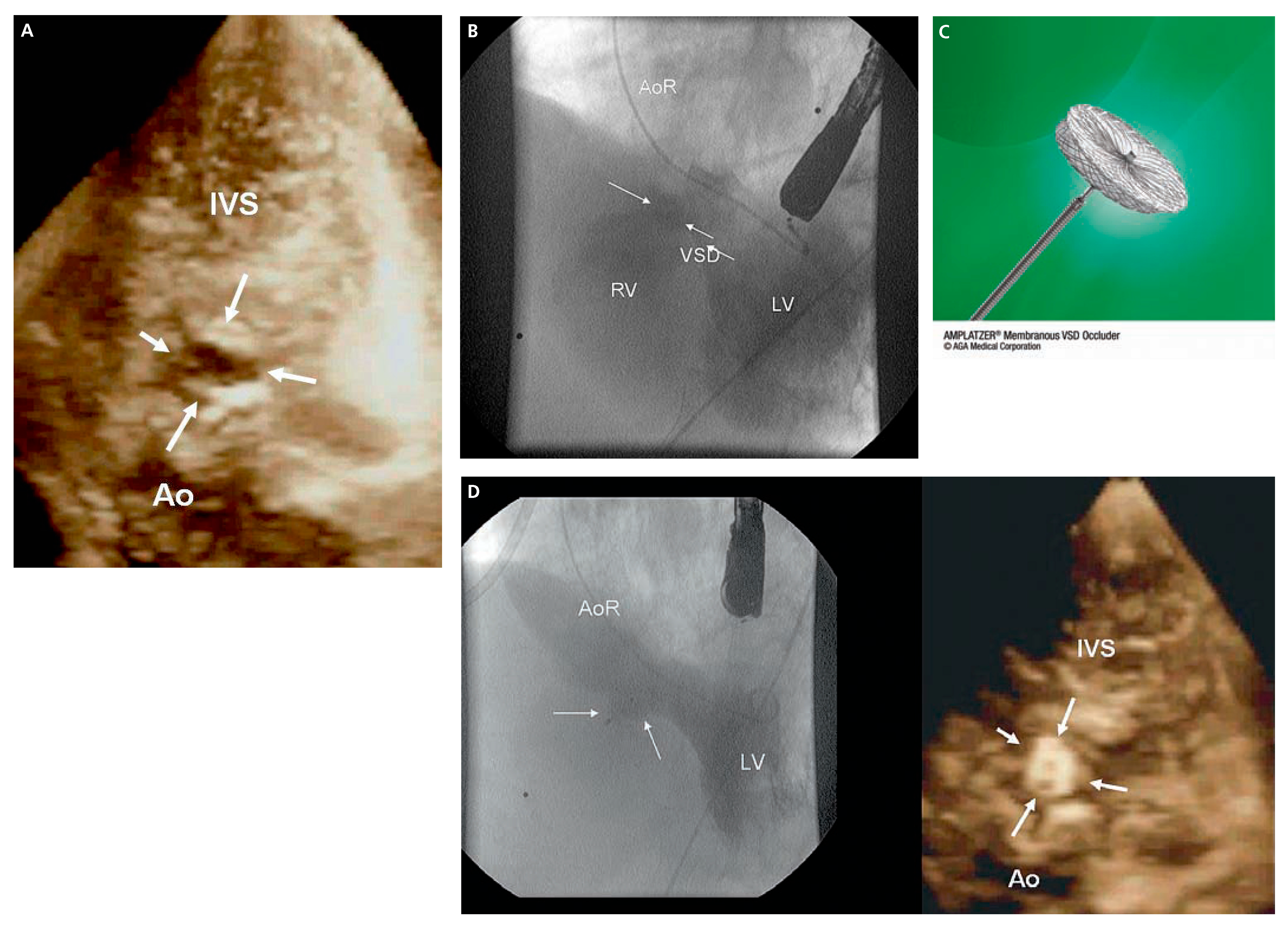
Figure 1. A Echocardiographic 3-D “en face” view of the large perimembranous septal defect (arrows). B Pigtail 5 F catheter in the left ventricle. By the LV angiogram the contrast medium flowing toward the right ventricle represents the large perimembranous septal defect. C The membranous VSD occluder has an eccentric shape, with the distal (left ventricular) disk exceeding the connecting waist by 1 mm in its superior part and by 5.5 mm in its inferior part; the proximal (right ventricular) disk is 2 mm larger than the connecting waist, which is only 1.5 mm in length. As for the others Amplatzer® devices, polyester fabric is incorporated within the nitinol wire mesh of both disks and the connecting waist. The device is secured to a delivery cable and inserted into a delivery sheath ranging from 6 F to 9 F in diameter. D Final result after releasing of the occluder device. Angiographic and echocardiographic 3-D “en face” view. The device (arrow) is in place, stable, and by the final ventriculography there is only a very small residual shunt. AoR = aortic root; LV = left ventricle; RV = right ventricle; VSD = ventricular septal defect; IVS = interventricular septum; Ao = Aorta.
Cardiovascmed 11 00016 g001
Figure 2. A 8 F guiding catheter introduced in a retrograde way through the aortic valve and then into the right ventricle through the perimebranous defect (arrows). B The muscular VSD occluder is a self-expandable device made of nitinol, consisting of 2 flat disks with a diameter 8 mm larger than their central connecting waist; waist diameter, from 4 to 18 mm, determines the size of the device. C Deployment of the 10 mm muscular Amplatzer® (arrow) device through the 8 F sheath introduced in a retrograde way through the aortic valve into the right ventricle.
Cardiovascmed 11 00016 g002