You are currently viewing a new version of our website. To view the old version click .
Cardiovascular Medicine
  • Cardiovascular Medicine is published by MDPI from Volume 28 Issue 1 (2025). Previous articles were published by another publisher in Open Access under a CC-BY (or CC-BY-NC-ND) licence, and they are hosted by MDPI on mdpi.com as a courtesy and upon agreement with Editores Medicorum Helveticorum (EMH).
  • Interesting Images
  • Open Access

27 April 2007

Mysterious Floating Structure in the Left Atrium After Coronary Artery Bypass Grafting

,
and
Cardiovascular Center Cardiology, University Hospital, CH-8091 Zurich, Switzerland
*
Author to whom correspondence should be addressed.
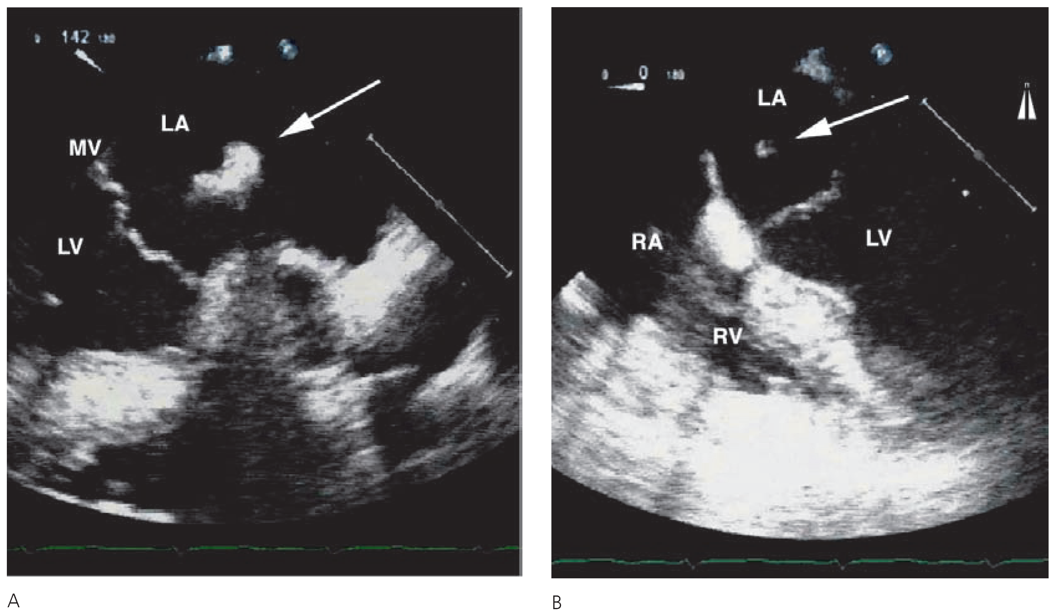
A 60-year-old male patient was referred to our hospital for coronary artery bypass graft surgery due to prior subacute myocardial infarction and subsequently angiographically diagnosed 3-vessel coronary artery disease. Preoperative transoesophageal echocardiography (TEE) revealed wall motion abnormalities, but was otherwise unremarkable. During stepwise reduction of the extracorporal circulatory assistance after uneventful course of the surgical procedure, the routinely conducted TEE revealed a well-defined homogeneous floating structure in the left atrium (Figure 1A,B). Diagnosis of a perioperative thrombus formation in the left atrium was proposed. Hence, the left atrium was incised to explore the structure leading to the diagnosis of an inverted left atrial appendage. Inversion of the left atrial appendage following cardiac surgery is a rare but important complication that can be transient showing spontaneous eversion. It can, however, result in serious complications such as necrosis with potential rupture of the left atrial wall. Therefore decision was taken towards surgical reduction of the inverted left atrial appendage. In the case of peri- or postoperative appearance of a new clearly delineated floating structure in the left atrium, an inverted left atrial appendage has to be taken into account.
Figure 1. (A) Transoesophageal crosssectional long-axis view of the heart at 142°. (B) Four-chamber-view of the heart at 0°. Arrows indicate a clearly delineated homogeneous floating structure in the left atrium. LA = left atrium; LV = left ventricle; RA = right atrium; RV = right ventricle; MV = mitral valve.

Conflicts of Interest

The authors declare no conflict of interest.

Article Metrics

Citations

Article Access Statistics

Multiple requests from the same IP address are counted as one view.