Abstract
A quercetin derivative with remarkable biological performance was successfully synthesized by chemical modification of the flavonoid with docosahexaenoic acid to synthesize 2-(2,2-diphenylbenzo[d][1,3]dioxol-5-yl)-5,7-dihydroxy-4-oxo-4H-chromen-3-yl-(4Z,7Z,10Z,13Z,16Z,19Z)-docosa-4,7,10,13,16,19-hexaenoate (3), deeply characterized by NMR spetroscopy. Modified quercetin and pectin were involved in a grafting process by an ecofriendly radical procedure able to preserve the biological features of the quercetin derivative. Antioxidant performances of the conjugate were evaluated both in term of total phenolic amount and scavenger activity in organic and aqueous environments. Additionally, in vitro acute oral toxicity was also tested against Caco-2 cells and 3T3 fibroblasts, confirming that pectin conjugate does not have any effect on cell viability at the dietary use concentrations. Finally, in vitro experiments highlighted the ability of the conjugate to counteract the migratory properties of Caco-2 and HepG2 cells, indicating its feature in the reduction of the migration of tumour cells. These data showed that the covalent binding of the quercetin derivative to the pectin chain represents a very interesting strategy to improve the bioavailability of the quercetin, representing an effective means of protecting and to transporting polyphenol molecules.
1. Introduction
Nowadays, to have a healthy human body, people are paying more attention to their foods, preferring minimally processed ones, consuming a large number of fruits and vegetables, and also taking nutraceuticals and dietary supplements [1,2,3,4,5]. Considering that the definition of “healthy food” can be ascribed to improved immune function, preventing specific diseases, and reducing side effects and health care costs, nutraceuticals comprise prebiotics, polyunsaturated fatty acids, probiotics, herbal products, and antioxidants, and consequently, the nutraceutical industry has encountered large consumer compliance so far [6]. Additionally, with both a growing population and the prevalence of chronic diseases as the population ages, future demands for eicosapentaenoic acid/docosahexaenoic acid (EPA/DHA) will further increase [7]. Recently, during the COVID-19 pandemic, scientific shreds of evidence confirmed that quercetin [8], astaxanthin, lactoferrin, glycyrrhizin, hesperidin, and curcumin have shown encouraging data in reverting long COVID-19 phenomena, suggesting their use in preventing and counteracting the symptoms of the infection [9,10]. In this work, we carried out the synthesis of a hybrid molecule, obtained by a Steglich coupling between quercetin and DHA, at position 3, and its grafting with pectin, to obtain a functional polymer, endowing better antioxidant performance of parent compounds and probably improving bioavailability. Quercetin is a flavonol characterized by the presence of an oxogroup at the 4 position and a 2,3-double bond on ring C, which allows conjugation between rings A and B and strongly affects the redox properties of this compound. They differ from the flavones for the presence of a nonphenolic hydroxyl group at the 3 position. Quercetin is the most powerful antioxidant flavonoid in nature, and it is present in many vegetal sources, particularly belonging to Mangifera indica, Curcuma domestica valenton, and others. In vitro experiments highlighted the anticancer, antidiabetic, antifungal, antibacterial, anti-inflammatory, antiobesity, antiviral, and neuroprotective properties of quercetin and its derivatives, as well as its feature to accelerate wound healing. These findings allowed for the employment of quercetin as an ingredient in the preparation of different nutraceuticals and cosmeceuticals [11]. The absorption of quercetin, broadly found in foods, is extremely variable relative to the type—and particularly the position—of sugar linked to the aglycone. The absorption of quercetin is strictly correlated to the food matrix, whilst plasma concentration of quercetin was found higher after onion intake than in red wine or tea. Some authors have demonstrated that absorption rate is not affected of the glucose position (3′ and 4′) [12]. However, the main challenge related to the employment of the quercetin is its bioavailability, which appears particularly low (<10%). Poor water solubility, absorption profile, and chemical stability represent the main causes, and they could be overcome by encapsulating it in suitable macromolecular systems collected from food-grade ingredients. More effective nutraceuticals and functional foods can definitely be developed by improving the bioavailability of quercetin [13]. The nutraceutical effect of quercetin, assayed in vivo in two experiments, is measurable with the ability to work as an antioxidant and anti-inflammatory agent when the basal level of oxidative stress and inflammation is high. This scientific evidence confirms that quercetin supplementation is useful in people with diseases, but not in healthy ones. However, in chronic disorders, the safety of quercetin supplementation is still to be established [14]. In the last few years, there have been a lot of efforts to increase the bioavailability of active molecules, overcoming the problems often related to their poor solubility. Several systems have been developed to incorporate such compounds to improve stability, increase dispersion, and ensure maximum health benefits. There are many biopolymers already used for these purposes, and pectin displayed the best characteristics for the realization of macromolecular carriers for oral administration. Pectin is a linear heteropolysaccharide contained in the cell walls of plant tissues. More precisely, the name “pectin” refers to the compound extracted from fruit starting from the protopectin it contains. In particular, apples, apricots, and pears are rich in it (variable content from 1 to 1.5%), but also oranges (0.5–3.5%) and carrots (1.4%). Very high content of pectin is present in fruit peel; in that of oranges, the presence of the fibre in question is particularly high (about 30%) [15]. Alternative sources of pectin can be waste from the processing of sugar beet, mango, sunflowers, legumes, bananas, cabbage, carrots, and pomelo. In general, however, it would be more correct to speak of “pectins”, because in nature there are different structures, the most common of which is composed of monomeric units of D-galacturonic acid joined together through α-(1→4) glycosidic bonds to form a long chain to which sugar functions such as rhamnose, arabinose, galactose, and xylose can bind, thus forming the side chains [15]. In this work, pectin was involved in an ecofriendly radical process, to synthesize polymeric conjugates with remarkable biological activities. Macromolecular compounds were deeply analysed in term of antioxidant activity and toxicity against Caco-2 and HepG2 cells, while their ability to counteract the migratory properties of cells was also investigated.
2. Materials and Methods
2.1. Chemistry
The synthesis of compound 3 was realized starting from 1, which was previously protected as 3′,4′-diphenylmethylketal derivative by treating 1 with α,α-dichlorodiphenylmethane. The protected derivative 2 was then treated with docosahexaenoic acid in Steglich conditions to obtain the final compound, named 3.
2.1.1. Synthesis of 2-(2,2-Diphenylbenzo[d][1,3]dioxol-5-yl)-3,5,7-trihydroxy-4H-chromen-4-one (2)
Compound 2 was synthesized according to our previous reported procedures (Scheme 1). Spectroscopic data are in line with those reported [16,17].
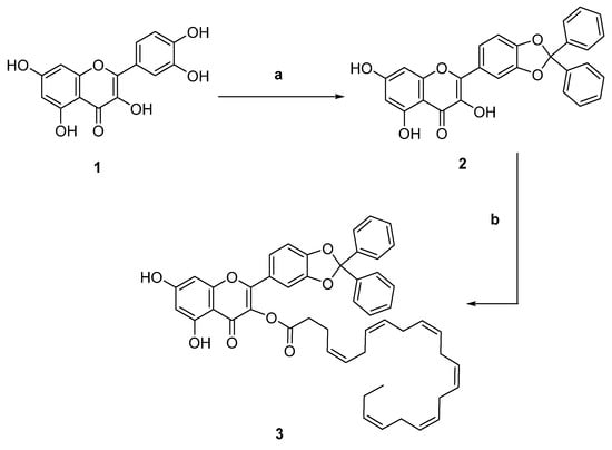
Scheme 1.
Reagents and conditions: (a) α,α-dichlorodiphenylmethane, 180 °C, 10 min; (b) cervonic acid, N,N’-dicyclohexylcarbodiimmide, dry dichloromethane, 0 °C to RT, 24 h.
2.1.2. Synthesis of 2-(2,2-Diphenylbenzo[d][1,3]dioxol-5-yl)-5,7-dihydroxy-4-oxo-4H-chromen-3-yl (4Z,7Z,10Z,13Z,16Z,19Z)-Docosa-4,7,10,13,16,19-hexaenoate (3)
To a well-stirred solution of cervonic acid (128 µL, 0.386 mg) in dry DCM (2.0 mL) at 0 °C, N,N’-dicyclohexylcarbodiimmide (91.0 mg, 0.46 mmol) was added. The mixture was stirred for 1 h at 0 °C, after which 2 (180.0 mg, 0.39 mmol) was dropped and the resulting mixture warmed to RT and stirred for 23 h. The mixture was filtered on Celite® and treated with 5 mL of saturated NaHCO3. The organic layer was dried, filtered, and evaporated under reduced pressure. The title compound 3 was purified through column chromatography (eluent n-hexanes/ethyl acetate 5:1 v/v) (Scheme 1). Yellow oil, 56% yield. 1H NMR (300 MHz, DMSO-d6) δ 11.5 (bs, 1H), 9.60 (bs, 1H), 7.7–7.3 (m, 11H), 7.25 (s, 1H), 7.10 (s, 1H), 6.90 (s, 1H), 6.20 (s, 1H), 5.5–5.0 (m, 12H), 4.1–3.8 (m, 9H), 2.9–2.6 (m, 2H), 2.4–2.2 (m, 2H), 2.0–1.9 (m, 2H), 1.3–1.0 (m, 2H). 13C NMR (75 MHz, DMSO-d6) δ 171.1, 170.1, 167.0, 159.8, 157.1, 154.0, 150.2, 143.0, 139.1, 133.3, 132.1, 131.9, 130.6, 129.1 (4C), 128.6, (3C), 128.3 (4C), 126.1 (3C), 126.0 (3C), 127.5 (2C), 122.0 (6C), 110.4, 98.8, 93.5, 39.10 (4C), 33.9, 25.6, 22.8, 20.5, 14.5. ESI-MS 775 [M-H]−.
2.2. Disposable Phenolic Equivalents by Folin–Ciocalteu Procedure
Total phenolic groups were evaluated employing Folin–Ciocalteu procedure, according to the literature protocol with some changes [18]. In a volumetric flask 6.0 mL, of an aqueous solution of the active species was prepared, then 1.0 mL of the Folin–Ciocalteu reagent was added and mixed thoroughly. After 3 min, 3.0 mL of Na2CO3 (7.5% w/w) was added, and the mixture was intermittently shaken for 2 h. The absorbance was measured at 760 nm. Total phenolic equivalents were expressed as quercetin (Q) equivalent concentration (8.0, 16.0, 24.0, 32.0, and 40.0 μM). Least-squares method was employed to calculate a calibration curve, slope, correlation coefficient (R2 = 0.9943), and intercept.
2.3. Total Antioxidant Activity
Total antioxidant activity of active species was investigated according to a literature protocol with some changes [19]. Briefly, 0.3 mL of an aqueous solution of the active species was mixed with 1.2 mL of reagent solution (28.0 M Na3PO4, 4.0 M (NH4)2MoO4 and 0.6 M H2SO4,) and then incubated for 150 min at 95 °C. The solutions were analysed by a UV–Vis spectrophotometer (at 695 nm). The total antioxidant activities were expressed as catechin (CT) equivalent concentration, by recording a calibration curve at different antioxidants (8.0, 16.0, 24.0, 32.0, and 40.0 μM) and employing the method of least squares to calculate correlation coefficient (R2), intercept, and slope of the regression equation.
2.4. Scavenging Activity against DPPH Radicals
Antioxidant species reacted with 2,2′-diphenyl-1- picrylhydrazyl radical (DPPH) free radicals to record their scavenging properties [20]. In each experiment, 12.5 mL of an aqueous solution of the active species was added to 12.5 mL of an ethanol solution of DPPH (200 μM) at 25 °C, and after 30 min, the absorbance was recorded (517 nm). The scavenging activity was expressed as percent inhibition of DPPH radicals, according to Equation (1):
where A0 is the absorbance recorded in absence of active species, and A1 is the absorbance recorded in presence of antioxidant compounds.
Inhibition (%) = (A0 − A1)/A0 × 100
2.5. Scavenging Activity agaist ABTS Radical Cations
Scavenging properties of active species in the aqueous environment were evaluated against 2,2′-azino-bis(3-etylbenzotiazolin-6-sulphonic) radicals (ABTS) according to a literature protocol with some modifications [21,22]. An aqueous ABTS+ solution able to guarantee an absorbance of 0.970 ± 0.020 at 734 nm was prepared. To evaluate the scavenging effect of the antioxidant compounds, 0.5 mL of each macromolecule solution was mixed with 2.0 mL of the ABTS radical solution and the mixture was incubated at 37 °C for 5 min. The antioxidant activity was expressed as a percentage of scavenging activity on the ABTS radical according to Equation (1).
2.6. Synthesis of Conjugates
Pectin conjugates were synthesized following a literature protocol with some modifications [23]. In a general procedure, 1.0 mL of H2O2 5.0 M (5.0 mmol) and 0.25 g of ascorbic acid (1.4 mmol) were added to 50.0 mL of pectin solution (10 mg mL−1) at 25 °C. After 2 h, a suitable amount of extract corresponding to 0.150 mg of quercetin equivalent concentrations was added to the solution. After the reaction time, the mixture was dialysed by using dialysis membranes of 6–27/32” Medicell International LTD (MWCO: 10–12,000 Da). Purified pectin conjugates were checked to be free of unreacted low-molecular-weight molecules by liquid chromatography analysis (LC-DAD) of the washing medium. The purified solution was frozen and dried (Freeze drier Micro Modulyo, Edwards was employed) with a freeze drier to afford a vaporous solid. Additionally, blank pectin, exploited as a control, was prepared when the grafting process was carried out in absence of the antioxidant molecules.
2.7. Cell Culture
Balb/c 3T3 clone A31 cells were cultured in DMEM supplemented with 10% calf bovine serum and 1% penicillin-streptomycin. Caco-2 were maintained in MEM with fetal bovine serum (FBS) 20% (w/w), non-essential amino acids (1% w/w), L-glutamine (1% w/w) and penicillin–streptomycin (1% w/w), while HepG2 was cultured in MEM culture medium, supplemented with 10% FBS, non-essential amino acids (1% w/w), L-glutamine (1% w/w) and penicillin–streptomycin (1% w/w). The cells were grown in a 5% CO2 atmosphere at 37 °C until 80% confluence, and subcultured twice a week. All cell lines were purchased from ATCC, Manassas, VA, USA.
2.8. Neutral Red Uptake (NRU)
The in vitro NRU test was described by the ISO 10993-5:2009 “Biological evaluation of medical devices-Part 5: Tests for in vitro cytotoxicity” on 3T3 cells. Cells with dimensions of 2.5 × 10⁴ 3T3 were treated with multiple concentrations of PB, P_2, and P_3 overnight, in DMEM for 24 h at 37 °C and 5% CO2 atmosphere. Cell viability was evaluated by neutral red uptake (NRU) assay, which included an incubation (3 h) with neutral red (50 μg/mL) followed by extraction with acetic acid, ethanol, and water (1:50:49 v/v/v) [24]. The absorbance was measured at 540 nm in a microplate reader Epoch (BioTek, Winooski, VT, USA). A percentage of viability was calculated as follows:
%Viability = [Abs(540 nm)test material – Abs(540 nm)blank]/[Abs(540 nm)control – Abs(540 nm)blank]
2.9. Cell Viability Assay
Cell viability was measured by the 3-(4,5-dime-thylthiazol-2-yl)-2,5-diphenyltetrazolium (MTT) assay. The cells were seeded in 96-well plates at a density of 1 × 10⁴ for Caco-2 and 2 × 10⁴ for HepG2 and were synchronized in serum-free media (SFM) for 12 h. The cells were treated with increasing doses of PB, P_2, and P_3. After 24 h, 20 μL of MTT (5 mg mL−1) was added to the cell media for 4 h. Finally, 200 μL of DMSO was added to each well, and the optical density was measured at 570 nm using a Beckman Coulter microplate reader [25]. Eight replicates were performed for each sample.
2.10. Wound-Healing Scratch Assay
Caco-2 and HepG2 cells were maintained in SFM for 12 h. The monolayers were scratched with a 100 µL pipet tip, as previously described [25]. After a wash with PBS, cells were treated with PB, P_2, and P_3 for 24 h, and the resulting wound healing was photographed at 24 h at 4× magnifications using phase-contrast microscopy.
2.11. Statistical Analysis
Statistical analysis was performed by Student’s t test using the GraphPad Prism 8.3.0 (GraphPad Software, Inc., San Diego, CA, USA). Antioxidant tests of the samples were assayed in triplicate. Data were expressed as means ± SD and analysed using the Wilcoxon test. A value of p < 0.05 was considered statistically significant. All analyses were conducted using GraphPad Prism 8.3.0 (GraphPad Software, Inc., San Diego, CA, USA).
3. Results
3.1. Antioxidant Performances
Antioxidant performances of 2 and 3 were deeply investigated in terms of total phenolic content, total antioxidant activity and scavenger activity against hydrophilic (ABTS) and lipophilic (DPPH) radical species, and the results are reported in Table 1.

Table 1.
Antioxidant performances of 2 and 3.
As expected, the chemical modification of 2 to 3 produced a significant reduction (29.6% lower) in phenolic content, and the total antioxidant capacity decreased by 33.0%. Additionally, the scavenging activities measured against DPPH and ABTS radical species displayed a substantial increase in IC50 values, which was more remarkable in the organic environment, concerning the aqueous medium. This loss of antioxidant performance can be attributed to the introduction of the docosahexaenoic acid in C3, which is the disappearance of one of the reactive sites. At the same time, the spatial conformation of the hydrocarbon chain can hinder the interaction between the other hydroxylic groups and the radical species.
Widely used in the food industry, pectin is safe, passes through the gastrointestinal tract without any changes, and can be easily degraded by bacterial microflora in the colon. Among the many synthetic strategies proposed in the literature, for the synthesis of biopolymers coupled with antioxidant compounds, free-radical grafting is one of the most popular methods used due to its ability to avoid the generation of toxic by-products and perform chemical reactions at room temperature, thus preserving the molecules from degradation processes. Macromolecular conjugates were synthesized involving pectin as the polymeric structure, 2 and 3 as active molecules, and using the acid redox couple ascorbic acid/hydrogen peroxide, which constitutes a biocompatible and soluble radical initiation system [26]. The hydroxyl radical that starts the reaction is formed by the oxidation of ascorbic acid by hydrogen peroxide with the production of the ascorbate radical (Figure 1).
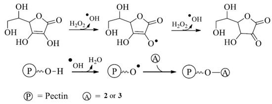
Figure 1.
Schematic representation of the synthesis of the macromolecular conjugate.
The opportunity to graft antioxidant structures in a biomacromolecule by an ecofriendly procedure is an innovative strategy that significantly improves the performance of the natural compounds, opening new applications in the pharmaceutical field and in the food industry. The employment of the redox pair in the synthesis of the grafted biopolymer represents a synthetic strategy that allows for the preservation of antioxidant molecules from the damages related to the high temperatures. Additionally, these compounds avoid the production of toxic products, allowing for the employment of the final product in the food industry [27]. Finally the synthesis of polymeric antioxidants was a special deal able to guarantee a class of compounds having high stability and a slow degradation rate compared to the low-molecular-weight compounds [28].
The activation of the pectin toward radical reactions and the grafting of active species expects that the radical initiators preferably react with the macromolecule, avoiding self reactions. In Figure 1, a probable mechanism of interaction between active species and polysaccharide chains is proposed. Specifically, hydroxyl radicals attack the sensible residues in the side chains of the sugar, producing macroradical species in the sugar chain. The reaction of these radicals with the antioxidant molecules allows for the formation of a covalent bond between the antioxidant and the pectin. Literature data propose that ortho- and parapositions relative to the hydroxyl group are the main target of the free-radical macromolecule chains on the phenolic ring [29]. The heteroatom-centred radicals in the side chains of the pectin preferentially interact in some of the above-mentioned positions. This experimental strategy allowed for the synthesis of two pectin-based conjugates, labelled with the acronyms P_2 and P_3. Additionally, a blank polymer (PB) was also synthesized, with pectin undergoing the same reaction but without any antioxidant molecules. Similarly, pectin–polyphenol conjugates were proposed by Karaki et al. that performed a grafting reaction via laccase catalysis methodology, employing ferulic acid as source on antioxidant moieties [30]. Ferulic acid was also involved in the synthesis of pectin conjugate by Wang et al., which activated reactive sites of ultrasound-treated pectins by vitamin C/hydrogen peroxide redox pair to obtain chain conformations more flexible than those of native sugar [31].
The antioxidant molecules’ inclusion in the backbone of the polysaccharide was verified by measuring the antioxidant activity of the conjugates by the Folin–Ciocalteu test, total antioxidant capacity, and scavenger activity against DPPH and ABTS radicals, and the results are reported in Table 2.
Table 2.
Antioxidant performances of pectin conjugates.
P_2 displayed the greatest antiradical activity: the polyphenolic content was significantly higher than P_3, which recorded a more than 30% increase in total antioxidant capacity. Disposable phenolic groups were in the same order of magnitude of the pectin–ferulic acid conjugates synthetized by employing ultrasound-treated pectins (0.335–0.386 meq of ferulic acid/g) that were able to ensure more flexible chains in respect to the native pectin [31]. Furthermore, between the two polymers, P_2 was the only one able to reach the IC50, while for P_3 only 44.1% of inhibition was observed, at a concentration of 1.3 mg mL−1. Wang et al. also recorded scavenging properties with IC50 values of about 0.5 mg mL−1, while a reduced activity was recorded in the pectin conjugates synthesized by enzyme-catalysed grafting, due to the decrease in free hydroxyl groups that are involved in the covalent linkage. In the hydrophilic environment, against ABTS radicals, both polymers were poorly responsive; P_2 reached an IC30 value, while P_3 showed a 50% lower efficacy. This finding was confirmed by literature data that analysed the antioxidant activity of similar pectin conjugates [30,31]. Finally, PB did not show any activity.
3.2. Toxicity Evaluation
3.2.1. NRU Test
The effect of PB, P_2, and P_3 on 3T3 cells was detected by in vitro NRU assay (Figure 2) The treatment with increasing doses of PB, P_2, and P_3 did not alter cell viability versus control, in all treatments.
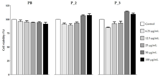
Figure 2.
3T3 cells’ viability (%) after treatment with increased concentrations of PB, P_2, and P_3 (µg mL−1). Each column represents the mean + SD of 3 wells/group.
3.2.2. Cell Viability
As previously reported, quercetin has effects on the cell viability of tumour cell lines [32,33]. In this study, the cellular effects of PB, P_2, and P_3 on cell motility were evaluated by MTT assay. This assay evaluates mitochondrial activity through the formation of the purple formazan in metabolically active cells, an NADP-dependent reaction catalysed by succinate dehydrogenase. This reaction was evaluated by measuring absorbance using a spectrophotometer [34]. Figure 3 shows the treatments with increasing concentrations of PB, P_2, and P_3. The results indicate a dose-dependent reduction in cell viability after treatment with P_2 and P_3. On the contrary, the treatment with PB did not produce any effect.
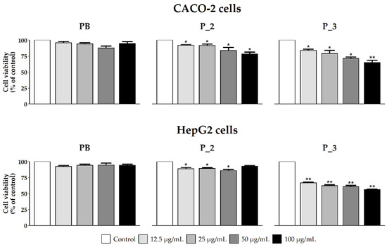
Figure 3.
Effect of PB, P_2, and P_3 on Caco-2 and HepG2 cells’ viability. Each column represents the mean + SD of 3 wells/group. * p < 0.01; ** p < 0.005 treated vs. control.
3.2.3. Cells’ Migratory Capability
Many studies report the effects of quercetin on cell motility in different intestinal cancer cell lines [32,33]. The capacity of PB, P_2, and P_3 to oppose the migratory ability of Caco-2 and HepG2 cells is shown in Figure 4. The results obtained showed that P_2 and P_3 reduce the migration of tumour cells, while PB has no effect.
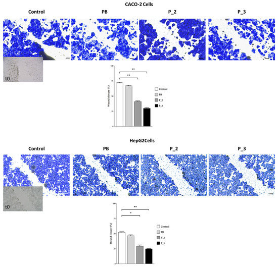
Figure 4.
A wound-healing assay on Caco-2 and HepG2 cells after treatment with of PB, P_2, and P_3 (50 µg mL−1). The percentage of closure was quantified by ImageJ after 24 h. Scale bars: 100 µm, * p < 0.01; ** p < 0.005 treated vs. control.
4. Conclusions
Quercetin derivatives displayed a remarkable antioxidant performance, as well as scavenger activity both in organic and aqueous environments. The inclusion of the synthesized molecules into a polymeric matrix is a useful tool to improve their application in the pharmaceutical and nutraceutical fields. In this regard, pectin was chosen as the polymeric backbone due to its chemical and biological characteristics compatible with our purposes, and an ecofriendly strategy was selected to preserve the biological features of the quercetin derivative. In vitro tests performed on the synthesized polymers highlighted a reduction in the antioxidant activity that was retained. However, our results indicated that quercetin reduced cell viability and migration, as indicated in previous reports [35,36]. These data suggest that quercetin slowed the growth and migration of Caco-2 and Hepg-2 cell lines. These data appear very interesting because they provide a useful alternative aimed to improve the bioavailability of quercetin-derivatives through their covalent binding to the pectin chain, which becomes a very versatile and effective means to transport polyphenol compounds, both in terms of protection of the polyphenol itself and in terms of biological activity manifested in the matrices in which it will be used.
Author Contributions
Conceptualization, U.G.S., G.C., D.R. and F.A.; methodology, G.C., U.G.S. and R.M.; validation, U.G.S., F.A. and R.M.; formal analysis, U.G.S., G.C., V.R. and M.F.M.; investigation, U.G.S., G.C. and R.M.; resources, D.R. and F.A.; data curation, G.C., U.G.S., V.R. and D.L.; writing—original draft preparation, U.G.S., G.C., R.M. and F.A.; writing—review and editing, U.G.S., G.C., V.R., D.L., D.R. and F.A.; visualization, G.C. and V.R.; supervision, D.R. and F.A.; funding acquisition, D.R. and F.A. All authors have read and agreed to the published version of the manuscript.
Funding
This research received no external funding.
Institutional Review Board Statement
Not applicable.
Informed Consent Statement
Not applicable.
Data Availability Statement
Not applicable.
Conflicts of Interest
The authors declare no conflict of interest.
References
- Carullo, G.; Ahmed, A.; Fusi, F.; Sciubba, F.; Di Cocco, M.E.; Restuccia, D.; Spizzirri, U.G.; Saponara, S.; Aiello, F. Vasorelaxant Effects Induced by Red Wine and Pomace Extracts of Magliocco Dolce cv. Pharmaceuticals 2020, 13, 87. [Google Scholar] [CrossRef]
- Carullo, G.; Ahmed, A.; Trezza, A.; Spiga, O.; Brizzi, A.; Saponara, S.; Fusi, F.; Aiello, F. A multitarget semi-synthetic derivative of the flavonoid morin with improved in vitro vasorelaxant activity: Role of CaV1.2 and KCa1.1 channels. Biochem. Pharmacol. 2021, 185, 114429. [Google Scholar] [CrossRef] [PubMed]
- Carullo, G.; Mazzotta, S.; Koch, A.; Hartmann, K.M.; Friedrich, O.; Gilbert, D.F.; Vega-Holm, M.; Schneider-Stock, R.; Aiello, F. New oleoyl hybrids of natural antioxidants: Synthesis and in vitro evaluation as inducers of apoptosis in colorectal cancer cells. Antioxidants 2020, 9, 1077. [Google Scholar] [CrossRef]
- Mazzotta, S.; Governa, P.; Borgonetti, V.; Marcolongo, P.; Nanni, C.; Gamberucci, A.; Manetti, F.; Pessina, F.; Carullo, G.; Brizzi, A.; et al. Pinocembrin and its linolenoyl ester derivative induce wound healing activity in HaCaT cell line potentially involving a GPR120/FFA4 mediated pathway. Bioorg. Chem. 2021, 108, 104657. [Google Scholar] [CrossRef]
- Carullo, G.; Ahmed, A.; Trezza, A.; Spiga, O.; Brizzi, A.; Saponara, S.; Fusi, F.; Aiello, F. Design, synthesis and pharmacological evaluation of ester-based quercetin derivatives as selective vascular KCa1.1 channel stimulators. Bioorg. Chem. 2020, 105, 104404. [Google Scholar] [CrossRef] [PubMed]
- Hoti, G.; Matencio, A.; Pedrazzo, A.R.; Cecone, C.; Appleton, S.L.; Monfared, Y.K.; Caldera, F.; Trotta, F. Nutraceutical Concepts and Dextrin-Based Delivery Systems. Int. J. Mol. Sci. 2022, 23, 4102. [Google Scholar] [CrossRef] [PubMed]
- Panchal, S.K.; Brown, L. Addressing the insufficient availability of epa and dha to meet current and future nutritional demands. Nutrients 2021, 13, 2855. [Google Scholar] [CrossRef] [PubMed]
- Imran, M.; Thabet, H.K.; Alaqel, S.I.; Alzahrani, A.R.; Abida, A.; Alshammari, M.K.; Kamal, M.; Diwan, A.; Asdaq, S.M.B.; Alshehri, S. The Therapeutic and Prophylactic Potential of Quercetin against COVID-19: An Outlook on the Clinical Studies, Inventive Compositions, and Patent Literature. Antioxidants 2022, 11, 876. [Google Scholar] [CrossRef]
- Alesci, A.; Aragona, M.; Cicero, N.; Lauriano, E.R. Can nutraceuticals assist treatment and improve COVID-19 symptoms? Nat. Prod. Res. 2022, 36, 2672–2691. [Google Scholar] [CrossRef] [PubMed]
- Pastor, N.; Collado, M.C.; Manzoni, P. Phytonutrient and nutraceutical action against COVID-19: Current review of characteristics and benefits. Nutrients 2021, 13, 464. [Google Scholar] [CrossRef] [PubMed]
- Azeem, M.; Hanif, M.; Mahmood, K.; Ameer, N.; Chughtai, F.R.S.; Abid, U. An insight into anticancer, antioxidant, antimicrobial, antidiabetic and anti-inflammatory effects of quercetin: A review. Polym. Bull. 2022. [Google Scholar] [CrossRef] [PubMed]
- Barreca, D.; Trombetta, D.; Smeriglio, A.; Mandalari, G.; Romeo, O.; Felice, M.R.; Gattuso, G.; Nabavi, S.M. Food flavonols: Nutraceuticals with complex health benefits and functionalities. Trends Food Sci. Technol. 2021, 117, 194–204. [Google Scholar] [CrossRef]
- Kandemir, K.; Tomas, M.; McClements, D.J.; Capanoglu, E. Recent advances on the improvement of quercetin bioavailability. Trends Food Sci. Technol. 2022, 119, 192–200. [Google Scholar] [CrossRef]
- Boots, A.W.; Haenen, G.R.M.M.; Bast, A. Health effects of quercetin: From antioxidant to nutraceutical. Eur. J. Pharmacol. 2008, 585, 325–337. [Google Scholar] [CrossRef] [PubMed]
- Sriamornsak, P. Application of pectin in oral drug delivery. Expert Opin. Drug Deliv. 2011, 8, 1009–1023. [Google Scholar] [CrossRef] [PubMed]
- Badolato, M.; Carullo, G.; Perri, M.; Cione, E.; Manetti, F.; Di Gioia, M.L.; Brizzi, A.; Caroleo, M.C.; Aiello, F. Quercetin/oleic acid-based G-protein-coupled receptor 40 ligands as new insulin secretion modulators. Future Med. Chem. 2017, 9, 1873–1885. [Google Scholar] [CrossRef]
- Carullo, G.; Perri, M.; Manetti, F.; Aiello, F.; Caroleo, M.C.; Cione, E. Quercetin-3-oleoyl derivatives as new GPR40 agonists: Molecular docking studies and functional evaluation. Bioorg. Med. Chem. Lett. 2019, 29, 1761–1764. [Google Scholar] [CrossRef]
- Spizzirri, U.G.; Abduvakhidov, A.; Caputo, P.; Crupi, P.; Muraglia, M.; Oliviero Rossi, C.; Clodoveo, M.L.; Aiello, F.; Restuccia, D. Kefir Enriched with Carob (Ceratonia siliqua L.) Leaves Extract as a New Ingredient during a Gluten-Free Bread-Making Process. Fermentation 2022, 8, 305. [Google Scholar] [CrossRef]
- Restuccia, D.; Giorgi, G.; Gianfranco Spizzirri, U.; Sciubba, F.; Capuani, G.; Rago, V.; Carullo, G.; Aiello, F. Autochthonous white grape pomaces as bioactive source for functional jams. Int. J. Food Sci. Technol. 2019, 54, 1313–1320. [Google Scholar] [CrossRef]
- Spizzirri, U.G.; Carullo, G.; De Cicco, L.; Crispini, A.; Scarpelli, F.; Restuccia, D.; Aiello, F. Synthesis and characterization of a (+)-catechin and L-(+)-ascorbic acid cocrystal as a new functional ingredient for tea drinks. Heliyon 2019, 5, e02291. [Google Scholar] [CrossRef]
- Carullo, G.; Spizzirri, U.G.; Loizzo, M.R.; Leporini, M.; Sicari, V.; Aiello, F.; Restuccia, D. Valorization of red grape (Vitis vinifera cv. Sangiovese) pomace as functional food ingredient. Ital. J. Food Sci. 2020, 32, 367–385. [Google Scholar] [CrossRef]
- Restuccia, D.; Sicari, V.; Pellicanò, T.M.; Spizzirri, U.G.; Loizzo, M.R. The impact of cultivar on polyphenol and biogenic amine profiles in Calabrian red grapes during winemaking. Food Res. Int. 2017, 102, 303–312. [Google Scholar] [CrossRef]
- Carullo, G.; Scarpelli, F.; Belsito, E.L.; Caputo, P.; Oliviero Rossi, C.; Mincione, A.; Leggio, A.; Crispini, A.; Restuccia, D.; Spizzirri, U.G.; et al. Formulation of New Baking (+)-Catechin Based Leavening Agents: Effects on Rheology, Sensory and Antioxidant Features during Muffin Preparation. Foods 2020, 9, 1569. [Google Scholar] [CrossRef]
- Stokes, W.S.; Casati, S.; Strickland, J.; Paris, M. Neutral Red Uptake Cytotoxicity Tests for Estimating Starting Doses for Acute Oral Toxicity Tests. Curr. Protoc. Toxicol. 2008, 36, 20–24. [Google Scholar] [CrossRef] [PubMed]
- Bossio, S.; Perri, A.; Malivindi, R.; Giordano, F.; Rago, V.; Mirabelli, M.; Salatino, A.; Brunetti, A.; Greco, E.A.; Aversa, A. Oleuropein Counteracts Both the Proliferation and Migration of Intra- and Extragonadal Seminoma Cells. Nutrients 2022, 14, 2323. [Google Scholar] [CrossRef] [PubMed]
- Spizzirri, U.G.; Carullo, G.; Aiello, F.; Paolino, D.; Restuccia, D. Valorisation of olive oil pomace extracts for a functional pear beverage formulation. Int. J. Food Sci. Technol. 2021, 56, 5497–5505. [Google Scholar] [CrossRef]
- Mishra, A.; Clark, J.H.; Vij, A.; Daswal, S. Synthesis of graft copolymers of xyloglucan and acrylonitrile. Polym. Adv. Technol. 2008, 19, 99–104. [Google Scholar] [CrossRef]
- Pan, J.-Q.; Liu, N.C.; Lau, W.W.Y. Preparation and properties of new antioxidants with higher MW. Polym. Degrad. Stab. 1998, 62, 165–170. [Google Scholar] [CrossRef]
- Kitagawa, M.; Tokiwa, Y. Polymerization of vinyl sugar ester using ascorbic acid and hydrogen peroxide as a redox reagent. Carbohydr. Polym. 2006, 64, 218–223. [Google Scholar] [CrossRef]
- Karaki, N.; Aljawish, A.; Muniglia, L.; Humeau, C.; Jasniewski, J. Physico-chemical characterization of pectin grafted with exogenous phenols. Food Hydrocoll. 2016, 60, 486–493. [Google Scholar] [CrossRef]
- Wang, C.; Cai, W.-D.; Yao, J.; Wu, L.-X.; Li, L.; Zhu, J.; Yan, J.-K. Conjugation of ferulic acid onto pectinaffected the physicochemical, functionaland antioxidant properties. J. Sci. Food Agric. 2020, 100, 5352–5362. [Google Scholar] [CrossRef] [PubMed]
- Han, M.; Song, Y.; Zhang, X. Quercetin suppresses the migration and invasion in human colon cancer caco-2 cells through regulating toll-like receptor 4/nuclear factor-kappa B pathway. Pharmacogn. Mag. 2016, 12, 237–244. [Google Scholar] [CrossRef]
- Zhou, J.; Fang, L.; Liao, J.; Li, L.; Yao, W.; Xiong, Z.; Zhou, X. Investigation of the anti-cancer effect of quercetin on HepG2 cells in vivo. PLoS ONE 2017, 12, e0172838. [Google Scholar] [CrossRef] [PubMed]
- Liu, Y.; Nair, M.G. An Efficient and Economical MTT Assay for Determining the Antioxidant Activity of Plant Natural Product Extracts and Pure Compounds. J. Nat. Prod. 2010, 73, 1193–1195. [Google Scholar] [CrossRef] [PubMed]
- Fernández-Palanca, P.; Fondevila, F.; Méndez-Blanco, C.; Tuñón, M.J.; González-Gallego, J.; Mauriz, J.L. Antitumor effects of quercetin in hepatocarcinoma in vitro and in vivo models: A systematic review. Nutrients 2019, 11, 2875. [Google Scholar] [CrossRef] [PubMed]
- Wu, L.; Li, J.; Liu, T.; Li, S.; Feng, J.; Yu, Q.; Zhang, J.; Chen, J.; Zhou, Y.; Ji, J.; et al. Quercetin shows anti-tumor effect in hepatocellular carcinoma LM3 cells by abrogating JAK2/STAT3 signaling pathway. Cancer Med. 2019, 8, 4806–4820. [Google Scholar] [CrossRef] [PubMed]


Publisher’s Note: MDPI stays neutral with regard to jurisdictional claims in published maps and institutional affiliations. |
© 2022 by the authors. Licensee MDPI, Basel, Switzerland. This article is an open access article distributed under the terms and conditions of the Creative Commons Attribution (CC BY) license (https://creativecommons.org/licenses/by/4.0/).