Article

Heart Rate and Oxygen Uptake During Recovery from High-Intensity Interval Training: A Retrospective Analysis

by
Todd A. Astorino
1,*,
Gregory C. Bogdanis
2 and
Eduardo C. Costa
3
1
Department of Kinesiology, California State University—San Marcos, San Marcos, CA 92096, USA
2
School of Physical Education and Sport Science, National and Kapodistrian University, 17237 Athens, Greece
3
ExCE Research Group, Department of Physical Education, Federal University of Rio Grande do Norte, Natal 59078-970, Brazil
*
Author to whom correspondence should be addressed.
Int. J. Environ. Res. Public Health 2025, 22(7), 999; https://doi.org/10.3390/ijerph22070999
Submission received: 18 May 2025 / Revised: 18 June 2025 / Accepted: 23 June 2025 / Published: 25 June 2025
(This article belongs to the Section Exercise and Health-Related Quality of Life)

Abstract

Background: Increases in maximal oxygen uptake (V̇O2max) occur with high-intensity interval training (HIIT), partially due to an extended duration spent at or near maximal V̇O2 or heart rate (HRmax). HIIT induces a delay in HR and V̇O2 during exercise, leading to consistently high HR/V̇O2 values in recovery between intervals. Purpose: This study compared the V̇O2 and HR response between exercise and recovery to various cycling HIIT protocols using data from seven prior studies. Methods: Healthy, active men and women (N = 104, age and V̇O2max 24 ± 5 yr and 40 ± 7 mL/kg/min) underwent HIIT protocols having different durations (30–60 s), intensities (70–85 percent of maximal workload (%Wmax), and recovery periods (10–75 s). V̇O2, HR, and blood lactate concentration (BLa) were assessed. Results: Across studies, peak HR was equal to 90.7 ± 6.2% HRmax. Results showed no significant difference in mean HR (159 ± 14 vs. 160 ± 15 b/min, p = 0.48) or V̇O2 (1.97 ± 0.47 vs. 1.98 ± 0.48 L/min, p = 0.82) between exercise and recovery. Conclusions: These data show elevated V̇O2 and HR during recovery from HIIT, suggesting a substantial, sustained load on the cardiovascular system in recovery from interval exercise.

1. Introduction

The American College of Sports Medicine recommends that all adults achieve 150 min/wk of moderate physical activity or 75 min/wk of vigorous activity to improve health and fitness status [1]. One widely reported adaptation demonstrated in response to regular physical activity is an increase in maximal oxygen uptake (V̇O2max) which is strongly associated with reductions in cardiovascular morbidity and mortality in adults [2,3]. Data from the HERITAGE study showed that 20 wk of moderate-intensity continuous training (MICT) led to a significant increase in V̇O2max in participants diverse in age, sex, ethnicity, and fitness level [4]. This response was accompanied by a significant reduction in resting and exercise heart rate (HR) and significant increase in stroke volume (SV) assessed during submaximal exercise [5], suggesting the incidence of adaptations within the cardiovascular system which enhance oxygen delivery to the working muscle.
Subsequent data in inactive adults demonstrated that higher-intensity MICT at 65–80% V̇O2peak elicits significantly greater increases in V̇O2max versus training at lower intensities (40–55% V̇O2peak) [6]. In addition, exercise training requiring greater time spent at or near V̇O2max or HRmax elicits greater increases in V̇O2max [7], suggesting the importance of vigorous exercise in optimizing the V̇O2max response to exercise training.
In the last three decades, there has been tremendous interest in the efficacy of high-intensity interval training (HIIT) for improving various health- and fitness-related outcomes similarly to MICT [8,9]. HIIT is defined as “intermittent exercise bouts performed above moderate intensity” [10] which elicit 77–95% HRmax. Although infinite permutations of HIIT exist, one widely used protocol consists of ten 1 min intervals, first used by Little et al. [11]. This regimen has been implemented in healthy adults [12] as well as those with chronic disease [13]. Prior data show that the 10 X 1 min protocol elicits peak HR and V̇O2 equal to 88 ± 6% HRmax and 71 ± 6% V̇O2max when intensity is equal to 75% Wmax [14]. Similar intensities (~86% HRmax) occur when this protocol is performed at 90 percent maximal treadmill velocity [15]. Yet, higher intensities are attained when exercise is performed at a higher workload equal to 85% Wmax (94% HRmax and 83% V̇O2max) [16].
It is evident that acute interval exercise places substantial stress on the cardiovascular system, represented by near-maximal values of HR and V̇O2. However, the structure of HIIT, consisting of relatively brief bursts (≤1 min) of exercise, leads to a lag in HR and V̇O2 whose magnitude of increase during each interval is not representative of the absolute exercise intensity. Consequently, the recovery period between short intervals demonstrates the maintenance of or increases in HR and V̇O2 [17], which is in accordance with the maintenance of stroke volume (SV) [16]. Although the acute HR and V̇O2 response to HIIT is well-described, few studies have explored how these variables change during the recovery period or compared recovery values to exercise interval responses. In 10 men, Farias-Junior et al. [18] described the HR, V̇O2, carbon dioxide production (V̇CO2), and minute ventilation (V̇E) responses to exercise and recovery periods during low-volume HIIT performed on a treadmill. Their results showed the maintenance of high V̇O2 (56–80% of the V̇O2 attained during the intervals) during the recovery period. In 11 trained cyclists, Pugh et al. [19] showed elevated V̇O2 in the 4.5 min recovery period following 30 s cycling sprints. It is apparent that the duration, intensity, and mode of recovery affect the energy expenditure and %HR/V̇O2max maintained between exercise intervals and, in turn, participant tolerance to subsequent intervals which, in turn, impacts the quality of the training session.
Due to the paucity of existing data examining responses in the recovery period from HIIT, this retrospective study aimed to compare the V̇O2 and HR response between exercise and recovery in response to various HIIT protocols previously performed in our laboratory by habitually active adults. A secondary aim was to examine correlates of the %V̇O2max response to HIIT to better understand potential variables which are related to training at high relative intensities. These data are useful to clinicians and scientists who implement HIIT protocols with a goal of optimizing the adaptive response to training. For example, if the maintenance of high HR/V̇O2 occurs in the recovery period from HIIT, it is possible that this factor needs to be considered when designing various HIIT regimens. Furthermore, the various benefits associated with HIIT should be explained not only by the near-maximal responses induced by these efforts but also by the high values sustained in the recovery period.

2. Materials and Methods
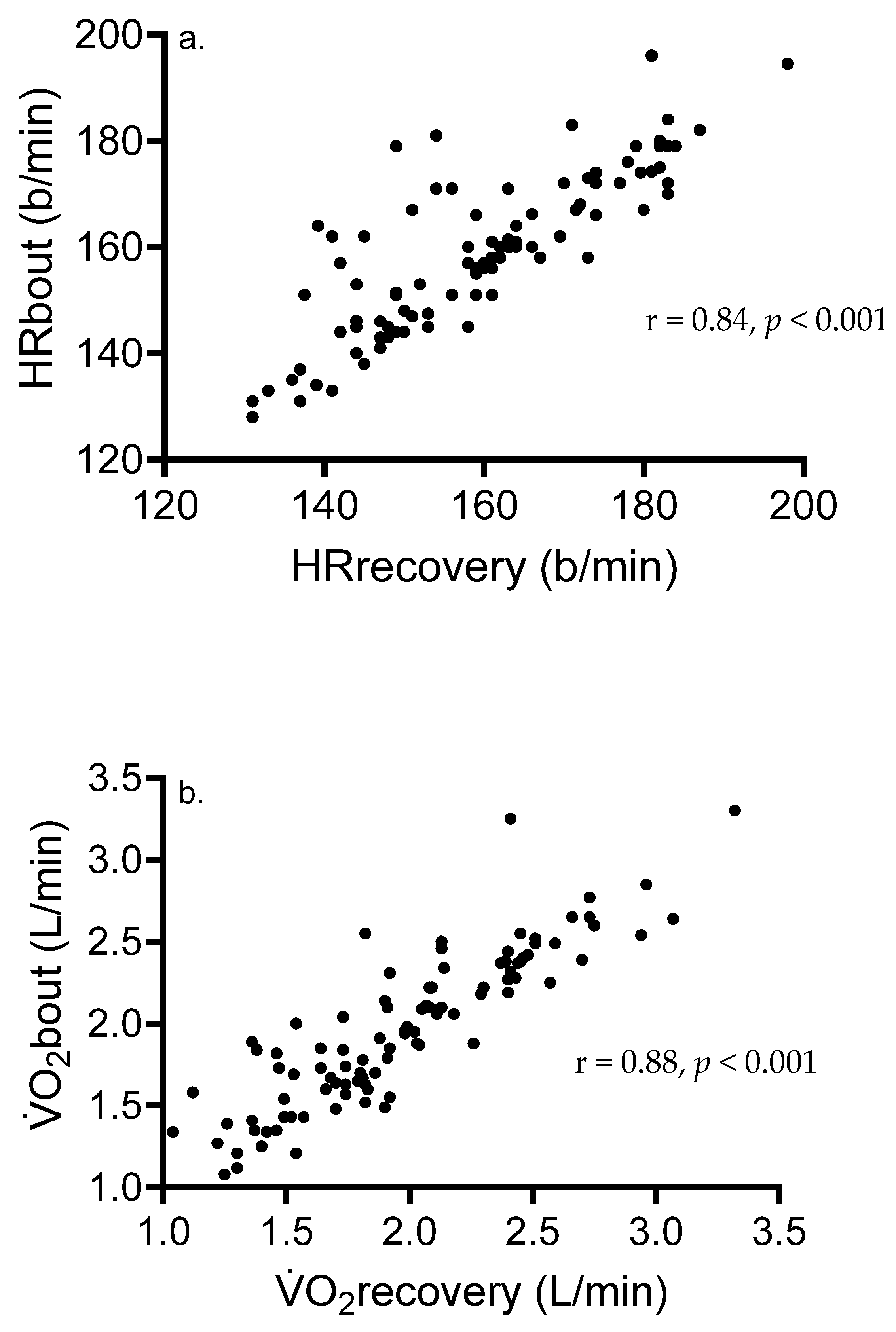
Experimental design and participants: All participants were informed of the procedures in accordance with the protocols and provided written informed consent, with each study approved by the California State University—San Marcos Institutional Review Board. The study was implemented in accordance with the Declaration of Helsinki. The protocol number and date of approval of the three most recent studies are denoted below, yet this information does not exist for the earlier HIIT studies: Astorino et al. (2019) [20] #1282557-1, 26 June 2018; Bogdanis et al. (2021) [21] #1326037-1, 23 October 2018; and Astorino and Emma (2021) [14] #1565370-1, 25 February 2020.
The results presented in this paper were acquired from seven studies using a within-subjects, randomized, crossover design. A total of 104 men and women (age ranging from 18 to 48 yr) underwent various low-volume HIIT protocols. All were physically active for a minimum of 12 mo and participated in non-competitive sport, resistance training, aerobic exercise, or surfing for ≥150 min/wk (duration = 6 ± 2 h/wk). They were non-obese, did not smoke, and were free from any joint issue which would preclude vigorous exercise.
Participants refrained from intense exercise for 36 h before all sessions and did not eat for a minimum of 3 h pre-trial, which were confirmed with a brief survey completed before exercise. Maximal oxygen uptake was initially determined on the cycle ergometer, and resultant values of maximal workload (Wmax) and HRmax were used to prescribe intensities for the HIIT sessions. During exercise, V̇O2, HR, and blood lactate concentration (BLa) were continuously acquired.
Assessment of V̇O2max: During this session, body mass and height were assessed using a scale and stadiometer to calculate body mass index (BMI). Subsequently, participants were prepared for incremental exercise on an electrically braked cycle ergometer (Velotron RacerMate, Quark, Spearfish, SD, USA), during which power output was increased in a ramp-like manner by 20–35 Watt/min after a 2 min warm-up at 40–70 Watt. Different warm-up and work-rate increments were used to accommodate the disparate sexes, body sizes, and fitness levels of our participants to elicit volitional fatigue in approximately 8–10 min. Volitional exhaustion occurred when pedal cadence was below 50 rev/min. Heart rate was determined using telemetry (Polar, Woodbury, NY, USA), and 15 s pulmonary gas exchange data (V̇O2, V̇CO2, V̇E, and RER) were obtained during exercise using a metabolic cart (ParvoMedics True One, Sandy, UT, USA) which was calibrated before testing following manufacturer guidelines. V̇O2max was identified as the mean of the two highest values in the last 60 s of exercise, and V̇O2max attainment was confirmed using the following criteria: change in V̇O2 < 0.15 L/min at V̇O2max; HRmax < 10 beats/minute of 220–age, and respiratory exchange ratio (RER) > 1.10 [22]. HRmax and Wmax were identified as the peak values consistent with exhaustion. Blood lactate concentration (BLa) was assessed 3 min post-exercise using a portable monitor and lancet.
Interval exercise protocols: Table 1 exhibits the characteristics of the HIIT protocols completed. During all protocols, gas exchange data and HR were acquired every 15 s using identical procedures to those described above for the testing of V̇O2max.
Across studies, the interval duration ranged from 0.5 to 1.0 min and there was a 0.16–1.25 min period of active recovery between each exercise interval. Intensity ranged from 70 to 85% Wmax to accommodate the average fitness of our participants. Protocols were low-volume, as the work duration ranged from 6 to 10 min across studies and the number of exercise intervals ranged from 8 to 12. All sessions were performed on an ergometer identical to that used for V̇O2max testing and required an initial warm-up for 4–5 min at low intensity ranging from 10 to 25% Wmax; then the desired %Wmax was selected and the HIIT protocol began. Target workload was fixed during exercise, and participants self-selected a suitable cadence on the cycle ergometer between 50 and 80 rev/min. Recovery intensity was maintained at the same intensity as the warm-up and ranged from 10 to 25% Wmax across studies. Verbal encouragement was provided during exercise.
Assessment of blood lactate concentration: Prior to exercise, after a 5 min seated rest, a 0.7 µL blood sample was taken from a fingertip using a lancet (Owen Mumford Inc., Marietta, GA, USA) and portable monitor (Lactate Plus, Sports Research Group, New Rochelle, NY, USA) to assess BLa. The fingertip was cleaned with a damp towel and dried, and then the first drop of blood was wiped away. This measure was repeated midway during exercise (for example, immediately at the end of bout 5 of the 10 X 1 protocol) and three minutes post-exercise following identical procedures.
Analysis of oxygen uptake, HR, and blood lactate concentration data: The mean V̇O2 and HR were determined as the average value from the entire bout, not including the warm-up, and were calculated from 15 s time-averaged values. V̇O2 and HR from each exercise interval were determined as the mean of the last two 15 s values and first value in the recovery period, with the exception of one study which only allotted a 10 s recovery period between exercise intervals. We used the first value in the recovery period as this approach was used in our prior studies to classify the HR/V̇O2 response to HIIT. The peak value was represented as the highest value from any exercise interval. Recovery V̇O2 and HR were calculated as the mean of the remaining 15 s values prior to the subsequent exercise interval, which included one (10 s recovery), three (1 min recovery), or four (1.25 min recovery) 15 s data points depending upon the duration of the recovery period. These outcomes are expressed in absolute (L/min and b/min) and relative units (%V̇O2max and HRmax). The BLa value reported is the highest value obtained at any point in response to the HIIT session. Energy expenditure (in kcal) was estimated using gas exchange data acquired from the metabolic cart.
Statistical analysis: Data are reported as means and standard deviations (SDs) and were analyzed using SPSS Version 27 (IBM, Armonk, NY, USA). The normality of data distributions was identified using the Shapiro–Wilks test. A two-way ANOVA with repeated measures was used to identify differences in HR and V̇O2, with one within-subjects factor (exercise interval vs. recovery = 2 levels) and one between-subjects factor (study = 7 levels). One-way ANOVA was used to identify differences in BLa across the seven studies. If a significant F ratio occurred, Tukey’s post hoc test was used to identify differences between means. The Greenhouse–Geisser adjustment was used if the sphericity assumption was violated. The dependent t-test was used to examine differences in mean/peak HR and V̇O2 between exercise and recovery. Pearson product moment correlation coefficient was used to identify pairwise relationships between variables. Cohen’s d was used as a measure of effect size, with a small, medium, and large effect equal to 0.2, 0.5, and 0.8, respectively [27]. Statistical significance was set at p < 0.05.

3. Results

Participant physical characteristics: The mean age, body mass, BMI, V̇O2max, HRmax, and Wmax in our participants (60 men and 44 women) were equal to 24 ± 5 yr, 72 ± 13 kg, 24 ± 3 kg/m2, 40 ± 7 mL/kg/min and 2.9 ± 0.7 L/min, 185 ± 10 b/min, and 273 ± 58 W, respectively.
Physiological data: Across all participants, peak intensity was equal to 78.5 ± 10.3% V̇O2max and 90.7 ± 6.2% HRmax, respectively, which reflects the vigorous nature of these HIIT protocols having a unique volume, intensity, number, duration, and recovery. Peak BLa and energy expenditure were equal to 9.9 ± 3.1 mM and 174 ± 46 kcal. Our data show no difference in mean HR (159 ± 14 vs. 160 ± 15 b/min, p = 0.48) or mean V̇O2 (1.97 ± 0.47 vs. 1.98 ± 0.48 L/min, p = 0.82) between interval exercise and the recovery period, respectively. Across studies, the mean exercise interval HR and V̇O2 ranged from 151 to 171 b/min and 1.62 to 2.19 L/min, which are equivalent to 82–91% HRmax and 63–72% V̇O2max (Table 2). Similar values were shown for HR and V̇O2 in the recovery period, equal to 81–92% HRmax and 62–77% V̇O2max. With the exception of one study [24], exercise interval and recovery HR and V̇O2 were similar in all studies.
For HR (b/min), repeated-measures ANOVA showed no main effect of time (exercise interval versus recovery period, p = 0.82, η2p = 0.01), yet there was a significant time X study interaction (p < 0.001, η2p= 0.73). Post hoc analyses showed significant differences in mean HR between various studies (p = 0.014, Table 2). In regard to mean V̇O2 (L/min), no main effect of time was shown (p = 0.68, η2p = 0.03), yet there was a significant time X study interaction (p < 0.001, η2p = 0.60). Significantly lower mean V̇O2 was shown in the Astorino and Emma [14] study compared to Wood et al. (p = 0.006) [26], Reigler et al. (p = 0.002) [25], and Astorino et al. (p = 0.01) [20].
Blood lactate concentration significantly varied across studies (p < 0.001), with values ranging from 6.6 to 13.2 mM (Table 2). The lowest value occurred in response to HIIT having the lowest absolute intensity and duration (70% Wmax and 30 s) [25], whereas the highest values were derived from studies in which self-paced HIIT was performed at 80% Wmax [20,23] and another study using constant intensity equal to 85% Wmax [24].
Correlation data: Significant and strong relationships were shown between mean HRbout and recovery (r = 0.84, p < 0.001) and mean V̇O2bout and recovery (Figure 1). Figure 2 shows associations between various variables and %HR/V̇O2max from all participants. Significant correlates of peak V̇O2 (%V̇O2max) included absolute V̇O2max (r = −0.40, p = 0.004), mean HR in b/min (r = 0.33, p = 0.001), peak HR (%HRmax) (r = 0.61, p < 0.001), mean HRbout (r = 0.48, p < 0.001), and mean HRrecovery (r = 0.20, p = 0.048). For peak V̇O2, there was no association between mean V̇O2 in L/min (r = −0.04, p = 0.71), mean V̇O2bout (r = 0.12, p = 0.23), mean V̇O2recovery (r = −0.12, p = 0.24), or BLa (r = 0.11, p = 0.27). Outcomes significantly associated with peak HR (%HRmax) included absolute V̇O2max (r = −0.42, p < 0.001), meanHRbout (r = 0.65, p < 0.001), meanHRrecovery (r = 0.56, p < 0.001), and BLa (r = 0.30, p = 0.003).

4. Discussion

This retrospective analysis used results from seven studies performed in our laboratory to portray the V̇O2 and HR response during the recovery period from HIIT, which is poorly understood. Prior studies show significant increases in V̇O2max [9,10] and glycemic control [28] as well as reductions in body fat [29] and blood pressure [30] with chronic HIIT. Compared to MICT, HIIT requires a higher intensity which elicits significantly higher HR, V̇O2, and BLa [24], which are thought to be associated with the adaptive response. The results of the present study demonstrate elevated HR and V̇O2 during the recovery period from low-volume HIIT, with these values not significantly different from values induced by the exercise intervals. Overall, a substantial portion of time spent at near-maximal intensity occurs in the recovery period between intervals, and further work is needed to discern whether this contributes to the widely reported long-term adaptive response.
Our results (Table 2) show no significant difference in HR or V̇O2 between the exercise interval and recovery period, with some studies showing slightly higher values during the recovery period versus the exercise interval. In a prior study in young men [17], gas exchange data were acquired during a 4 X 30 s sprint interval training (SIT) session. Results showed that V̇O2 increased from 53 to 72% V̇O2max during the exercise intervals, with significantly elevated values early (10–30 s) in the active recovery period equal to 88–99% V̇O2max. By minute 2 of the recovery period, V̇O2 decreased to approximately 38% V̇O2max, and it further declined to 33% V̇O2max during minutes 3 to 4 of the recovery period. These initial recovery V̇O2 values are substantially higher than those reported in the present study, likely due to the “all-out” nature of SIT requiring dramatically higher power outputs which, in turn, elicit a more prolonged and severe recovery period than HIIT. In addition, V̇O2 was recorded breath by breath, which augments the V̇O2 response compared to longer time-averaging intervals [31]. In a more recent study, Ksoll et al. [32] required 24 men (V̇O2max = 54 mL/kg/min) to complete two work-matched HIIT protocols: five sets of 3 min intervals at 80% V̇O2max followed by a 3 min recovery period, or five sets of six 30 s intervals at 80% V̇O2max followed by a 30 s recovery period. Despite equivalent mean V̇O2 between protocols, 3 min intervals elicited a significantly higher V̇O2 and total duration above 80% V̇O2max than the 30 s intervals, which exhibited significantly higher V̇O2 in the recovery period. In addition, HR and cardiac output (CO) were significantly higher between recovery from 30 s and 3 min intervals. Similar findings were observed by Farias-Junior et al. [18] in comparing HR and gas exchange data during running-based HIIT sessions with different work–recovery durations (10 X 1/1 min vs. 20 X 30/30 s) at 100% maximal velocity interspersed with passive recovery. The 1/1 min HIIT protocol elicited a greater amplitude (i.e., work–recovery differences) in physiological responses compared to the 30/30 s HIIT protocol.
Stanley and Buchheit [33] characterized the V̇O2 and stroke volume (SV) response to 3 X 3 min HIIT at 90% Wmax, interspersed with 2 min of active recovery at 30 or 60% Wmax, in trained cyclists. During the recovery period, results showed the maintenance of relatively high HR (~80 and 85% HRmax at 30 and 60% Wmax) and V̇O2 (~60 and 80% V̇O2max at 30 and 60% Wmax) as well as elevations in SV and CO. This sustained elevation in V̇O2 and hence CO throughout the entire exercise protocol, including the exercise intervals and recovery period, likely potentiates adaptations in slow-twitch muscle fibers and myocardial growth and, in turn, contributes to increases in V̇O2max when completed long-term [34]. Moreover, as acute intensity of exercise has been identified as a key regulator of mitochondrial biogenesis [35], and a maintenance of high V̇O2 in the recovery period, despite a relatively low external power output, may partially elicit these responses. Together, this elevation in V̇O2 and HR in the recovery period may be important to elicit an increase in V̇O2max in response to exercise training.
There are several possible explanations for the relatively high values of V̇O2 and HR in the recovery period reported in the present study. First, there is a marked delay in the V̇O2 response to short exercise intervals; moreover, V̇O2 is significantly lower in response to short versus long intervals at 100% Wmax when intensity and total work are matched [18,36]. Second, there are slower O2 kinetics observed in response to cycling versus running [37]. Third, nonathletic adults, such as those used in the present study, have slower O2 kinetics which may attenuate the V̇O2 gain during each exercise interval and cause V̇O2 to rise early in the recovery period. Fourth, the completion of each exercise interval accelerates O2 kinetics [38] and, combined with the V̇O2 slow component [39], may lead to elevated V̇O2 in the recovery period.
The importance of substantial elevations in V̇O2 and HR in the recovery period between intervals, as shown by our results and others, is relatively unclear due to the paucity of prior results revealing the potential impact of recovery V̇O2 on subsequent changes in health- and fitness-based outcomes. Prior data in trained cyclists [33] showed similar SV values in the recovery period from HIIT to those during the exercise intervals, causing the authors to conclude that part of the circulatory response to HIIT may be due to hemodynamic changes occurring in the recovery period allowing the maintenance of a high CO. In active men and women, Coe and Astorino [15] revealed the maintenance of maximal SV throughout 10 X 1 min and 4 X 4 min HIIT, yet this was not a training study, so no long-term changes in V̇O2max were assessed. In overweight men, Boyd et al. [40] compared changes in V̇O2max and cycling performance in response to nine sessions of HIIT requiring 1 min efforts at 70 or 100% Wmax. Training at 100% Wmax, which would likely elicit higher V̇O2 in each exercise interval and in the recovery period, revealed substantially greater increases in V̇O2max (28 vs. 11%) and cycling performance (14 vs. 9%) versus lower-intensity HIIT. Nevertheless, another study in inactive women [12] showed no significant differences in the V̇O2max response to 12 wk of HIIT at 60–80% or 80–90%Wmax. Further study is needed to monitor HR and V̇O2 in the recovery period from HIIT and identify their potential association with training-induced changes in V̇O2max and exercise performance.
Active recovery is typically prescribed after HIIT to accelerate metabolic recovery (BLa and H+ removal) and maintain a certain level of V̇O2 to expedite the attainment of near-maximal V̇O2 in successive intervals [34,40,41]. Nevertheless, it is apparent that active recovery may reduce exercise tolerance in subsequent intervals [34] despite allowing a substantial duration to be spent at or near V̇O2/HRmax [34]. Overall, the intensity and duration of recovery depend on the interval number, intensity, and duration, and it is likely that the specific characteristics of the recovery period can be manipulated to address different participant goals such as augmenting time at or near V̇O2max, the contribution of nonoxidative metabolism, and energy expenditure. For example, in the Olney et al. [24] study, V̇O2 in the recovery period was substantially lower than the exercise interval value due to the slightly longer recovery period (75 vs. 60 s) employed, which led to a greater decline in V̇O2 compared to the other studies having a shorter recovery period. Yet this longer recovery period was afforded to reduce fatigue and preserve performance, which is important in participants having average V̇O2max, such as the majority of participants enrolled in our studies.
Figure 1 shows strong, significant associations between exercise and recovery HR and V̇O2, supporting our data showing no significant difference in these outcomes. In addition, our results (Figure 2) show significant inverse associations between %V̇O2max and absolute V̇O2max, suggesting that individuals having lower cardiorespiratory fitness tend to exercise at higher fractions of %V̇O2max during HIIT than more-fit adults. Although speculative, the greater training-induced increase in V̇O2max observed in less-fit adults [8,9] may be partially associated with the higher V̇O2 and HR attained during the exercise interval and the recovery period. Nine male participants had V̇O2max ≥ 50 mL/kg/min, and their mean intensity attained during the exercise intervals was equal to 66.5% V̇O2max (57–70% V̇O2max). In contrast, 12 female participants with V̇O2max ≤ 31 mL/kg/min attained intensity equal to 86.8% V̇O2max (75–97% V̇O2max) during HIIT. Participants with lower V̇O2max likely exhibit a lower ratio of slow- to fast-twitch fibers [42] and a ventilatory or lactate threshold at lower relative intensities, in turn requiring a larger contribution of nonoxidative metabolism toward energy expenditure supporting interval exercise. It is recommended that in fit adults, interval protocols requiring intensities well above 85% Wmax, or durations > 1 min, are needed to optimize time spent at higher fractions of %V̇O2max. Another strategy to maximize the V̇O2 response in this population would be to reduce recovery duration and blunt the decline in V̇O2 seen with a longer recovery period.
This study faces several limitations. First, data were combined across all studies despite small differences in HIIT structure, intensity, and recovery, which limits the generalizability of our results. Although %Wmax varied across studies, peak BLa (6.6–13.2 mM) and relative intensity (83–92% HRmax) are representative of near-maximal exercise characteristic of HIIT, suggesting that methodological discrepancies have minimal impact upon our conclusions. The Olney et al. study [24] revealed similar HR and V̇O2 responses between high- (6 X 2 min at 70% Wmax) and low-volume HIIT (8 X 1 min at 85% Wmax), suggesting a similar cardiorespiratory load despite different exercise volumes, intensities, and recovery periods. Second, the results were acquired from young, healthy, habitually active adults, so data cannot be applied to other populations. The mean V̇O2max ranged from 37 to 42 mL/kg/min across studies, which represents average cardiorespiratory fitness for young men and women [43]. The relative similarity in participant V̇O2max across studies suggests a minimal impact of fitness on our results. Third, these results only apply to cycling-based HIIT, and different results may occur when treadmill [21,44] or upper-body interval exercise [14,45] is performed. Fourth, in all studies, we implemented active recovery at low intensities, and different results could occur in response to passive recovery or different modes or intensities of active recovery. Lastly, HR and V̇O2 estimates calculated from each exercise interval included the first value in the recovery period, per methods used in our prior work. Nevertheless, if this data point was not used, it is likely that the exercise interval V̇O2 would have been substantially lower than values acquired in the recovery period. However, this does not change the fact that V̇O2 in the recovery period comprises a substantial portion of the total oxygen consumption and duration spent at or near V̇O2max inherent to HIIT. Yet, this study is strengthened by the large sample of diverse sexes and fitness levels, the use of standardized procedures for the assessment of HR/V̇O2max, and the identical methods used to analyze the data.

5. Conclusions

In conclusion, our results from a large sample of adults undergoing various low-volume HIIT regimens reveal no difference in V̇O2 or HR between the exercise interval and subsequent recovery period. This suggests that the active recovery period between intervals maintains a substantial stress on the cardiovascular system. The impact of this additional cardiovascular load during recovery periods on cardiovascular and/or fitness-related adaptations is unclear and deserves further investigation.

Author Contributions

Conceptualization, T.A.A.; methodology, T.A.A. and G.C.B.; software, T.A.A.; validation, T.A.A., G.C.B. and E.C.C.; formal analysis, T.A.A.; investigation, T.A.A. and G.C.B.; resources, T.A.A. and G.C.B.; data curation, T.A.A. and G.C.B.; writing—original draft preparation, T.A.A.; writing—review and editing, T.A.A., G.C.B. and E.C.C.; visualization, T.A.A.; supervision, T.A.A. and G.C.B.; project administration, T.A.A. and G.C.B. All authors have read and agreed to the published version of the manuscript.

Funding

This research received no external funding.

Institutional Review Board Statement

The studies described in this manuscript were conducted in accordance with the Declaration of Helsinki and approved by the Institutional Review Board of California State University—San Marcos, IRB #1282557-1, 26 June 2018.

Informed Consent Statement

Informed consent was obtained from all subjects involved in the study.

Data Availability Statement

All pertinent data concerning this manuscript is presented here.

Acknowledgments

The authors thank countless individuals for their assistance in collecting the data reported in this manuscript.

Conflicts of Interest

The authors declare no conflicts of interest.

Abbreviations

The following abbreviations are used in this manuscript:
b/minbeats per minute
BLablood lactate concentration
COcardiac output
HIIThigh-intensity interval training
hrhours
HRheart rate
MICTmoderate-intensity continuous training
minminute
RERrespiratory exchange ratio
sseconds
SDstandard deviation
SITsprint interval training
SVstroke volume
V̇CO2volume of carbon dioxide produced
Eventilation
V̇O2volume of oxygen consumed
V̇O2maxmaximal oxygen uptake
wkweeks
Wmaxmaximal workload
yryears

References

  1. Garber, C.E.; Blissmer, B.; Deschenes, M.R.; Deschenes, M.R.; Franklin, B.A.; Lamonte, M.J.; Lee, I.-M.; Nieman, D.C.; Swain, D.P. American College of Sports Medicine position stand. Quantity and quality of exercise for developing and maintaining cardiorespiratory, musculoskeletal, and neuromotor fitness in apparently healthy adults: Guidance for prescribing exercise. Med. Sci. Sports Exerc. 2011, 43, 1334–1359. [Google Scholar] [CrossRef] [PubMed]
  2. Kodama, S.; Saito, K.; Tanaka, S.; Maki, M.; Yachi, Y.; Asumi, M.; Sugawara, A.; Totsuka, K.; Shimano, H.; Ohashi, Y.; et al. Cardiorespiratory fitness as a quantitative predictor of all-cause mortality and car diovascular events in healthy men and women: A meta-analysis. J. Am. Med. Assoc. 2009, 301, 2024–2035. [Google Scholar] [CrossRef]
  3. Ross, R.; Blair, S.N.; Arena, R.; Church, T.S.; Després, J.P.; Franklin, B.A.; Haskell, W.L.; Kaminsky, L.A.; Levine, B.D.; Lavie, C.J.; et al. Importance of assessing cardiorespiratory fitness in clinical practice: A case for fitness as a clinical vital sign: A scientific statement from the American Heart Association. Circulation 2016, 134, e653–e699. [Google Scholar] [CrossRef] [PubMed]
  4. Skinner, J.S.; Jaskólski, A.; Jaskólska, A.; Krasnoff, J.; Gagnon, J.; Leon, A.S.; Rao, D.C.; Wilmore, J.H.; Bouchard, C. Age, sex, race, initial fitness, and response to training: The HERITAGE Family Study. J. Appl. Physiol. 2001, 90, 1770–1776. [Google Scholar] [CrossRef]
  5. Wilmore, J.H.; Stanforth, P.R.; Gagnon, J.; Rice, T.; Mandel, S.; Leon, A.S.; Rao, D.C.; Skinner, J.S.; Bouchard, C. Cardiac output and stroke volume changes with endurance training: The HERITAGE Family Study. Med. Sci. Sports Exerc. 2001, 33, 99–106. [Google Scholar] [CrossRef] [PubMed]
  6. Duscha, B.D.; Slentz, C.A.; Johnson, J.L.; Houmard, J.A.; Bensimhon, D.R.; Knetzger, K.J.; Kraus, W.E. Effects of exercise training amount and intensity on peak oxygen consumption in middle-age men and women at risk for cardiovascular disease. Chest 2005, 128, 2788–2793. [Google Scholar] [CrossRef]
  7. Midgley, A.W.; McNaughton, L.R. Time at or near V̇O2max during continuous and intermittent running. A review with special reference to considerations for the optimisation of training protocols to elicit the longest time at or near V̇O2max. J. Sports Med. Phys. Fit. 2006, 46, 16596093. [Google Scholar]
  8. Milanovic, Z.; Sporis, G.; Weston, M. Effectiveness of high-intensity interval training (HIT) and continuous endurance training for V̇O2max improvements: A systematic review and meta-analysis of controlled trials. Sports Med. 2015, 45, 1469–1481. [Google Scholar] [CrossRef]
  9. Weston, M.; Taylor, K.L.; Batterham, A.M.; Hopkins, W.G. Effects of low-volume high-intensity interval training (HIT) on fitness in adults: A meta-analysis of controlled and non-controlled trials. Sports Med. 2014, 44, 1005–1017. [Google Scholar] [CrossRef]
  10. Coates, A.; Joyner, M.J.; Little, L.P.; Jones, A.M.; Gibala, M.J. A perspective on high-intensity interval training for performance and health. Sports Med. 2023, 53 (Suppl. S1), s85–s96. [Google Scholar] [CrossRef]
  11. Little, J.P.; Safdar, A.; Wilkin, G.P.; Tarnopolsky, M.A.; Gibala, M.J. A practical model of low-volume high-intensity interval training induces mitochondrial biogenesis in human skeletal muscle: Potential mechanisms. J. Physiol. 2010, 588, 1011–1022. [Google Scholar] [CrossRef]
  12. Gosselin, L.E.; Kozlowski, K.F.; Devinney-Boymel, L.; Hambridge, C. Metabolic response of different high-intensity aerobic interval exercise protocols. J. Strength Cond. Res. 2012, 26, 2866–2871. [Google Scholar] [CrossRef]
  13. Little, J.P.; Gillen, J.B.; Percival, M.E.; Safdar, A.; Tarnopolsky, M.A.; Punthakee, Z.; Jung, M.E.; Gibala, M.J. Low-volume high-intensity interval training reduces hyperglycemia and increases muscle mitochondrial capacity in patients with type 2 diabetes. J. Appl. Physiol. 2011, 111, 1554–1560. [Google Scholar] [CrossRef] [PubMed]
  14. Astorino, T.A.; Emma, D. Differences in physiological and perceptual responses to high intensity interval exercise between arm and leg cycling. Front. Physiol. 2021, 12, 700294. [Google Scholar] [CrossRef]
  15. Frazão, D.T.; de Farias Junior, L.F.; Dantas, T.C.B.; Krinski, K.; Elsangedy, H.M.; Prestes, J.; Hardcastle, S.J.; Costa, E.C. Feeling of pleasure to high-intensity interval exercise is dependent of the number of work bouts and physical activity status. PLoS ONE 2016, 11, e0152752. [Google Scholar]
  16. Coe, L.N.; Astorino, T.A. Sex differences in hemodynamic response to high intensity interval exercise. Scand. J. Med. Sci. Sports 2024, 34, e14495. [Google Scholar] [CrossRef] [PubMed]
  17. Hazell, T.J.; Olver, T.D.; Macpherson, R.E.; Hamilton, C.D.; Lemon, P.W. Sprint interval exercise elicits near maximal peak V̇O2 during repeated bouts with a rapid recovery within 2 minutes. J. Sports Med. Phys. Fit. 2014, 54, 750–756. [Google Scholar]
  18. Farias-Junior, L.F.; Dantas Macêdo, G.A.; Vieira Browne, R.A.; Freire, Y.A.; Oliveira-Dantas, F.F.; Schwade, D.; Mortatti, A.L.; Santos, T.M.; Costa, E.C. Physiological and psychological responses during low-volume high-intensity interval training sessions with different work-recovery durations. J. Sports Sci. Med. 2019, 18, 181–190. [Google Scholar]
  19. Pugh, C.F.; Paton, C.D.; Ferguson, R.A.; Driller, M.W.; Beaven, C.M. Acute physiological responses of blood flow restriction between high-intensity interval repetitions in trained cyclists. Eur. J. Sports Sci. 2024, 24, 777–787. [Google Scholar] [CrossRef]
  20. Astorino, T.A.; Sherrick, S.; Mariscal, M.; Jimenez, V.C.; Stetson, K.; Courtney, D. No effect of meal intake on physiological or perceptual responses to self-selected high intensity interval exercise (HIIE). Biol. Sport 2019, 36, 225–231. [Google Scholar] [CrossRef]
  21. Bogdanis, G.C.; Mallios, V.J.; Katsikas, C.; Fouseki, T.; Holman, I.; Smith, C.; Astorino, T.A. Effects of exercise structure and modality on physiological and perceptual responses to exercise. J. Strength Cond. Res. 2021, 35, 2427–2432. [Google Scholar] [CrossRef] [PubMed]
  22. Nolan, P.B.; Beaven, M.K.; Dalleck, L.C. Comparison of intensities and rest periods for V̇O2max verification testing procedures. Int. J. Sports Med. 2014, 35, 1024–1029. [Google Scholar] [CrossRef]
  23. Kellogg, E.; Cantacessi, C.; McNamer, O.; Holmes, H.; von Bargen, R.; Ramirez, R.; Gallagher, D.; Vargas, S.; Santia, B.; Rodriguez, K.; et al. Comparison of psychological and physiological responses to imposed vs. self-selected high-intensity interval training. J. Strength Cond. Res. 2019, 33, 2445–2952. [Google Scholar] [CrossRef] [PubMed]
  24. Olney, N.; Wertz, T.; LaPorta, Z.; Mora, A.; Serbas, J.; Astorino, T.A. Comparison of acute physiological and psychological responses between moderate intensity continuous exercise and three regimes of high intensity training. J. Strength Cond. Res. 2018, 32, 2130–2158. [Google Scholar] [CrossRef]
  25. Riegler, M.; Stotz, G.; Fitzgerald, K.; Munoz, C.K.; Lewis, J.; Ring, S.; Astorino, T.A. Acute responses to the 7-Minute workout. J. Strength Cond. Res. 2017, 31, 2572–2578. [Google Scholar] [CrossRef] [PubMed]
  26. Wood, K.M.; Olive, B.; LaValle, K.; Thompson, H.; Greer, K.; Astorino, T.A. Dissimilar physiological and perceptual responses between sprint interval training and high-intensity interval training. J. Strength Cond. Res. 2016, 30, 234–240. [Google Scholar] [CrossRef]
  27. Cohen, J. A power primer. Psychol. Bull. 1992, 112, 155–159. [Google Scholar] [CrossRef]
  28. Jelleyman, C.; Yates, T.; O’Donovan, G.; Gray, L.J.; King, J.A.; Khunti, K.; Davies, M.J. The effects of high-intensity interval training on glucose regulation and insulin resistance: A meta-analysis. Obes. Rev. 2015, 16, 942–961. [Google Scholar] [CrossRef]
  29. Maillard, F.; Pereira, B.; Boisseau, N. Effect of high-intensity interval training on total, abdominal and visceral fat mass: A meta-analysis. Sports Med. 2018, 48, 269–288. [Google Scholar] [CrossRef]
  30. Carpes, L.; Costa, R.; Schaarschmidt, B.; Reichert, T.; Ferrari, R. High-intensity interval training reduces blood pressure in older adults: A systematic review and meta-analysis. Exp. Gerontol. 2022, 158, 111657. [Google Scholar] [CrossRef]
  31. Robergs, R.A.; Dwyer, D.; Astorino, T.A. Recommendations for improved data processing from expired gas analysis indirect calorimetry. Sports Med. 2010, 40, 95–111. [Google Scholar] [CrossRef] [PubMed]
  32. Ksoll, K.S.H.; Muhlberger, A.; Stocker, F. Central and peripheral oxygen distribution in two different modes of interval training. Metabolites 2021, 11, 790. [Google Scholar] [CrossRef] [PubMed]
  33. Stanley, J.; Buchheit, M. Moderate recovery unnecessary to sustain high stroke volume during interval training. A brief report. J. Sports Sci. Med. 2014, 13, 393–396. [Google Scholar]
  34. Buchheit, M.; Laursen, P.B. High-intensity interval training, solutions to the programming puzzle: Part I: Cardiopulmonary emphasis. Sports Med. 2013, 43, 313–338. [Google Scholar] [CrossRef] [PubMed]
  35. MacInnis, M.J.; Gibala, M.J. Physiological adaptations to interval training and the role of exercise intensity. J. Physiol. 2016, 595, 2915–2930. [Google Scholar] [CrossRef]
  36. Bogdanis, G.C.; Stavrinou, P.S.; Tsirigkakis, S.; Mougios, V.; Astorino, T.A.; Mastorakos, G. Attenuated metabolic and cardiorespiratory responses to isoenergetic high-intensity interval exercise of short versus long bouts. Med. Sci. Sports Exerc. 2022, 52, 1199–1209. [Google Scholar] [CrossRef]
  37. Hill, D.W.; Halcomb, J.N.; Stevens, E.C. Oxygen uptake kinetics during severe intensity running and cycling. Eur. J. Appl. Physiol. 2003, 89, 612–618. [Google Scholar] [CrossRef]
  38. Åstrand, I.; Åstrand, P.; Christensen, E.H.; Hedman, R. Intermittent muscular work. Acta Physiol. Scand. 1960, 48, 448–453. [Google Scholar] [CrossRef]
  39. Dorado, C.; Sanchis-Moysi, J.; Calbet, J.A. Effects of recovery mode on performance, O2 uptake, and O2 deficit during high-intensity intermittent exercise. Can. J. Appl. Physiol. 2004, 29, 227–244. [Google Scholar] [CrossRef]
  40. Boyd, J.C.; Simpson, C.A.; Jung, M.E.; Gurd, B.J. Reducing the intensity and volume of interval training diminishes cardiovascular adaptation but not mitochondrial biogenesis in overweight/obese men. PLoS ONE 2013, 8, e68091. [Google Scholar] [CrossRef]
  41. Belcastro, A.N.; Bonen, A. Lactic acid removal rates during controlled and uncontrolled recovery exercise. J. Appl. Physiol. 1975, 39, 932–936. [Google Scholar] [CrossRef]
  42. Gollnick, P.D.; Armstrong, R.B.; Saubert, C.W.; Piehl, K.; Saltin, B. Enzyme activity and fiber composition in skeletal muscle of untrained and trained men. J. Appl. Physiol. 1972, 33, 312–319. [Google Scholar] [CrossRef] [PubMed]
  43. Kaminsky, L.A.; Imboden, M.T.; Arena, R.; Myers, J. Reference standards for cardiorespiratory fitness measured with cardiopulmonary exercise testing using cycle ergometry: Data from the fitness registry and the importance of exercise national database (FRIEND) registry. Mayo Clin. Proceed. 2017, 92, 228–233. [Google Scholar] [CrossRef] [PubMed]
  44. Neves, L.N.S.; Gasparini-Neto, V.H.; Leite, R.D.; Carletti, L. Acute cardiopulmonary response of high-intensity interval training with elastic resistance vs. high-intensity interval training on a treadmill in healthy adults. Int. J. Environ. Res. Public Health 2023, 20, 6061. [Google Scholar] [CrossRef] [PubMed]
  45. Zinner, C.; Morales-Alamo, D.; Ørtenblad, N.; Larsen, F.J.; Schiffer, T.A.; Willis, S.J.; Gelabert-Rebato, M.; Perez-Valera, M.; Boushel, R.; Calbet, J.A.; et al. The physiological mechanisms of performance enhancement with sprint interval training differ between the upper and lower extremities in humans. Front. Physiol. 2016, 7, 426. [Google Scholar] [CrossRef] [PubMed]
Figure 1. Associations between (a) mean HRbout and mean HR in the recovery period and (b) mean V̇O2bout and mean V̇O2 in the recovery period.
Ijerph 22 00999 g001
Figure 2. Associations between (a) peak V̇O2 (%V̇O2max) and V̇O2max, r = −0.40, p = 0.004; (b) peak V̇O2 (%V̇O2max) and mean HR, r = 0.33, p = 0.001; (c) peak V̇O2 (%V̇O2max) and peak HR (%HRmax), r = 0.61, p < 0.001; (d) peak V̇O2 (%V̇O2max) and mean HRbout, r = 0.48, p < 0.001; and (e) peak V̇O2 (%V̇O2max) and mean HRrecovery, r = 0.20, p = 0.048.
Ijerph 22 00999 g002
Table 1. High-intensity interval training protocols completed in the present study.
StudySubjects and Age (yr)V̇O2max (mL/kg/min)HRmax (b/min)HIIT ProtocolIntensityRecovery Period (min)Recovery Period (%Wmax)
Astorino and Emma [14]23 M/W
25 ± 6
37 ± 6185 ± 1210 X 1 min75% Wmax1.010
Astorino et al. [20]17 M/W
26 ± 6
39 ± 4186 ± 810 X 1 min~79% Wmax1.010
Bogdanis et al. [21]5 M/W
23 ± 4
40 ± 8183 ± 310 X 1 minVT + 20% 1.020
Kellogg et al. [23]14 M/W
24 ± 3
42 ± 9188 ± 88 X 1 min80% Wmax1.010
Olney et al. [24]19 M/W
24 ± 3
40 ± 6188 ± 88 X 1 min85% Wmax1.2520
Reigler et al. [25]14 M/W
25 ± 8
40 ± 6185 ± 1212 X 30 s70% Wmax0.1620
Wood et al. [26]12 M/W
24 ± 6
41 ± 4179 ± 108 X 1 min85% Wmax1.025
M = men; W = women; V̇O2max = maximal oxygen uptake; HR = heart rate; b/min = beats per minute; %Wmax = percent maximal workload; min = minute; s = seconds.
Table 2. Mean heart rate (HR), oxygen uptake (V̇O2), and blood lactate response to interval exercise and recovery (mean ± SD and minimum to maximum value).
StudyHRmeanbout (b/min)HRmeanrecovery (b/min)V̇O2meanbout (L/min)V̇O2meanrecovery
(L/min)
BLa (mM)
Astorino and Emma [14]151 ± 14155 ± 141.62 ± 0.321.66 ± 0.287.6 ± 2.7
128–172131–1831.27–2.471.22–2.183.6–12.5
Astorino et al. [20]165 ± 14 a171 ± 14 a2.01 ± 0.41 a2.17 ± 0.47 a13.2 ± 1.7 a
141–195147–1981.43–2.651.47–3.079.8–15.6
Bogdanis et al. [21]166 ± 10168 ± 102.12 ± 0.492.19 ± 0.529.2 ± 2.9
151–174156–1811.54–2.651.49–2.735.0–12.3
Kellogg et al. [23]157 ± 17163 ± 171.90 ± 0.502.05 ± 0.4711.3 ± 3.2 a
131–179133–1841.08–2.551.25–2.706.8–15.2
Olney et al. [24]171 ± 12153 ± 132.10 ± 0.501.75 ± 0.419.8 ± 3.1 b
151–196137–1811.34–3.251.04–2.413.8–15.3
Reigler et al. [25]159 ± 15160 ± 152.19 ± 0.44 a2.23 ± 0.44 a6.6 ± 2.1 b
137–182137–1871.43–2.851.57–2.963.6–9.7
Wood et al. [26]154 ± 9153 ± 92.19 ± 0.40 a2.17 ± 0.42 a11.3 ± 3.3 a
144–164144–1701.70–3.301.64–3.326.7–17.0
HR = heart rate; V̇O2 = oxygen uptake; BLa = blood lactate concentration; a = p < 0.05 vs. Astorino et al. (2021) [14]; b = p < 0.05 vs. Astorino et al. (2019) [20].
Disclaimer/Publisher’s Note: The statements, opinions and data contained in all publications are solely those of the individual author(s) and contributor(s) and not of MDPI and/or the editor(s). MDPI and/or the editor(s) disclaim responsibility for any injury to people or property resulting from any ideas, methods, instructions or products referred to in the content.

