Article

Metabolite Formation by Fungal Pathogens of Potatoes (Solanum tuberosum L.) in the Presence of Bioprotective Agents

by
Aleksandra Steglińska
1,2,*,
Michael Sulyok
3,
Regina Janas
4,
Mieczysław Grzesik
4,
Wiktoria Liszkowska
1,
Dorota Kręgiel
1 and
Beata Gutarowska
1
1
Department of Environmental Biotechnology, Lodz University of Technology, Wólczańska 171/173, 90-950 Łódź, Poland
2
Interdisciplinary Doctoral School, Lodz University of Technology, Żeromskiego 116, 90-924 Łódź, Poland
3
Department of Agrobiotechnology (IFA-Tulln), Institute of Bioanalytics and Agro-Metabolomics, University of Natural Resources and Life Sciences, Vienna, Konrad-Lorenz-Strasse 20, 3430 Tulln, Austria
4
The National Institute of Horticultural Research, Konstytucji 3 Maja 1/3, 96-100 Skierniewice, Poland
*
Author to whom correspondence should be addressed.
Int. J. Environ. Res. Public Health 2023, 20(6), 5221; https://doi.org/10.3390/ijerph20065221
Submission received: 7 February 2023 / Revised: 9 March 2023 / Accepted: 14 March 2023 / Published: 22 March 2023
(This article belongs to the Special Issue Microbiological Contamination and Food Safety)

Abstract

:
The potato is a crop of global importance for the food industry. This is why effective protection against pathogens is so important. Fungi as potato pathogens are responsible for plant diseases and a significant reduction in yields, as well as for the formation of mycotoxins. This study focuses on the effect of three natural biocides, yeast Metschnikowia pulcherrima, lactic acid bacteria Lactiplantibacillus plantarum, and aqueous garlic extract, on the improvement of the physiology of planted potato tubers and the reduction in mycotoxin formation. The secondary metabolites produced by the fungal pathogens of genera Fusarium, Alternaria, Colletotrichum, Rhizoctonia, and Phoma in the presence of these biocontrol agents were compared to profiles obtained from contaminated potatoes. Analysis of liquid chromatography coupled with tandem mass spectrometry data showed the presence of 68 secondary metabolites, including the mycotoxins: alternariol, alternariol methyl ether, altertoxin-I, aurofusarin, beauvericin, diacetoxyscirpenol, enniatin B, and sterigmatocystin. The studies showed that the applied biocontrol agents had a positive effect on the physiological parameters of potatoes (including root growth, stem growth, gas exchange, and chlorophyll content index) and on the reduction in the production of mycotoxins and other secondary metabolites by Fusarium, Alternaria, and Phoma.

1. Introduction

Potato (Solanum tuberosum L.) tubers are the world’s fourth most-consumed crop, cultivated in 160 countries, with 322 Mt of yield annually [1]. Poland, Germany, France, Romania, the Netherlands, and Belgium are the leading countries in potato production in the European Union with 55.3 Mt of tubers produced in total [2]. Crop pathogenic microorganisms are responsible for diseases, which cause reductions in yield and the quality of potatoes (worldwide loss estimate is 17.2%). This presents a great economic risk and a threat to global food security [3]. The most common fungal pathogens of potato tubers include the Alternaria spp., the Fusarium spp., Phoma exigua, Helminthosporium solani, Colletotrichum coccodes, Rhizoctonia solani, and Phytophthora infestans [4]. Some of them are able to produce mycotoxins [5].
Mycotoxins are low-molecular-weight toxic secondary metabolites (the majority are <700 Da) produced by certain fungi (Fusarium, Alternaria, Penicillium, Aspergillus, and other species), naturally present in foodstuffs of plant origin such as seeds, fodder, cereal, fruit, and hazelnuts. More than 400 mycotoxins have already been identified, some of which can be produced by more than one fungus genus. Certain species of fungi are able to synthesise several mycotoxins [6]. The most common mycotoxins include aflatoxins (AFB1, AFB2, AFG1, AFG2, and AM1), ochratoxin A (OTA), zearalenone (ZEN), deoxynivalenol (DON), T-2 and HT-2 toxins, patulin (PAT), and fumonisins (FB1 and FB2) [7]. Mycotoxins produced by fungi can be detrimental to both humans and animals with carcinogenic, neurotoxic, hepatotoxic, and teratogenic effects [8,9]. In European Union countries, Commission Regulation (EC) No. 1881/2006 of 19 December 2006 determines the maximum permissible mycotoxin level in certain commodity groups [10]. There are only few studies which have described mycotoxin occurrence in potato plants, mainly concerning the presence of mycotoxins produced by the Fusarium spp. [11,12,13]. There is a lack of research on the production of mycotoxins on potatoes by genera of other fungi. Most available research focuses on assessing mycotoxin contamination in maize, wheat, soybean, rice, and barley [7,14].
Most mycotoxins are heat-stable and difficult to remove from contaminated products. The threat to human and animal health is associated with their high ability to bioaccumulate in crop plants [8]. Mycotoxins occur mainly in agricultural fields, from sowing to harvesting; however, they can also be produced during inappropriate storage conditions [15]. Potato storage conditions, such as relative humidity (80–90%), temperature, long storage time (up to 8 months), and soil contamination are favourable to mould growth [16].
An effective way to reduce mycotoxin contamination in seed potatoes at an early stage is to prevent the development of mycotoxin-producing fungi. This can be achieved through various biological approaches, e.g., the use of microorganisms and their metabolites, plant extracts, or combined methods with certain synthetic compounds [8]. In our previous research, we described the inhibition of phytopathogen growth using three biological control agents: lactic acid bacteria [17], yeast Metschnikowia pulcherrima [18], and garlic water extract [19].
The potential application of lactic acid bacteria (LAB) as a biological control agent against plant pathogens has been previously described for several pathogens ranging from the Fusarium, Alternaria, Colletotrichum, and Rhizoctonia to the Phoma species [17,20,21,22]. Lactic and acetic acid, hydrogen peroxidase, cyclic dipeptides, phenyllactic acid, and 3-hydroxy fatty acids are mainly responsible for the antimicrobial activities of LAB [23,24,25]. In addition to direct antifungal activity, some LAB strains have demonstrated the ability to convert already produced mycotoxins into less or nontoxic compounds [4,6] or to remove them entirely by binding to LAB cell walls, both living and dead. The mechanism of mycotoxin removal is based on the adsorption abilities linked to the presence of peptidoglycans, proteins, and polysaccharides in LAB cell walls [6,26].
Yeast Metschnikowia pulcherrima is another biological control agent that we used for potato protection against mycotoxins produced by fungi. These yeasts are the natural microbiota of fruit, fruit juices, flowers (nectar), and flower insects [27,28]. Their main antimicrobial activity is connected to the secretion of pulcherriminic acid that forms pulcherrimin—a red complex with iron ions. This nonenzymatic reaction removes iron, an essential element for pathogen growth [29]. The published data provide information about the antifungal activity of yeast M. pulcherrima against potato pathogens from the Fusarium, Alternaria, Rhizoctonia, Phoma, and Colletotrichum species [18,30,31]. The composition of the culture medium and the conditions for the cultivation of M. pulcherrima yeasts and selected lactic acid bacteria used as biopreservatives were developed in earlier studies conducted by Steglińska et al. [17,18,32,33].
In this work, we evaluated the effect of aqueous garlic extract as a plant biopesticide against mycotoxin-producing fungi that infect potatoes. Several works confirmed the antifungal activity of garlic extracts on Alternaria, Rhizoctonia, Fusarium, Colletotrichum, and Phoma fungi [19,34,35,36]. The antimicrobial activity of garlic extracts is linked to the production of organosulphur compounds such as allicin, ajoenes, diallyl disulphide, methyl allyl disulphide, and others [37].
Due to the global production of potatoes, the climatic change favouring the development of potato phytopathogens, the high degree of infestation during storage, and the withdrawal of many pesticides from the market due to their accumulative and toxic abilities, new ecological methods of protection need to be developed. Therefore, selected natural active ingredients of biopreparations based on microorganisms and plants may be a solution. Their high effectiveness in inhibiting mould growth was proven in earlier studies [17,18,19]. There are no data, however, considering the effect of biopreparations on the formation of mycotoxins by moulds on potato tubers, as well as on the growth and physiological activity of potatoes. Therefore, the aim of this research was to evaluate the profiles of secondary metabolites in potato seeds in response to different fungal phytopathogens. In addition, the alteration of these profiles in the presence of selected biocontrol agents of microbial and plant origin was evaluated. Hence, we assessed the possibility of using M. pulcherrima yeast, lactic acid bacteria, and aqueous garlic extract as preservatives for potatoes. Additionally, the impact of the use of these biopreparations on the growth and physiological activities of potatoes was assessed.

2. Materials and Methods

2.1. Biological Material Used in the Research

2.1.1. Plant Material

The experiments were performed on the Impresja variant of potato tubers (Solanum tuberosum L.). This is an early potato, widespread in Poland with potential for cultivation in different countries. Potatoes were obtained from the Hodowla Ziemniaka Zamarte Sp. z o. o. (IHAR Group, Kamieńsk Krajeński, Poland; 53°35′58″ N, 17°28′54″ E).

2.1.2. Potato Phytopathogens

Five fungal potato phytopathogens, Fusarium sambucinum DSM 62397, Alternaria tenuissima DSM 63360, Colletotrichum coccodes DSM 62126, Phoma exigua DSM 62040, and Rhizoctonia solani DSM 22843, were used in the experiments. The strains were purchased from the German Collection of Microorganisms and Cell Cultures GmbH (DSMZ, Braunschweig, Germany). All strains were activated on Potato Dextrose Agar (PDA; Merck, Darmstadt, Germany) and stored at 4 °C. Pathogen suspensions were prepared from the pure cultures on the PDA agar plates and adjusted to a final concentration of 106 CFU/mL in saline solution (0.85% NaCl).
For fungal metabolite analysis (Section 2.3), samples of fungal cultures on Malt Extract Agar (MEA; Merck, Darmstadt, Germany) plates and potatoes infected with phytopathogens were prepared.

2.1.3. Lactic Acid Bacteria Biopreparation (LpB)

The bacterial strain Lactiplantibacillus plantarum KB2 LAB 03 isolated from sauerkraut was used to obtain bacterial preparations against potato phytopathogens with a previously described spectrum of activity [17]. Lactic acid bacteria were identified using molecular methods based on sequencing of the 16S rRNA gene fragment. Genomic DNA was isolated using the Genomic Mini kit (A&A Biotechnology, Poland), according to the methodology provided by the manufacturer. The reaction mixture was prepared in a volume of 50 µL containing 25 µL of REDTaq™ ReadyMix™ (Sigma, Saint Louis, MO, USA) polymerase (1.5 units), 0.4 µL of each primer solution (at a concentration of 100 µM), 25 µL of water, and 20 ng of template DNA. The 16S rRNA gene was amplified by PCR in the MJ Mini Gradient Thermal Cycler (Bio-Rad, Hercules, CA, USA) using the universal primers 5′–AGAGTTTGATCCTGGCTCAGGA–3′ (forward) and 5′–GGAGGTGATCCAGCGGC–3′ (reverse). The cycle consisted of pre-denaturation at 94 °C for 2 min, denaturation at 94 °C for 1 min, primer annealing at 50 °C for 1 min (34 repetitions), elongation at 72 °C for 3 min, and final elongation at 72 °C for 3 min. The obtained PCR products were analysed by 1% (w/v) agarose gel electrophoresis. The amplified PCR products were purified using the Clean-Up AX kit (A&A Biotechnology, Gdansk, Poland) and then subjected to a sequencing reaction at an external company—GENOMED S.A. (Warsaw).
The strain was activated and then cultured for further experiments on an acid–whey-based medium supplemented with 0.8% yeast extract (BTL, Łódź, Poland), 1.4% peptone K, 0.2% ammonium citrate (Chempur, Poland), 0.2% dipotassium phosphate (Chempur, Poland), 0.5% sodium acetate (Chempur, Poland), 0.02% magnesium sulphate heptahydrate (Chempur, Poland), and 0.005% magnesium sulphate tetrahydrate (Chempur, Poland) for 48 h at 30 °C [17]. The acid–whey was obtained from JOGO–Łódź Dairy Cooperative (Kraszewo, Poland).

2.1.4. Metschnikowia Pulcherrima Yeast Biopreparation (MpB)

The strain Metschnikowia pulcherrima TK1 isolated from strawberry flowers was used to obtain yeast preparations against potato phytopathogens with a previously described spectrum of activity [18]. A pure culture of yeast was identified by MALDI-TOF MS analysis, described in detail in Steglińska et al. [18]. Briefly, a 24 h culture of yeast cultured on an MEA plate (Merck, Darmstadt, Germany) was analysed with the AXIMA-iD Plus Confidence MALDI-TOF MS system (Kratos Analytical Ltd. and Shimadzu Corporation, Kyoto, Japan) and SARAMIS PREMIUM software (Spectral Archive And Microbial Identification System, bioMérieux, Marcy l’Étoile, France). The direct formic acid smear method was used. The yeast colony was spread on an analytical plate with a sterile loop. After that, 0.5 µL of 25% formic acid was spotted onto cells, mixed, and left until almost dry. Then, 1 µL of saturated α-cyano-4-hydroxycinnamic acid solution in an acetonitrile, ethanol, and water (1:1:1; v/v) mixture containing 3% trifluoroacetic acid was added onto the yeast cells, mixed, and dried. The mass spectra were obtained and processed using Launchpad 2.9 software (Kratos Analytical Ltd. and Shimadzu Corporation, Kyoto, Japan) in the SARAMIS linear positive mode, with a laser frequency of 50 Hz in a mass-to-charge ratio (m/z) range from 2000 to 20,000 Da (laser power: 90; 200 per sample; five shots accumulated per profile) for each mass spectrum. E. coli DH5 (TAKARA BIO INC.) cells were used as a calibrator of the AXIMA-iD Plus Confidence MALDI-TOF MS system and internal control of the identification process, in accordance with the recommendations of the manufacturer.
The strain was activated and then cultured for further experiments on an acid–whey-based medium supplemented with 0.13% yeast extract, 0.25% peptone, and 1% glucose for 72 h at 25 °C on a shaker (Unimax 1010, Heidolph, Germany) at 160 rpm [18]. The acid–whey was obtained from JOGO–Łódź Dairy Cooperative (Kraszewo, Poland).

2.1.5. Aqueous Garlic Extract Biopreparation (GB)

Garlic (Allium sativum L.) was obtained from Dary Natury Sp. z o. o., Grodzisk, Poland. The aqueous plant extract was prepared by pouring 50 g of finely ground material in 500 mL of water at 100 °C, which was left covered without stirring for 1 h. After that, the garlic extract was sonicated (40 kHz, 25 °C, 30 min) and filtered under reduced pressure [19].

2.2. Sample Inoculation with Phytopathogens and Application of Bioprotective Agents

“Impresja” seed potatoes were rinsed in sterile distilled water and left to dry in air. Then, 5 mm wide and 5 mm diameter cuts were made into each potato with a cork bore. Potato biopreparations were made according to Section 2.1.3, Section 2.1.4 and Section 2.1.5. Samples were immersed in LpB, MpB, and GB for 5 min and then left to dry. After that, 20 µL of each fungal suspension was applied to the cuts. Control potatoes were only inoculated with pathogen suspensions. Samples were incubated for 14 days at a temperature of 25 °C.

2.3. Fungal Metabolite Analysis by LC–MS/MS

For fungal metabolite analysis, plugs were cut from the investigated potatoes and MEA plates with a sterile cork bore, and then analysed according to the validated method described in detail by Sulyok et al. [38] for LC–MS/MS analysis of >500 mycotoxins and other secondary metabolites in food crops. Samples weighing 1–3 g were extracted with 5 or 10 mL (depending on the weight of the sample) by extraction solvent (acetonitrile/water/formic acid 79:20:1, v/v/v) for 90 min on a GFL rotary shaker (GFL, Burgwedel, Germany). After extraction, aliquots of the prepared extracts were diluted 1:1 using a dilution solvent (acetonitrile/water/formic acid 20:79:1, v/v/v). Then, 5 µL of the diluted extracts were injected into the column of the liquid chromatography apparatus, Agilent 1290 Series HPLC System (Agilent Technologies, Waldbronn, Germany), coupled with a QTrap 5500 m (Sciex, Foster City, CA, USA) equipped with a Turbo Ion Spray ESI source. The compounds were separated using a Gemini® C18 column, 150 × 4.6 mm i.d., 5 μm particle size from Phenomenex (Torrance, CA, USA) protected by a C18 security guard cartridge, 4 × 3 mm i.d. (Phenomenex, Torrance, CA, USA) at 25 °C. The eluent was a methanol/water gradient containing 1% acetic acid and 5 mM ammonium acetate. The elution rate was 1 mL/min. Scheduled multiple reaction monitoring (sMRM) in both negative and positive mode after two separate chromatographic runs per sample was performed for data acquisition. Confirmation of the identity of the metabolites was obtained through the comparison of retention time and intensity ratio to a multianalyte standard (SANTE/12089/2016). External quantification was performed on the basis of the linear calibration curves from serial dilutions of a multianalyte standard. The accuracy of the method is verified on a routine basis by participation in a proficiency testing scheme organised by BIPEA (Genneviliers, France). Currently, the rate of satisfactory results of −2 < z < 2 is 95.6% with >1900 results submitted. Limits of detection (LOD) and limits of quantification (LOQ) are presented in Table S2.

2.4. Growth and Physiological Parameter Analysis

2.4.1. Methods of Plant Cultivation

For greenhouse experiments, a mix of phytopathogen suspensions was used to inoculate potatoes after biopreparation treatment. After incubation, potatoes were subjected to negative selection and cultivated in a ventilated greenhouse (20 °C), in modified plant microcosms with dimensions 30 × 40 × 10 cm (height × width × depth, respectively). The front walls of plant microcosms were made from transparent glass, which enabled constant observation of tuber germination, root growth and quality, and the dynamics of stem growth [39]. The microcosms were filled with 10 L of standard horticultural substrate mixed with a universal complete fertiliser containing macro- and microelements (YaraMila Complex; Yara) at a dose of 2 kg/m3. The plants were watered with tap water as needed throughout the growing season [40,41].

2.4.2. Assessment of Growth and Physiological Activity Parameters

After potatoes were planted in microcosms, the growth and physiological activity of the plants were checked every 2 days for the first 24 days, and every 6 days for the remaining period [41]. The following parameters were measured:
  • Biological condition of the seed potatoes—their vigour, turgor, rotting, and infection by phytopathogens [42];
  • The percentage of germinating tubers [42];
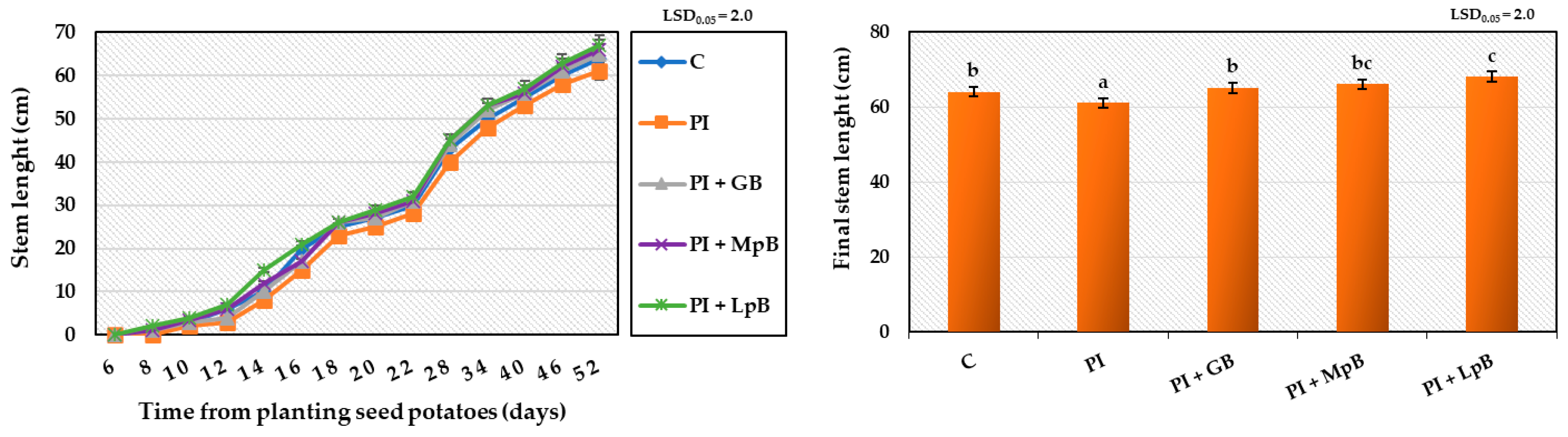
  • Growth kinetics of the plant, by measuring the length of shoots every 2 days during the first 24 days, and every 6 days for the remaining period [43];
  • Quality of the shoots on a five-point valuation scale, where 5 indicates shoots growing well, full of vigour, while 1 indicates dried out shoots [44];
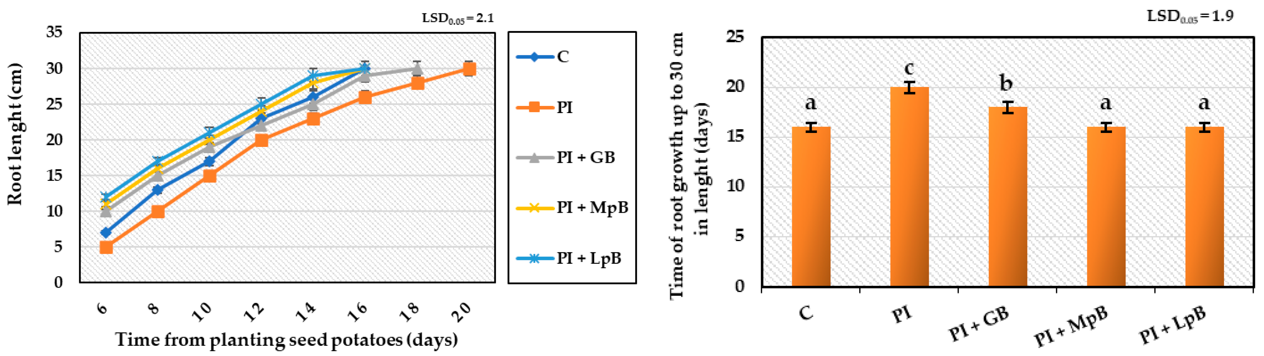
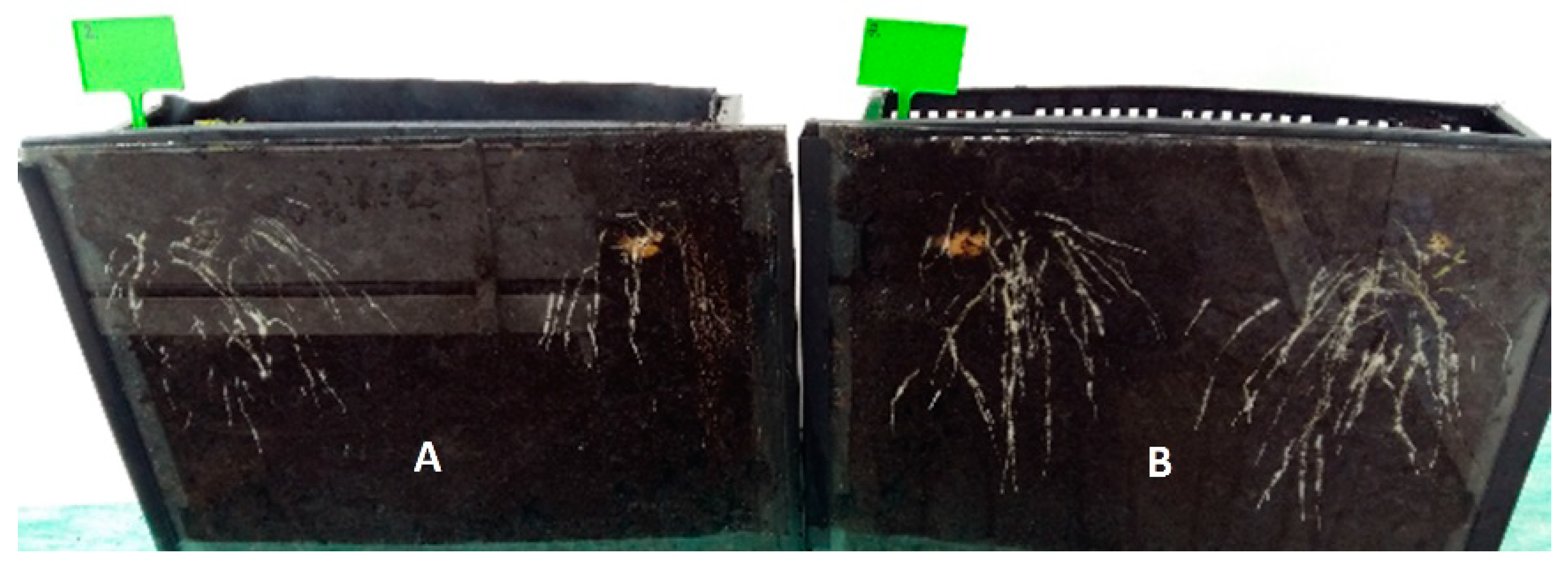
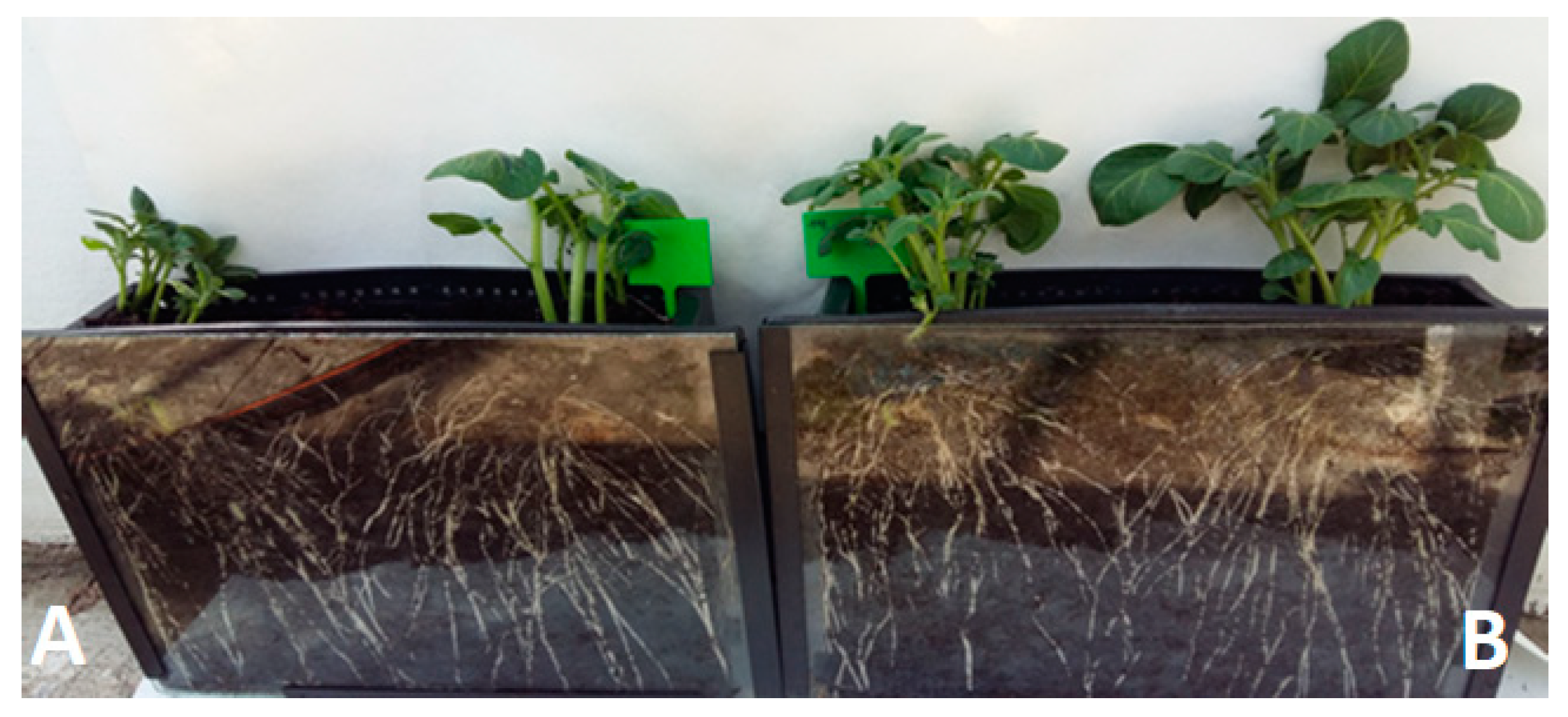
  • Quality and growth kinetics of root, by measuring root length every 2 days until they reached 30 cm [42];
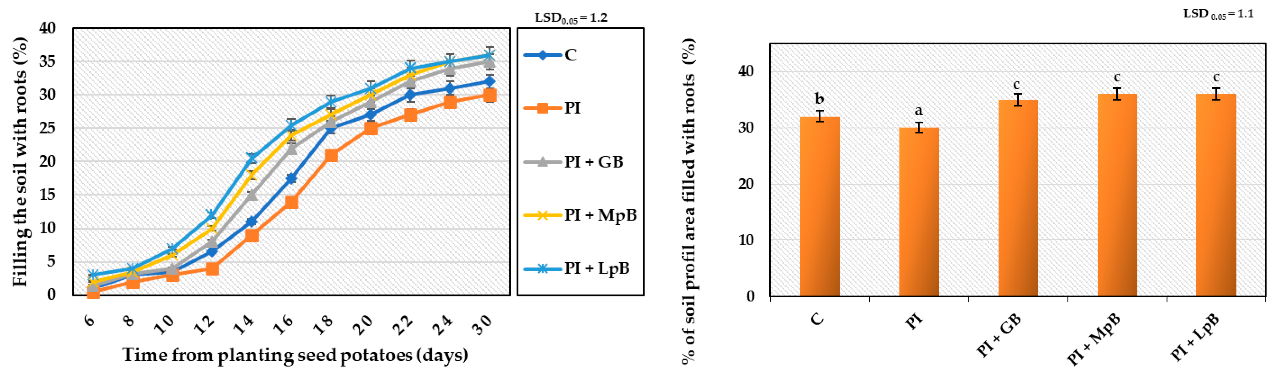
  • Growth kinetics of the root system, by assessing every 2 days the percentage of the soil profile area filled by the roots [45];
  • Quality of plants on a five-point rating scale, where 5 indicates plants with high vigour, coloured and growing properly, and 1 indicates dried out plants [8];
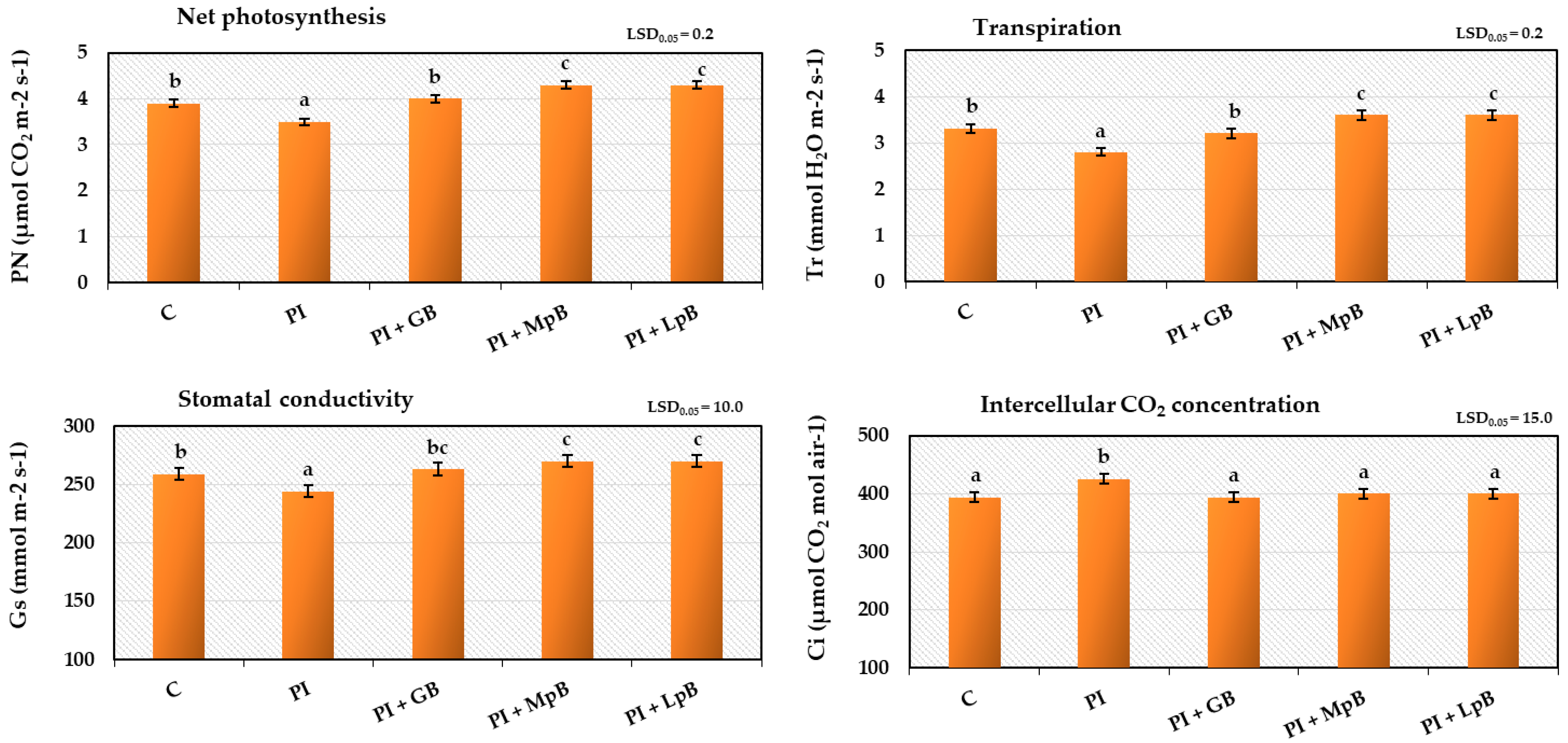
  • Intensity of gas exchange (transpiration, net photosynthesis, intercellular CO2 content, and stomata conductivity), measured with a TPS-2 apparatus (PP Systems, USA) in the highest positioned fully developed leaves, during the period of most dynamic plant growth [39];
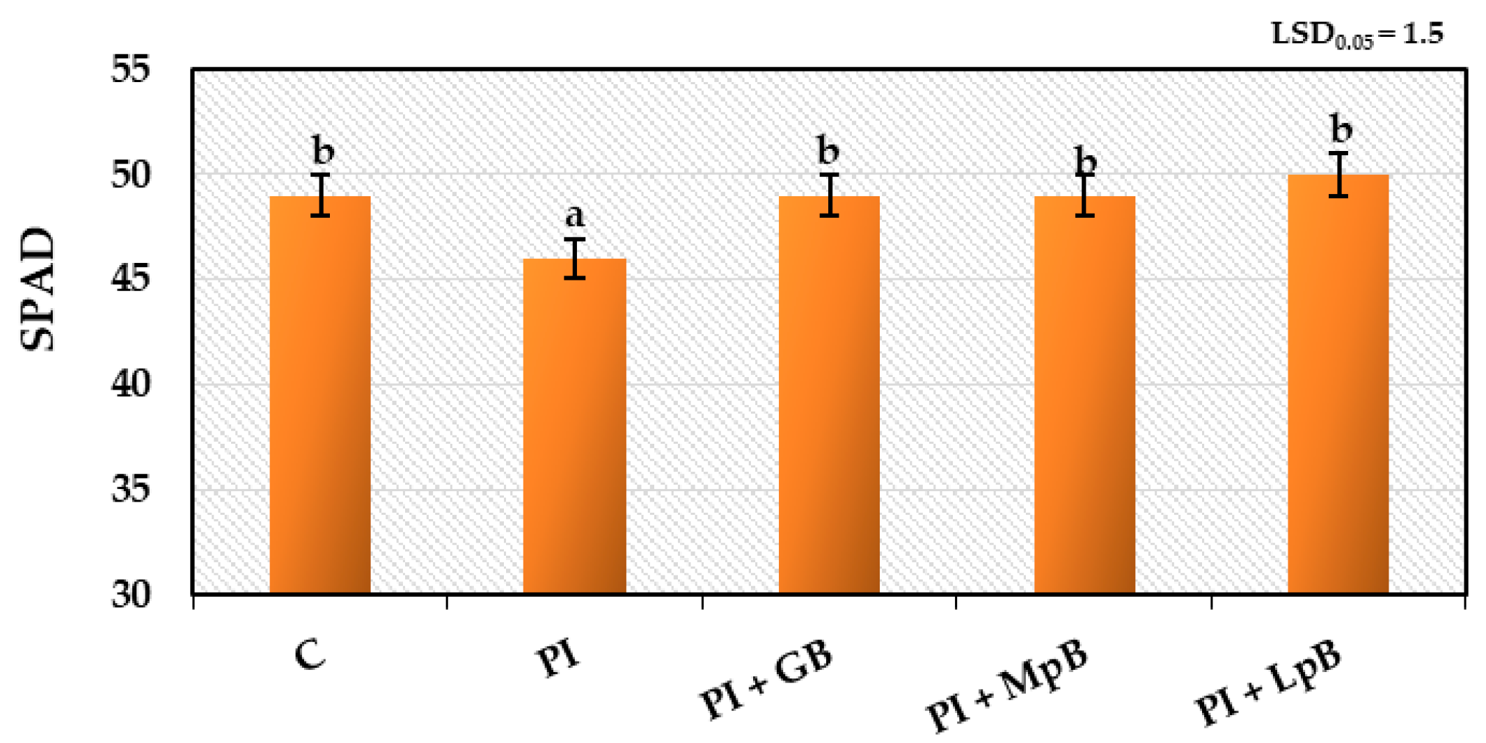
  • Index of chlorophyll content, measured with a Minolta SPAD-502 apparatus (Japan) in the highest positioned fully developed leaves, during the period of most dynamic plant growth [44].
In each experimental variant, the plants were grown in three repetitions of four plants in each. This means that 12 plants were evaluated in each variant.

2.5. Statistical Analysis

Statistical analysis was performed with Statistica 13.1 (Statsoft, Tulsa, OK, USA). All measurements of plant growth and physiological activity were performed in triplicates of four plants in each repetition, and the results are reported as the mean ± standard deviation. For each value, a one-way ANOVA with Duncan’s multiple ranges was performed to determine significance at a confidence level of p < 0.05. Measurements of secondary metabolites were compared using a one-way analysis of variance (ANOVA) at a significance level of 0.05. When a statistical difference was detected (p < 0.05), the means were compared using Tukey’s post hoc procedure at a significance level of 0.05.

3. Results and Discussion

3.1. Fungal Metabolite Analysis by LC–MS/MS

In our work, the secondary metabolite profile of Fusarium sambucinum, Alternaria tenuissima, Colletotrichum coccodes, Phoma exigua, and Rhizoctonia solani was determined by LC–MS/MS. Fungi were cultivated on Malt Extract Agar (MEA) plates as pure cultures on the potatoes. The effect of the use of biological treatments on the profile of metabolites (especially mycotoxins), including aqueous garlic extract biopreparation (GB), Metschnikowia pulcherrima TK1 biopreparation (MpB), and Lactiplantibacillus plantarum KB2 LAB 03 (LpB) biopreparation was assessed. The results are presented in Table 1, Table 2, Table 3, Table 4 and Table 5 and Table S1. In total, 68 different metabolites were quantified by LC–MS/MS. Among them were 18 Fusarium metabolites (fusaric acid (FSA), acuminatum B and C, aurofusarin (AUR), beauvericin (BEA), bikaverin, chrysogin, cyclosporin A, B, D and H, deoxygerfelin, diacetoxyscirpenol (DAS), enniatin B (Enn B), equisetin, gibepyron D, sambutoxin and siccanol; eight from the Alternaria species: 4-hydroxyalternariol, alteichin (ALTCH), alternariol (AOH), altersetin (ALN), alternariolmethylether (AME), altertoxin-I (ATX 1), radicinin (RAD), and tentoxin (TX)), 14 derived from other fungi (asterric acid, beauvericin A, cytochalasin B and E, isosulochrin, monocerin, rosellichalasin, secalonic acid D, sterigmatocystin, sulochrin, terragine, trichodimerol, trichotetronine, and xanthoquinoidin A1), three bacterial metabolites (dinactin, monactin, and nonactin), eight of plant origin (abscisic acid, daidzein, daidzin, genistein, genistin, glycitein, glycitin, and solanine), two from lichens (vulpinic acid and lecanoric acid), and 15 unspecific ones (brevianamide F, citreorosein, cordycepin, cyclo(L-Leu-L-Pro), cyclo(L-Pro-L-Tyr), cyclo(L-Pro-L-Val), emodin, endocrocin, fallacinol, fellutanine A, iso-rhodoptilometrin, rugulusovin, tryptophol, and violaceol I and II) (Table 1, Table 2, Table 3, Table 4 and Table 5; Table S1).
The results for secondary metabolites of Fusarium sambucinum growing on MEA plates and potatoes are presented in Table 1. Eight metabolites were determined in a pure F. sambucinum culture on MEA plates of which beauvericin, chrysogin, and diacetoxyscirpenol are recognised as Fusarium mycotoxins [15,46,47] and two more components (acuminatum B and C) are recognised as specific for the Fusarium species. Paciolla et al. [48] reported phytotoxic activity (by disruption of the ascorbate system) of bauvericin in tomato protoplasts. This compound has also exhibited cytotoxic properties on mammalian cells by affecting the cell membranes and mitochondria [49]. Diacetoxyscirpenol belongs to the trichothecene group of mycotoxins. Their toxic activity results from blocking protein synthesis [15]. For potatoes infected with F. sambucinum, the metabolite profile was different from the pure culture on MEA. It consisted of 18 compounds, including four mycotoxins/phytotoxins (aurofusarin, beauvericin, sambutoxin, and fusaric acid) and two more specific compounds (bikaverin and deoxygerfelin) (Table 1). Sambutoxin has been already determined in rotten potato tubers infected with F. sambucinum [50]. Kim and Lee work confirmed its toxic activity in a rat-feeding test [51]. Fusaric acid was reported as a phytotoxin that contributed to the pathogenesis of Fusarium in tomatoes by reducing the activity of antioxidant enzymes and increasing reactive oxygen species (ROS). It eventually led to plant cell death [52]. We observed a possible interesting interaction between GB and Fusarium, which resulted in a several-fold increase in the amount of produced fusaric acid. The LC–MS/MS analysis did not show the presence of other mycotoxins specific to Fusarium spp. commonly found in the published data [12,13,46], such as DON, nivalenol (NIV), or ZEN in any of the potato samples inoculated with F. sambucinum. Furthermore, rubrofusarin, usually occurring with aurofusarin [53], was not marked in our research. The fact that twofold more components were identified in potato samples than in MEA may be the result of the presence of natural potato microflora, differences in fungi metabolism depending on the available substrates for growth, and some possible cross-contamination in the potato samples which might originates from the soil. Similarly, Kokkonen et al. [54] observed the distinctive influence of the culture medium on mycotoxin production by some Penicillium species. Westphal et al. [55] described significant differences in the metabolite profile of four Fusarium species, depending on the manufacturer of the Potato Dextrose Agar (PDA), a commonly used medium for fungi cultivation.
Overall, the incorporation of the biological treatment had a positive effect on reducing the fungal contamination of potatoes with selected Fusarium mycotoxins (Table 1). Compared to the biologically untreated potatoes, the presence of aurofusarin was not detected in potato samples treated with GB and MpB. Aurofusarin is a golden-yellow pigment with described genotoxic, cytotoxic, and oxidative stress-inducing abilities in human colon cells [56]. Fusaric acid and sambutoxin were absent in potatoes treated with MpB and LpB. The occurrence of beauvericin, however, did not change. Furthermore, equisetin and sterigmatocystin appeared in potatoes treated with MpB (Table 1). Sterigmatocystin is, however, a cytotoxic compound of Aspergillus origin [57], present in our research as a probable soil contamination.
To the best of our knowledge, this is the first study to determine the secondary metabolite profile and mycotoxin contamination of potatoes inoculated with Alternaria spp. The results for the metabolite profile of Alternaria tenuissima growing on MEA plates and potatoes are presented in Table 2. Nine metabolites were detected in pure A. tenuissima cultured on MEA plates. Among them, three are classified as Alternaria species mycotoxins: AOH, AME, and ALX-I [46,58,59], which expressed genotoxic and strongly cytotoxic effects for humans in micromolar concentrations [60]. Alternaria spp. mycotoxins, AOH and AME, are the most dangerous in terms of risk to human health [61]. ALX-II, related to ALX-I, was demonstrated as a highly effective and selective compound against the Ewing sarcoma cell lines [59]. The metabolite profile of potatoes infected with A. tenuissima also included nine compounds, two of which (AME and TX) are known as metabolites of Alternaria origin [46,58].
As a probable result of cross-contamination originated from the soil, the presence of three Fusarium-derived mycotoxins was detected. The treatment of the LpB biopreparation resulted in reducing the contamination of Alternaria mycotoxins, and samples were free from AME in comparison to control potatoes (Table 2). On the contrary, the presence of ALTCH, a mycotoxin related to ALX-I, was only observed in potatoes treated with GB and LpB and then inoculated with A. tenuissima. Mycotoxin secretion is induced by various stimuli that are still under investigation. It is already known that several secondary metabolites are formed as a fungal adaptation to stress, e.g., too low/high temperature and pH changes, UV light, limited availability of nutrients, or the presence of fungicides [62], most likely including the biological treatments used in our work.
The metabolite profile of Rhizoctonia solani growing on MEA plates and potatoes is presented in Table 3. The production of mycotoxins by R. solani is still unknown; however, Li et al. [63] recently identified 3-methoxyphenylacetic acid as a phytotoxin that is linked to tobacco leaf necrosis. In our work, only two unspecific metabolites were determined in the pure culture of R. solani growing on the MEA plate. The metabolite profile of fungal culture growing on potatoes included 10 compounds, half of which are unspecific or of plant origin. Four compounds, bikaverin, beauvericin, fusaric acid, and sambutoxin, are a probable cause of cross-contamination with Fusarium moulds, originated from the environment in which potatoes were grown. Fusarium mycotoxins identified in control potato samples were also present in samples treated with biopreparations (Table 3).
The results for secondary metabolites of Phoma exigua growing on MEA plates and potatoes are presented in Table 4. Five metabolites were identified in the pure culture of P. exigua growing on MEA plates. Among them, only cytochalasin B was recognised as a mycotoxin of Phoma origin [64], while the other compounds were unspecific. The metabolite profile of potatoes inoculated with P. exigua was more complex and contained 14 compounds, including bikaverin and toxic cytochalasin B, but mostly other unspecific or compounds of plant, lichen, and bacterial origin. The MpB biopreparation had a positive influence on reducing potato contamination with Phoma mycotoxin cytochalasin B (Table 4). Samples treated with biopreparations, however, contained Fusarium spp. mycotoxins.
The metabolite profile of Colletotrichum coccodes growing on MEA plates and potatoes is presented in Table 5. The production of mycotoxins by C. coccodes is still unknown. Nine compounds were present in the pure culture of C. coccodes growing on MEA plates. They were mainly unspecific components with the exception of bacterial monocerin and cytochalasin E, originally isolated from the Aspergillus species [65], as a probable effect of cross-contamination. Eleven metabolites were identified in potatoes inoculated with C. coccodes, mostly unrelated to fungal activity, except for several compounds originally isolated from the Fusarium species, such as beauvericin, bikaverin, and deoxygerfelin (Table 5). Potato samples treated with biopreparations contained Fusarium spp. mycotoxins (fusaric acid, beauvericin, and sambutoxin). The presence of mycotoxins and other secondary metabolites that are not produced by the fungi inoculated in the potato originated from the environment, i.e., the soil where potatoes were grown.
Chaconine and solanine, common potato peel metabolites, were determined in all potato samples (Table 1, Table 2, Table 3, Table 4 and Table 5). Their concentration varied significantly across individual tubers. The results of LC–MS/MS analysis of the potato control sample indicated high mycotoxin contamination, especially characteristic of the Fusarium spp. (Table 1, Table 2, Table 3, Table 4 and Table 5; Table S1), even if visible symptoms of infection on the tubers were not observed.

3.2. Growth and Physiological Parameter Analysis

3.2.1. Germination of Seed Potatoes and Stem and Root Development

Untreated seed potatoes (control; C), stored at 4 °C from harvest until 1 April 2022 and then for 2 weeks at 15 °C and 80% RH, were characterised by high healthiness, typical phenotypic characteristics, and high turgor. After planting in the ground at 20 °C on 14 April, they germinated at 100%. Seed potatoes treated with biological agents, such as aqueous garlic extract biopreparation (GB), Metschnikowia pulcherrima TK1 biopreparation (MpB), and Lactiplantibacillus plantarum KB2 LAB 03 biopreparation (LpB), and infected with a mixture of phytopathogens, did not lose the turgor, germination capacity, and phenotypic properties of the mother tubers. During planting into the substrate, the seed potatoes did not show any mechanical damage, and no visible symptoms of disease were observed. This indicates that the treatment of seed potatoes with the biological preparations used and the inoculation of phytopathogens resulted in their proper germination, without damaging the sprouts mechanically or pathogenically, as was shown by Kirk and Gachango [66] for Fusarium contamination.
The research showed that the treatment of seed potatoes with biological agents prevented the development of diseases initiated by the inoculated mixtures of phytopathogens. The pathogenic activity of phytopathogens and the fungistatic activity of the biopreparations applied to seed potatoes affected the growth, development, and physiological activity of stems and roots obtained from them to a different extent. The biggest impact of these treatments was observed in the growth and development of the root system, assessed by the dynamics of elongation of individual roots, filling the soil profile with them (Figure 1, Figure 2 and Figure 3). Inoculation of seed potatoes with a mixture of phytopathogens (PI) significantly delayed and reduced the growth dynamics of particular roots and their filling of the soil profile in comparison to the control sample without phytopathogens (C) and biological agent treatment. The root growth up to 30 cm in length lasted 20 days for the PI sample in comparison to 16 days in the case of the C sample. The percentage of soil profile area filled with roots was 30% for the PI sample and 32% for the C sample. Seed potato treatment with biopreparations before applying the mixture of phytopathogens had a significantly positive effect on the growth and development of the root system (Figure 1, Figure 2 and Figure 3). In particular, this beneficial effect was achieved for the treatment of seed potatoes with LpB and MpB. Root growth up to 30 cm in length lasted for 16 days for the MpB and LpB samples, and this was the same for control sample C. The percentage of soil profile area filled with roots was 36% for both the MpB and the LpB samples. The use of MpB and LpB biopreparations allowed entirely eliminating the negative effect of phytopathogen development on root growth and even stimulating it when the soil profile filling with roots was taken into account. In addition, Hamed et al. [67] and Abdel-Aziz et al. [22] observed a positive effect of lactic acid bacteria treatment on root development in tomato plants (increases of 216–358% and 271–305% over the control, respectively).
The treatment of seed potatoes with GB resulted in the prevention of the negative impact of the applied phytopathogens, but to a lesser extent. These results indicate the high sensitivity of the root system to various stimuli, including the treatment of seed potatoes with biological compounds and pathogenic mycoflora, which significantly affected the growth, development, and yield of plants (Figure 1, Figure 2 and Figure 3). The biopreparations used for seed potato biological protection and the content of inoculated mixtures of phytopathogens caused different growth dynamics of the root system, indicating the possibility of a degree of improvement or deterioration of the physiological condition of sprouts. Compared to stems, resting and sprouting sprouts in moist soil are particularly exposed to the phytopathogenic activity of pathogens that completely or partially contaminate the surface of seed potatoes, which can then infect other parts of the plant. For this reason, as well as earlier development compared to other plant organs, root growth may be a useful marker for the assessment of the pathogenic activity of mycoflora contaminating seed potatoes [66,68]. The high sensitivity of the root system to the various factors applied to seeds and plants, and the usefulness of this test in assessing the reaction of plants to these treatments have also been indicated in earlier studies by the authors. This method of testing plant reactions to stimuli is noninvasive and more accurate, in addition to allowing a quicker indication of plant response to treatments than, for example, measurements of the height and physiological activity of stems, whose growth depends on the development of the roots [40,69,70,71].
Treatment of seed potatoes with GB, MpB, and LpB, and then infecting them with a mixture of phytopathogens affected the growth and physiological activity of the stems (Figure 4 and Figure 5), but to a much lesser extent than the development of the root system. The plants obtained from untreated seed potatoes (C) began to emerge 6 days after planting and grew for the next 46 days (52 days in total), reaching a final height of 64 cm and forming flower buds. The stems obtained from seed potatoes infected with phytopathogens (PI) grew slightly more slowly than the control samples (final stem length was 61 cm), albeit without adversely affecting their flowering and vigour. Treatment of seed potatoes with biological agents in the period prior to inoculation with phytopathogens prevented the negative impact of microorganisms, which led to more dynamic stem growth. As observed for the roots, treatment of seed potatoes with MpB (final stem length was 66 cm) and LpB (final stem length was 68 cm) was the most beneficial. The obtained results indicate that the applied biological shield of biopreparations and the inoculation of phytopathogens into seed potatoes may affect the growth of stems. This effect, however, depends on the degree of infection of seed potato sprouts with pathogens and the degree of infection of the growing stems. In this case, the degree of sprout infection seems not to be sufficiently dangerous, leading to a slight decrease in stem growth dynamics, and it was clearly dependent on the biopreparations and the mixture of phytopathogens applied to the seed potatoes [66,68].

3.2.2. Gas Exchange in Leaves and Index of Chlorophyll Content in Leaves

The study of gas exchange activity in leaves (net photosynthesis, transpiration, stomatal conductivity, and intercellular CO2 content) and the index of chlorophyll contentconfirmed the correlation between the methods of biological treatment of seed potatoes and the development of roots and stems, as discussed in Section 3.2.1. (Figure 6 and Figure 7). Inoculation of seed potatoes with phytopathogens adversely affected gas exchange and the index of chlorophyll content in leaves. Treatment of seed potatoes with biopreparations, especially MpB and LpB, significantly increased this activity. The obtained results confirmed the close, verified by the literature, relationship between the tested physiological activity and plant growth, which depends on gas exchange in the leaves and the content of the photosynthetic pigment [72]. These parameters are important in the case of the potato, whose leaves did not show pathogenic symptoms visible to the naked eye, while their measurements demonstrated a beneficial effect of the appropriate biopreparations and a harmful pathogenic effect of phytopathogens [73].
The applied methods of biological treatment of the Impresja variant of seed potatoes had a similar effect on the quality of stems and leaves, assessed using a five-point rating scale. During the 52 days of the study, no significant changes in their vigour and colouration were found.

4. Conclusions

Biological treatment exhibited an overall positive effect on the inhibition of metabolite production by the tested fungi on potatoes. In comparison to the control potatoes, after MpB treatment, aurofusarin, sambutoxin, and fusaric acid were not present in Fusarium-contaminated potatoes, while cytochalasin B was not formed in Phoma-inoculated samples. In potatoes treated with LpB and contaminated by Alternaria spores, no presence of alternariolmethylether was noted. Likewise, fusaric acid and sambutoxin were not determined in Fusarium-inoculated potatoes. In the GB treatment, aurofusarin was absent in Fusarium-contaminated potatoes in comparison to the control. Treatment with biopreparations, however, did not reduce the formation of all mycotoxins. Some mycotoxins were observed exclusively in samples treated with LpB (alteichin in the sample inoculated with A. tenuissima), MpB (equisetin and sterigmatocystin in the sample inoculated with F. sambucinum), and GB (alteichin in the sample inoculated with A. tenuissima). It is worth mentioning that biological treatment with MpB, LpB, and GB increased the growth of potato roots and stems, as well as improved the basic parameters of potato physiology: gas exchange and the index of chlorophyll content. Knowledge about the cellular biology underlying interactions between microbes in their natural environments is still relatively scarce. The tested bioprotective agents are environmentally friendly and suitable for use at an early stage for reducing contamination caused by fungi. Our promising results, however, require further examination in order to determine the optimal composition of biopreparations for the effective and long-lasting biological control of potato pathogens.

Supplementary Materials

The following supporting information can be downloaded at https://www.mdpi.com/article/10.3390/ijerph20065221/s1: Table S1. LC–MS/MS secondary metabolite analysis of control samples—MEA and potato; Table S2. Analytical parameters of LC–MS/MS.

Author Contributions

Conceptualisation, A.S., D.K. and B.G.; formal analysis, A.S., M.S., R.J. and M.G.; funding acquisition, A.S. and B.G.; investigation, R.J. and W.L.; methodology, A.S., M.S., R.J. and M.G.; resources, M.S. and B.G.; supervision, D.K. and B.G.; visualisation, A.S.; writing—original draft, A.S.; writing—review and editing, D.K. and B.G. All authors have read and agreed to the published version of the manuscript.

Funding

This work was supported by the Agency for Restructuring and Modernisation of Agriculture, Poland [grant number 00010.DDD.6509.00016.2018.05] and the Polish National Agency for Academic Exchange under the PROM programme, co-financed by the European Social Fund under the Knowledge Education Development Operational Programme, implemented by the Łódź University of Technology. The APC was funded by the Łódź University of Technology, Poland.

Acknowledgments

The authors express their gratitude to Hannes Gratzl and Patrick Rennhofer for their help in the LC–MS/MS analysis.

Conflicts of Interest

The authors declare no conflict of interest. The funders had no role in the design of the study; in the collection, analyses or interpretation of the data; in the writing of the manuscript; or in the decision to publish the results.

References

  1. Camire, M.E.; Kubow, S.; Donnelly, D.J. Potatoes and Human Health. Crit. Rev. Food Sci. Nutr. 2009, 49, 823–840. [Google Scholar] [CrossRef]
  2. FAO. Top Countries in Potatoes Production. Potatoes Production—Food and Agriculture Organization of the United Nations; FAO: Rome, Italy, 2019; Available online: https://www.fao.org/faostat/en/ (accessed on 6 January 2023).
  3. Savary, S.; Willocquet, L.; Pethybridge, S.J.; Esker, P.; McRoberts, N.; Nelson, A. The global burden of pathogens and pests on major food crops. Nat. Ecol. Evol. 2019, 3, 430–439. [Google Scholar] [CrossRef] [PubMed]
  4. Liu, J.; Sun, Z.; Zou, Y.; Li, W.; He, F.; Huang, X.; Lin, C.; Cai, Q.; Wisniewski, M.; Wu, X. Pre- and postharvest measures used to control decay and mycotoxigenic fungi in potato (Solanum tuberosum L.) during storage. Crit. Rev. Food Sci. Nutr. 2021, 62, 415–428. [Google Scholar] [CrossRef]
  5. Kebede, H.; Liu, X.; Jin, J.; Xing, F. Current status of major mycotoxins contamination in food and feed in Africa. Food Control 2020, 110, 106975. [Google Scholar] [CrossRef]
  6. Muhialdin, B.J.; Saari, N.; Meor Hussin, A.S. Review on the Biological Detoxification of Mycotoxins Using Lactic Acid Bacteria to Enhance the Sustainability of Foods Supply. Molecules 2020, 25, 2655. [Google Scholar] [CrossRef]
  7. Gruber-Dorninger, C.; Jenkins, T.; Schatzmayr, G. Global Mycotoxin Occurrence in Feed: A Ten-Year Survey. Toxins 2019, 11, 375. [Google Scholar] [CrossRef] [PubMed] [Green Version]
  8. Liu, Y.; Galani Yamdeu, J.H.; Gong, Y.Y.; Orfila, C. A review of postharvest approaches to reduce fungal and mycotoxin contamination of foods. Compr. Rev. Food Sci. Food Saf. 2020, 19, 1521–1560. [Google Scholar] [CrossRef] [PubMed]
  9. Kamle, M.; Mahato, D.K.; Devi, S.; Lee, K.E.; Kang, S.G.; Kumar, P. Fumonisins: Impact on Agriculture, Food, and Human Health and their Management Strategies. Toxins 2019, 11, 328. [Google Scholar] [CrossRef] [Green Version]
  10. Commission Regulation (EC). No 1881/2006 of 19 December 2006 Setting Maximum Levels for Certain Contaminants in Foodstuffs. 2006. Available online: https://eur-lex.europa.eu/LexUriServ/LexUriServ.do?uri=OJ:L:2006:364:0005:0024:EN:PDF (accessed on 6 January 2023).
  11. Xue, H.L.; Bi, Y.; Tang, Y.M.; Zhao, Y.; Wang, Y. Effect of cultivars, Fusarium strains and storage temperature on trichothecenes production in inoculated potato tubers. Food Chem. 2014, 151, 236–242. [Google Scholar] [CrossRef]
  12. Stefańczyk, E.; Sobkowiak, S.; Brylińska, M.; Śliwka, J. Diversity of Fusarium spp. associated with dry rot of potato tubers in Poland. Eur. J. Plant Pathol. 2016, 145, 871–884. [Google Scholar] [CrossRef] [Green Version]
  13. Delgado, J.A.; Schwarz, P.B.; Gillespie, J.; Rivera-Varas, V.V.; Secor, G.A. Trichothecene mycotoxins associated with potato dry rot caused by fusarium graminearum. Phytopathology 2010, 100, 290–296. [Google Scholar] [CrossRef] [PubMed] [Green Version]
  14. Rodrigues, I.; Naehrer, K. A Three-Year Survey on the Worldwide Occurrence of Mycotoxins in Feedstuffs and Feed. Toxins 2012, 4, 663–675. [Google Scholar] [CrossRef] [PubMed]
  15. Perincherry, L.; Lalak-Kańczugowska, J.; Stępień, Ł. Fusarium-Produced Mycotoxins in Plant-Pathogen Interactions. Toxins 2019, 11, 664. [Google Scholar] [CrossRef] [PubMed] [Green Version]
  16. Canadian Horticultural Council. Control of Potato Storage Conditions for the Management of Post-Harvest Losses due to Diseases; Canadian Horticultural Council: Ottawa, ON, Canada, 2017. [Google Scholar]
  17. Steglińska, A.; Kołtuniak, A.; Motyl, I.; Berłowska, J.; Czyżowska, A.; Cieciura-Włoch, W.; Okrasa, M.; Kręgiel, D.; Gutarowska, B. Lactic Acid Bacteria as Biocontrol Agents against Potato (Solanum tuberosum L.) Pathogens. Appl. Sci. 2022, 12, 7763. [Google Scholar] [CrossRef]
  18. Steglińska, A.; Kołtuniak, A.; Berłowska, J.; Czyżowska, A.; Szulc, J.; Cieciura-Włoch, W.; Okrasa, M.; Kręgiel, D.; Gutarowska, B. Metschnikowia pulcherrima as a Biocontrol Agent against Potato (Solanum tuberosum) Pathogens. Agronomy 2022, 12, 2546. [Google Scholar] [CrossRef]
  19. Steglińska, A.; Bekhter, A.; Wawrzyniak, P.; Kunicka-Styczyńska, A.; Jastrząbek, K.; Fidler, M.; Śmigielski, K.; Gutarowska, B. Antimicrobial Activities of Plant Extracts against Solanum tuberosum L. Phytopathogens. Molecules 2022, 27, 1579. [Google Scholar] [CrossRef]
  20. El-Mabrok, A.S.W.; Hassan, Z.; Mokhtar, A.M.; Hussin, K.M.A. Antifungal activity of Lactobacillus plantarum LAB-C5 and LAB-G7 isolated from Malaysian fruits. Acta Biol. Malaysiana 2013, 2, 22–30. [Google Scholar] [CrossRef]
  21. Canpolat, E.; Doğaner, M.M.; Derviş, S.; Ulubaş Serçe, Ç. Antifungal Activity of Some Lactic Acid Bacteria Against Several Soil-borne Fungal Pathogens Isolated from Strawberry Plants. Turkish J. Agric.-Food Sci. Technol. 2018, 6, 1163. [Google Scholar] [CrossRef]
  22. Abdel-Aziz, S.M.; Moustafa, Y.A.; Hamed, H.A. Lactic Acid Bacteria in the Green Biocontrol against some Phytopathogenic Fungi: Treatment of Tomato Seeds. J. Basic. Appl. Sci. Res 2014, 4, 1–9. [Google Scholar]
  23. Šušković, J.; Kos, B.; Beganović, J.; Pavunc, A.L.; Habjanič, K.; Matoć, S. Antimicrobial activity—The most important property of probiotic and starter lactic acid bacteria. Food Technol. Biotechnol. 2010, 48, 296–307. [Google Scholar]
  24. Gerez, C.L.; Torres, M.J.; Font de Valdez, G.; Rollán, G. Control of spoilage fungi by lactic acid bacteria. Biol. Control 2013, 64, 231–237. [Google Scholar] [CrossRef]
  25. Arena, M.P.; Silvain, A.; Normanno, G.; Grieco, F.; Drider, D.; Spano, G.; Fiocco, D. Use of Lactobacillus plantarum Strains as a Bio-Control Strategy against Food-Borne Pathogenic Microorganisms. Front. Microbiol. 2016, 7, 464. [Google Scholar] [CrossRef] [Green Version]
  26. Perczak, A.; Goliński, P.; Bryła, M.; Waśkiewicz, A. The efficiency of lactic acid bacteria against pathogenic fungi and mycotoxins. Arh. Hig. Rada Toksikol. 2018, 69, 32–45. [Google Scholar] [CrossRef] [PubMed] [Green Version]
  27. Mendonca-Hagler, L.C.; Hagler, A.N.; Kurtzman, C.P. Phylogeny of Metschnikowia Species Estimated from Partial rRNA Sequences. Int. J. Syst. Bacteriol. 1993, 43, 368–373. [Google Scholar] [CrossRef] [Green Version]
  28. Sipiczki, M. Metschnikowia pulcherrima and related pulcherrimin-producing yeasts: Fuzzy species boundaries and complex antimicrobial antagonism. Microorganisms 2020, 8, 1029. [Google Scholar] [CrossRef] [PubMed]
  29. Sipiczki, M. Metschnikowia strains isolated from botrytized grapes antagonize fungal and bacterial growth by iron depletion. Appl. Environ. Microbiol. 2006, 72, 6716–6724. [Google Scholar] [CrossRef] [Green Version]
  30. Guo, D.; Zhu, L.; Hou, X. Combination of UV-C Treatment and Metschnikowia pulcherrimas for Controlling Alternaria Rot in Postharvest Winter Jujube Fruit. J. Food Sci. 2015, 80, M137–M141. [Google Scholar] [CrossRef]
  31. Tian, Y.Q.; Li, W.; Jiang, Z.T.; Jing, M.M.; Shao, Y.Z. The preservation effect of Metschnikowia pulcherrima yeast on anthracnose of postharvest mango fruits and the possible mechanism. Food Sci. Biotechnol. 2018, 27, 95–105. [Google Scholar] [CrossRef]
  32. No. P.441896, Method of Bio-Protection of Seed Potato Tubers against the Development of Bacterial and Fungal Phytopathogens. Pat. Appl. Pat. Off. Pol. 2022. Available online: https://uprp.gov.pl/pl (accessed on 6 January 2023).
  33. No. P.441895: Method of Bio-Protection of Seed Potato Tubers against the Development of Fungal Phytopathogens. Pat. Appl. Pat. Off. Pol. 2022. Available online: https://uprp.gov.pl/pl (accessed on 6 January 2023).
  34. Sharma, A.; Rajendran, S.; Srivastava, A.; Sharma, S.; Kundu, B. Antifungal activities of selected essential oils against Fusarium oxysporum f. sp. lycopersici 1322, with emphasis on Syzygium aromaticum essential oil. J. Biosci. Bioeng. 2017, 123, 308–313. [Google Scholar] [CrossRef] [PubMed]
  35. Abd-El-Khair, H.; El-Gamal Nadia, G. Effects of aqueous extracts of some plant species against Fusarium solani and Rhizoctonia solani in Phaseolus vulgaris plants. Arch. Phytopathol. Plant Prot. 2011, 44, 1–16. [Google Scholar] [CrossRef]
  36. Nashwa, S.M.A.; Abo-Elyou, K.A.M. Evaluation of various plant extracts against the early blight disease of tomato plants under greenhouse and field conditions. Plant Prot. Sci. 2012, 48, 74–79. [Google Scholar] [CrossRef] [Green Version]
  37. Martins, N.; Petropoulos, S.; Ferreira, I.C.F.R. Chemical composition and bioactive compounds of garlic (Allium sativum L.) as affected by pre- and post-harvest conditions: A review. Food Chem. 2016, 211, 41–50. [Google Scholar] [CrossRef] [PubMed] [Green Version]
  38. Sulyok, M.; Stadler, D.; Steiner, D.; Krska, R. Validation of an LC-MS/MS-based dilute-and-shoot approach for the quantification of > 500 mycotoxins and other secondary metabolites in food crops: Challenges and solutions. Anal. Bioanal. Chem. 2020, 412, 2607–2620. [Google Scholar] [CrossRef] [Green Version]
  39. Romanowska-Duda, Z.; Janas, R.; Grzesik, M. Application of Phytotoxkit in the quick assessment of ashes suitability as fertilizers in sorghum crops. Int. Agrophysics 2019, 33, 145–152. [Google Scholar] [CrossRef]
  40. Grzesik, M.; Janas, R.; Steglińska, A.; Kręgiel, D.; Gutarowska, B. Influence of microclimatic conditions during year-long storage of ‘Impresja’ potato tubers (Solanum tuberosum L.) on the emergence, growth, physiological activity and yield of plants. J. Stored Prod. Res. 2022, 99, 102033. [Google Scholar] [CrossRef]
  41. Steglińska, A.; Pielech-Przybylska, K.; Janas, R.; Grzesik, M.; Borowski, S.; Kręgiel, D.; Gutarowska, B. Volatile Organic Compounds and Physiological Parameters as Markers of Potato (Solanum tuberosum L.) Infection with Phytopathogens. Molecules 2022, 27, 3708. [Google Scholar] [CrossRef] [PubMed]
  42. Janas, R.; Górnik, K.; Grzesik, M.; Romanowska-Duda, Z.; van Duijn, B. Effectiveness of pulsed radio frequency in seed quality improvement of vegetable plant species. Int. Agrophysics 2019, 33, 463–471. [Google Scholar] [CrossRef] [PubMed]
  43. Romanowska-Duda, Z.; Szufa, S.; Grzesik, M.; Piotrowski, K.; Janas, R. The Promotive Effect of Cyanobacteria and Chlorella sp. Foliar Biofertilization on Growth and Metabolic Activities of Willow (Salix viminalis L.) Plants as Feedstock Production, Solid Biofuel and Biochar as C Carrier for Fertilizers via Torrefaction Proce. Energies 2021, 14, 5262. [Google Scholar] [CrossRef]
  44. Grzesik, M.; Górnik, K.; Janas, R.; Lewandowki, M.; Romanowska-Duda, Z.; Duijn, B. van High efficiency stratification of apple cultivar Ligol seed dormancy by phytohormones, heat shock and pulsed radio frequency. J. Plant Physiol. 2017, 219, 81–90. [Google Scholar] [CrossRef] [PubMed]
  45. Romanowska-Duda, Z.; Grzesik, M.; Janas, R. Maximal Efficiency of PSII as a Marker of Sorghum Development Fertilized With Waste From a Biomass Biodigestion to Methane. Front. Plant Sci. 2019, 9, 1920. [Google Scholar] [CrossRef] [Green Version]
  46. Spanic, V.; Katanic, Z.; Sulyok, M.; Krska, R.; Puskas, K.; Vida, G.; Drezner, G.; Šarkanj, B. Multiple fungal metabolites including mycotoxins in naturally infected and fusarium-inoculated wheat samples. Microorganisms 2020, 8, 578. [Google Scholar] [CrossRef] [Green Version]
  47. Wu, Q.; Kuca, K.; Nepovimova, E.; Wu, W. Type A Trichothecene Diacetoxyscirpenol-Induced Emesis Corresponds to Secretion of Peptide YY and Serotonin in Mink. Toxins 2020, 12, 419. [Google Scholar] [CrossRef] [PubMed]
  48. Paciolla, C.; Dipierro, N.; Mulè, G.; Logrieco, A.; Dipierro, S. The mycotoxins beauvericin and T-2 induce cell death and alteration to the ascorbate metabolism in tomato protoplasts. Physiol. Mol. Plant Pathol. 2004, 65, 49–56. [Google Scholar] [CrossRef]
  49. Mallebrera, B.; Prosperini, A.; Font, G.; Ruiz, M.J. In vitro mechanisms of Beauvericin toxicity: A review. Food Chem. Toxicol. 2018, 111, 537–545. [Google Scholar] [CrossRef] [PubMed]
  50. Kim, J.; Lee, Y.; Yu, S. Sambutoxin-producing isolates of fusarium species and occurrence of sambutoxin in rotten potato tubers. Appl. Environ. Microbiol. 1995, 61, 3750–3751. [Google Scholar] [CrossRef] [Green Version]
  51. Kim, J.C.; Lee, Y.W. Sambutoxin, a new mycotoxin produced by toxic Fusarium isolates obtained from rotted potato tubers. Appl. Environ. Microbiol. 1994, 60, 4380–4386. [Google Scholar] [CrossRef] [Green Version]
  52. Singh, V.K.; Upadhyay, R.S. Fusaric acid induced cell death and changes in oxidative metabolism of Solanum lycopersicum L. Bot. Stud. 2014, 55, 66. [Google Scholar] [CrossRef] [Green Version]
  53. Frandsen, R.J.N.; Schütt, C.; Lund, B.W.; Staerk, D.; Nielsen, J.; Olsson, S.; Giese, H. Two Novel Classes of Enzymes Are Required for the Biosynthesis of Aurofusarin in Fusarium graminearum. J. Biol. Chem. 2011, 286, 10419–10428. [Google Scholar] [CrossRef] [Green Version]
  54. Kokkonen, M.; Jestoi, M.; Rizzo, A. The effect of substrate on mycotoxin production of selected Penicillium strains. Int. J. Food Microbiol. 2005, 99, 207–214. [Google Scholar] [CrossRef]
  55. Westphal, K.R.; Heidelbach, S.; Zeuner, E.J.; Riisgaard-Jensen, M.; Nielsen, M.E.; Vestergaard, S.Z.; Bekker, N.S.; Skovmark, J.; Olesen, C.K.; Thomsen, K.H.; et al. The effects of different potato dextrose agar media on secondary metabolite production in Fusarium. Int. J. Food Microbiol. 2021, 347, 109171. [Google Scholar] [CrossRef]
  56. Jarolim, K.; Wolters, K.; Woelflingseder, L.; Pahlke, G.; Beisl, J.; Puntscher, H.; Braun, D.; Sulyok, M.; Warth, B.; Marko, D. The secondary Fusarium metabolite aurofusarin induces oxidative stress, cytotoxicity and genotoxicity in human colon cells. Toxicol. Lett. 2018, 284, 170–183. [Google Scholar] [CrossRef] [PubMed]
  57. Segaran, G.; Sathiavelu, M. Fungal endophytes: A potent biocontrol agent and a bioactive metabolites reservoir. Biocatal. Agric. Biotechnol. 2019, 21, 101284. [Google Scholar] [CrossRef]
  58. De Souza, G.D.; Mithöfer, A.; Daolio, C.; Schneider, B.; Rodrigues-Filho, E. Identification of Alternaria alternata mycotoxins by LC-SPE-NMR and their cytotoxic effects to soybean (Glycine max) cell suspension culture. Molecules 2013, 18, 2528–2538. [Google Scholar] [CrossRef] [Green Version]
  59. Robles, A.J.; Dai, W.; Haldar, S.; Ma, H.; Anderson, V.M.; Overacker, R.D.; Risinger, A.L.; Loesgen, S.; Houghton, P.J.; Cichewicz, R.H.; et al. Altertoxin II, a Highly Effective and Specific Compound against Ewing Sarcoma. Cancers 2021, 13, 6176. [Google Scholar] [CrossRef]
  60. Wang, H.; Guo, Y.; Luo, Z.; Gao, L.; Li, R.; Zhang, Y.; Kalaji, H.M.; Qiang, S.; Chen, S. Recent Advances in Alternaria Phytotoxins: A Review of Their Occurrence, Structure, Bioactivity and Biosynthesis. J. Fungi 2022, 8, 168. [Google Scholar] [CrossRef]
  61. Aichinger, G.; Del Favero, G.; Warth, B.; Marko, D. Alternaria toxins—Still emerging? Compr. Rev. Food Sci. Food Saf. 2021, 20, 4390–4406. [Google Scholar] [CrossRef]
  62. Pusztahelyi, T.; Holb, I.J.; PÃ3csi, I. Secondary metabolites in fungus-plant interactions. Front. Plant Sci. 2015, 6, 573. [Google Scholar] [CrossRef] [Green Version]
  63. Li, X.; Hou, H.; Liu, H.; Wang, H.; Cai, L.; An, M.; Zhang, C. Identification of 3-Methoxyphenylacetic Acid as a Phytotoxin, Produced by Rhizoctonia solani AG-3 TB. Molecules 2023, 28, 790. [Google Scholar] [CrossRef] [PubMed]
  64. Chang, H.-T.; Chou, C.-T.; Chen, I.-S.; Yu, C.-C.; Lu, T.; Hsu, S.-S.; Shieh, P.; Jan, C.-R.; Liang, W.-Z. Mechanisms underlying effect of the mycotoxin cytochalasin B on induction of cytotoxicity, modulation of cell cycle, Ca2+ homeostasis and ROS production in human breast cells. Toxicology 2016, 370, 1–19. [Google Scholar] [CrossRef]
  65. Mendes, G.; Baltazar, L.M.; Souza, D.G.; Sá, N.P.; Rosa, L.H.; Rosa, C.A.; Souza-Fagundes, E.M.; Ramos, J.P.; Alves-Silva, J.; Cota, B.B.; et al. Effects of cytochalasin E on Paracoccidioides brasiliensis. J. Appl. Microbiol. 2018, 125, 1296–1307. [Google Scholar] [CrossRef]
  66. Kirk, W.; Gachango, E.; General Seed-Rots Can Pose Problems in Potatoes. Michigan State Univ. Ext. Dep. Plant Pathol. 2011. Available online: https://www.canr.msu.edu/news/general_seed_rots_can_pose_problems_in_potatoes (accessed on 20 December 2022).
  67. Hamed, H.A.; Moustafa, Y.A.; Abdel-Aziz, S.M. In vivo efficacy of lactic acid bacteria in biological control against Fusarium oxysporum for protection of tomato plant. Life Sci. J. 2011, 8, 462–468. [Google Scholar]
  68. Koroney, A.S.; Plasson, C.; Pawlak, B.; Sidikou, R.; Driouich, A.; Menu-Bouaouiche, L.; Vicré-Gibouin, M. Root exudate of Solanum tuberosum is enriched in galactose-containing molecules and impacts the growth of Pectobacterium atrosepticum. Ann. Bot. 2016, 118, 797–808. [Google Scholar] [CrossRef] [Green Version]
  69. Badek, B.; Romanowska-Duda, Z.; Grzesik, M.; Duijn, B. van Rapid Evaluation of Germinability of Primed China Aster (Callistephus Chinensis Ness.) Seeds with Physiological and Biochemical Markers. J. Hortic. Res. 2014, 22, 5–18. [Google Scholar] [CrossRef] [Green Version]
  70. Badek, B.; Romanowska-Duda, Z.; Grzesik, M.; Kuras, A. Physiological Markers for Assessing Germinability of Lycopersicon esculentum Seeds Primed by Environment-Friendly Methods. Polish J. Environ. Stud. 2016, 25, 1831–1838. [Google Scholar] [CrossRef] [PubMed]
  71. Fu, Q.; Fu, J.; Chen, Z.; Chen, C.; Zhang, J.; Ren, L. Measurement and Analysis of Root Anchorage Effect on Stalk Forces in Lodged Corn Harvesting. Front. Plant Sci. 2022, 13, 852375. [Google Scholar] [CrossRef] [PubMed]
  72. Grzesik, M.; Romanowska-Duda, Z.; Kalaji, H.M. Effectiveness of cyanobacteria and green algae in enhancing the photosynthetic performance and growth of willow (Salix viminalis L.) plants under limited synthetic fertilizers application. Photosynthetica 2017, 55, 510–521. [Google Scholar] [CrossRef] [Green Version]
  73. Ghosh, S.C.; Asanuma, K.; Kusutani, A.; Toyota, M. Gas Exchange Properties of Potato Plants under Different Nitrogen Regimes. Pakistan J. Biol. Sci. 2000, 3, 1654–1657. [Google Scholar] [CrossRef]
Figure 1. Growth kinetics and growth period of roots up to 30 cm in length, obtained from Impresja seed potatoes treated with biopreparations and then inoculated with phytopathogens. C—control potato, not treated with any biopreparations and not inoculated with pathogens; PI—potato sample only inoculated with pathogens; PI + GB—potato sample treated with aqueous garlic extract biopreparation and inoculated with pathogens; PI + MpB—potato sample treated with M. pulcherrima TK1 biopreparation and inoculated with pathogens; PI + LpB—potato sample treated with L. plantarum KB2 LAB 03 biopreparation and inoculated with pathogens. Means marked with the same letters do not differ statistically at the significance level p = 0.05; LSD—least significant difference.
Ijerph 20 05221 g001
Figure 2. Soil profile overgrown by developing root system obtained from seed potatoes: (A) infected with phytopathogens; (B) biologically treated and then infected with phytopathogens.
Ijerph 20 05221 g002
Figure 3. Kinetics of filling the soil profile with roots and final filling after 30 days from planting Impresja seed potatoes treated with biopreparations and inoculated with phytopathogens. C—control potato, not treated with any biopreparations and not inoculated with pathogens; PI—potato sample only inoculated with pathogens; PI + GB—potato sample treated with aqueous garlic extract biopreparation and inoculated with pathogens; PI + MpB—potato sample treated with M. pulcherrima TK1 biopreparation and inoculated with pathogens; PI + LpB—potato sample treated with L. plantarum KB2 LAB 03 biopreparation and inoculated with pathogens. Means marked with the same letters (a–c) do not differ statistically at the significance level p = 0.05; LSD—least significant difference.
Ijerph 20 05221 g003
Figure 4. The kinetics of stem growth and their final length after 52 days from planting Impresja seed potatoes treated with biopreparations and inoculated with phytopathogens. C—control potato, not treated with any biopreparations and not inoculated with pathogens; PI—potato sample only inoculated with pathogens; PI + GB—potato sample treated with aqueous garlic extract biopreparation and inoculated with pathogens; PI + MpB—potato sample treated with M. pulcherrima TK1 biopreparation and inoculated with pathogens; PI + LpB—potato sample treated with L. plantarum KB2 LAB 03 biopreparation and inoculated with pathogens. Means marked with the same letters (a–c) do not differ statistically at the significance level p = 0.05; LSD—least significant difference.
Ijerph 20 05221 g004
Figure 5. Plants obtained from seed potatoes: (A) infected with phytopathogens; (B) biologically treated and then infected with phytopathogens.
Ijerph 20 05221 g005
Figure 6. Gas exchange in leaves obtained from Impresja seed potatoes treated with biopreparations and inoculated with phytopathogens. C—control potato, not treated with any biopreparations and not inoculated with pathogens; PI—potato sample only inoculated with pathogens; PI + GB—potato sample treated with aqueous garlic extract biopreparation and inoculated with pathogens; PI + MpB—potato sample treated with M. pulcherrima TK1 biopreparation and inoculated with pathogens; PI + LpB—potato sample treated with L. plantarum KB2 LAB 03 biopreparation and inoculated with pathogens. Means marked with the same letters (a–c) do not differ statistically at the significance level p = 0.05; LSD—least significant difference.
Ijerph 20 05221 g006
Figure 7. Index of chlorophyll content in leaves obtained from Impresja seed potatoes treated with biopreparations and inoculated with phytopathogens. C—control potato, not treated with any biopreparations and not inoculated with pathogens; PI—potato sample only inoculated with pathogens; PI + GB—potato sample treated with aqueous garlic extract biopreparation and inoculated with pathogens; PI + MpB—potato sample treated with M. pulcherrima TK1 biopreparation and inoculated with pathogens; PI + LpB—potato sample treated with L. plantarum KB2 LAB 03 biopreparation and inoculated with pathogens. Means marked with the same letters (a,b) do not differ statistically at the significance level p = 0.05; LSD—least significant difference.
Ijerph 20 05221 g007
Table 1. Secondary metabolites of Fusarium sambucinum cultured on MEA plates and potatoes treated with biopreparations.
Components Concentration Mean ± SD [ng/g]
F. sambucinum Culture on MEA Plate (n = 3)F. sambucinum Culture on Potato (n = 3)Potatoes Treated with Biological Preparations and Infected with F. sambucinum
GB (n = 3)LpB (n = 3)MpB (n = 3)
Abscisic acid<LOD b<LOD b<LOD b<LOD b30.2 ± 20.4 a
Acuminatum B20,500 ± 584 a<LOD b<LOD b<LOD b<LOD b
Acuminatum C9380 ± 862 a<LOD b<LOD b<LOD b<LOD b
Asterric acid<LOD a83.3 ± 112 a<LOD a50.9 ± 9.73 a<LOD a
Aurofusarin<LOD b7.27 ± 2.49 a<LOD b3.50 ± 0.0718 a<LOD b
Beauvericin16.3 ± 2 a27.5 ± 17.8 a73.8 ± 63.6 a11.8 ± 15.9 a2.21 ± 1.55 a
Bikaverin 1<LOD a505 ± 510 a5.04 ± 1.40 a<LOD a31.1 ± 29.5 a
Brevianamide F 220.8 ± 1.13 a<LOD b<LOD b<LOD b<LOD b
Chaconin<LOD a<LOD a1320 ± 1070 a2240 ± 1740 ab5560 ± 2080 b
Chrysogin14.8 ± 4.59 a<LOD b<LOD b<LOD b<LOD b
Citreorosein<LOD b<LOD b<LOD b1.54 ± 0.128 a<LOD b
Cordycepin 1,2<LOD a65.6 ± 11 a106 ± 120 a169 ± 144 a43.5 ± 7.32 a
cyclo(L-Pro-L-Tyr) 267.2 ± 14.9 a<LOD b<LOD b<LOD b<LOD b
cyclo(L-Pro-L-Val) 2216 ± 10.7 a<LOD b<LOD b<LOD b<LOD b
Cyclosporin A<LOD b<LOD b948 ± 407 a<LOD b<LOD b
Cyclosporin B<LOD b<LOD b114 ± 80.2 a<LOD b<LOD b
Cyclosporin D<LOD b<LOD b50.8 ± 27.4 a<LOD b<LOD b
Cyclosporin H<LOD b<LOD b783 ± 338 a<LOD b<LOD b
Deoxygerfelin 1<LOD a34.3 ± 33.2 a<LOD a0.668 ± 0.679 a10.8 ± 10.6 a
Diacetoxyscirpenol147.1 ± 17.3 a<LOD b<LOD b<LOD b<LOD b
Dinactin<LODb<LOD b<LOD b<LOD b3.04 ± 1.72 a
Emodin 1<LOD a4.31 ± 2.90 a<LOD a1.65 ± 2.35 a7.64 ± 7.36 a
Endocrocin<LOD ab203 ± 113 cd<LOD ae31.9 ± 3.61 ace63.7 ± 64.8 bde
Equisetin<LOD b<LOD b<LOD b<LOD b1.67 ± 1.07 a
Fallacinol<LOD b<LOD b<LOD b2.78 ± 2.08 a<LOD b
Fusaric acid<LOD a370 ± 133 a7870 ± 6990 a<LOD a<LOD a
Gibepyron D<LOD b<LOD b46.5 ± 34.3 a<LOD b<LOD b
Iso-Rhodoptilometrin<LOD a24.4 ± 21 a<LOD a3.92 ± 2.56 a4.87 ± 5.47 a
Isosulochrin 1<LOD a25.9 ± 26.8 a<LOD a28.2 ± 40.7 a<LOD a
Lecanoric acid 1<LOD a232 ± 262 a<LOD a27.6 ± 34.1 a218 ± 305 a
Monactin<LOD b<LOD b<LOD b<LOD b26.7 ± 256 a
Nonactin<LOD a<LOD a<LOD a<LOD a2.61 ± 2.04 a
Radicinin<LOD b<LOD b1.14 ± 0.16 a<LOD b<LOD b
Sambutoxin<LOD b0.29 ± 0.15 a0.0220 ± 0.0113 b<LOD b<LOD b
Secalonic acid D<LOD a<LOD a<LOD a3.53 ± 1.93 b2.57 ± 2.24 ab
Solanin 1<LOD a3250 ± 2000 ab462 ± 393 a1970 ± 1670 ab7820 ± 5010 b
Sterigmatocystin<LOD b<LOD b<LOD b<LOD b0.238 ± 0.228 a
Sulochrin 1<LOD a60.9 ± 68.2 a<LOD a91.9 ± 108 a<LOD a
Terragine 1<LOD a1310 ± 795 a2830 ± 3080 a8350 ± 13,300 a944 ± 181 a
Tryptophol 1,2<LOD b<LOD b<LOD b<LOD b14.2 ± 4.75 a
Violaceol I<LOD a1670 ± 1970 a<LOD a1120 ± 1380 a6080 ± 7270 a
Violaceol II 1<LOD a17.7 ± 20.3 a<LOD a<LOD a<LOD a
Vulpinic acid<LOD b<LOD b<LOD b<LOD b0.0993 ± 0.0542 a
Xanthoquinoidin A1<LOD a<LOD a<LOD a<LOD a24.8 ± 18.5 a
1 Also present in potato control sample; the exact concentration is presented in Table S1. 2 Also present in MEA control sample; the exact concentration is presented in Table S1. <LOD—below level of detection; GB—aqueous garlic extract biopreparation; MpB—M. pulcherrima TK1 biopreparation; LpB—L. plantarum KB2 LAB 03 biopreparation. Underlined compounds are specific for a given mould. Decreases in the concentration of components in GB, LpB, and MpB samples compared to potatoes inoculated with mould and not treated with biopreparation are blue shading. n—number of replicates. Results with different letters (a–e) for each compound are significantly different (Tukey’s test, α = 0.05). * Statistically significant differences between the GB/LpB/MpB samples and the fungal culture on potato samples (one-way ANOVA, p < 0.05).
Table 2. Secondary metabolites of Alternaria tenuissima growing on MEA plates and potatoes treated with biopreparations.
Components Concentration Mean ± SD [ng/g]
A. tenuissima Culture on MEA Plate (n = 3)A. tenuissima Culture on Potato (n = 3)Potatoes Treated with Biological Preparations and Infected with A. tenuissima
GB (n = 3)LpB (n = 3)MpB (n = 3)
4-Hydroxyalternariol32.7 ± 10.2 a<LOD b<LOD b<LOD b<LOD b
Abscisic acid<LOD a<LOD a40.6 ± 10.2 d21.9 ± 14.4 ad33.9 ± 4.57 d
Alteichin<LOD c<LOD c8.98 ± 1.14 a3.97 ± 1.83 b<LOD c
Alternariol (AOH)935 ± 90.7 a<LOD b11.8 ± 12.2 b<LOD b0.451 ± 0.222 b
Alternariolmethylether (AME)18.6 ± 4.54 a0.57 ± 0.55 b11.4 ± 12.6 ab<LOD b0.889 ± 0.352 b
Altertoxin-I (ALX-1)1030 ± 9.32 a<LOD c15.4 ± 2.79 b2.98 ± 0.85 bc5.45 ± 3.44 bc
Asterric acid<LOD a<LOD a58.3 ± 75.1 a192.5 ± 148.6 a91.7 ± 111 a
Beauvericin1<LOD a46.3 ± 68.6 a8.98 ± 8.08 a6.41 ± 6.18 a4.91 ± 3.23 a
Bikaverin1<LOD b16.6 ± 19.2 b265 ± 79.4 a78.1 ± 109.2 b86.9 ± 53.8 b
Brevianamid F 29.36 ± 0.353 a<LOD b<LOD b<LOD b<LOD b
Chaconin<LOD a<LOD a5710 ± 700 b5460 ± 2060 b7680 ± 2510 b
Citreorosein<LOD b<LOD b1.87 ± 1.54 ab2.34 ± 0.20 ab0.993 ± 0.0613 ab
Cordycepin 1,213.8 ± 1.68 a128 ± 120 a44.0 ± 4.51 a50.6 ± 7.87 a32.9 ± 4.91 a
cyclo(L-Leu-L-Pro)4.57 ± 1.54 a<LOD b<LOD b<LOD b<LOD b
cyclo(L-Pro-L-Tyr) 258.6 ± 10.2 a<LOD b<LOD b<LOD b<LOD b
Cytochalasin B5.81 ± 3.27 a<LOD b<LOD b<LOD b<LOD b
Deoxygerfelin 1<LOD a<LOD a7.46 ± 7.71 a4.28 ± 5.23 a3.60 ± 2.96 a
Dinactin<LOD a<LOD a<LOD a<LOD a0.871 ± 1.08 a
Emodin 1<LOD b<LOD b2.92 ± 2.40 ab3.92 ± 1.20 a2.34 ± 1.58 ab
Endocrocin<LOD a<LOD a<LOD a54.7 ± 52.3 a<LOD a
Fallacinol<LOD b<LOD b1.20 ± 1.63 ab2.57 ± 0.38 a1.50 ± 0.975 ab
Fusaric acid<LOD a3820 ± 3850 a23.4 ± 15.1 a82.2 ± 65.2 a<LOD a
Iso-Rhodoptilometrin<LOD b0.478 ± 0.0493 bc0.91 ± 0.65 bc2.66 ± 1.49 ac1.07 ± 1.10 bc
Isosulochrin 1<LOD a<LOD a7.26 ± 9.28 a31.2 ± 39.1 a0.80 ± 0.70 a
Lecanoric acid1<LOD a<LOD a66.6 ± 42.3 a32.4 ± 24.1 a43.8 ± 56.9 a
Sambutoxin<LOD a0.0484 ± 0.0235 a0.0511 ± 0.045 a<LOD a0.0290 ± 0.0222 a
Secalonic acid D<LOD a<LOD a12.7 ± 13.5 a<LOD a15.6 ± 11.7 a
Solanin 1<LOD a31,500 ± 38,500 a7090 ± 1310 a9110 ± 6350 a19,100 ± 17,000 a
Sulochrin 1<LOD a<LOD a13.0 ± 17.1 a65.0 ± 82.9 a12.8 ± 15.0 a
Tentoxin (TX)<LOD a27.2 ± 30 a12.3 ± 6.79 a11.3 ± 5.52 a31.4 ± 45.2 a
Terragine 1<LOD a<LOD a<LOD a716 ± 179 b685 ± 25.5 b
Tryptophol 1,2<LOD a<LOD a18.0 ± 5.83 b9.42 ± 4.42 ab17.0 ± 2.95 b
Violaceol I<LOD a<LOD a5730 ± 6340 a1850 ± 1570 a8280 ± 13,100 a
1 Also present in potato control sample; the exact concentration is presented in Table S1. 2 Also present in MEA control sample; the exact concentration is presented in Table S1. <LOD—below level of detection; GB—aqueous garlic extract biopreparation; MpB—M. pulcherrima TK1 biopreparation; LpB—L. plantarum KB2 LAB 03 biopreparation. Underlined compounds are specific for a given mould. Decreases in the concentration of components in GB, LpB, and MpB samples compared to potatoes inoculated with mould and not treated with biopreparation are blue shading. n—number of replicates. Results with different letters (a–d) for each compound are significantly different (Tukey’s test, α = 0.05). * Statistically significant differences between the GB/LpB/MpB samples and the fungal culture on potato samples (one-way ANOVA, p < 0.05).
Table 3. Secondary metabolites of Rhizoctonia solani growing on MEA plates and potatoes treated with biopreparations.
Components Concentration Mean ± SD [ng/g]
R. solani Culture on MEA Plate (n = 3)R. solani Culture on Potato (n = 3)Potatoes Treated with Biological Preparations and Infected with R. solani
GB (n = 3)LpB (n = 3)MpB (n = 3)
Abscisic acid<LOD a<LOD a30.8 ± 11.5 a37.5 ± 43.0 a27.2 ± 6.88 a
Beauvericin A<LOD b<LOD b0.33 ± 0.20 b1.27 ± 0.39 a0.299 ± 0.102 b
Beauvericin 1<LOD a36.1 ± 31.8 a7.15 ± 3.43 a53.7 ± 50.7 a24.5 ± 24.6 a
Bikaverin 1<LOD a23.6 ± 26.3 a116 ± 131 a48.8 ± 59.9 a164 ± 77.1 a
Brevianamide F 22.47 ± 1.69 a<LOD b<LOD b<LOD b<LOD b
Chaconine<LOD b<LOD b9320 ± 3060 a7890 ± 1910 a7760 ± 3100 a
Citreorosein<LOD b<LOD b<LOD b<LOD b2.17 ± 0.49 a
Cordycepin 1,2<LOD cd124 ± 34.1 a43.9 ± 6.3 b c60.2 ± 28.1 b38.8 ± 17.3 bd
cyclo(L-Leu-L-Pro)7.97 ± 0.92 a<LOD b<LOD b<LOD b<LOD b
Deoxygerfelin 1<LOD b<LOD b11.8 ± 6.83 a1.03 ± 0.76 b6.53 ± 3.54 ab
Dinactin<LOD b<LOD b<LOD b0.71 ± 0.13 a0.16 ± 0.13 b
Emodin 1<LOD a<LOD a1.74 ± 0.89 a0.80 ± 0.77 a2.08 ± 1.26 a
Endocrocin<LOD a49 ± 49.3 a29.2 ± 7.76 a<LOD a39.4 ± 14.1 a
Enniatin B<LOD b<LOD b0.111 ± 0.0169 a<LOD b<LOD b
Fallacinol<LOD b<LOD b<LOD b<LOD b2.45 ± 0.403 a
Fusaric acid<LOD a2730 ± 3720 a<LOD a265.9 ± 116.3 a114 ± 63.1 a
Iso-Rhodoptilometrin<LOD b2.32 ± 2.52 ab2.53 ± 1.20 ab1.32 ± 1.14 b5.55 ± 1.37 a
Isosulochrin 1<LOD a<LOD a1.96 ± 2.25 a<LOD a9.69 ± 14.7 a
Lecanoric acid 1<LOD a<LOD a21 ± 22.6 a6.15 ± 4.10 a37.8 ± 34.8 a
Monactin<LOD b<LOD b3.37 ± 4.26 ab11.6 ± 5.40 a2.25 ± 1.91 ab
Nonactin 1<LOD b<LOD b0.60 ± 0.34 b1.43 ± 0.47 a<LOD b
Sambutoxin<LOD a0.45 ± 0.44 a<LOD a0.0691 ± 0.0620 a0.149 ± 0.133 a
Secalonic acid D<LOD a<LOD a<LOD a25.7 ± 17.8 a46.9 ± 45.3 a
Solanine 1<LOD a17,400 ± 10,300 a27,700 ± 17,500 a11,000 ± 5030 a22,500 ± 23,700 a
Sulochrin 1<LOD a<LOD a<LOD a<LOD a26.0 ± 31.4 a
Terragine 1<LOD a2640 ± 1800 a1780 ± 1270 a1520 ± 1480 a1390 ± 1360 a
Tryptophol 1,2<LOD b13.8 ± 5.52 ab15.0 ± 4.31 ab25.1 ± 15.1 a10.4 ± 5.16 a b
Violaceol I<LOD a<LOD a1870 ± 1720 a246 ± 29.2 a2070 ± 2480 a
Violaceol II 1<LOD a<LOD a41.6 ± 30.5 a10.2 ± 0.739 a30.7 ± 28.9 a
Xanthoquinoidin A1<LOD a<LOD a<LOD a22.9 ± 31.1 a<LOD a
1 Also present in potato control sample; the exact concentration is presented in Table S1. 2 Also present in MEA control sample; the exact concentration is presented in Table S1. <LOD—below level of detection; GB—aqueous garlic extract biopreparation; MpB—M. pulcherrima TK1 biopreparation; LpB—L. plantarum KB2 LAB 03 biopreparation. Underlined compounds are specific for a given mould. Decreases in the concentration of components in GB, LpB, and MpB samples compared to potatoes inoculated with mould and not treated with biopreparation are blue shading. n—number of replicates. Results with different letters (a–d) for each compound are significantly different (Tukey’s test, α = 0.05).
Table 4. Secondary metabolites of Phoma exigua growing on MEA plates and potatoes treated with biopreparations.
Components Concentration Mean ± SD [ng/g]
P. exigua Culture on MEA Plate (n = 3)P. exigua Culture on Potato (n = 3)Potatoes Treated with Biological Preparations and Infected with P. exigua
GB (n = 3)LpB (n = 3)MpB (n = 3)
Abscisic acid<LOD c<LOD c33.2 ± 7.68 ab46.3 ± 7.62 a29.0 ± 3.03 b
Beauvericin A<LOD a<LOD a<LOD a0.334 ± 0.314 a<LOD a
Beauvericin 1<LOD a21.7 ± 32.1 a11.1 ± 1.27 a7.60 ± 11.9 a1.49 ± 0.461 a
Bikaverin 1<LOD a103 ± 142 a137 ± 55.4 a17.0 ± 5.17 a42.45 ± 2.96 a
Brevianamide F 211.0 ± 0.67 a<LOD b<LOD b<LOD b<LOD b
Chaconine<LOD c<LOD c10,400 ± 662 ab7700 ± 1010 a13,500 ± 3720 b
Citreorosein<LOD c<LOD c2.34 ± 0.944 b4.70 ± 1.18 a<LOD c
Cordycepin 1,22.76 ± 0.562 a86.0 ± 90.7 a35.4 ± 3.97 a23.2 ± 8.53 a38.1 ± 9.51 a
cyclo(L-Pro-L-Tyr) 2347 ± 42.9 a<LOD b<LOD b<LOD b<LOD b
cyclo(L-Pro-L-Val) 2269 ± 11.7 a<LOD b<LOD b<LOD b<LOD b
Cytochalasin B75,000 ± 5890 a238 ± 305 b9.98 ± 4.06 b552 ± 857 b<LOD b
Deoxygerfelin 1<LOD a1.83 ± 0.304 a7.36 ± 7.81 a2.26 ± 2.60 a3.41 ± 3.87 a
Dinactin<LOD a<LOD a1.51 ± 1.19 a0.186 ± 0.104 a1.37 ± 0.613 a
Emodin 1<LOD b0.830 ± 0.349 ab1.67 ± 1.11 ab1.17 ± 0.49 a2.07 ± 1.08 a b
Endocrocin<LOD b<LOD b<LOD b<LOD b49.5 ± 7.45 a
Enniatin B<LOD b<LOD b<LOD b9.30 ± 0.0543 a<LOD b
Fallacinol<LOD a<LOD a<LOD a2.07 ± 2.76 a<LOD a
Iso-Rhodoptilometrin<LOD b1.98 ± 1.67 ab0.98 ± 0.69 b1.05 ± 0.792 ab6.92 ± 4.23 a
Isosulochrin 1<LOD a<LOD a2.98 ± 3.73 a<LOD a1.42 ± 0.60 a
Lecanoric acid 1<LOD a8.51 ± 3.38 a34.2 ± 47.1 a16.0 ± 14.3 a5.44 ± 4.13 a
Monactin<LOD b<LOD b19.8 ± 14.2 a12.4 ± 2.56 ab6.05 ± 0.88 ab
Nonactin 1<LOD a0.97 ± 1.27 a2.42 ± 1.55 a1.40 ± 1.30 a0.52 ± 0.05 a
Secalonic acid D<LOD d<LOD cd5.94 ± 2.96 b c d5.89 ± 3.93 b23.6 ± 6.92 a
Siccanol<LOD b<LOD b69.8 ± 17.7 b190 ± 77.1 a<LOD b
Solanine 1<LOD b14,800 ± 20,800 ab31,000 ± 13,900 ab8890 ± 2700 ab44,300 ± 9400 a
Sulochrin 1<LOD b<LOD b<LOD b<LOD b2.07 ± 0.68 a
Terragine 1<LOD a2400 ± 3020 a1130 ± 518 a718 ± 320 a<LOD a
Tryptophol 1,2<LOD a69.9 ± 85.1 a12.7 ± 2.76 a18.5 ± 4.31 a18.3 ± 4.44 a
Violaceol I<LOD a<LOD a3460 ± 5550 a453 ± 606 a326 ± 279 a
Violaceol II 1<LOD a<LOD a93.9 ± 113 a547 ± 10.5 a<LOD a
Xanthoquinoidin A1<LOD a45.0 ± 57.3 a<LOD a<LOD a<LOD a
1 Also present in potato control sample; the exact concentration is presented in Table S1. 2 Also present in MEA control sample; the exact concentration is presented in Table S1. <LOD—below level of detection; GB—aqueous garlic extract biopreparation; MpB—M. pulcherrima TK1 biopreparation; LpB—L. plantarum KB2 LAB 03 biopreparation. Underlined compounds are specific for a given mould. Decreases in the concentration of components in GB, LpB, and MpB samples compared to potatoes inoculated with mould and not treated with biopreparation are blue shading. n—number of replicates. Results with different letters (a–d) for each compound are significantly different (Tukey’s test, α = 0.05).
Table 5. Secondary metabolites of Colletotrichum coccodes growing on MEA plates and potatoes treated with biopreparations.
Components Concentration Mean ± SD [ng/g]
C. coccodes Culture on MEA Plate (n = 3)C. coccodes Culture on Potato (n = 3)Potatoes Treated with Biological Preparations and Infected with C. coccodes
GB (n = 3)LpB (n = 3)MpB (n = 3)
Abscisic acid<LOD a<LOD a<LOD a<LOD a26.4 ± 17.1 a
Asterric acid<LOD a<LOD a<LOD a<LOD a98.7 ± 158 a
Beauvericin 1<LOD a1.15 ± 0.984 a11.5 ± 9.68 a13.0 ± 13.9 a3.85 ± 4.31 a
Bikaverin 1<LOD b23.7 ± 23.4 ab99.4 ± 49.8 a44.5 ± 40.1 ab14.6 ± 1.92 b
Brevianamid F 231.2 ± 2.81 a<LOD b<LOD b<LOD b<LOD b
Chaconin<LOD b<LOD b5110 ± 1100 a3480 ± 618 a5450 ± 2230 a
Citreorosein<LOD b<LOD b<LOD b<LOD b2.10 ± 0.479 a
Cordycepin 1,2<LOD b58.1 ± 23.5 ab92.3 ± 61.3 a88.3 ± 4.15 a33.2 ± 5.96 ab
cyclo(L-Pro-L-Tyr) 2490 ± 229 a<LOD b<LOD b<LOD b<LOD b
cyclo(L-Pro-L-Val) 2297 ± 81.3 a<LOD b<LOD b<LOD b<LOD b
Cytochalasin E 138,000 ± 12,800 a<LOD b<LOD b<LOD b<LOD b
Deoxygerfelin 1<LOD a1.55 ± 0.359 a12.0 ± 14.4 a3.88 ± 5.11 a3.06 ± 3.24 a
Dinactin<LOD a<LOD a<LOD a<LOD a1.78 ± 1.71 a
Emodin 16.13 ± 4.42 a<LOD a8.66 ± 11.5 a0.47 ± 0.29 a5.45 ± 3.98 a
Endocrocin370 ± 18.1 a<LOD b<LOD b28.2 ± 15.3 b23.8 ± 19.5 b
Fallacinol<LOD b<LOD b<LOD b<LOD b1.33 ± 1.07 a
Fusaric acid<LOD a<LOD a<LOD a4090 ± 5540 a<LOD a
Iso-Rhodoptilometrin0.384 ± 0.181 a0.413 ± 0.561 a8.96 ± 11.2 a1.36 ± 1.32 a1.88 ± 0.49 a
Isosulochrin 1<LOD a<LOD a<LOD a6.33 ± 10.4 a10.5 ± 12.3 a
Lecanoric acid 1<LOD a<LOD a31.1 ± 49.0 a13.40 ± 16.6 a27.6 ± 31.1 a
Monactin<LOD a10.3 ± 11.3 a<LOD a<LOD a18.6 ± 14.8 a
Monocerin 11760 ± 487 a4.24 ± 5.32 b24.0 ± 9.74 b41.6 ± 33.3 b4.15 ± 3.62 b
Nonactin 1<LOD a1.11 ± 1.42 a<LOD a<LOD a1.59 ± 1.12 a
Rosellichalasin 1458 ± 65.0 a<LOD b<LOD b11.0 ± 0.65 b<LOD b
Sambutoxin<LOD a<LOD a<LOD a0.0807 ± 0.113 a<LOD a
Solanin 1<LOD a6140 ± 1690 a5250 ± 1870 a2950 ± 908 a9590 ± 9020 a
Sulochrin 1<LOD a<LOD a<LOD a<LOD a11.1 ± 15.6 a
Terragine 1<LOD b780 ± 830 ab3110 ± 1740 a1430 ± 156 ab1060 ± 578 ab
Trichodimerol<LOD b<LOD b<LOD b486 ± 298 a22.5 ± 18.9 b
Trichotetronine<LOD b<LOD b<LOD b40.3 ± 9.79 a<LOD b
Tryptophol 1,2<LOD c6.07 ± 2.16 ab10.4 ± 2.88 a<LOD c5.52 ± 0.89 b
Violaceol I<LOD a<LOD a2330 ± 3680 a1090 ± 1650 a1820 ± 2090 a
Violaceol II 1<LOD a<LOD a<LOD a38.8 ± 43.2 a47.0 ± 45.6 a
1 Also present in potato control sample; the exact concentration is presented in Table S1. 2 Also present in MEA control sample; the exact concentration is presented in Table S1. <LOD—below level of detection; GB—aqueous garlic extract biopreparation; MpB—M. pulcherrima TK1 biopreparation; LpB—L. plantarum KB2 LAB 03 biopreparation. Underlined compounds are specific for a given mould. Decreases in the concentration of components in GB, LpB, and MpB samples compared to potatoes inoculated with mould and not treated with biopreparation are blue shading. n—number of replicates. Results with different letters (a–c) for each compound are significantly different (Tukey’s test, α = 0.05).
Disclaimer/Publisher’s Note: The statements, opinions and data contained in all publications are solely those of the individual author(s) and contributor(s) and not of MDPI and/or the editor(s). MDPI and/or the editor(s) disclaim responsibility for any injury to people or property resulting from any ideas, methods, instructions or products referred to in the content.

