Article

Association of Maternal Smoking during Pregnancy with Neurophysiological and ADHD-Related Outcomes in School-Aged Children

by
Karina Jansone
1,*,
Anna Eichler
2,
Peter A. Fasching
3,
Johannes Kornhuber
4,
Anna Kaiser
1,
Sabina Millenet
1,
Tobias Banaschewski
1,†,
Frauke Nees
1,5,† and
on behalf of the IMAC-Mind Consortium
1
Department of Child and Adolescent Psychiatry and Psychotherapy, Medical Faculty Mannheim, Central Institute of Mental Health, University of Heidelberg, 68159 Mannheim, Germany
2
Department of Child and Adolescent Mental Health, University Hospital Erlangen, Friedrich-Alexander University Erlangen-Nürnberg, 91054 Erlangen, Germany
3
Department of Obstetrics and Gynecology, University Hospital Erlangen, Friedrich-Alexander University Erlangen-Nürnberg, 91054 Erlangen, Germany
4
Department of Psychiatry and Psychotherapy, University Hospital Erlangen, Friedrich-Alexander University Erlangen-Nürnberg, 91054 Erlangen, Germany
5
Institute of Medical Psychology and Medical Sociology, University Medical Center Schleswig-Holstein, Kiel University, 24105 Kiel, Germany
*
Author to whom correspondence should be addressed.
Shared last authorship.
Membership of the IMAC-Mind Consortium is provided in the Acknowledgements.
Int. J. Environ. Res. Public Health 2023, 20(6), 4716; https://doi.org/10.3390/ijerph20064716
Submission received: 11 February 2023 / Revised: 26 February 2023 / Accepted: 1 March 2023 / Published: 7 March 2023

Abstract

:
Data of a longitudinal cohort study were analyzed to investigate the association between prenatal tobacco exposure and electroencephalographical (EEG) power spectrum in healthy, school-aged children as well as its relationship with attention deficit hyperactivity disorder (ADHD)-related symptoms. Group comparisons (exposed, non-exposed) were performed to test whether prenatal tobacco exposure was associated with brain activity and ADHD symptoms, with adjustments made for covariates including child’s sex, child’s age, maternal age, maternal smoking habit before pregnancy, alcohol consumption during pregnancy, gestation age, and maternal psychopathology. Tobacco-exposed children showed higher brain activity in the delta and theta frequency bands. This effect was independent of the considered covariates. However, the effects on hyperactivity were found to significantly depend on maternal age and alcohol consumption during pregnancy, but not on the amount of exposure. In summary, smoking during pregnancy significantly affected the resting-state brain activity in children, independent of socio-demographic factors, indicating potential long-lasting effects on brain development. Its impact on ADHD-related behavior was shown to be influenced by socio-demographic confounding factors, such as maternal alcohol consumption and the age of the mother.

1. Introduction

Pregnancy is a period that is vital for the physiological and psychological development of a child. Health-related risk behaviors of the mother during this period can therefore have highly relevant consequences for the child [1,2]. A growing body of literature over the past decades has provided evidence that specifically, maternal smoking during pregnancy results in an increased risk of adverse neurodevelopmental and health outcomes during childhood and adolescence (e.g., [3,4]).
Tobacco smoke contains thousands of known toxic components including nicotine and carbon monoxide. These can cause early life changes at the level of enzymes and hormones as well as on the expression of genes, micro RNAs, and proteins in the child [5], and are potential mechanisms underlying an increased risk of adverse outcomes observed in children exposed to maternal smoking during pregnancy. Prenatal smoking was shown to lead to deleterious effects on cognitive [6] and neurobehavioral developmental processes [7] during childhood including externalizing behaviors of oppositional defiant problems, hyperactivity, inattention [8], and impairments in intellectual functioning associated with auditory processing, reading, and language development [9]. In this respect, several review articles [10,11] have also described an association between maternal smoking and diagnoses of mental disorders, pointing to higher rates of conduct disorder and ADHD in prenatally tobacco-exposed compared to non-exposed children (e.g., [12,13,14]). Nevertheless, some studies suggest that exposure to maternal cigarette smoking in pregnancy might only be indirectly linked to ADHD symptoms, and rather reflect an interaction with factors related to socio-environmental load such as family-related ones of parental psychopathology including an additional involvement of genetic factors in the case of parental ADHD [15]. Another study by Langley et al. (2011) compared the risks of maternal and paternal smoking during pregnancy on offspring ADHD. The study demonstrated that ADHD symptoms were significantly associated with exposure to both maternal and paternal smoking during pregnancy. Even when paternal smoking was examined in the absence of maternal smoking, the associations remained significant [16]. Moreover, paternal smoking was still significant after controlling for genetic and household-level factors.
It should be noted that previous research has demonstrated the impact of prenatal smoking exposure on brain development. For instance, an example of such effects can be seen in studies that have found that exposure to prenatal tobacco can lead to a reduction in brain volume including regional thinning in areas such as the superior frontal, superior parietal, lateral occipital, and precentral cortices [17,18], white matter [19], and subcortical regions such as the amygdala, cerebellum, and the corpus callosum in the brains of newborns [20]. These structural changes were also observed to be accompanied by functional and behavioral alterations, for instance, changes in the cerebellum were found to relate to emotional, impulse control, and attentional processes [21].
Importantly, these early changes in the brain and alterations in behavior and psychopathology might persist into adolescence and adulthood. For example, Holz et al. [22] showed that maternal smoking is associated with changes in brain regions related to behavioral response inhibition, together with ADHD symptomatology. The results of the study indicated an inverse relationship between inferior frontal gyrus activity and ADHD symptoms in prenatally tobacco-exposed children.
Aside from fMRI, some studies on maternal smoking during pregnancy also used electroencephalography (EEG) as a measure of brain functions [23]. Due to its non-invasive nature and high millisecond temporal resolution, EEG represents a specifically valid measure not only with respect to brain development [24], but also as an indicator of behavioral control [25]. The few available data on EEG and maternal smoking during pregnancy stem from investigations during childhood. In these studies, a consistent and synchronized pattern of activity across different brain regions involved in efficient communication and information processing has been found. In studies that investigated individuals with ADHD, this pattern of brain activity was shown to be disrupted and less synchronization between regions was found. In a prospective cohort study, Shuffrey et al. (2020) [26] found significantly increased right-central beta (19–24 Hz), low gamma (28–36 Hz), and increased right-parietal low gamma (28–36 Hz) and high gamma (37–45 Hz) EEG power in prenatally tobacco-exposed neonates compared to non-exposed infants [26]. Such negative effects of fetal exposure to parental tobacco smoking might further cumulate negatively with smoking during lactation and with second-hand smoking exposure [2]. Investigations of the influence of prenatal smoking on electrical brain activity, assessed with EEG, in the offspring are therefore limited so far, and did not investigate the effects into childhood.
Nevertheless, previous studies have indicated associations between behavioral symptoms of ADHD with EEG power spectra [27,28,29]. School-aged children diagnosed with ADHD showed increases in the theta activity and the theta/beta ratio, increased frontal delta, reduced global alpha and frontal beta activity during an eyes-closed resting condition [30] compared to healthy controls. Frontal theta activity correlated with inattention, while the theta/beta ratio correlated with hyperactivity-impulsivity. Similarly, the results of the study of Rodríguez-Martínez et al. observed an increase in the delta power and decreased beta power in children and adolescents with ADHD compared to control subjects [31].
In the present study, we aimed to (a) examine the influence of prenatal smoking on EEG brain activity in school-aged children, taking into consideration the amount of smoked cigarettes during pregnancy and (b) analyze its relations to ADHD-associated behaviors. For ADHD, particularly higher rates in hyperactive and impulsive behavior might be related to prenatal smoking as indicated by earlier work [14]. Moreover, ADHD is also characterized by functional changes in brain regions that were found to be affected by prenatal tobacco exposure in the newborn brain, including reduced delta power in the fronto-central and parietal regions [26]. Therefore, we also hypothesize the main changes in these brain regions in our adolescent sample. Because of previous findings [15,16,32], maternal psychopathology and alcohol consumption during pregnancy as well as the child’s sex, child’s age, maternal age, and maternal smoking habit before pregnancy and gestation age were included in the analysis as additional cofounders in order to investigate the true risk effect of prenatal tobacco exposure.

2. Materials and Methods

We used data from the longitudinal Franconian Maternal Health Evaluation Studies (FRAMES, Erlangen, Bavaria, Germany; see [33]) and the follow-up Franconian Cognition and Emotion Studies (FRANCES, Erlangen, Bavaria, Germany [34]). The FRAMES data were obtained between the years 2005 and 2007 and the total sample on the baseline assessments consisted of 1100 pregnant women older than 18 years. These women had all reached a gestational age of at least 30 full weeks. Perinatal maternal health data were collected at the Department of Obstetrics and Gynecology [33,35].
Between the years 2012 and 2015, when the children attended primary school, 618 women were contacted again via telephone for participation in the follow up FRANCES assessments. Finally, n = 245 FRAMES mother–child dyads (39.6%; child age: M = 7.74, SD = 0.74, range 6.00–9.90) agreed to take part in the FRANCES I follow-up wave. These assessments took place at the Department of Child and Adolescent Mental Health in Erlangen, Germany, where the mothers filled out the questionnaires and children were tested for cognitive abilities and the EEG measurements were performed [36]. The studies are consistent with the Declaration of Helsinki and were approved by the Local Ethics Committee of the University Hospital Erlangen (no. 3374 (FRAMES) and 4596 (FRANCES)). Before participation, all subjects received detailed information about the study and gave their written informed consent.

2.1. Assessment of Maternal Prenatal Smoking

Maternal smoking behavior was evaluated at two time points, first in the FRAMES study and then in the FRANCES study. In the FRAMES study, the maternal smoking behavior was assessed through a gestational questionnaire (face-to-face interview), where women were asked to self-report their smoking behavior during pregnancy. The interview consisted of a series of questions related to the frequency and amount of cigarettes smoked by the women during pregnancy. In the FRANCES study, maternal prenatal smoking was assessed through a retrospective self-report screening questionnaire. This questionnaire included questions about the number of cigarettes smoked per week during each of the three trimesters of pregnancy, as well as before pregnancy and during lactation. The study also gathered information on the father’s smoking behavior and its potential impact on the mother’s exposure to secondhand smoke during pregnancy. Overall, the smoking assessments in both studies provide a comprehensive picture of the maternal smoking behavior over time, and the responses regarding smoking during pregnancy were consistent across the overlapping items from the face-to-face-interview and the retrospective questionnaire.

2.2. Assessment and Pre-Processing of Resting-State EEG Activity

Children completed 2.5 min of eyes-open and 2.5 min of eyes-closed resting-state EEG. During the eyes-open resting state, participants fixated on a point in front of them and were encouraged to minimize both ocular and other bodily movements. EEG activity was recorded from 25 sites (10–20 system plus additional midline electrodes and mastoid electrodes; recording reference: Fcz, ground electrode: CP2), with standard electrode caps with sintered Ag/AgCl electrodes (Easycap, Herrsching, Germany). The raw EEG data were inspected, pre-processed, and analyzed offline using a BrainVision Analyzer (Version 2.2.0). Filter bandwidth was set to 0.016–120 Hz; the sampling frequency was 500 Hz. The resistance of the electrodes was kept below 20 kΩ. The EEG data were pre-processed using several steps including downsampling to 250 Hz, filtering, and the removal of artifacts caused by eye blinking, heart beating, muscle movements, or loose/broken electrodes. To ensure more sensitive and reliable analyses, rejection techniques such as filtering were applied. The low pass filter was set to 0.1 Hz to capture the basic cortical rhythms underlying higher brain functions, while the high pass filter was set to 70 Hz. In order to attenuate artifacts caused by external devices such as the electrical power supply of devices in the recording room, we used a notch filter of around 50 Hz. After artifact rejection, frequency analysis was performed in order to explore the EEG data gathered during the two resting state conditions of “eyes open” and “eyes closed”. Ocular (blinks and saccades) and any other remaining artifacts such as muscular or cardiac effects were isolated and rejected via independent component analysis (ICA) on the continuous data. Components for rejection were selected manually. Using a Hanning window with a 10% taper length, fast Fourier transformations were conducted with non-overlapping 2.048 s epochs of corrected data. By using a built-in algorithm in Brain Vision Analyzer, fast Fourier transform (FFT) was applied to transform the time–domain EEG epochs into equivalent frequency–domain epochs. Afterward, the obtained FFT values of delta, theta, alpha, and beta were extracted using the FFT band export option. After artifact detection and rejection, the averaged data consisted of 74% of good segments from the eyes-open resting-state condition and 68% of the good segments from the eyes-closed condition.
Finally, we averaged the participants’ data individually across the epochs for each electrode site and for the frequency bands (delta, theta, alpha, beta) and the mean absolute power was computed and exported in the form of text files. Participants were only included if their EEG data contained at least 50% of artifact-free segments. The recorded activity was divided into three regions using an average value for each region. The posterior region represented the averaged activity in electrodes T5, P3, O1, P2, T6, P4, O2; the frontal region averaged activity from Fp1, Fp2, F3, F7, FCz, Fz, F4, F8, and the average of the T3, C3, Cz, T4, and C4 electrodes was used as an indicator of the central area. For both resting-state conditions, “eyes open” and “eyes closed”, the absolute power was obtained for four frequency bands: theta (0.5–3.5 Hz), delta (3.5–7.5 Hz), alpha (7.5–12.5 Hz), and beta (12.5–30 Hz).

2.3. Assessment of ADHD-Related Behavioral Difficulties

For behavioral difficulties, we referred to the German screening instrument Parent-assessment ADHD (DISPYPS-II, Fremdbeurteilungsbogen ADHS, FBB-ADHD [37]), a 20-item questionnaire (here: mother-report) that captures the following three dimensions: inattention, impulsivity, and hyperactivity. It includes an assessment of the severity and perceived burden of each dimension and the sum of all dimensions represents the total score of ADHD.

2.4. Statistical Analysis

The statistical analyses were performed using two types of software: SPSS 27.0 (IBM SPSS Statistics, IBM Corporation, Armonk, NY, USA) and R. In R, the “mgcv” package (Version 1.8–28) was utilized, which is specifically designed for generalized additive modeling (GAM) [38,39]. The “mgcv” package includes GAM using a variety of smoothing functions such as cubic splines or penalized regression splines, and allowed for a more flexible and nuanced analysis of the present data, taking into account the potential nonlinear effects of the predictor variables on the EEG measures. In the first step, we performed analyses for the sample characteristics with respect to smoking-exposed vs. non-exposed children including correlations between our outcome variables (EEG power spectrum and ADHD-related symptoms). For the main hypotheses, we then examined (a) the effects of prenatal smoking as a dichotomous variable (prenatally tobacco exposed vs. unexposed) on the EEG power spectrum and FBB-ADHD scales of impulsivity, attention deficit, and hyperactivity, and (b) in the children who experienced prenatal tobacco exposure, we explored the linear and nonlinear associations between the estimated averaged amount of weekly smoked cigarettes of the mother during pregnancy and brain frequencies as well as the ADHD-related outcomes. We used a series of generalized additive mixed models (GAMMs) [40,41], adjusting for fixed and random effects (spline models) [42]. Here, 1.5% winsorization was applied to convert extreme outliers. This approach follows prior studies on the effects of prenatal alcohol exposure on the psychological, behavioral, and neurodevelopmental outcomes in children (e.g., [43]). For all analyses, we ran covariate-unadjusted and -adjusted models, using the following covariates for the adjusted models: child’s sex, child’s age, maternal age at child birth or FRANCES assessment, maternal psychopathology (interview question with dichotomous self-rating, FRANCES assessment), maternal smoking before pregnancy, maternal alcohol drinking during pregnancy (assessed retrospective via interview with dichotomous self-rating in the third trimester), and week of pregnancy at birth.

3. Results

The FRANCES cohort included 248 parents and children each. For our data analyses, we only used full datasets, which resulted in a final sample of N = 142 parents and children each. The mothers aged 19 to 41 (M = 32.68, SD = 4.32) at the child’s birth, fathers aged 23 to 52 (M = 35.25, SD = 5.50) at the child’s birth, and children aged 6 to 9 years (M = 7.73, SD = 0.67) at FRANCES assessment. Subject characteristics were described as frequency, mean, and standard deviation (see Table 1). None of the children had a history of clinically significant developmental or intellectual disorders or clinically significant somatic abnormalities. The information on parental psychopathology was obtained with trained psychologists. According to the maternal responses, 34 mothers reported the presence of at least one psychiatric diagnosis.
For the mean values of the maternal smoking behavior and the number of weekly smoked cigarettes for active and passive smoking (for illustration see Table 2), we found that, in total, 26 women smoked during pregnancy and 116 women were in the non-smoker group. Thereby, 35 women from the non-smoker group reported passive smoking during pregnancy. For the main data analyses, we used the mean value of three obtained values from each trimester with respect to the averaged quantity of cigarettes smoked per week during pregnancy.

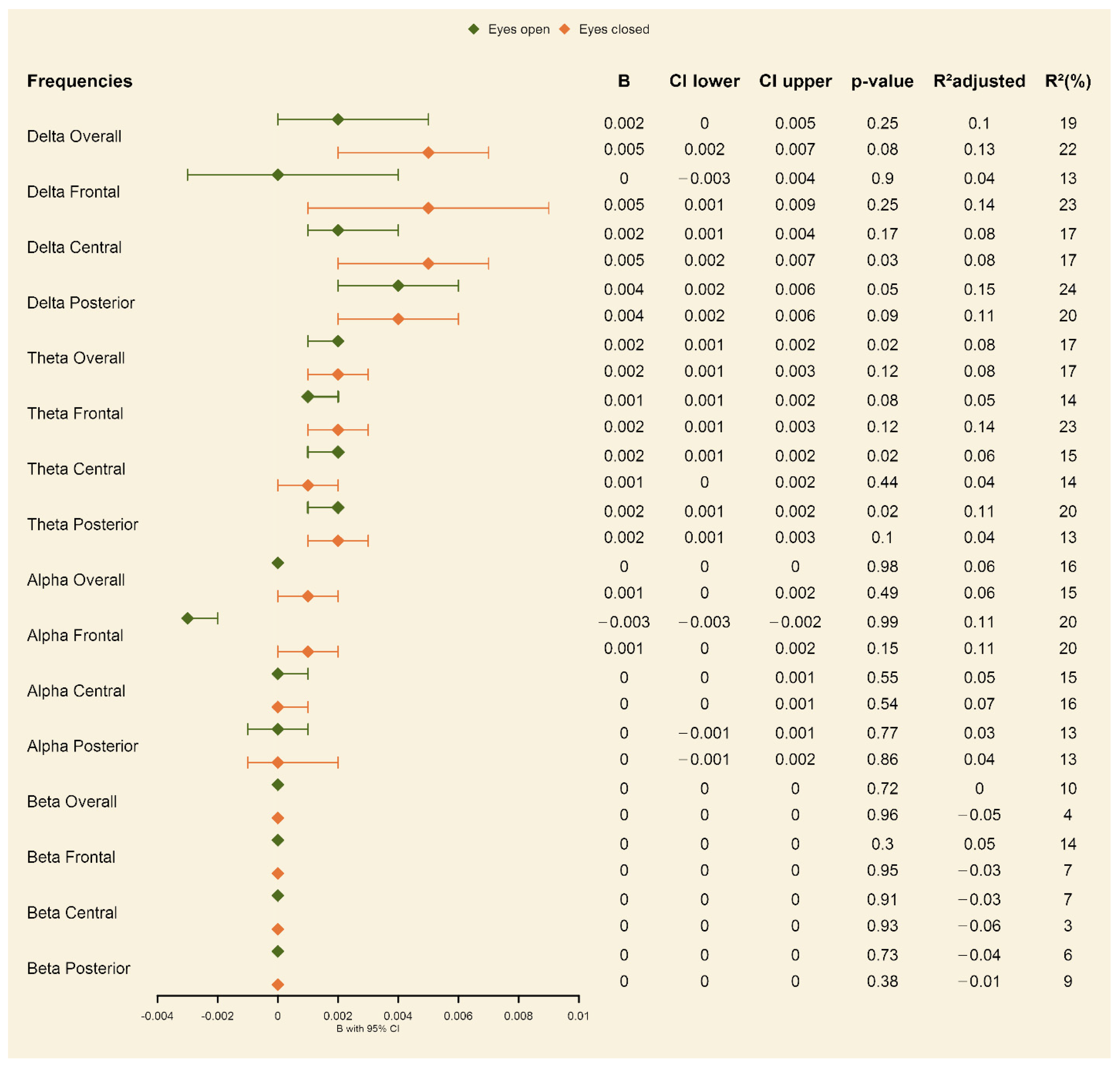
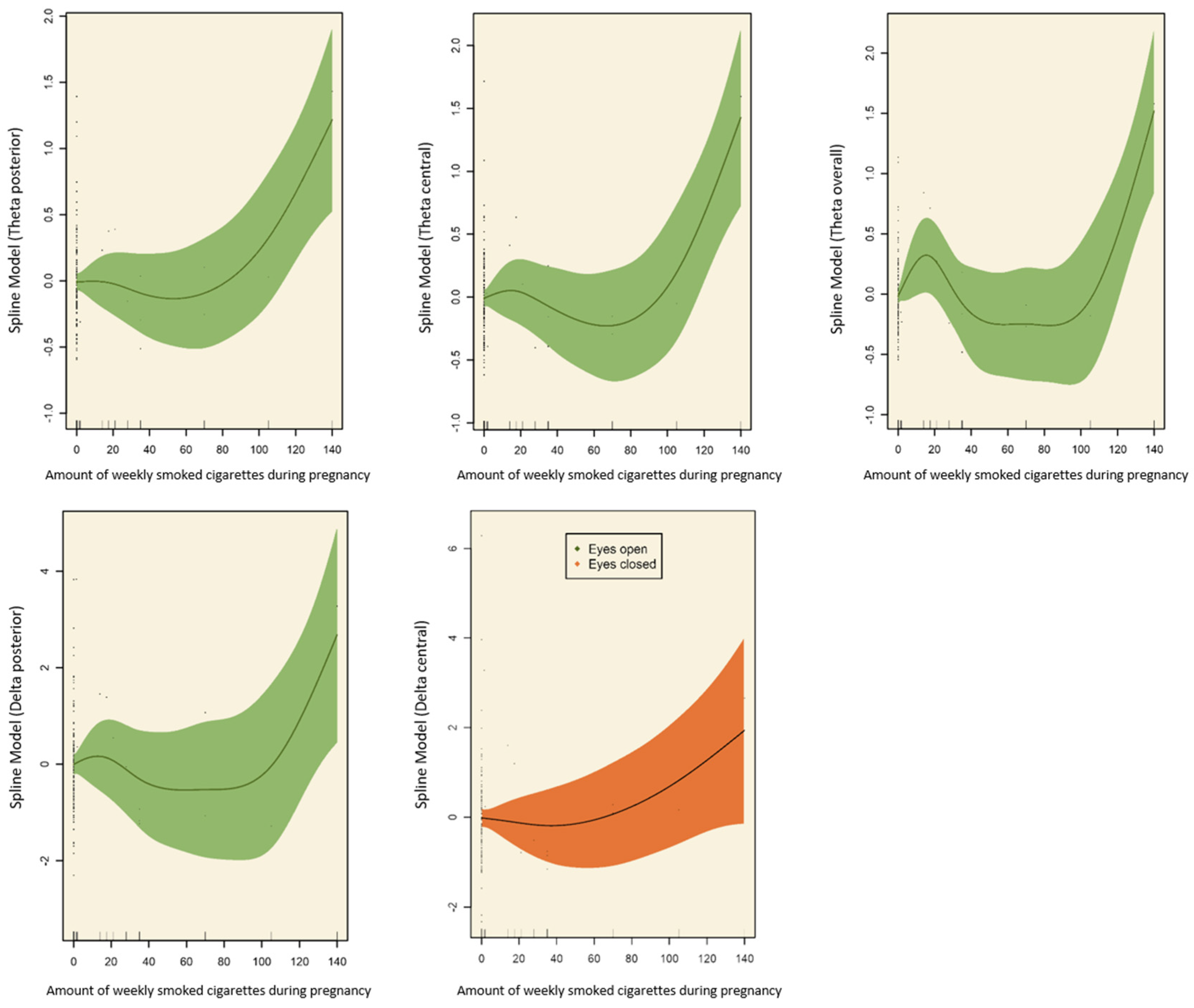
3.1. Effects of Prenatal Tobacco Exposure on Brain Activity

Exposure vs. non-exposure. For the covariate-unadjusted models, we found significant effects of maternal smoking during pregnancy on the delta central, posterior and overall in the frontal, posterior, and overall theta as well as on the frontal, central, and overall alpha brain activity in the eyes-closed resting-state condition (see Table S3 in the online supplement). Here, the prenatally exposed group obtained significantly higher brain activity. In the eyes-open resting-state condition, significant effects were shown in the frontal, central, posterior, and overall theta as well as in the central alpha (see Table S3 in the online Supplementary Materials) brain activity, with prenatally tobacco-exposed children compared to non-exposed children also showing significantly higher brain activity in these areas. When adjusting for the covariates (child’s sex, child’s age, maternal age; maternal psychopathology; maternal smoking before pregnancy; maternal alcohol drinking, week of pregnancy at birth), the effects for the delta and theta frequency bands in both the eyes-open and eyes-closed conditions remained significant (see forest plot in Figure 1; for the effects of the included covariates, see Table S5 in the online Supplementary Materials).
Linear and nonlinear associations. For the covariate-unadjusted models, we found both linear and nonlinear associations between the estimated averaged number of weekly smoked cigarettes during pregnancy and brain activity, with linear associations for the delta central, and nonlinear associations for the theta posterior, theta central, theta overall, and delta posterior. These associations remained significant when adjusting for the covariates (see spline models in Figure 2).

3.2. Effects of Prenatal Tobacco Exposure on ADHD

Exposure vs. non-exposure. For the unadjusted models, we observed a significant effect of maternal smoking during pregnancy on the hyperactivity scores of the children where prenatally exposed children (M = 5.33, SD = 1.45) showed higher rates of hyperactivity compared to prenatally unexposed (M = 4.75, SD = 2.28) children (see Table S3 in the online Supplementary Materials). No significant effects were observed for impulsivity (exposed: M = 5.15, SD = 2.49; unexposed: M = 5.14, SD = 2.05), attention deficits (exposed: M = 5.85, SD = 1.41; unexposed: M = 5.72; SD = 1.59), and the total FBB-ADHD score (exposed: M = 5.58, SD = 1.38; unexposed: M = 5.19, SD = 1.65). When adjusting for the covariates, the effect on hyperactivity did not survive (see Table S4 in the online Supplementary Materials).
Linear and nonlinear associations. For both the covariate-adjusted and -unadjusted models, we did not observe any significant associations between the estimated number of smoked cigarettes of the mother and ADHD-related symptoms.

3.3. Interaction of Prenatal Tobacco Exposure, Brain Activity, and ADHD Symptoms

With respect to the associations between the outcome variables, brain activity, and ADHD-related symptoms, in the eyes-open condition, we found significant partial correlations between impulsivity and frontal (r = 0.21, p < 0.05), central (r = 0.22, p < 0.05) alpha brain activity, central (r = 0.31, p < 0.05), frontal (r = 0.20, p < 0.05), posterior (r = 0.20, p < 0.05), overall delta (r = 0.32, p < 0.05) frequency power, and frontal (r = 0.21, p < 0.05) theta brain activity. Hyperactivity and the total ADHD score were significantly associated with frontal (r = 0.25, p < 0.01; r = 0.22, p < 0.01), central (r = 0.33, p < 0.01; r = 0.31, p < 0.01), posterior (r = 0.23, p < 0.01; r = 0.21, p < 0.01), and overall (r = 0.34, p < 0.01; r = 0.30, p < 0.01) delta brain activity. No significant correlation was observed between attention deficit and any of the frequencies in the eyes-open condition. In the eyes-closed condition, significant correlations were observed between the central delta frequency power and impulsivity (r = 0.22, p < 0.05), hyperactivity (r = 0.22, p < 0.05), and the total ADHD score (r = 0.20, p < 0.05). Posterior delta frequency power was also significantly associated with the total ADHD score (r = 0.21, p < 0.05). As for the eyes open condition, no significant correlation was observed between attention deficit and any of the frequencies in the eyes-closed condition (see Figure S1 in the online Supplementary Materials).
Since brain activity in the alpha, delta, and theta frequencies was positively associated with ADHD-related symptoms (hyperactivity, impulsivity, total ADHD score), mediation analysis was performed in order to test whether there were brain-activity-mediated associations between prenatal maternal smoking and behavioral outcomes (FBB-ADHD scales) in the covariate-adjusted models. Here, differential mediation was observed, where prenatal smoking was significantly associated with brain activity (alpha, delta, and theta frequencies), and brain activity (alpha and delta frequencies) was significantly associated with impulsivity, whereas brain activity did not mediate the relationship between prenatal smoking and ADHD-related behavior (see Tables S5–S10 in the online Supplementary Materials).

4. Discussion

In the present study, we aimed to investigate the influence of prenatal smoking on (a) EEG brain activity, (b) ADHD-related symptoms in school-aged children, and (c) their interaction, taking into account the number of smoked cigarettes during pregnancy as well as considering potential confounding factors including the child’s sex, child’s age, maternal age at the child’s birth, maternal psychopathology, maternal smoking before pregnancy, maternal alcohol drinking during pregnancy, and week of pregnancy at birth.
Previous studies have shown that maternal smoking during pregnancy is a major prenatal risk factor for child development: excessive prenatal tobacco exposure has been related to adverse behavioral outcomes including hyperactivity and impulsivity to deficits in cognitive abilities such as auditory and visual attention performance accuracy, and to changes in the brain’s structure such as a significant reduction in cortical gray matter in young children [11,44,45]. With respect to changes in the brain, the majority of these studies has so far focused either on fetal structural and functional brain development or on brain correlates in newborns [26,45,46,47], mainly using MRI [22,44,48]. Moreover, studies have so far been partly restricted by relatively small sample sizes [26,46,47], and proximal risk factors such as socio-demographical characteristics may have co-determined or masked effects (e.g., [49]).
Capitalizing on the EEG, behavioral, and clinical data with socio-demographical relations such as maternal psychopathology from a larger sample of mothers and their children, we found significant changes in the resting-state EEG as a response to prenatal tobacco, with increased delta and theta brain activity in the resting-state EEG in school-aged exposed compared to non-exposed children. Brain activity in these frequency bands was also significantly related to the number of cigarettes smoked. This is in line with the previously above-mentioned studies that reported an association between prenatal tobacco exposure and changes in the brain of newborns and young children [11,44,45]. In our study, we also found that these effects were independent of potential confounding covariates—both the covariate-unadjusted and -adjusted models were significant. This adds information to the previous literature highlighting that there might be some single specific effects that are rather independent of proximal risk factors. Furthermore, we explored the effects of prenatal maternal smoking on the behavior and clinical symptoms. For the ADHD-related symptoms, the effects of prenatal maternal smoking were only significant in the unadjusted models including potential confounding factors; when controlling for covariates, the effects did not remain significant. This indicates the need to take into account the proximal risk factors, particularly when it comes to the effects of prenatal tobacco exposure on the behavior and clinical symptoms. Some previous studies have also made this point, highlighting that the association of ADHD with prenatal tobacco exposure symptoms might not be directly affected, but might rather covary with intra-household-related factors [15,16]. Rice et al. (2018) stated that the negative causal link between maternal smoking and physiological characteristics such as child birth weight seemed to be a rather direct effect [50], consistent across various study designs [16,51]; however, when it comes to children’s psychopathology, a broader set of risk factors including socioeconomic status (e.g., income conditions), parental psychopathology, maternal stress [50], and maternal age [52] come into play. This was also indicated by a large prospective birth cohort study, where the association between prenatal tobacco exposure and offspring ADHD was found to be significantly related to the confounding factors. The association may therefore not necessarily be indicative of causal intrauterine effects [53].
These and our findings raise several questions for future studies, for example, whether or to which degree these are divergent pathways, and at which point during development factors might strongly interact. Associations between prenatal smoking exposure during pregnancy and behavior problems might not be simply direct or causal (see, e.g., [15]).
In the present study, we also observed significant partial correlations between impulsivity and hyperactivity and frontal alpha (for impulsivity only) as well as frontal, central, posterior, and overall delta frequencies (also for the total ADHD score). Such associations have also been previously reported, for example, increased alpha and theta activity [27,54,55] as well as increased delta activity [54] has been found in children with an ADHD diagnosis compared to typically developing children. This emphasizes that the developmental pathway of prenatal tobacco exposure is complex and involves not only brain–behavior interactions, but also socio-demographic and psychosocial factors. Since the present study involved typically developing children without an ADHD diagnosis, this might have an effect, even at lower rates of ADHD symptoms.
The present study needs to be seen in the light of some limitations. First, we assessed smoking behavior during pregnancy retrospectively with a self-report questionnaire. This could have resulted in a bias, for example, in that parents might underreport smoking during pregnancy due to social desirability. However, double checking the women’s answers regarding their smoking behavior in both the FRAMES and FRANCES assessments provides a reliable measure of their smoking behavior over time. In particular, the fact that the answers were consistent across the two assessments increases the confidence in the validity of the self-reported data. This helps to ensure that the data accurately reflect the smoking behavior of the women during pregnancy and after childbirth. Second, the ADHD scores in our sample were rather low. While on one hand one could argue that this may dampen the generalizability of the findings, on the other hand, we could provide added insights into a low-risk sample, which are also interesting with respect to brain-related associations. Additionally, with only 26 children exposed to prenatal tobacco, the study may be underpowered to detect significant effects on ADHD-related symptoms. While our findings suggest that prenatal tobacco exposure was not significantly associated with hyperactivity in this sample, larger studies may be needed to confirm this result and explore potential interactions with other covariates.

5. Conclusions

In summary, to the best of our knowledge, this is one of the first studies that examined the effects of maternal smoking during pregnancy on brain activity assessed with EEG and behavioral data. While smoking during pregnancy had an impact on the resting-state brain activity in children, which persisted irrespective of socio-demographic factors and hence may indicate long-lasting effects on brain development, the effects on ADHD-related behavior appeared to significantly depend on socio-demographic confounding factors such as maternal alcohol consumption and age of the mother. Future research should aim to investigate the effects of maternal smoking on the oscillatory power in offspring over different time points to better understand the temporal dynamics of the effects and thus enable the characterization of developmental changes in disorder specificity in EEG profiles. Additionally, future studies should consider different ADHD symptom scores and ADHD diagnosis longitudinally. Furthermore, it is crucial to disentangle and integrate socio-demographic and cultural circumstances in a more comprehensive manner.

Supplementary Materials

The following supporting information can be downloaded at: https://www.mdpi.com/article/10.3390/ijerph20064716/s1, Table S1: Brain activity descriptive characteristics for EEG resting-state conditions; Table S2: Association of prenatal tobacco smoking of any severity with EEG brain activity and FBB-ADHD scales among school-aged children, unadjusted for covariates; Table S3: Association of prenatal tobacco smoking of any severity with FBB-ADHD scales among school-aged children, adjusted for covariates; Table S4: Significant associations between covariates included in GAMMs and outcomes; Table S5: Mediation Analyses—FBB-ADHD: Hyperactivity in eyes closed resting state condition; Table S6: Mediation Analyses—FBB-ADHD: Hyperactivity in eyes open resting state condition; Table S7: Mediation Analyses—FBB-ADHD: Inattention in eyes closed resting state condition; Table S8: Mediation Analyses—FBB-ADHD: Inattention in eyes open resting state condition; Table S9: Mediation Analyses—FBB-ADHD: Impulsivity in eyes closed resting state condition; Table S10: Mediation Analyses—FBB-ADHD: Impulsivity in eyes open resting state condition; Figure S1: Partial correlation matrix of EEG resting state conditions and FBB-ADHD questionnaire dimensions; Figure S2: Pearson correlation matrix of Eyes open resting state condition, maternal smoking and FBB-ADHD questionnaire dimensions; Figure S3: Pearson correlation matrix of Eyes closed resting state condition, maternal smoking and FBB-ADHD questionnaire dimensions.

Author Contributions

Conceptualization, K.J., F.N. and T.B.; Formal analysis, K.J.; Funding acquisition, T.B.; Investigation, A.E., P.A.F. and J.K.; Methodology, K.J.; Project administration, A.E., P.A.F. and J.K.; Software, K.J.; Supervision, T.B. and F.N.; Validation, K.J.; Visualization, K.J.; Writing—original draft, K.J.; Writing—review and editing, K.J., A.E., P.A.F., J.K., A.K., S.M., T.B., F.N. and IMAC-Mind Consortium. All authors have read and agreed to the published version of the manuscript.

Funding

The trial was funded by the Federal Ministry of Education and Research Germany (BMBF 01GL1745F, subproject 6). With the public-funded research project IMAC-Mind: Improving Mental Health and Reducing Addiction in Childhood and Adolescence through Mindfulness: Mechanisms, Prevention and Treatment (2017–2021), the Federal Ministry of Education and Research contributes to improving the prevention and treatment of children and adolescents with substance use disorders and associated mental disorders. The project coordination was realized by the German Center of Addiction Research in Childhood and Adolescence at the University Medical Center Hamburg–Eppendorf.

Institutional Review Board Statement

The study was consistent with the Declaration of Helsinki and approved by the Local Ethics Committee of the University Hospital Erlangen (no. 3374 (FRAMES) and 4596 (FRANCES)).

Informed Consent Statement

Informed consent was obtained from all subjects involved in the study.

Data Availability Statement

Due to ethical, legal, or privacy issues are present, the data should not be shared.

Acknowledgments

We thank all members of the IMAC-Mind consortium for their contribution. The consortium comprises seven projects in Germany. The Principle Investigators are: Rainer Thomasius (Coordinator, University Medical Center Hamburg–Eppendorf), Tobias Banaschewski and Herta Flor (both Central Institute for Mental Health, Mannheim), Johannes Kornhuber (Friedrich-Alexander-Universität Erlangen–Nürnberg), Michael Klein (Catholic University of Applied Sciences, Cologne), Olaf Reis (University Medicine of Rostock), Tanja Legenbauer (Ruhr-University Bochum), and Antonia Zapf (University Medical Center Hamburg–Eppendorf). Further members of the consortium are: Nicolas Arnaud, Austermann Maria, Christiane Baldus, Anne Daubmann, Léa Josette Laurenz, Sabrina Kunze, Sophie Luise Schiller, and Michael Supplieth (University Medical Center Hamburg–Eppendorf), Frauke Nees, Karl Gottfried, Stella Guldner, Sabina Millenet, and Maren Prignitz (Central Institute for Mental Health, Mannheim), Bernd Lenz, Peter Fasching, Matthias Beckmann, Hartmut Heinrich, Verena Nadine Buchholz, Anna Eichler, Lothar Häberle, Christiane Mühle, Patrick Stelzl, and Bernhard Volz (Friedrich-Alexander-University Erlangen–Nürnberg), Katharina Ise and Diana Moesgen (Catholic University of Applied Sciences, Cologne), Lucie Waedel (University Medicine of Rostock) as well as Martin Holtmann, Regina Herdering, Carina Maria Huhn, and Laura Mokros (Ruhr-University Bochum). For more information, please visit our homepage: www.IMAC-Mind.de.

Conflicts of Interest

Banaschewski has served in an advisory or consultancy role for Lundbeck, Medice, Neurim Pharmaceuticals, Oberberg GmbH, Shire. He has received conference support or speaker’s fees from Lilly, Medice, Novartis, and Shire. He has been involved in clinical trials conducted by Shire and Viforpharma. He has received royalties from Hogrefe, Kohlhammer, CIP Medien, and Oxford University Press. The present work is unrelated to the above grants and relationships. All authors report no biomedical financial interests or potential conflicts of interest.

References

  1. Rogers, J.M. Smoking and pregnancy: Epigenetics and developmental origins of the metabolic syndrome. Birth. Defects Res. 2019, 111, 1259–1269. [Google Scholar] [CrossRef] [PubMed]
  2. Banderali, G.; Martelli, A.; Landi, M.; Moretti, F.; Betti, F.; Radaelli, G.; Lassandro, C.; Verduci, E. Short and long term health effects of parental tobacco smoking during pregnancy and lactation: A descriptive review. J. Transl. Med. 2015, 13, 327. [Google Scholar] [CrossRef] [PubMed] [Green Version]
  3. Modabbernia, A.; Reichenberg, A.; Ing, A.; Moser, D.A.; Doucet, G.E.; Artiges, E.; Banaschewski, T.; Barker, G.J.; Becker, A.; Bokde, A.L.W.; et al. IMAGEN Consortium.: Linked patterns of biological and environmental covariation with brain structure in adolescence: A population-based longitudinal study. Mol. Psychiatry 2021, 26, 4905–4918. [Google Scholar] [CrossRef] [PubMed]
  4. Orleans, C.T.; Johnson, R.W.; Barker, D.C.; Kaufman, N.J.; Marx, J.F. Helping pregnant smokers quit: Meeting the challenge in the next decade. West. J. Med. 2001, 174, 276–281. [Google Scholar] [CrossRef] [Green Version]
  5. Braun, M.; Klingelhöfer, D.; Oremek, G.M.; Quarcoo, D.; Groneberg, D.A. Influence of Second-Hand Smoke and Prenatal Tobacco Smoke Exposure on Biomarkers, Genetics and Physiological Processes in Children-An Overview in Research Insights of the Last Few Years. Int. J. Environ. Res. Public Health 2020, 17, 3212. [Google Scholar] [CrossRef]
  6. Clifford, A.; Lang, L.; Chen, R. Effects of maternal cigarette smoking during pregnancy on cognitive parameters of children and young adults: A literature review. Neurotoxicology Teratol. 2012, 34, 560–570. [Google Scholar] [CrossRef]
  7. Laucht, M.; Schmidt, M.H. Mütterliches Rauchen in der Schwangerschaft: Risikofaktor für eine ADHS des Kindes? Maternal smoking during pregnancy: Risk factor for ADHD in the offspring? Z. Kinder Jugendpsychiatr. Psychother. 2004, 32, 177–185. [Google Scholar] [CrossRef]
  8. Melchior, M.; Hersi, R.; van der Waerden, J.; Larroque, B.; Saurel-Cubizolles, M.J.; Chollet, A.; Galéra, C.L. EDEN Mother-Child Cohort Study Group. Maternal tobacco smoking in pregnancy and children’s socio-emotional development at age 5: The EDEN mother-child birth cohort study. Eur. Psychiatry 2015, 30, 562–568. [Google Scholar] [CrossRef]
  9. Weitzman, M.; Byrd, R.S.; Aligne, C.A.; Moss, M. The effects of tobacco exposure on children’s behavioral and cognitive functioning: Implications for clinical and public health policy and future research. Neurotoxicol. Teratol. 2002, 24, 397–406. [Google Scholar] [CrossRef]
  10. Neuman, R.J.; Lobos, E.; Reich, W.; Henderson, C.A.; Sun, L.W.; Todd, R.D. Prenatal Smoking Exposure and Dopaminergic Genotypes Interact to Cause a Severe ADHD Subtype. Biol. Psychiatry 2007, 61, 1320–1328. [Google Scholar] [CrossRef]
  11. Dong, T.; Hu, W.; Zhou, X.; Lin, H.; Lan, L.; Hang, B.; Lv, W.; Geng, Q.; Xia, Y. Prenatal exposure to maternal smoking during pregnancy and attention-deficit/hyperactivity disorder in offspring: A meta-analysis. Reprod. Toxicol. 2018, 76, 63–70. [Google Scholar] [CrossRef]
  12. D’Onofrio, B.M.; Van Hulle, C.A.; Waldman, I.D.; Rodgers, J.L.; Harden, K.P.; Rathouz, P.J.; Lahey, B.B. Smoking during pregnancy and offspring externalizing problems: An exploration of genetic and environmental confounds. Dev. Psychopathol. 2008, 20, 139–164. [Google Scholar] [CrossRef] [Green Version]
  13. Sciberras, E.; Mulraney, M.; Silva, D.; Coghill, D. Prenatal risk factors and the etiology of ADHD—Review of existing evidence. Curr. Psychiatry Rep. 2017, 19, 1. [Google Scholar] [CrossRef]
  14. Tarver, J.; Daley, D.; Sayal, K. ADHD: An updated review of the essential facts. Child Care Health Dev. 2014, 40, 762–774. [Google Scholar] [CrossRef]
  15. Thapar, A.; Rice, F.; Hay, D.; Boivin, J.; Langley, K.; van den Bree, M.; Rutter, M.; Harold, G. Prenatal smoking might not cause attention-deficit/hyperactivity disorder: Evidence from a novel design. Biol. Psychiatry 2009, 66, 722–727. [Google Scholar] [CrossRef] [Green Version]
  16. Langley, K.; Heron, J.; Smith, G.D.; Thapar, A. Maternal and paternal smoking during pregnancy and risk of ADHD symptoms in offspring: Testing for intrauterine effects. Am. J. Epidemiol. 2012, 176, 261–268. [Google Scholar] [CrossRef] [Green Version]
  17. El Marroun, H.; Schmidt, M.N.; Franken, I.H.; Jaddoe, V.W.; Hofman, A.; van der Lugt, A.; Verhulst, F.C.; Tiemeier, H.; White, T. Prenatal tobacco exposure and brain morphology: A prospective study in young children. Neuropsychopharmacology 2014, 39, 792–800. [Google Scholar] [CrossRef] [Green Version]
  18. Derauf, C.; Lester, B.M.; Neyzi, N.; Kekatpure, M.; Gracia, L.; Davis, J.; Kallianpur, K.; Efird, J.T.; Kosofsky, B. Subcortical and cortical structural central nervous system changes and attention processing deficits in preschool-aged children with prenatal methamphetamine and tobacco exposure. Dev. Neurosci. 2012, 34, 327–341. [Google Scholar] [CrossRef] [Green Version]
  19. Paus, T.; Nawazkhan, I.; Leonard, G.; Perron, M.; Pike, G.B.; Pitiot, A.; Richer, L.; Veillette, S.; Pausova, Z. Corpus callosum in adolescent offspring exposed prenatally to maternal cigarette smoking. Neuroimage 2008, 40, 435–441. [Google Scholar] [CrossRef]
  20. Ekblad, M.; Korkeila, J.; Lehtonen, L. Smoking during pregnancy affects foetal brain development. Acta Paediatr. 2015, 104, 12–18. [Google Scholar] [CrossRef]
  21. Ekblad, M.; Korkeila, J.; Parkkola, R.; Lapinleimu, H.; Haataja, L.; Lehtonen, L.; PIPARI Study Group. Maternal Smoking during Pregnancy and Regional Brain Volumes in Preterm Infants. J. Pediatr. 2010, 156, 185–190.e1. [Google Scholar] [CrossRef] [PubMed]
  22. Holz, N.E.; Boecker, R.; Baumeister, S.; Hohm, E.; Zohsel, K.; Buchmann, A.F.; Blomeyer, D.; Jennen-Steinmetz, C.; Hohmann, S.; Wolf, I.; et al. Effect of prenatal exposure to tobacco smoke on inhibitory control: Neuroimaging results from a 25-year prospective study. JAMA Psychiatry 2014, 71, 786–796. [Google Scholar] [CrossRef] [PubMed]
  23. Baillet, S.; Mosher, J.C.; Leahy, R.M. “Electromagnetic Brain Mapping,” Signal Processing Magazine. IEEE 2001, 18, 14–30. [Google Scholar] [CrossRef]
  24. Brown, T.T.; Jernigan, T.L. Brain development during the preschool years. Neuropsychol. Rev. 2012, 22, 313–333. [Google Scholar] [CrossRef] [PubMed] [Green Version]
  25. Bridwell, D.A.; Cavanagh, J.F.; Collins, A.G.E.; Nunez, M.D.; Srinivasan, R.; Stober, S.; Calhoun, V.D. Moving Beyond ERP Components: A Selective Review of Approaches to Integrate EEG and Behavior. Front. Hum. Neurosci. 2018, 12, 106. [Google Scholar] [CrossRef] [Green Version]
  26. Shuffrey, L.C.; Shuffrey, L.C.; Myers, M.M.; Isler, J.R.; Lucchini, M.; Sania, A.; Pini, N.; Nugent, J.D.; Condon, C.; Ochoa, T.; et al. PASS Network. Association Between Prenatal Exposure to Alcohol and Tobacco and Neonatal Brain Activity: Results From the Safe Passage Study. JAMA Netw. Open 2020, 3, e204714. [Google Scholar] [CrossRef]
  27. Barry, R.J.; Clarke, A.R.; Johnstone, S.J.; Brown, C.R.; Bruggemann, J.M.; van Rijbroek, I. Caffeine effects on resting-state arousal in children. Int. J. Psychophysiol. 2009, 73, 355–361. [Google Scholar] [CrossRef]
  28. Machado, C.; Estévez, M.; Leisman, G.; Melillo, R.; Rodríguez, R.; DeFina, P.; Hernández, A.; Pérez-Nellar, J.; Naranjo, R.; Chinchilla, M.; et al. QEEG spectral and coherence assessment of autistic children in three different experimental conditions. J. Autism Dev. Disord. 2015, 45, 406–424. [Google Scholar] [CrossRef] [Green Version]
  29. Karakaş, S. A comparative review of the psychophysiology of attention in children with and without attention deficit hyperactivity disorder. Int. J. Psychophysiol. 2022, 177, 43–60. [Google Scholar] [CrossRef]
  30. Clarke, A.R.; Barry, R.J.; McCarthy, R. Correlation Between EEG Activity and Behavior in Children with Attention-Deficit/Hyperactivity Disorder. J. Neurother. 2011, 15, 193–199. [Google Scholar] [CrossRef] [Green Version]
  31. Rodríguez-Martínez, E.I.; Angulo-Ruiz, B.Y.; Arjona-Valladares, A.; Rufo, M.; Gómez-González, J.; Gómez, C.M. Frequency coupling of low and high frequencies in the EEG of ADHD children and adolescents in closed and open eyes conditions. Res. Dev. Disabil. 2020, 96, 103520. [Google Scholar] [CrossRef]
  32. Langley, K.; Turic, D.; Rice, F.; Holmans, P.; van den Bree, M.B.; Craddock, N.; Kent, L.; Owen, M.J.; O’Donovan, M.C.; Thapar, A. Testing for Gene x Environment Interaction Effects in Attention Deficit Hyperactivity Disorder and Associated Antisocial Behavior. Am. J. Med. Genet. Part B 2008, 147B, 49–53. [Google Scholar] [CrossRef]
  33. Reulbach, U.; Bleich, S.; Knorr, J.; Burger, P.; Fasching, P.A.; Kornhuber, J.; Beckmann, M.W.; Goecke, T.W. Pre-, peri- and postpartal depression. Fortschr. Neurol. Psychiat 2009, 77, 708–713. [Google Scholar] [CrossRef]
  34. Eichler, A.; Walz, L.; Grunitz, J.; Grimm, J.; van Doren, J.; Raabe, E.; Goecke, T.W.; Fasching, P.A.; Beckmann, M.W.; Kornhuber, J.; et al. Children of prenatally depressed mothers: Externalizing and internalizing symptoms are accompanied by reductions in specific social-emotional competencies. J. Child Fam. Stud. 2017, 26, 3135–3144. [Google Scholar] [CrossRef]
  35. Hein, A.; Rauh, C.; Engel, A.; Häberle, L.; Dammer, U.; Voigt, F.; Fasching, P.A.; Faschingbauer, F.; Burger, P.; Beckmann, M.W.; et al. Socioeconomic status and depression during and after pregnancy in the Franconian Maternal Health Evaluation Studies (FRAMES). Arch. Gynecol. Obstet. 2014, 289, 755–763. [Google Scholar] [CrossRef]
  36. Eichler, A.; Hudler, L.; Grunitz, J.; Grimm, J.; Raabe, E.; Goecke, T.W.; Fasching, P.A.; Beckmann, M.W.; Kratz, O.; Moll, G.H.; et al. Effects of prenatal alcohol consumption on cognitive development and ADHD-related behaviour in primary-school age: A multilevel study based on meconium ethyl glucuronide. J. Child Psychol. Psychiatry 2018, 59, 110–118. [Google Scholar] [CrossRef]
  37. Breuer, D.; Döpfner, M. Entwicklung eines Fragebogens zur Erfassung von Aufmerksamkeitsdefizit-/Hyperaktivitätsstörungen (ADHS) bei Vorschulkindern im Eltern- und im Erziehungsurteil. Z. Für Entwickl. Und Pädagogische Psychol. 2008, 40, 40–48. [Google Scholar] [CrossRef]
  38. Wood, S.N. Stable and efficient multiple smoothing parameter estimation for generalized additive models. J. Amer. Statist. Ass. 2004, 99, 673–686. [Google Scholar] [CrossRef] [Green Version]
  39. Wood, S.N. Fast stable restricted maximum likelihood and marginal likelihood estimation of semiparametric generalized linear models. J. R. Stat. Soc. B 2011, 73, 3–36. [Google Scholar] [CrossRef] [Green Version]
  40. Chen, C. Generalized additive mixed models. Commun. Stat. Theory Methods 2000, 29, 1257–1271. [Google Scholar] [CrossRef]
  41. Baayen, H.; Vasishth, S.; Kliegl, R.; Bates, D. The cave of shadows: Addressing the human factor with generalized additive mixed models. J. Mem. Lang. 2017, 94, 206–234. [Google Scholar] [CrossRef] [Green Version]
  42. Lin, X.; Zhang, D. Inference in generalized additive mixed models by using smoothing splines. J. R. Stat. Soc. Ser. B Stat. Methodol. 1999, 61, 381–400. [Google Scholar] [CrossRef] [Green Version]
  43. Lees, B.; Mewton, L.; Jacobus, J.; Valadez, E.A.; Stapinski, L.A.; Teesson, M.; Tapert, S.F.; Squeglia, L.M. Association of Prenatal Alcohol Exposure With Psychological, Behavioral, and Neurodevelopmental Outcomes in Children From the Adolescent Brain Cognitive Development Study. Am. J. Psychiatry 2020, 177, 1060–1072. [Google Scholar] [CrossRef] [PubMed]
  44. Rivkin, M.J.; Davis, P.E.; Lemaster, J.L.; Cabral, H.J.; Warfield, S.K.; Mulkern, R.V.; Robson, C.D.; Rose-Jacobs, R.; Frank, D.A. Volumetric MRI study of brain in children with intrauterine exposure to cocaine, alcohol, tobacco, and marijuana. Pediatrics 2008, 121, 741–750. [Google Scholar] [CrossRef] [Green Version]
  45. Albers, L.; Sobotzki, C.; Kuß, O.; Ajslev, T.; Batista, R.F.; Bettiol, H.; Brabin, B.; Buka, S.L.; Cardoso, V.C.; Clifton, V.L.; et al. Maternal smoking during pregnancy and offspring overweight: Is there a dose-response relationship? An individual patient data meta-analysis. Int. J. Obes. 2018, 42, 1249–1264. [Google Scholar] [CrossRef]
  46. Havlicek, V.; Childiaeva, R.; Chernick, V. EEG Frequency spectrum characteristics of sleep states in infants ofalcoholic mothers. Neuropadiatrie 1977, 8, 360–373. [Google Scholar] [CrossRef]
  47. Loffe, S.; Childiaeva, R.; Chernick, V. Prolonged effects of maternal alcohol ingestion on the neonatal electroencephalogram. Pediatrics 1984, 74, 330–335. [Google Scholar] [CrossRef]
  48. Jacobsen, L.K.; Slotkin, T.A.; Mencl, W.E.; Frost, S.J.; Pugh, K.R. Gender-specific effects of prenatal and adolescent exposure to tobacco smoke on auditory and visual attention. Neuropsychopharmacology 2007, 32, 2453–2464. [Google Scholar] [CrossRef] [Green Version]
  49. Cornelius, M.D.; Day, N.L. Developmental consequences of prenatal tobacco exposure. Curr. Opin. Neurol. 2009, 22, 121–125. [Google Scholar] [CrossRef] [Green Version]
  50. Rice, F.; Langley, K.; Woodford, C.; Davey Smith, G.; Thapar, A. Identifying the contribution of prenatal risk factors to offspring development and psychopathology: What designs to use and a critique of literature on maternal smoking and stress in pregnancy. Dev. Psychopathol. 2018, 30, 1107–1128. [Google Scholar] [CrossRef]
  51. Abraham, M.; Alramadhan, S.; Iniguez, C.; Duijts, L.; Jaddoe, V.W.; Den Dekker, H.T.; Crozier, S.; Godfrey, K.M.; Hindmarsh, P.; Vik, T.; et al. A systematic review of maternal smoking during pregnancy and fetal measurements with meta-analysis. PLoS ONE 2017, 12, e0170946. [Google Scholar] [CrossRef] [Green Version]
  52. Gustavson, K.; Ystrom, E.; Stoltenberg, C.; Susser, E.; Surén, P.; Magnus, P.; Knudsen, G.P.; Smith, G.D.; Langley, K.; Rutter, M.; et al. Smoking in Pregnancy and Child ADHD. Pediatrics 2017, 139, e20162509. [Google Scholar] [CrossRef] [Green Version]
  53. Moylan, S.; Gustavson, K.; Øverland, S.; Karevold, E.B.; Jacka, F.N.; Pasco, J.A.; Berk, M. The impact of maternal smoking during pregnancy on depressive and anxiety behaviors in children: The Norwegian Mother and Child Cohort Study. BMC Med. 2015, 3, 13–24. [Google Scholar] [CrossRef] [Green Version]
  54. Kitsune, G.L.; Cheung, C.H.; Brandeis, D.; Banaschewski, T.; Asherson, P.; McLoughlin, G.; Kuntsi, J. A matter of time: The influence of recording context on EEG spectral power in adolescents and young adults with ADHD. Brain Topogr. 2015, 28, 580–590. [Google Scholar] [CrossRef] [Green Version]
  55. Tye, C.; Rijsdijk, F.; McLoughlin, G. Genetic overlap between ADHD symptoms and EEG theta power. Brain Cogn. 2014, 87, 168–172. [Google Scholar] [CrossRef]
Figure 1. Forest plot showing the association between smoking during pregnancy and offspring EEG activity when comparing the exposed and non-exposed groups, adjusted for covariates (child’s sex, child’s age, maternal age, maternal psychopathology, maternal smoking before pregnancy, maternal alcohol drinking, week of pregnancy at birth).
Ijerph 20 04716 g001
Figure 2. Spline models demonstrating a significant relationship between the estimated number of smoked cigarettes of the mother during pregnancy and offspring brain activity (delta and theta frequency bands), adjusted for covariates (child’s sex, child’s age, maternal age, maternal psychopathology, maternal smoking before pregnancy, maternal alcohol drinking, week of pregnancy at birth).
Ijerph 20 04716 g002
Table 1. Sample characteristics with children divided into two groups of the tobacco non-exposed and prenatal tobacco-exposed children.
CharacteristicTobacco Unexposed
Children
(n = 116)
Children With
Prenatal
Tobacco
Exposure
(n = 26)
Youth Variables
n%n%p
Sex 0.434
Male
Female
59
57
50.9
49.1
11
15
42.3
57.7
Prenatal alcohol
exposure
0.562
No
Yes
95
21
81.9
18.1
20
6
76.9
23.1
MeanSDMeanSDp
Age (years)7.720.657.760.740.781
Gestational age39.411.3339.231.450.533
Birth weight3453.91494.283370.77520.340.446
Intelligence quotient *104.2310.25104.317.180.972
Parent Variables
Age at giving birth 32.864.1231.885.150.299
n%n%p
Prenatal marital status 0.068
Married
Single parent
113
3
97.4
2.6
23
3
88.5
11.5
Maternal
Psychopathology

0.536
No
Yes
Unknown
68
29
19
58.6
25.0
16.4
17
5
4
65.4
19.2
15.3
Paternal
Psychopathology

0.461
No
Yes
Unknown
92
12
12
79.3
10.3
8.3
20
2
4
76.9
7.7
15.3
* Intelligence quotient was assessed with the Intelligence and Development Scales (IDS) test battery (Grob, Meyer, 2013).
Table 2. Detailed information on maternal smoking behavior.
SmokedDid Not SmokePassive Smoking
n (%)
[Min, Max]
M (SD)
n (%)n (%)
[Min, Max]
M (SD)
Before pregnancy40 (28.2)
[2, 300]
25.21 (50.72)
102 (71.8)33 (32.4)
[3, 210]
102.39 (57.44)
1. Trimester26 (18.3)
[2, 140]
5.25 (19.28)
116 (81.7)40 (34.5)
[4, 210]
98.16 (57.09)
2. Trimester24 (16.9)
[2, 140]
5.07 (19.25)
118 (83.1)37 (31.4)
[4, 210]
99.05 (56.39)
3. Trimester24 (16.9)
[2, 140]
4.83 (19.01)
118 (83.1)35 (29.7)
[4, 210]
99.00 (56.48)
During pregnancy26 (18.3)
[2, 140]
5.09 (19.23)
116 (81.7)35 (30.2)
[4, 210]
100.51 (55.27)
Disclaimer/Publisher’s Note: The statements, opinions and data contained in all publications are solely those of the individual author(s) and contributor(s) and not of MDPI and/or the editor(s). MDPI and/or the editor(s) disclaim responsibility for any injury to people or property resulting from any ideas, methods, instructions or products referred to in the content.

