Article

Salinity Moderated the Toxicity of Zinc Oxide Nanoparticles (ZnO NPs) towards the Early Development of Takifugu obscurus

1
Center for Eco-Environmental Research, Nanjing Hydraulic Research Institute, Nanjing 210029, China
2
State Key Laboratory of Hydrology-Water Resources and Hydraulic Engineering, Nanjing Hydraulic Research Institute, Nanjing 210029, China
3
Yangtze Institute for Conservation and Green Development, Nanjing 210029, China
4
China Three Gorges Corporation, Wuhan 430010, China
*
Author to whom correspondence should be addressed.
These authors contribute equally to this paper.
Int. J. Environ. Res. Public Health 2023, 20(4), 3209; https://doi.org/10.3390/ijerph20043209
Submission received: 5 November 2022 / Revised: 30 January 2023 / Accepted: 10 February 2023 / Published: 12 February 2023

Abstract

:
ZnO nanoparticles (ZnO NPs) have been applied in a wide range of fields due to their unique properties. However, their ecotoxicological threats are reorganized after being discharged. Their toxic effect on anadromous fish could be complicated due to the salinity fluctuations during migration between freshwater and brackish water. In this study, the combined impact of ZnO NPs and salinity on the early development of a typical anadromous fish, obscure puffer (Takifugu obscurus), was evaluated by (i) observation of the nanoparticle characterization in salt solution; (ii) quantification of the toxicity to embryos, newly hatched larvae, and larvae; and (iii) toxicological analysis using biomarkers. It is indicated that with increased salinity level in brackish water (10 ppt), the toxicity of ZnO NPs decreased due to reduced dissolved Zn2+ content, leading to higher hatch rate of embryos and survival rate of larvae than in freshwater (0 ppt). The irregular antioxidant enzyme activity changes are attributed to the toxic effects of nanoparticles on CAT (catalase), but further determination is required. The results of present study have the significance to guide the wildlife conservation of Takifugu obscurus population.

1. Introduction

Nanoparticles (NPs) are commonly referred to particles with at least one dimension less than 100 nm [1]. The small sizes can lead to several unique physiochemical properties, and NPs are enabled to achieve outstanding optical sensitivity, reactivity, and conductivity compared with the bulk materials [2]. According to Bouwmeester et al. [3], with the revolutionary development of nanotechnology, various types of NPs are increasingly used in numerous commercial and industrial products. Among different types of NPs, the metal oxide nanoparticles (MNPs) have been mass produced for over a decade and applied in consumer goods, agriculture, and industry [4]. In particular, zinc oxide nanoparticles (ZnO NPs) are considered to be one of the most widely employed, which have shown the capabilities of photocatalysis and photooxidation [5]. As mentioned by Keller, McFerran, Lazareva, and Suh [6], the annual global production of ZnO NPs is supposed to be over 30,000 metric tons, and the final disposals are landfills, soils, water bodies, or the atmosphere, which could be habitats for massive organisms. Therefore, the potential ecotoxicological risks of ZnO NPs has recently become a hot topic.
ZnO NPs released from various sources such as transport, industry, and agriculture appear to eventually accumulate in aquatic systems. The contaminated water bodies could lead to a large amount of interactions with the aquatic ecosystems [7]. Previous studies have shown the toxicity of ZnO NPs to different kinds of aquatic organisms including vertebrates, invertebrates, algae, plants, and bacteria [8,9,10]. The main attention of these prior toxicity studies has been paid to fish, especially the zebrafish, because of its comprehensively understood biological mechanism as one of the model test organisms approved by regulatory agencies [11]. For example, the experiment conducted by Bai et al. [12] based on zebrafish embryos 96 h after fertilization (hpf) showed that exposure to ZnO NPs (30 nm) could cause the effects of killing embryos, retarding hatching, reducing larval length, and tail deformities. In addition, ZnO NPs can accumulate to toxic concentrations in mammals through aquatic and terrestrial food chains [13]. As summarized by Golbamaki et al. [14], ZnO NPs could be cytotoxic and genotoxic to human epidermal, liver, and kidney cells, as significant cell and DNA damages were observed post exposure in several previous research. Regarding the possible toxicity mechanisms, Zhu et al. [15] suggested the combined influence of nanoparticle aggregates and dissolved Zn2+, while Yu et al. [16] proposed that aggregation and precipitation could contribute to the reduction of toxicity. According to Bacchetta et al. [17], the soluble Zn2+ passing through the cell membrane and combining with mitochondria can activate the apoptotic pathway to promote the production of reactive oxygen species (ROS) and trigger the autophagy process upon oxidative stress. Thus, ZnO NPs undoubtedly pose great potential hazards to aquatic organisms and the ecosystem and are thus regarded to be “extremely toxic” [18].
The current ZnO NPs hazard assessments are commonly based on traditional toxic effect tests under one designed condition, which can lead to biased results for overlooking the differences of the environment in each region and time period [17]. There are types of aquatic ecosystems worldwide, including freshwater, marine, lake, river, pond, and wetlands, with distinct characteristics of temperature, salinity, and pH [19]. Hence, it should be noted that research on the toxic effects of ZnO NPs under the impact of different environmental factors could be important for further understanding and assessing their ecotoxicological risks [9]. According to Wagner et al. [20], ZnO NPs can be less toxic under pH 6.5 to pH 8.5 since their control of nanoparticles’ surface charge could decrease the solubility, thereby facilitating aggregation and lowering stability. Additionally, the study using Thalassiosira pseudonana carried out by Yung et al. [21]. indicated that ZnO NPs at higher temperature and salinity could constitute aggregates in larger volume and produce Zn2+ in lower concentration, resulting in lowered toxicity. The experiment conducted by Lai et al. [22] obtained a similar result that high salinity could reduce the toxic effects of ZnO NPs to the copepod Tigriopus japonicus, which is possibly because of the improved osmotic pressure of copepods and ion dissolution with lower salinity. Based on relevant experimental results, raising salinity is considered to be a potential moderator against the toxicity of metal oxide nanoparticles [23]. Therefore, the influences of salinity on the physical and chemical properties of ZnO NPs could have a great research value.
In order to explore the combined toxic effects of salinity and ZnO NPs in different aquatic ecosystems, the optimal sample to ensure the accuracy of the experimental results is anadromous migratory fish due to its strong ability adapting to salinity changes during migration processes [24]. As a typical anadromous fish mainly distributed in the Chinese coastal waters and the middle and lower Yangtze River, juveniles of obscure puffer (Takifugu obscurus) move from rivers and lakes to offshore seas after being hatched and fatten, indicating large fluctuations in salinity levels for the juveniles [23,25]. The environmental stress during their vulnerable early development periods could cause a negative impact on the growth, migration, and reproduction processes of individuals or the population [26]. Hence, it is necessary to understand the capacity and response of obscure puffer embryos and larvae to the nanoparticles’ potential toxicity under the influence of salinity. As for the specific defense mechanism against the oxidative stress caused by ZnO NPs, the research of Kim et al. [27] on the resistance of T. obscurus to cadmium toxicity confirmed the function of highly expressed antioxidant genes. The produced harmful superoxide anion could be converted into H2O2 through superoxide dismutase (SOD) and further decomposed into H2O and O2 through catalase (CAT) [28,29]. According to Frankič et al. [30], malondialdehyde (MDA) is one of the ultimate products of lipids peroxidation and has been evaluated in dozens of related studies due to its reflection ability of cellular damages through toxins. Furthermore, the adenosine triphosphatases (ATPases) index is another important biochemical parameter for obscure puffer juveniles because of their undeveloped intestinal osmoregulation system and dominated Na+/K+ ion-transport regulation [31]. Therefore, the determination of those four biochemical indicators could contribute to the further identification of the toxicity mechanism of nanoparticles on the obscure puffer juveniles.
To evaluate the potential impacts of ZnO NPs and salinity on the early development of T. obscurus, this work aimed to quantify the toxic effects via the hatching rate of embryos and the survival rate of larvae as well as determine the toxicity mechanism based on the configuration of nanoparticles and the measurement of biochemical indices including CAT, SOD, MDA, and Na+/K+ ATPase.

2. Materials and Methods

All experiments in this study were approved by the ACUC (Animal Care and Use Committee) of Nanjing Normal University, Nanjing, China, and carried out according to its guidelines (Research Permit Number: SYXK2015-0028).

2.1. Preparation of Embryos and Larvae

After collection from a fish farm located at Yangzhong City, Jiangsu Province, China, the T. obscurus adults were conveyed to lab. The adult fish were temporarily stored in a fish tank (100 L freshwater), whose environmental circumstance were similar to the fish farm. Based on the method of Yang and Chen [25], the luteinizing-hormone-releasing hormone analogue was applied on 3 males and 1 female of adult fish, and fertilized eggs were acquired. The eggs were then retained in a tank with temperature of 23 ± 1 °C, pH value of 7.1 ± 0.3, and constant aeration. Damaged and dead fertilized eggs was removed after 16 h via wide-mouth pipettes. Part of the healthy eggs were transported for subsequent embryo-related experiments. The eggs were slowly stirred with feathers to evenly dispense the eggs. The remaining eggs were used for the hatching process for collection of larvae. Larvae in good health were randomly selected for further experiments around 24 h after hatching.

2.2. Preparation of ZnO NPs

The ZnO NPs (diameter = 50 nm) were purchased from Nanjing Haitai Nanoparticles Ltd. The stock solution of ZnO NPs (0.5 g/L) was prepared before the exposure treatments. Nanoparticles with a diameter around 25 nm were suspended in Milli-Q water, which was further processed in an ice-water bath for half an hour using the ultrasonic vibration at 40 kHz. The concentration of the stock ZnO NPs solution was 0.5 g/L, which was then diluted to six levels, including 0 mg/L, 5 mg/L, 10 mg/L, 20 mg/L, 50 mg/L, and 100 mg/L. In order to assess the morphological configuration of ZnO NPs, H-7650 scanning electron microscopy (SEM, Hitachi, H-7650, Tokyo, Japan) was employed for the suspension.

2.3. Experimental Procedure

Based on a full factorial experiment with three replicates for each treatment and two elements, the salinity was employed as 0 ppt and 10 ppt, and the concentration of ZnO NPs was employed as 0 mg/L, 5 mg/L, 10 mg/L, 20 mg/L, 50 mg/L, and 100 mg/L. Tissue culture-treated (TCT) 6-well plates containing 3 mL of ZnO NPs solution in different concentrations were applied to rear 10 embryos or larvae per well. The body length of newly hatched larvae is generally less than 1 mm, suggesting that the capacity of 16.7 mL per well is adequate. For the adjustment process of embryos or larvae to the salinity changes, 2 ppt was raised per hour with continuous aeration for each solution under the salinity of 10 ppt. The replacement of half ZnO NPs solution with newly prepared solution was then carried out daily to ensure no significant alteration of the salinity and nanoparticles levels. The culture conditions were set for all tests with temperature at 23 ± 1 °C, dissolved oxygen content over 5.2 mg/L, pH value adjusted to 7.1 ± 0.3, and duration of light and dark to 12 h each. For each embryos-based treatment, the hatching rate of embryos and the survival rate of new larvae 24 h post hatching were recorded. For each larvae-based treatment, the dead larvae were removed, and the survival rate was recorded every 24 h for 96 h.

2.4. Determination of Biochemical Parameters

In order to determine the damage extent and toxicity mechanism of ZnO NPs to T. obscurus larvae, the larvae-based treatments with 50 mg/L ZnO NPs (the concentration approximately corresponds to lethal dose 50%) under 0 ppt and 10 ppt of salinity condition were selected for the measurement of biomarkers. Ten experimental larvae were respectively and randomly selected at 0, 24, 48, 72, and 96 h. According to Sun et al. [32], the biomarkers of hatched larvae with relatively less body length could be determined through homogenates of whole fish. Prior to the homogenization process in a Dounce homogenizer, larvae were rinsed using 0.7% normal saline with a temperature around 0 °C and dried through filter papers. After that, the larvae were each transferred in 2 mL of 0.7% normal saline. The centrifugation (4000× g) procedure at 4 °C was then performed for 10 min for the elimination of homogenate’s fragments of cartilages and cells, and the supernatants were used for the measurement of biomarkers. In this study, the diagnostic reagent kits for the determination of CAT (U/mg protein), SOD (U/mg protein), MDA (nmol/mg protein), and Na+/K+ ATPase (μmol Pi/mg protein/h) were purchased from the NanJing Jian Cheng Bioengineering Institute (China). The diameters of ZnO NPs at 0.5, 1, 6, 12, and 24 h were detected by Dynamic Light Scattering (DLS, Nicomp, Z3000, Tokyo, Japan).

2.5. Statistical Analysis

Two levels of salinity were selected in the exposure experiment―0 ppt represents freshwater, and 10 ppt represents brackish water. Four days of exposure was determined due to no living larvae observed after 96 h of cultivation with 100 mg/L ZnO NPs under 0 ppt. Differences between treatments are calculated by one-way ANOVA and Tukey’s multiple comparison. Each result was displayed as mean ± standard error (SE), and p < 0.05 was considered as statistically significant. All data analysis and graphs were conducted with SigmaPlot 11.0.

3. Results

3.1. Characterization of ZnO NPs in Salinity Solution

As shown in Figure 1, nanoparticles aggregated into diameter of about several hundred nanometers in various shapes and sizes. Brackish water could lead to more aggregates with relatively larger dimensions and less dissolution than freshwater (Figure 1).

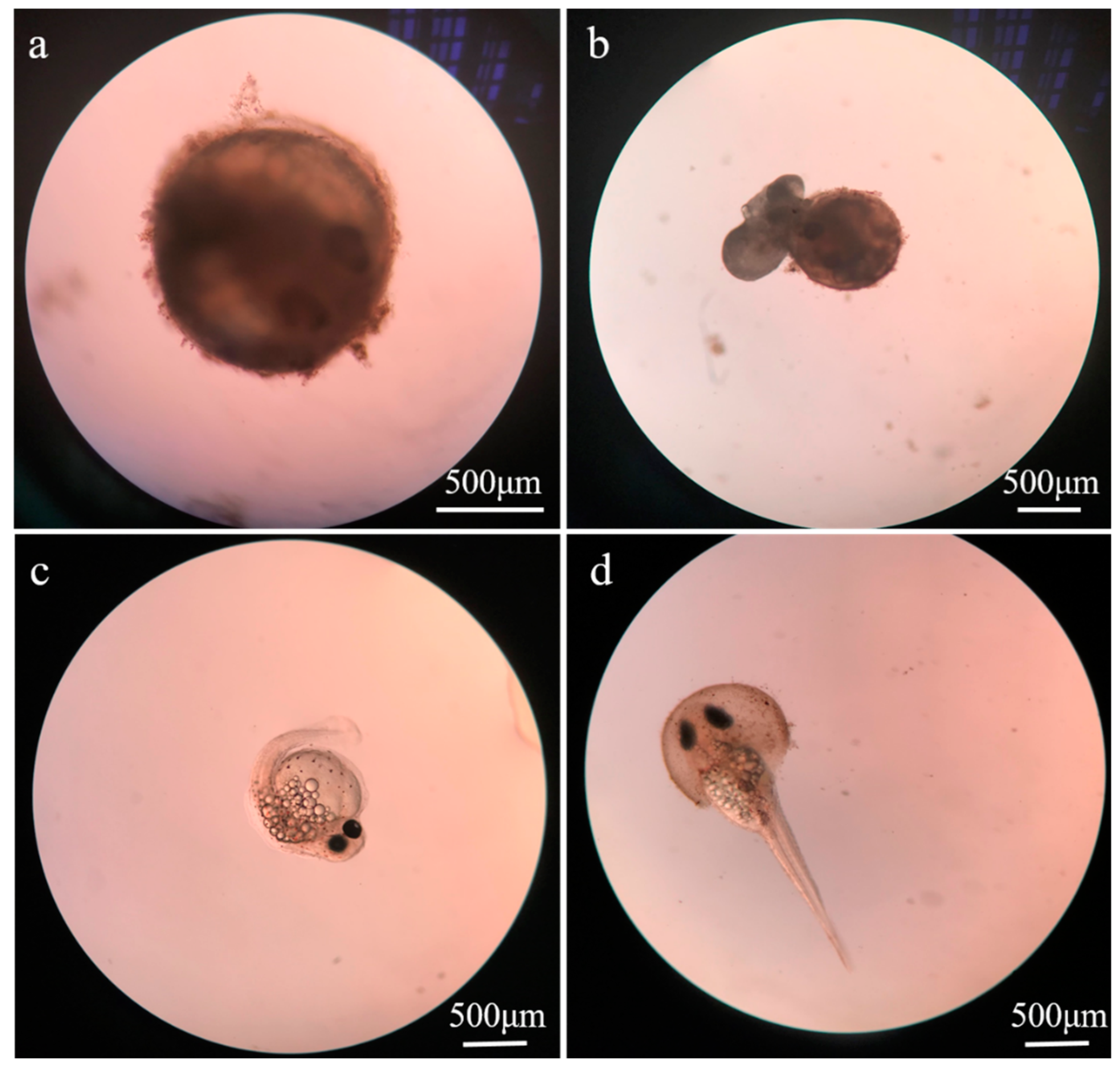
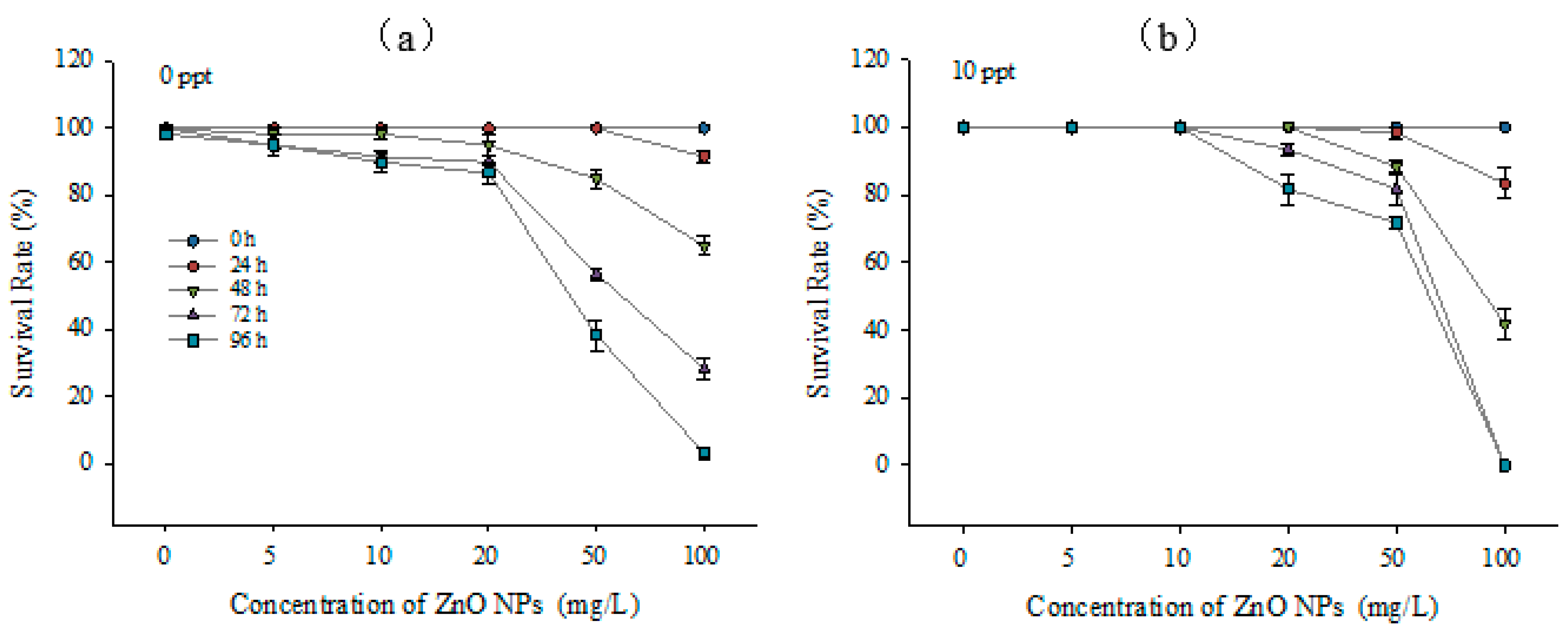
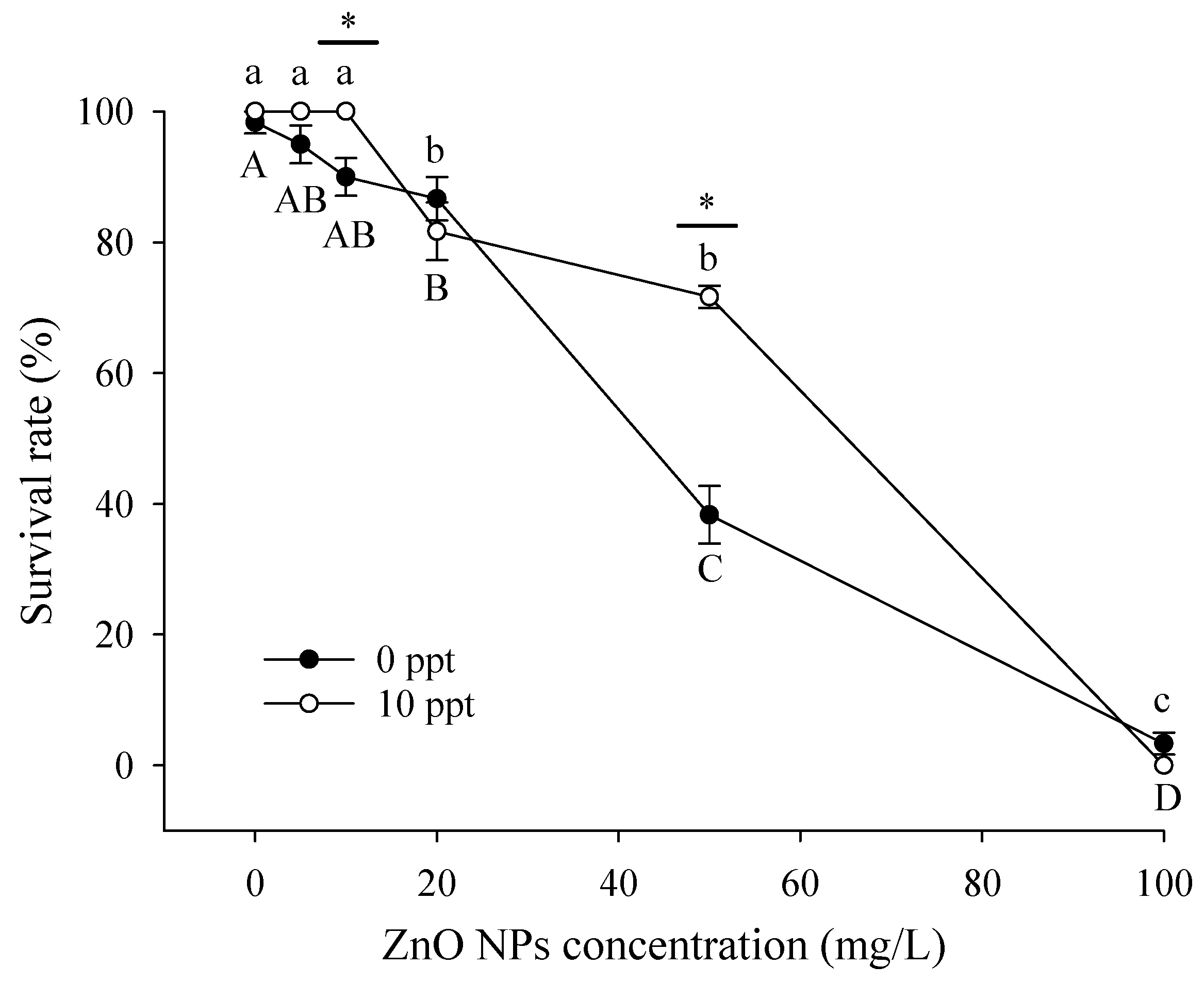
3.2. Toxic Effects of ZnO NPs on Embryos

ZnO NPs fully covered the surface of the embryo (blastocystic egg of approximately 24 h), resulting in the overflow of embryo yolk sac (Figure 2). As a result, the hatching of larvae (healthy young fish surviving within 24 h after hatching out) was affected, and the larvae had deformities. The toxicity to T. obscurus embryos was evaluated based on the hatching rate of eggs and the survival rates of newly hatched larvae. According to Figure 3a, the hatching rate of embryos decreased significantly with increasing ZnO NPs concentration and the decrease of salinity. The hatch rate was 0 with 50 mg/L ZnO NPs in freshwater, while it was around 13.3 ± 0.8% with the same nanoparticles concentration in brackish water. Higher ZnO NPs concentrations and lower salinity level could lead to lower survival rate of larvae (Figure 3b), which was consistent with the results of hatch rate. Under 0 ppt, no larvae were hatched with over 50 mg/L ZnO NPs; thus, the survival rate was regarded to be 0. In contrast, the survival rates in saline water were higher than 50%.

3.3. Toxic Effects on Larvae

The survival rates of T. obscurus larvae after exposing to a range of ZnO NPs concentrations for different time periods in freshwater and brackish water are displayed in Figure 4. Generally, the survival rate decreased significantly with higher ZnO NPs concentrations and longer exposure time. The survival rate of each treatment was higher than 80% after 24 h of exposure, while it decreased to 0 with 100 mg/L ZnO NPs after 96 h of exposure. As for the impacts of salinity levels on the toxicity, it can be observed that the death of T. obscurus larvae was observed with over 10 mg/L ZnO NPs under 10 ppt. In contrast, death was observed without the presence of nanoparticles under 0 ppt. No survival was observed after cultured for 96 h under both salinity conditions with 100 mg/L solution. Similarly, survival was hardly observed for the treatment in brackish water with the same concentration already after 72 h.
In order to determine the influence of salinity, Figure 5 shows the survival rate of T. obscurus larvae after 96 h of exposure to different ZnO NP concentrations and salinity. The survival rates of higher salinity condition were significantly higher than that under 0 ppt with ZnO NPs concentrations of 0, 5, 10, and 50 mg/L. It should be noted that the surviving larvae had varying extents of diseases, including pericardial effusion and edema.

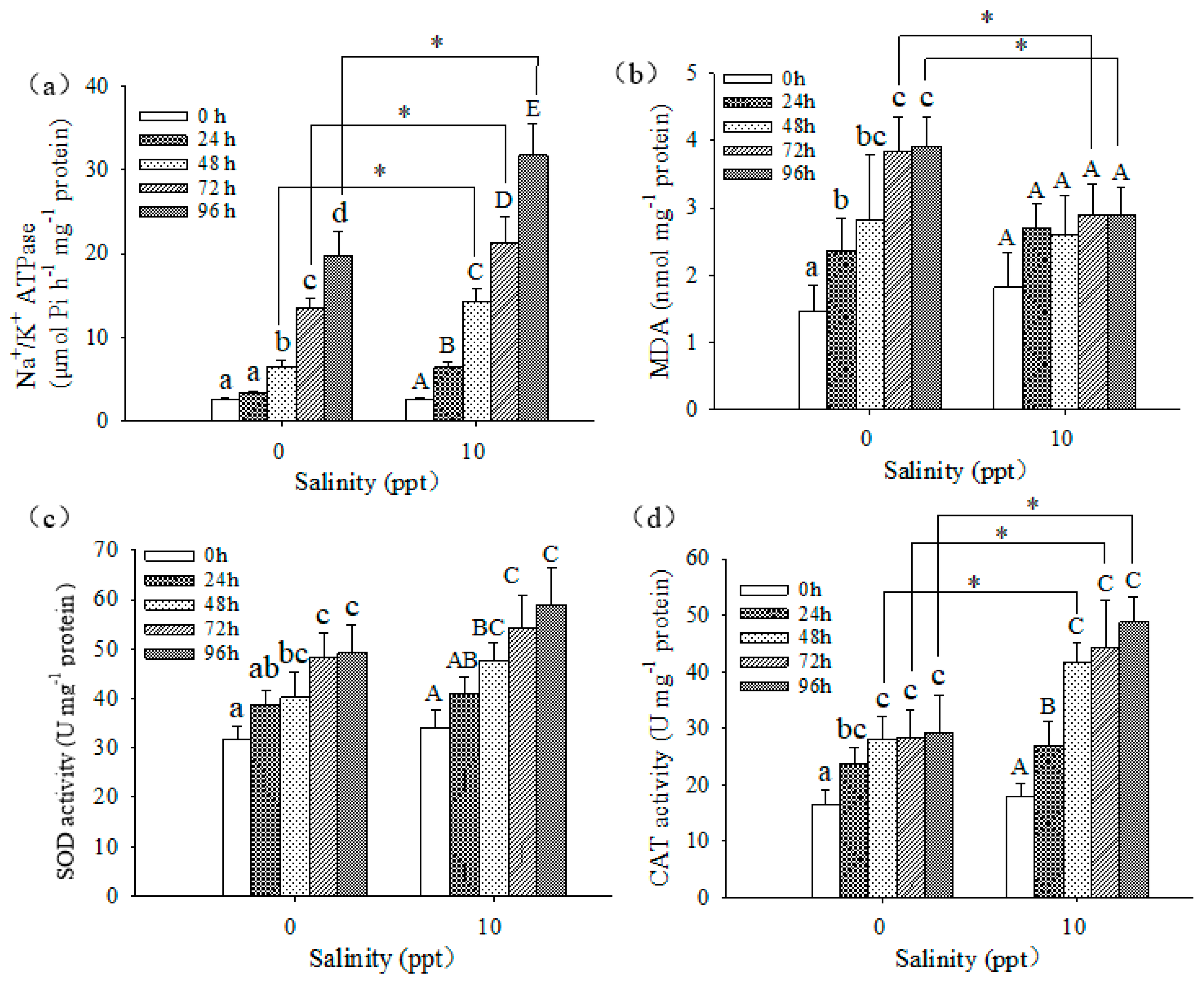
3.4. Biochemical Responses of Larvae

Figure 6 shows the Na+/K+ ATPase values under exposure to 50 mg/L of ZnO NPs with different salinity levels. Na+/K+ ATPase values increased significantly with the increase of salinity level and exposed time. Similar to Na+/K+ ATPase, CAT activity also showed significant higher values in brackish water than freshwater after 48 h of exposure. Another antioxidant indicator, SOD activity also had higher values with 10 ppt of salinity at each duration time although not statistically significant. In contrast, MDA showed a different pattern, with no significant differences for various exposure time in brackish water and significantly higher values under 0 ppt than 10 ppt after 72 h.

4. Discussion

4.1. Impacts of Salinity on the Characterization and Toxicity of ZnO NPs

ZnO NPs aggregated in higher extent in brackish water than freshwater (Figure 1), indicating the change of characterization of ZnO NPs in solution by salinity. Similar results are demonstrated in several examples of previous research. For example, Yung et al. [33] concluded that increased salinity could lead to greater diameters and lower concentration of dissolved Zn2+. According to Kim et al. [34], aggregation could be achieved through counteracting with the particle entropy and electrostatic energy barrier. However, higher salinity in brackish water can eliminate most of the particle electrostatic repulsion in the solution [35], which results in larger aggregates. The salt could provide extra chloride ions for the complexation reaction with the dissolved metal ion and reduce the concentration of Zn2+ [36,37]. Therefore, with increased salinity, the amount of aggregation increases, and the content of Zn2+ decreases.
Many studies showed that the dissolved metal ions are responsible for their negative impacts on aquatic organisms, which means that the relatively lower concentration of dissolved Zn2+ in brackish water could lead to the lower toxic effects of ZnO NPs than in freshwater [9,38,39]. The experiment conducted by Adam et al. [40] tested the chronic toxic effects of ZnO NPs and dissolved Zn2+ to Daphnia magna separately for 21 days and emphasized that the ZnO NPs’ toxicity is highly likely due to the dissolution instead of the aggregations or nanoparticles. The toxicological effects experiments on T. obscurus in this study showed similar results, as the survival rates of larvae were lower under 0 ppt than 10 ppt with 10 and 50 mg/L ZnO NPs (Figure 4), indicating the main contribution of zinc ions to the toxicity.

4.2. Embryos and Newly Hatched Larvae

As the most vulnerable stage in the life cycle of T. obscurus, the embryo could be easily affected by various environmental stresses, including the combined effects of salinity and ZnO NPs [41]. In the present study, the concentration of zinc oxide nanoparticles was positive correlated with the mortality of embryo (Figure 3). However, the research of Browning et al. [42] showed that the toxic effects of gold nanoparticles on zebrafish embryos was not related with their concentration, and about 74% of the embryos managed to develop into juveniles without deformation. The difference could be due to the distinct physiochemical properties of different nanoparticles [43]. Thus, further selection of nanomaterials regarding toxicity is needed before applications [44].
Hua et al. [45] explored the toxicity of different shapes of ZnO NPs to zebrafish embryos and suggested that nanoparticles in each suspension could be more toxic than dissolved Zn2+. According to Zhao et al. [46], Zn2+ dissolution promotes the toxic effects of ZnO NPs to some extent, while pure nanoparticles could cause oxidative stress, DNA damages, and developmental toxicity on embryos. Therefore, in this study, the significant decrease of embryos’ hatching rate and of survival rate of the newly hatched larvae can be mainly attributed to the toxicity of nanoparticles.

4.3. Biochemical Responses of Larvae

As a membrane protein, Na+/K+ ATPase promotes the active exchange between K+ ions and Na+ ions, provides energy for ion transport, and maintains the osmotic pressure across the membranes [47]. In our study, the Na+/K+ ATPase activity in brackish water was significantly higher than in freshwater for each treatment with 50 mg/L ZnO NPs, indicating its adaptive changes to salinity. Na+/K+ ATPase activity also increased significantly with exposure time at the same salinity level (Figure 6). Similar results were reported in several previous studies. For example, the experiment using rainbow trout carried out by Richards [48] showed that Na+/K+ ATPase α1b mRNA (one of the isoforms) increased rapidly after transferred to 80% seawater and continued to rise with time. Kong et al. [49] also showed a similar combined effect of exposure time, temperature, and salinity on the Na+/K+ ATPase activities of mud crab muscles. Hence, in the present study, salinity and ZnO NP had joint toxicity to the survival of T. obscurus embryos and larvae.
The effects of nanoparticles can stimulate or inhibit the activities of various antioxidant enzymes [17,50,51]. As the first defense line, SOD converts superoxide anion O2- into H2O2 and oxygen, and the generated H2O2 is further converted into H2O and oxygen by CAT, minimizing the adverse effects of ROS [51]. One previous study demonstrated that ZnO and CuO nanoparticles in Cumumis sativus could lead to alterations of oxidative stress, resulting in statistically significant increased SOD and CAT activities [52]. In the present study, SOD and CAT had higher activities in brackish water than freshwater (Figure 5). Under brackish condition, the activities of both SOD and CAT increased significantly with time, indicating antioxidative properties, whereas CAT activity remained constant and SOD activity increased under freshwater condition. Similar results have been demonstrated in previous studies. The inhibition of CAT activity under the influence of ZnO NPs can lead to the incomplete removal of H2O2, which can result in the accumulation of ROS in cells [46,53]. The changes of peroxidase activity could indicate that the ROS stress is greater for T. obscurus larvae in freshwater, which can be further confirmed with MDA content. MDA is one of the products of the reaction in cell membrane based on unsaturated fatty acids and free radicals [46]. It is generally regarded as an indicator of the oxidative stress and the extent of cell destruction under stress [54]. The experiment conducted by Xiong et al. [55] showed the increased MDA content with increased TiO2 NPs concentration in zebrafish. In this study, MDA content was significantly higher in freshwater than brackish water (Figure 5) and consistent with the results of hatch rate and survival rate of embryos and newly hatched larvae (Figure 3). These results indicated that embryos were under less stress in brackish water than in freshwater. Therefore, it is proposed that the toxic effects of ZnO NPs to T. obscurus larvae could hinder the activities of antioxidant enzymes (mostly CAT) and cause abnormal function, which could be the reason for the high mortality of embryos and larvae with the concentration of 50 mg/L ZnO NPs.

5. Conclusions

In conclusion, this study evaluated the impact of ZnO NPs on the early development of T. obscurus based on the quantification and analysis of their toxic effects on embryos and larvae. The toxicity is associated with exposure time and nanoparticle concentration. Salinity negatively affected the toxicity of ZnO NPs. The influence of increased salinity on ZnO NPs was mainly manifested in the formation of aggregations with greater size and less content of dissolved Zn2+. Thus, salinity is an important factor to be considered when evaluating the impact of nanoparticles on aquatic organisms.

Author Contributions

Conceptulization, Y.L. and J.W. Methodology, J.W., H.D. and Q.C.; software, F.M. and H.Y.; Validation and formal analysis, Y.L., J.W. and M.C.; investigation, Y.L. and J.W.; supervision, Q.C. All authors have read and agreed to the published version of the manuscript.

Funding

This research was financially supported by the National Natural Science Foundation of China (52121006, 52009082), the National Key Research and Development Program of China (2022YFC3203900, 2019YFC0408901), Fundamental Research Funds for the Central Universities (Y919025), and Postdoctoral Research Foundation of China (2019M651889).

Institutional Review Board Statement

This study were approved by the ACUC (Animal Care and Use Committee) of Nanjing Normal University, Nanjing, China, and carried out according to its guidelines (Research Permit Number: SYXK2015-0028).

Informed Consent Statement

Not applicable.

Data Availability Statement

Data is available based on proper request to the corresponding author.

Conflicts of Interest

The authors declare no conflict of interest.

References

  1. Vert, M.; Doi, Y.; Hellwich, K.; Hess, M.; Hodge, P.; Kubisa, P.; Rinaudo, M.; Schué, F. Terminology for biorelated polymers and applications (IUPAC Recommendations 2012). Pure Appl. Chem. 2012, 84, 377–410. [Google Scholar] [CrossRef]
  2. Nel, A.; Xia, T.; Moedler, L.; Li, N. Toxic Potential of Materials at the Nanolevel. Science 2006, 311, 622–627. [Google Scholar] [CrossRef] [PubMed]
  3. Bouwmeester, H.; Dekkers, S.; Noordam, M.; Hagens, W.; Bulder, A.; de Heer, C.; ten Voorde, S.; Wijnhoven, S.; Marvin, H.; Sips, A. Review of health safety aspects of nanotechnologies in food production. Regul. Toxicol. Pharmacol. 2009, 53, 52–62. [Google Scholar] [CrossRef]
  4. Joo, S.; Zhao, D. Environmental dynamics of metal oxide nanoparticles in heterogeneous systems: A review. J. Hazard. Mater. 2017, 322, 29–47. [Google Scholar] [CrossRef]
  5. Szabó, T.; Németh, J.; Dékány, I. Zinc oxide nanoparticles incorporated in ultrathin layer silicate films and their photocatalytic properties. Colloids Surf. A Physicochem. Eng. Asp. 2003, 230, 23–35. [Google Scholar] [CrossRef]
  6. Keller, A.; McFerran, S.; Lazareva, A.; Suh, S. Global life cycle releases of engineered nanomaterials. J. Nanopart. Res. 2013, 15, 1692. [Google Scholar] [CrossRef]
  7. Casas-Beltran, D.; Hernández-Pedraza, M.; Alvarado-Flores, J. Estimation of the Discharge of Sunscreens in Aquatic Environments of the Mexican Caribbean. Environments 2020, 7, 15. [Google Scholar] [CrossRef]
  8. Bhatt, I.; Tripathi, B. Interaction of engineered nanoparticles with various components of the environment and possible strategies for their risk assessment. Chemosphere 2011, 82, 308–317. [Google Scholar] [CrossRef] [PubMed]
  9. Hou, J.; Wu, Y.; Li, X.; Wei, B.; Li, S.; Wang, X. Toxic effects of different types of zinc oxide nanoparticles on algae, plants, invertebrates, vertebrates and microorganisms. Chemosphere 2018, 193, 852–860. [Google Scholar] [CrossRef]
  10. García-Gómez, C.; Babin, M.; Obrador, A.; Álvarez, J.; Fernández, M. Toxicity of ZnO Nanoparticles, ZnO Bulk, and ZnCl2 on Earthworms in a Spiked Natural Soil and Toxicological Effects of Leachates on Aquatic Organisms. Arch. Environ. Contam. Toxicol. 2014, 67, 465–473. [Google Scholar] [CrossRef] [PubMed] [Green Version]
  11. Ma, H.; Williams, P.; Diamond, S. Ecotoxicity of manufactured ZnO nanoparticles–A review. Environ. Pollut. 2013, 172, 76–85. [Google Scholar] [CrossRef] [PubMed]
  12. Bai, W.; Zhang, Z.; Tian, W.; He, X.; Ma, Y.; Zhao, Y.; Chai, Z. Toxicity of zinc oxide nanoparticles to zebrafish embryo: A physicochemical study of toxicity mechanism. J. Nanopart. Res. 2009, 12, 1645–1654. [Google Scholar] [CrossRef]
  13. Clarkson, T. Environmental contaminants in the food chain. Am. J. Clin. Nutr. 1995, 61, 682S–686S. [Google Scholar] [CrossRef] [PubMed]
  14. Golbamaki, N.; Rasulev, B.; Cassano, A.; Marchese Robinson, R.; Benfenati, E.; Leszczynski, J.; Cronin, M. Genotoxicity of metal oxide nanomaterials: Review of recent data and discussion of possible mechanisms. Nanoscale 2015, 7, 2154–2198. [Google Scholar] [CrossRef]
  15. Zhu, X.; Wang, J.; Zhang, X.; Chang, Y.; Chen, Y. The impact of ZnO nanoparticle aggregates on the embryonic development of zebrafish (Danio rerio). Nanotechnology 2009, 20, 195103. [Google Scholar] [CrossRef]
  16. Yu, L.; Fang, T.; Xiong, D.; Zhu, W.; Sima, X. Comparative toxicity of nano-ZnO and bulk ZnO suspensions to zebrafish and the effects of sedimentation, OH production and particle dissolution in distilled water. J. Environ. Monit. 2011, 13, 1975. [Google Scholar] [CrossRef] [PubMed]
  17. Bacchetta, R.; Maran, B.; Marelli, M.; Santo, N.; Tremolada, P. Role of soluble zinc in ZnO nanoparticle cytotoxicity in Daphnia magna: A morphological approach. Environ. Res. 2016, 148, 376–385. [Google Scholar] [CrossRef] [PubMed]
  18. Mukherjee, A.; Pokhrel, S.; Bandyopadhyay, S.; Mädler, L.; Peralta-Videa, J.; Gardea-Torresdey, J. A soil mediated phyto-toxicological study of iron doped zinc oxide nanoparticles (Fe@ZnO) in green peas (Pisum sativum L.). Chem. Eng. J. 2014, 258, 394–401. [Google Scholar] [CrossRef]
  19. Alexander, D.; Fairbridge, R. Encyclopedia Of Environmental Science; Springer Science & Business Media: Berlin, Germany, 1999; pp. 1–23. [Google Scholar]
  20. Wagner, S.; Gondikas, A.; Neubauer, E.; Hofmann, T.; von der Kammer, F. Spot the Difference: Engineered and Natural Nanoparticles in the Environment-Release, Behavior, and Fate. Angew. Chem. Int. Ed. 2014, 53, 12398–12419. [Google Scholar] [CrossRef]
  21. Yung, M.; Kwok, K.; Djurišić, A.; Giesy, J.; Leung, K. Influences of temperature and salinity on physicochemical properties and toxicity of zinc oxide nanoparticles to the marine diatom Thalassiosira pseudonana. Sci. Rep. 2017, 7, 3662. [Google Scholar] [CrossRef] [Green Version]
  22. Lai, R.; Yung, M.; Zhou, G.; He, Y.; Ng, A.; Djurišić, A.; Shih, K.; Leung, K. Temperature and salinity jointly drive the toxicity of zinc oxide nanoparticles: A challenge to environmental risk assessment under global climate change. Environ. Sci. Nano 2020, 7, 2995–3006. [Google Scholar] [CrossRef]
  23. Park, J.; Kim, S.; Yoo, J.; Lee, J.; Park, J.; Jung, J. Effect of salinity on acute copper and zinc toxicity to Tigriopus japonicus: The difference between metal ions and nanoparticles. Mar. Pollut. Bull. 2014, 85, 526–531. [Google Scholar] [CrossRef]
  24. Chang, S.; Kou, G.; Liao, I. Salinity Adaptation and Subsidence Attributes in the Early Stages of Japanese Eel (Anguilla japonica). Acta Zool. Taiwanica 2003, 14, 33–44. [Google Scholar]
  25. Yang, Z.; Chen, Y. Induced ovulation using LHRHa in anadromous obscure puffer Takifugu obscurus cultured entirely in freshwater. Fish Physiol. Biochem. 2003, 29, 323–326. [Google Scholar] [CrossRef]
  26. Kwok, K.; Leung, K. Toxicity of antifouling biocides to the intertidal harpacticoid copepod Tigriopus japonicus (Crustacea, Copepoda): Effects of temperature and salinity. Mar. Pollut. Bull. 2005, 51, 830–837. [Google Scholar] [CrossRef] [PubMed]
  27. Kim, J.; Dahms, H.; Rhee, J.; Lee, Y.; Lee, J.; Han, K.; Lee, J. Expression profiles of seven glutathione S-transferase (GST) genes in cadmium-exposed river pufferfish (Takifugu obscurus). Comp. Biochem. Physiol. C Toxicol. Pharmacol. 2010, 151, 99–106. [Google Scholar] [CrossRef]
  28. Katusic, Z.; Vanhoutte, P. Superoxide anion is an endothelium-derived contracting factor. Am. J. Physiol-Heart C 1989, 257, H33–H37. [Google Scholar] [CrossRef]
  29. Ighodaro, O.; Akinloye, O. First line defence antioxidants-superoxide dismutase (SOD), catalase (CAT) and glutathione peroxidase (GPX): Their fundamental role in the entire antioxidant defence grid. Alex. J. Med. 2018, 54, 287–293. [Google Scholar] [CrossRef]
  30. Frankič, T.; Pajk, T.; Rezar, V.; Levart, A.; Salobir, J. The role of dietary nucleotides in reduction of DNA damage induced by T-2 toxin and deoxynivalenol in chicken leukocytes. Food Chem. Toxicol. 2006, 44, 1838–1844. [Google Scholar] [CrossRef] [PubMed]
  31. Rombough, P. The functional ontogeny of the teleost gill: Which comes first, gas or ion exchange? Comp. Biochem. Physiol. Part A Mol. Integr. Physiol. 2007, 148, 732–742. [Google Scholar] [CrossRef]
  32. Sun, H.; Lü, K.; Minter, E.; Chen, Y.; Yang, Z.; Montagnes, D. Combined effects of ammonia and microcystin on survival, growth, antioxidant responses, and lipid peroxidation of bighead carp Hypophthalmythys nobilis larvae. J. Hazard. Mater 2012, 221–222, 213–219. [Google Scholar] [CrossRef]
  33. Yung, M.; Wong, S.; Kwok, K.; Liu, F.; Leung, Y.; Chan, W.; Li, X.; Djurišić, A.; Leung, K. Salinity-dependent toxicities of zinc oxide nanoparticles to the marine diatom Thalassiosira pseudonana. Aquat. Toxicol. 2015, 165, 31–40. [Google Scholar] [CrossRef]
  34. Kim, I.; Taghavy, A.; DiCarlo, D.; Huh, C. Aggregation of silica nanoparticles and its impact on particle mobility under high-salinity conditions. J. Pet. Sci. Eng. 2015, 133, 376–383. [Google Scholar] [CrossRef]
  35. VandeSteeg, H.; Cohen Stuart, M.; De Keizer, A.; Bijsterbosch, B. Polyelectrolyte adsorption: A subtle balance of forces. Langmuir 1992, 8, 2538–2546. [Google Scholar] [CrossRef]
  36. Dong, Z.; Zhang, W.; Qiu, Y.; Yang, Z.; Wang, J.; Zhang, Y. Cotransport of nanoplastics (NPs) with fullerene (C60) in saturated sand: Effect of NPs/C60 ratio and seawater salinity. Water Res. 2019, 148, 469–478. [Google Scholar] [CrossRef] [PubMed]
  37. Verslycke, T.; Vangheluwe, M.; Heijerick, D.; De Schamphelaere, K.; Van Sprang, P.; Janssen, C. The toxicity of metal mixtures to the estuarine mysid Neomysis integer (Crustacea: Mysidacea) under changing salinity. Aquat. Toxicol. 2003, 64, 307–315. [Google Scholar] [CrossRef] [PubMed]
  38. Loza, K.; Diendorf, J.; Sengstock, C.; Ruiz-Gonzalez, L.; Gonzalez-Calbet, J.; Vallet-Regi, M.; Köller, M.; Epple, M. The dissolution and biological effects of silver nanoparticles in biological media. J. Mater. Chem. B 2014, 2, 1634. [Google Scholar] [CrossRef] [PubMed]
  39. Wong, S.; Leung, P.; Djurišić, A.; Leung, K. Toxicities of nano zinc oxide to five marine organisms: Influences of aggregate size and ion solubility. Anal. Bioanal. Chem. 2009, 396, 609–618. [Google Scholar] [CrossRef] [PubMed]
  40. Adam, N.; Schmitt, C.; Galceran, J.; Companys, E.; Vakurov, A.; Wallace, R.; Knapen, D.; Blust, R. The chronic toxicity of ZnO nanoparticles and ZnCl2 to Daphnia magna and the use of different methods to assess nanoparticle aggregation and dissolution. Nanotoxicology 2014, 8, 709–717. [Google Scholar]
  41. Zander, D.; Thompson, J.; Lane, M. Perturbations in Mouse Embryo Development and Viability Caused by Ammonium Are More Severe after Exposure at the Cleavage Stages1. Biol. Reprod. 2006, 74, 288–294. [Google Scholar] [CrossRef] [PubMed]
  42. Browning, L.; Lee, K.; Huang, T.; Nallathamby, P.; Lowman, J.; Nancy Xu, X. Random walk of single gold nanoparticles in zebrafish embryos leading to stochastic toxic effects on embryonic developments. Nanoscale 2009, 1, 138. [Google Scholar] [CrossRef] [PubMed]
  43. Bar-Ilan, O.; Albrecht, R.; Fako, V.; Furgeson, D. Toxicity Assessments of Multisized Gold and Silver Nanoparticles in Zebrafish Embryos. Small 2009, 5, 1897–1910. [Google Scholar] [CrossRef]
  44. Asharani, P.; Lianwu, Y.; Gong, Z.; Valiyaveettil, S. Comparison of the toxicity of silver, gold and platinum nanoparticles in developing zebrafish embryos. Nanotoxicology 2010, 5, 43–54. [Google Scholar] [CrossRef]
  45. Hua, J.; Vijver, M.; Richardson, M.; Ahmad, F.; Peijnenburg, W. Particle-specific toxic effects of differently shaped zinc oxide nanoparticles to zebrafish embryos (Danio rerio). Environ. Toxicol. Chem. 2014, 33, 2859–2868. [Google Scholar] [CrossRef] [PubMed]
  46. Zhao, X.; Wang, S.; Wu, Y.; You, H.; Lv, L. Acute ZnO nanoparticles exposure induces developmental toxicity, oxidative stress and DNA damage in embryo-larval zebrafish. Aquat. Toxicol. 2013, 136–137, 49–59. [Google Scholar] [CrossRef] [PubMed]
  47. Garner, W.; Garner, M.; Spector, A. H2O2-induced uncoupling of bovine lens Na+, K+-ATPase. Proc. Natl. Acad. Sci. USA 1983, 80, 2044–2048. [Google Scholar] [CrossRef] [PubMed]
  48. Richards, J. Na+/K+-ATPase α-isoform switching in gills of rainbow trout (Oncorhynchus mykiss) during salinity transfer. J. Exp. Biol. 2003, 206, 4475–4486. [Google Scholar] [CrossRef] [PubMed]
  49. Kong, X.; Wang, G.; Li, S. Effects of low temperature acclimation on antioxidant defenses and ATPase activities in the muscle of mud crab (Scylla paramamosain). Aquaculture 2012, 370–371, 144–149. [Google Scholar] [CrossRef]
  50. Sharma, V.; Anderson, D.; Dhawan, A. Zinc oxide nanoparticles induce oxidative DNA damage and ROS-triggered mitochondria mediated apoptosis in human liver cells (HepG2). Apoptosis 2012, 17, 852–870. [Google Scholar] [CrossRef]
  51. Olsvik, P.; Kristensen, T.; Waagbø, R.; Rosseland, B.; Tollefsen, K.; Baeverfjord, G.; Berntssen, M. mRNA expression of antioxidant enzymes (SOD, CAT and GSH-Px) and lipid peroxidative stress in liver of Atlantic salmon (Salmo salar) exposed to hyperoxic water during smoltification. Comp. Biochem. Physiol. C Toxicol. Pharmacol. 2005, 141, 314–323. [Google Scholar] [CrossRef]
  52. Kim, S.; Lee, S.; Lee, I. Alteration of Phytotoxicity and Oxidant Stress Potential by Metal Oxide Nanoparticles in Cucumis sativus. Water Air Soil Pollut. 2012, 223, 2799–2806. [Google Scholar] [CrossRef]
  53. Gomes, T.; Pinheiro, J.; Cancio, I.; Pereira, C.; Cardoso, C.; Bebianno, M. Effects of Copper Nanoparticles Exposure in the Mussel Mytilus galloprovincialis. Environ. Sci. Technol. 2011, 45, 9356–9362. [Google Scholar] [CrossRef] [PubMed]
  54. Vinagre, C.; Madeira, D.; Narciso, L.; Cabral, H.; Diniz, M. Effect of temperature on oxidative stress in fish: Lipid peroxidation and catalase activity in the muscle of juvenile seabass, Dicentrarchus labrax. Ecol. Indic. 2012, 23, 274–279. [Google Scholar] [CrossRef]
  55. Xiong, D.; Fang, T.; Yu, L.; Sima, X.; Zhu, W. Effects of nano-scale TiO2, ZnO and their bulk counterparts on zebrafish: Acute toxicity, oxidative stress and oxidative damage. Sci. Total Environ. 2011, 409, 1444–1452. [Google Scholar] [CrossRef] [PubMed]
Figure 1. SEM images of ZnO NPs suspension (50 mg/L) under 10 ppt (a) and 0 ppt (b).
Ijerph 20 03209 g001
Figure 2. Effects of ZnO NPs (50 mg/L) on embryos and larvae. (a) The surface of embryo was covered with ZnO NPs; (b) the yolk sac of embryo was overflowed; (c) abnormal larvae with caudal vertebra bending; (d) the larvae could not completely hatch out.
Ijerph 20 03209 g002
Figure 3. (a) Hatch rate of T. obscurus embryos under different ZnO NPs concentrations and salinities and (b) survival rate of T. obscurus larvae 24 h post hatch under different ZnO NP concentrations and salinities. Different letters represent significant differences (p < 0.05) between different ZnO NPs concentrations under the same salinity. The asterisks above the black lines represent significant differences (p < 0.05) between different salinities under the same concentration of ZnO NPs (n = 3).
Ijerph 20 03209 g003
Figure 4. Survival rate of T. obscurus larvae under different salinities (a) 0 ppt, and (b) 10 ppt, exposure time, and ZnO NPs concentrations (n = 3).
Ijerph 20 03209 g004
Figure 5. Survival rate of T. obscurus larvae under different ZnO NP concentrations and salinity levels after 96 h of exposure. Different letters represent significant differences (p < 0.05) between different ZnO NPs concentrations under the same salinity. The asterisks above the black lines represent significant differences (p < 0.05) between different salinities under the same concentration of ZnO NPs (n = 3).
Ijerph 20 03209 g005
Figure 6. Na+/K+ ATPase (a), MDA (b), SOD (c), and CAT (d) in the whole T. obscurus larvae with exposure to 50 mg/L ZnO NPs for 96 h under two salinities (0 ppt and 10 ppt). Different letters represent significant differences (p < 0.05) between different exposure times under the same salinity. The asterisks above the black lines represent significant differences (p < 0.05) between different salinities under the same ZnO NPs concentration and exposure time (n = 3).
Ijerph 20 03209 g006
Disclaimer/Publisher’s Note: The statements, opinions and data contained in all publications are solely those of the individual author(s) and contributor(s) and not of MDPI and/or the editor(s). MDPI and/or the editor(s) disclaim responsibility for any injury to people or property resulting from any ideas, methods, instructions or products referred to in the content.

