
A Very Rare Basidiobolomycosis Case Presented with Cecal Perforation and Concomitant Hepatic Involvement in an Elderly Male Patient: A Case Study

 ,
,  , , and
Abstract
:1. Introduction
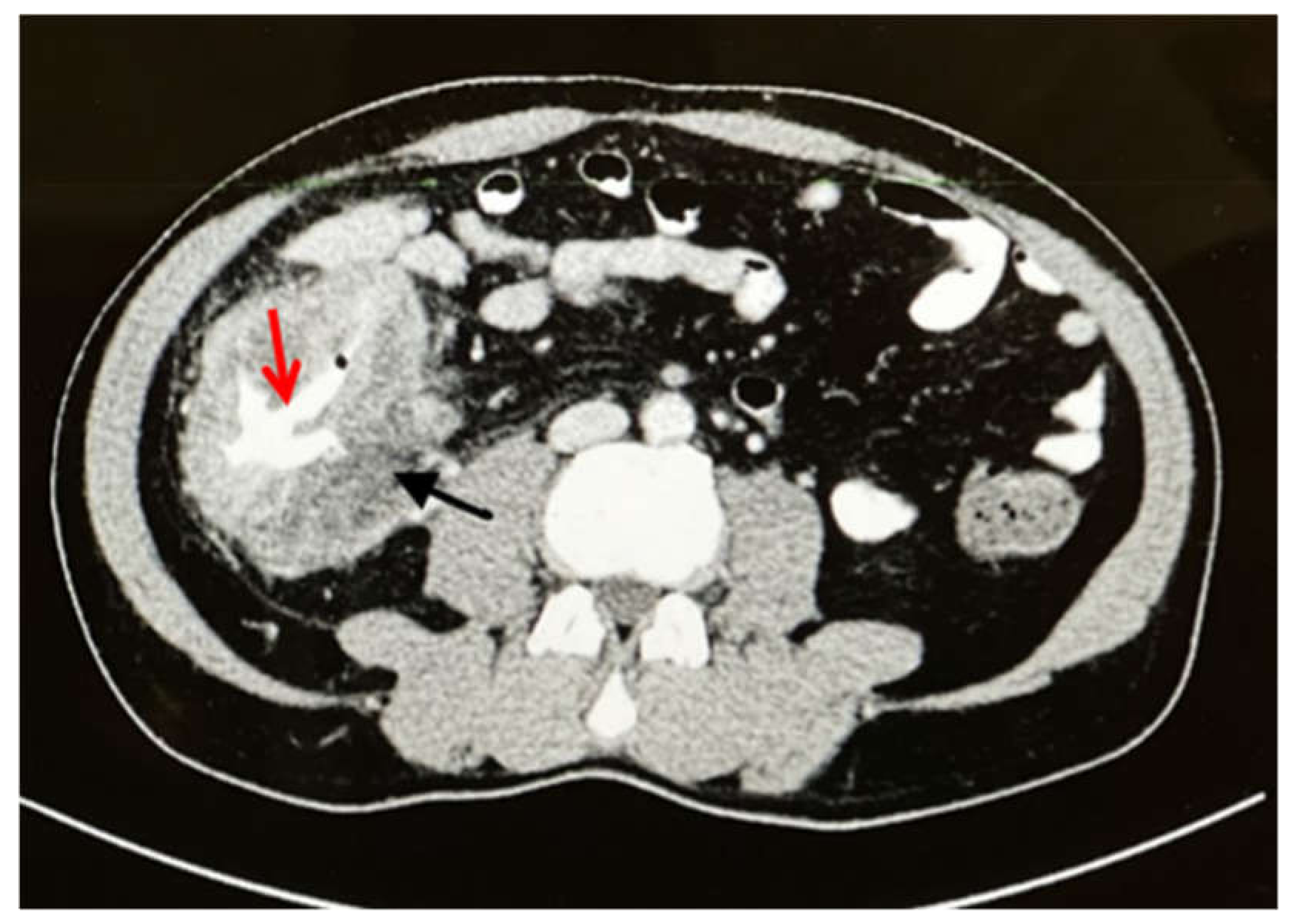
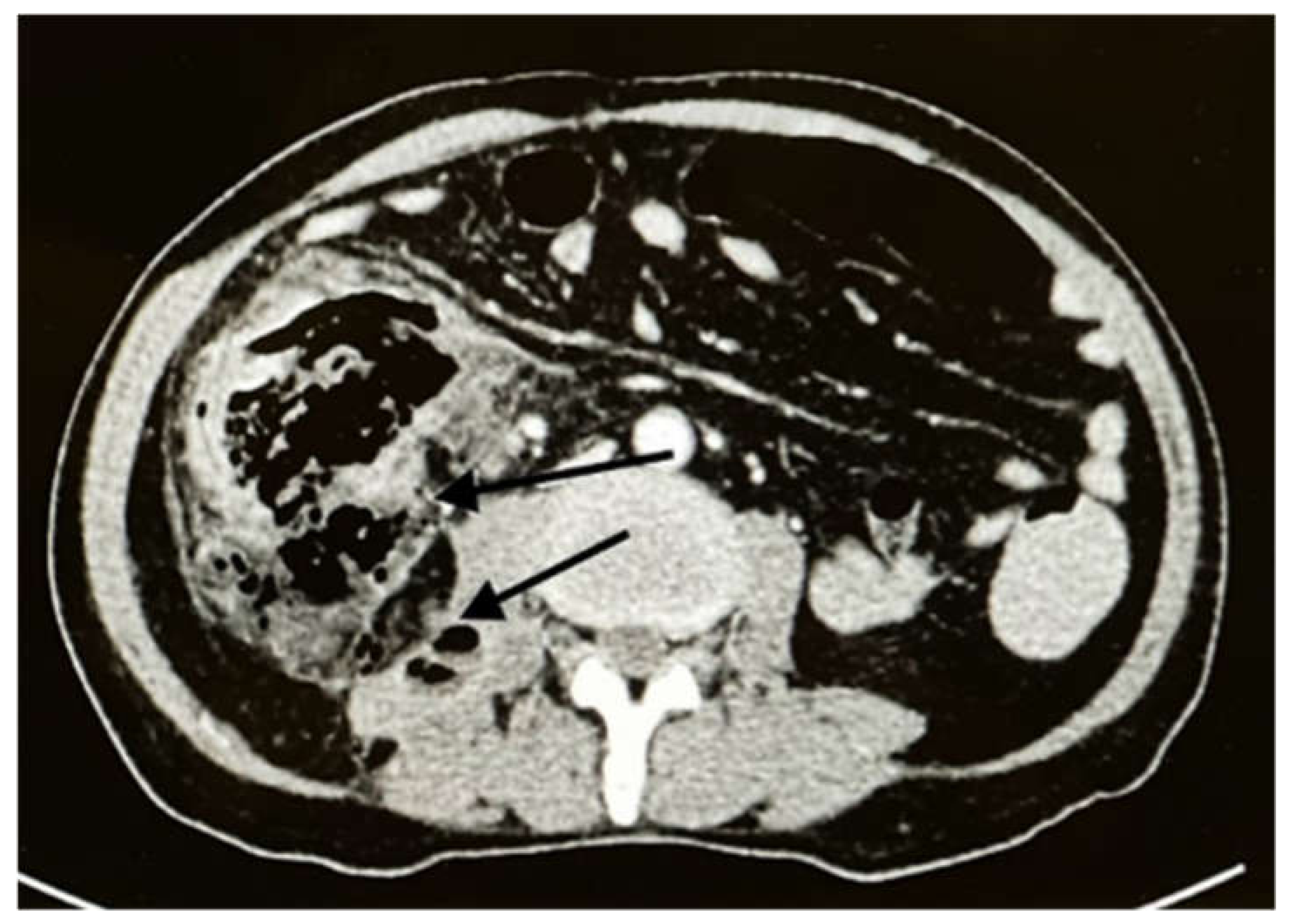
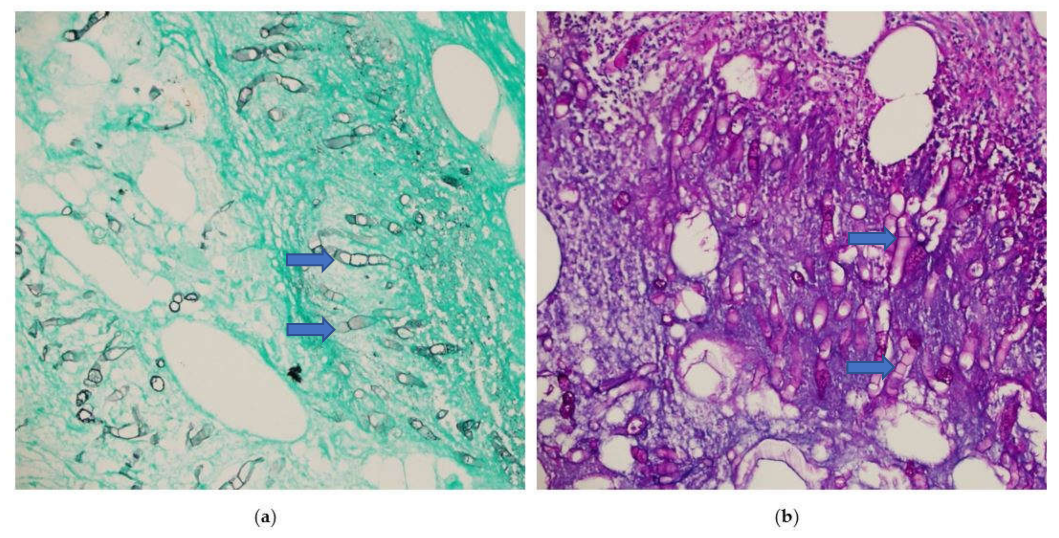
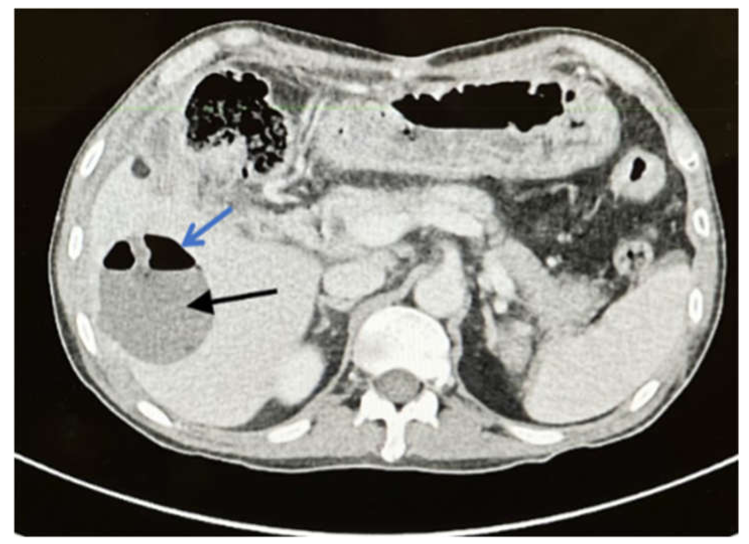
2. Case
3. Discussion
4. Conclusions
Author Contributions
Funding
Institutional Review Board Statement
Informed Consent Statement
Acknowledgments
Conflicts of Interest
References
- Al-Shanafey, S.; AlRobean, F.; Hussain, I.B. Surgical management of gastrointestinal Basidiobolomycosis in pediatric patients. J. Pediatr. Surg. 2012, 47, 949–951. [Google Scholar] [CrossRef] [PubMed]
- Alsharidah, A.; Mahli, Y.; Alshabyli, N.; Alsuhaibani, M. Invasive Basidiobolomycosis Presenting as Retroperitoneal Fibrosis: A Case Report. Int. J. Environ. Res. Public Health 2020, 17, 535. [Google Scholar] [CrossRef] [PubMed] [Green Version]
- Mohta, A.; Neogi, S.; Das, S. Gastrointestinal mucormycosis in an infant. Indian J. Pathol. Microbiol. 2011, 54, 664. [Google Scholar] [CrossRef] [PubMed]
- Shaikh, N.; Hussain, K.; Petraitiene, R.; Schuetz, A.; Walsh, T. Entomophthoramycosis: A neglected tropical mycosis. Clin. Microbiol. Infect. 2016, 22, 688–694. [Google Scholar] [CrossRef] [PubMed] [Green Version]
- Sethy, M.; Sahu, S.; Sachan, S. Basidiobolomycosis: Case report and literature overview. Indian Dermatol. Online J. 2021, 12, 307. [Google Scholar] [CrossRef] [PubMed]
- Albishri, A.; Shoukeer, M.A.; Hader, H.; Ashour, M.H.M.; Alsherbiny, H.; Ghazwani, E.; Alkedassy, K. Gastrointestinal Basidiobolomycosis. J. Pediatr. Surg. Case Rep. 2020, 55, 101411. [Google Scholar] [CrossRef]
- Takrouni, A.O.; Schammut, M.H.; Al-Otaibi, M.; Al-Mulla, M.; Privitera, A. Disseminated intestinal Basidiobolomycosis with mycotic aneurysm mimicking obstructing colon cancer. BMJ Case Rep. CP 2019, 12, e225054. [Google Scholar] [CrossRef] [PubMed] [Green Version]
- Mohammadi, R.; Chaharsoghi, M.A.; Khorvash, F.; Kaleidari, B.; Sanei, M.; Ahangarkani, F.; Abtahian, Z.; Meis, J.F.; Badali, H. An unusual case of gastrointestinal Basidiobolomycosis mimicking colon cancer; literature and review. J. Mycol. Med. 2019, 29, 75–79. [Google Scholar] [CrossRef] [PubMed]
- Van den Berk, G.E.; Noorduyn, L.A.; van Ketel, R.J.; van Leeuwen, J.; Bemelman, W.A.; Prins, J.M. A fatal pseudo-tumour: Disseminated Basidiobolomycosis. BMC Infect. Dis. 2006, 6, 140. [Google Scholar] [CrossRef] [PubMed] [Green Version]
- Saeed, M.A.; Al Khuwaitir, T.S.; Attia, T.H. Gastrointestinal Basidiobolomycosis with hepatic dissemination: A case report. JMM Case Rep. 2014, 1, e003269. [Google Scholar] [CrossRef] [PubMed] [Green Version]
- Ejtehadi, F.; Anushiravani, A.; Bananzadeh, A.; Geramizadeh, B. Gastrointestinal Basidiobolomycosis accompanied by liver involvement: A case report. Iran. Red Crescent Med. J. 2014, 16, e14109. [Google Scholar] [CrossRef] [PubMed] [Green Version]
- Zekavat, O.R.; Abdolkarimi, B.; Pouladfar, G.; Fathpour, G.; Mokhtari, M.; Shakibazad, N. Colonic Basidiobolomycosis with liver involvement masquerading as gastrointestinal lymphoma: A case report and literature review. Rev. Soc. Bras. Med. Trop. 2017, 50, 712–714. [Google Scholar] [CrossRef] [PubMed] [Green Version]
- El-Shabrawi, M.H.; Kamal, N.M. Gastrointestinal Basidiobolomycosis in children: An overlooked emerging infection? J. Med. Microbiol. 2011, 60, 871–880. [Google Scholar] [CrossRef] [PubMed] [Green Version]
- Shreef, K.; Saleem, M.; Saeedd, M.A.; Eissa, M. Gastrointestinal Basidiobolomycosis: An emerging, and a confusing, disease in children (A multicenter experience). Eur. J. Pediatr. Surg. 2018, 28, 194–199. [Google Scholar] [PubMed]
- Almoosa, Z.; Alsuhaibani, M.; AlDandan, S.; Alshahrani, D. Pediatric gastrointestinal Basidiobolomycosis mimicking malignancy. Med. Mycol. Case Rep. 2017, 18, 31–33. [Google Scholar] [CrossRef] [PubMed]

| Patient Information & Disease | Antecedents | Hospitalization Cause? | Type of Infection | Treatment | Follow-Up Patient | References |
|---|---|---|---|---|---|---|
| 61-year-old man (Division of Infectious Diseases, Tropical Medicine and AIDS unit in Amsterdam Hospital (Netherlands)). | Progressive left abdominal pain and constipation for a few months. Colonoscopy showed an obstructing tumor in the descending colon, and a hemicolectomy was performed. Histology showed inflammation, possibly caused by a fungal or parasitic infection, without definite identification of an organism. A few weeks postoperatively a CT scan, made because of abdominal discomfort, revealed a liver mass (6 cm). Treatment with metronidazole, directed against an amoebic liver abscess was unsuccessful. | Gastrointestinal Basidiobolomycosis with an obstructing colon tumor and a large hepatic mass. | A presumptive diagnosis of Basidiobolus spp. infection after autopsy Basidiobolus ranarum was cultured from liver, gallbladder and colon | Treated with amphotericin B (itraconazole contraindicated because of renal insufficiency). | A few days later the patient died of septic shock. | [9] |
| 12-year-old boy (Yemeni boy living in Abha, Aseer, Saudi Arabia). | 2-month history of diffuse abdominal pain, non-bilious vomiting, poor appetite, and weight loss. | The initial provisional diagnosis was intestinal lymphoma, and a right hemicolectomy was carried out, but histopathological assessment ruled out lymphoma and suggested intestinal tuberculosis. Two weeks after starting antituberculosis medications, the patient was referred to our hospital because of fever and right upper abdominal discomfort. There was leukocytosis with marked eosinophilia, and a liver biopsy showed evidence of B. ranarum infection. | Gastrointestinal Basidiobolomycosis with hepatic dissemination. | Itraconazole treatment was started immediately at a dose of 100 mg twice daily. | The patient was healthy. | [10] |
| 41-year-old woman (Shiraz, Iran). | She was complaining of abdominal pain, nausea, and experienced significant weight loss for one month. Past medical history and personal history were not significant, and she had no specific risk factor exposure. | Suffered from Basidiobolomycosis with concomitant lesions in the cecum and liver involvement. Physical examination temperature was 38.5 °C (101.3 °F) and signs of anemia, including pale conjunctiva were obvious. Also, she had mild generalized abdominal tenderness with no peritoneal sign. | Gastrointestinal Basidiobolomycosis infection with concomitant lesions in the cecum and liver involvement. | Patient was treated with itraconazole 200 mg twice a day for 4 months. The patient health status showed significant improvement, and no evidence of active liver lesion was detected in follow-up imaging study. | In the next year of follow-up, the patient was healthy and symptom free. | [11] |
| 5-year-old boy (Bushehr, Iran). | 2-month history of diffuse abdominal pain, non-bilious vomiting, poor appetite, weight loss, and a detectable mass on abdominal sonography. | This study presents a boy with colonic BM involving the liver, masquerading as gastrointestinal lymphoma. Physical examination, he seemed ill and emaciated, his body temperature was 39 °C, and his liver was tender on palpation 3 cm inferior to the costal margin. Multiple lymphadenopathies were detected. The mass was similar to Castleman’s disease, lymphoma, or tuberculosis. | Colonic BM involving the liver, masquerading as gastrointestinal lymphoma. | Posaconazole as an effective single agent treatment with minimum complications during a prolonged treatment plan. Treatment with amphotericin B (intravenous 1 mg/kg/day for 2 months) and Posaconazole (200 mg by mouth four times per day). | The patient was followed closely by means of physical exams and abdominal computed tomography scans, which revealed no recurrence 6 months after starting therapy with Posaconazole. | [12] |
| 18-year-old women (Jazan, Saudi Arabia). | 2-week history of right lower quadrant abdominal pain associated with nausea, vomiting and anorexia. Medical and surgical history were unremarkable, and she was not on any medications. | Obstructing cecal mass initially suspected to be malignant. | Surgical resection was complicated by bowel perforation, histology and cultures confirmed Basidiobolomycosis infection. | The postoperative course was complicated by an enterocutaneous fistula, fungal intraabdominal abscesses, liver and lung abscesses, formation of mycotic hepatic artery aneurysm and meningoencephalitis. This was treated successfully with vacuum-assisted closure device and total parenteral nutrition (TPN). | Despite aggressive intensive care unit and antimicrobial treatment, she expired due to septic shock. | [7] |
| 39-year-old woman (Southern part of Saudi Arabia). | Hypertension, for which she was being treated with amlodipine. She had visited several hospitals and had provisionally been diagnosed as having either a retroperitoneal malignancy or retroperitoneal fibrosis before being referred to hospital. | Severe left-sided abdominal pain and weight loss. | Retroperitoneal Basidiobolomycosis infection. | Antifungal treatment. This led to significant improvement, without surgical intervention. | Currently, she is still on treatment and undergoing follow-up. | [2] |
Publisher’s Note: MDPI stays neutral with regard to jurisdictional claims in published maps and institutional affiliations. |
© 2022 by the authors. Licensee MDPI, Basel, Switzerland. This article is an open access article distributed under the terms and conditions of the Creative Commons Attribution (CC BY) license (https://creativecommons.org/licenses/by/4.0/).
Share and Cite
Abduh, M.S.; Aldaqal, S.M.; Almaghrabi, J.; Aljiffry, M.M.; Elbadrawy, H.A.; Alsahafi, M.A. A Very Rare Basidiobolomycosis Case Presented with Cecal Perforation and Concomitant Hepatic Involvement in an Elderly Male Patient: A Case Study. Int. J. Environ. Res. Public Health 2022, 19, 3412. https://doi.org/10.3390/ijerph19063412
Abduh MS, Aldaqal SM, Almaghrabi J, Aljiffry MM, Elbadrawy HA, Alsahafi MA. A Very Rare Basidiobolomycosis Case Presented with Cecal Perforation and Concomitant Hepatic Involvement in an Elderly Male Patient: A Case Study. International Journal of Environmental Research and Public Health. 2022; 19(6):3412. https://doi.org/10.3390/ijerph19063412
Chicago/Turabian StyleAbduh, Maisa S., Saleh M. Aldaqal, Jaudah Almaghrabi, Murad M. Aljiffry, Hany A. Elbadrawy, and Majid A. Alsahafi. 2022. "A Very Rare Basidiobolomycosis Case Presented with Cecal Perforation and Concomitant Hepatic Involvement in an Elderly Male Patient: A Case Study" International Journal of Environmental Research and Public Health 19, no. 6: 3412. https://doi.org/10.3390/ijerph19063412
APA StyleAbduh, M. S., Aldaqal, S. M., Almaghrabi, J., Aljiffry, M. M., Elbadrawy, H. A., & Alsahafi, M. A. (2022). A Very Rare Basidiobolomycosis Case Presented with Cecal Perforation and Concomitant Hepatic Involvement in an Elderly Male Patient: A Case Study. International Journal of Environmental Research and Public Health, 19(6), 3412. https://doi.org/10.3390/ijerph19063412