Article

Maximal Fat Oxidation during Incremental Upper and Lower Body Exercise in Healthy Young Males

1
Centre for Sport, Exercise and Life Sciences, School of Life Sciences, Coventry University, Coventry CV1 5FB, UK
2
Department of Psychology, Sport and Geography, University of Hertfordshire, Hatfield AL10 9AB, UK
3
Alliance for Research in Exercise, Nutrition and Activity, Campus Central—City East, University of South Australia, GPO Box 2471, Adelaide 5001, Australia
*
Author to whom correspondence should be addressed.
Int. J. Environ. Res. Public Health 2022, 19(22), 15311; https://doi.org/10.3390/ijerph192215311
Submission received: 7 October 2022 / Revised: 12 November 2022 / Accepted: 14 November 2022 / Published: 19 November 2022
(This article belongs to the Section Exercise and Health)

Abstract

:
The aim of this study is to determine the magnitude of maximal fat oxidation (MFO) during incremental upper and lower body exercise. Thirteen non-specifically trained male participants (19.3 ± 0.5 y, 78.1 ± 9.1 kg body mass) volunteered for this repeated-measures study, which had received university ethics committee approval. Participants undertook two incremental arm crank (ACE) and cycle ergometry (CE) exercise tests to volitional exhaustion. The first test for each mode served as habituation. The second test was an individualised protocol, beginning at 40% of the peak power output (POpeak) achieved in the first test, with increases of 10% POpeak until volitional exhaustion. Expired gases were recorded at the end of each incremental stage, from which fat and carbohydrate oxidation rates were calculated. MFO was taken as the greatest fat oxidation value during incremental exercise and expressed relative to peak oxygen uptake (% V ˙ O2peak). MFO was lower during ACE (0.44 ± 0.24 g·min−1) than CE (0.77 ± 0.31 g·min−1; respectively, p < 0.01) and occurred at a lower exercise intensity (53 ± 21 vs. 67 ± 18% V ˙ O2peak; respectively, p < 0.01). Inter-participant variability for MFO was greatest during ACE. These results suggest that weight loss programs involving the upper body should occur at lower exercise intensities than for the lower body.

1. Introduction

During the initial intensities of incremental exercise, both carbohydrate and fat oxidation increase [1]. However, in subsequent stages of submaximal exercise, carbohydrate oxidation continues to increase, whereas fat oxidation reaches maximal values and begins to decrease [1,2]. Within such an incremental exercise model, the greatest fat oxidation value observed is termed maximal fat oxidation (MFO). The exercise intensity at which the MFO occurs, typically a percentage of maximal or peak oxygen uptake ( V ˙ O2peak), has been termed ‘Fatmax’ [3]. The intensity at which Fatmax occurs has been shown to be affected by factors such as endurance training status [4], prior carbohydrate ingestion [5], habitual physical activity and maximal oxygen uptake ( V ˙ O2max) [6]. Furthermore, few studies have directly compared males and females, with MFO tending to be greater in males but Fatmax tending to be greater in females [7]. Although the determination of the exercise intensity at which Fatmax occurs has been reported to be reliable, there is often large inter- and intra-individual variation, as demonstrated by large coefficients of variation and not all participants demonstrating a clear Fatmax intensity [6,8].
Previous studies examining Fatmax have predominantly considered lower body exercise. However, for the general population, upper body exercise elicits lower V ˙ O2peak than for lower body exercise modes [9,10,11,12]. For example, values for V ˙ O2peak during arm crank ergometry (ACE) and cycle ergometry (CE) in non-specifically trained individuals have been reported as 34 and 48 mL·kg·−1min.−1, respectively [10], with upper body values generally representing ~70% of those during CE [10,11,13]. When V ˙ O2peak during ACE (e.g., 28–39 mL·kg·−1min.−1) is compared to that during treadmill running (e.g., 44–59 mL·kg·−1min.−1) in similar non-specifically trained populations [9,12], the proportional difference between modes is increased, with ACE being ~60–65% of treadmill values [9,12]. Differences in V ˙ O2peak across exercise modes are predominantly due to differences in the size of the active muscle mass. The physiological range for metabolic values during upper body exercise is thus lower than for other exercise modes and likely includes a reduced range of fat metabolism.
Maximal fat oxidation and Fatmax during typical incremental CE and treadmill walking/running protocols have been directly compared in moderately trained men [14]. Maximal oxygen uptake and fat oxidation were lower during CE (64 mL·kg·−1min.−1 and 0.47 g·min−1, respectively) than for treadmill exercise (67 mL·kg·−1min.−1, and 0.65 g·min−1, respectively), as was fat oxidation over a range of submaximal exercise intensities. However, Fatmax was similar between modes (~60% V ˙ O2peak), as was the point where blood lactate increased from rest (i.e., the lactate threshold), suggesting no exercise mode differences for these specific variables. The participants in the above study were club/county standard endurance cyclists and, therefore, of greater aerobic training status than the general population, where fitness indicators such as lactate threshold are lower in ACE when compared to CE and treadmill exercise [12]. Differences in MFO and Fatmax across exercise modes may, therefore, be more pronounced for non-specifically trained individuals.
As well as eliciting lower aerobic fitness indicators when compared to lower body exercise modes, the upper body is also considered to possess a greater proportion of fast twitch muscle fibres [15,16]. Considering that fast twitch fibres are more glycolytic than slow twitch fibres and that lower training status elicits lower absolute MFO values at lower relative exercise intensities [4,6], the magnitude of both MFO and Fatmax is likely lower for upper body exercise. Studies that have investigated Fatmax during upper body exercise have examined individuals with diabetes and obesity, specifically focusing on mitochondrial function [17,18], or those with spinal cord injury, focusing upon substrate utilisation [19]. Within these studies, healthy or non-disabled controls have been utilised and matched for age and body mass [17,18].
Based on the above studies, our knowledge of MFO and Fatmax during upper body exercise is from a small number of studies of older (~30–40 years of age) and heavier participants (>90 kg) with low aerobic fitness; all factors associated with poor MFO and Fatmax intensities [20]. Therefore, the aim of this study is to determine the magnitude of MFO and Fatmax during upper body and lower body exercise in young, healthy, recreationally active males. Due to the likely differences in muscle mass, metabolic capacity and training status between the upper and lower body, it is hypothesised that MFO and Fatmax would be lower and more variable during upper body exercise.

2. Materials and Methods

2.1. Participants

Thirteen healthy, active males volunteered to participate in this study (age; 19.3 ± 0.5 y, body mass; 78.1 ± 9.1 kg; 4.5 ± 1.8 sessions per week in predominantly team sports), which had received university ethics committee approval (Ref: P35579) and was in accordance with the Declaration of Helsinki. A sub-group of participants (n = 9; age 19.2 ± 0.4 y; body mass 76.8 ± 8.9 kg) undertook an additional exercise testing session, as outlined below. All participants provided written informed consent. An a priori sample size calculation was undertaken using a desired power value of 0.8, an alpha level of p = 0.05 and effect size values based on the MFO and Fatmax data from Ara et al. [17] and Larsen et al. [18], who previously compared these variables during upper and lower body exercise. The calculation resulted in a recommended sample of n = 6. Therefore, our sample of n = 13 was sufficient to account for any potentially large variation in MFO and Fatmax responses and any participant drop-out, as well as being in line with previous studies (n = 10 and n = 7, respectively).
The study was a repeated measure design. Inclusion criteria were: young, healthy males who took part in structured exercise sessions at least three times each week. Exclusion criteria were: being specifically cycle or arm ergometry trained or endurance athletes. Recruitment occurred through existing researcher networks and participant friendship groups. Participants visited the university’s exercise physiology laboratory on four separate occasions. Testing sessions were undertaken in the morning, at the same time of day, with at least three days between sessions. Participants were requested not to consume any food within the two to three hours preceding each testing session and to keep the same diet on all testing days, which was confirmed verbally. Participants were also asked not to undertake any strenuous exercise or consume any alcohol or caffeinated products in the 24 h prior to testing. Participants were able to drink water ab libitum during each testing session. Laboratory conditions were maintained at approximately 19–20 °C and ~40% relative humidity. All participants wore similar attire on each laboratory visit, being: light-weight tracksuit trousers, shorts, sports socks and training shoes. Data were collected by the lead author, with considerable experience in both upper and lower body exercise testing.

2.2. Exercise Tests

The testing sessions involved two ACE tests, two incremental CE tests and, for the sub-group of participants, one submaximal ACE test. The incremental exercise tests were maximal aerobic tests to determine V ˙ O2peak and power output (POpeak). Lower body exercise tests were undertaken using a cycle ergometer (Monark 814E, Varberg, Sweden), whereas the upper body exercise tests were undertaken on an electronically braked Lode arm crank ergometer (Lode Angio, Groningen, the Netherlands). The first ACE and CE tests were preliminary tests to habituate the participants to each exercise mode and the testing environment. For ACE, the protocol began at 0 W, with increments of 20 W every 2-min [21], whereas the CE protocol began at 35 W and increased by 35 W every 3 min [10]. Power output was progressively increased until volitional exhaustion occurred or when the desired cadence (i.e., 70 rev·min−1) could not be maintained for 10 s. The second ACE and CE protocols were individualised protocols based on each participant’s performance in the preliminary tests. Participants initially warmed up for 5 min on the unloaded ergometer (0 and 35 W for ACE and CE, respectively) before beginning the main protocol at 40% of the POpeak achieved in the preliminary tests. After this stage, the exercise intensity increased by 10% of each participant’s initial POpeak every two minutes for ACE and every three minutes for CE until volitional exhaustion, using the same criteria as in the preliminary tests.
As previous studies of upper body exercise routinely used exercise protocols with stage durations of 2 min [10,21,22], it is possible that metabolic responses may not have adapted to the same extent as observed for CE when using 3 min exercise stage durations. Therefore, a sub-group of participants (detailed above) undertook a submaximal ACE protocol comparing 2- and 3-minute exercise responses. The submaximal protocol involved the participants undertaking the same individual exercise intensities for three minutes at each stage, up to a rating of perceived exertion (RPE, Borg Scale; [23]) for local muscular effort (RPEL) of 15 (equivalent to ‘Hard’ exercise on the 6–20 Borg scale). After all exercise tests, participants performed a cool-down on the unloaded ergometer (0 and 35 W for ACE and CE, respectively) at a self-selected cadence, typically 30 to 50 rev·min−1 for ACE and 50 to 60 rev·min−1 for CE, for at least five minutes to aid recovery. Due to individualised exercise protocols being undertaken, only the order of ACE and CE testing modes could be realistically counterbalanced.

2.3. Measurements

Heart rate (HR) was recorded using a Polar heart rate monitor (Polar beat, Kempele, Finland) consisting of a telemetry belt worn around the chest and a monitor. Values were recorded during seated rest and in the last 15 s of each exercise stage. Participants also wore a face mask connected to an online expired gas analysis system (Metamax 3b, Biophysik GmbH Leipzig, Germany) for the determination of oxygen uptake ( V ˙ O2) and carbon dioxide production ( V ˙ CO2), minute ventilation ( V ˙ E) and the respiratory exchange ratio (RER). Carbohydrate and fat oxidation were subsequently calculated according to standard stoichiometric equations [24] where:
C a r b o h y d r a t e   m e t a b o l i s m = ( 1.67 × V ˙ O 2 ) ( 1.67 × V ˙ C O 2 )   g · m i n 1 a n d
F a t   m e t a b o l i s m = ( 6.21 × V ˙ C O 2 ) ( 4.21 × V ˙ O 2 )   g · m i n 1
Fat metabolism was plotted against the exercise stage for each participant’s individualised ACE and CE protocols and the exercise intensity (% V ˙ O2peak) at which the MFO occurred was determined (i.e., Fatmax). Ratings of perceived exertion were recorded at the end of each exercise stage for RPEL and central (cardiorespiratory) exertion (RPEC). The RPE scale and verbal anchors were fully explained to each participant. Capillary blood samples (20 µL) were taken from the participant’s earlobe at rest, at volitional exhaustion and following 5 min of active recovery for the analysis of blood lactate concentration (Biosen, C_Line analyser, EFK Diagnostics, Cardiff, UK). Blood sampling was undertaken in accordance with the British Association of Sport and Exercise Science guidelines [25].

2.4. Data Analysis

Data are expressed as mean ± standard deviation (SD). All data were analysed using the Statistical Package for the Social Sciences (v22; IBM Inc., Chicago, USA). Data were checked for normality using the Shapiro–Wilkes test and the homogeneity of variance using Levene’s test. Peak physiological and perceptual responses, maximal carbohydrate oxidation, MFO and Fatmax were compared between exercise modes using paired t-tests. Data for physiological variables at each exercise stage and between exercise modes were analysed using factorial analysis of variance (ANOVA), with repeated measures on both factors (Mode: ACE, CE; × intensity; rest, 40, 50, 60, 70 80, 90, 100% POpeak). Where significance was achieved, Tukey’s post-hoc analysis was undertaken to determine the difference required between means for significance at the level of p < 0.05 [26].
Due to the nature of the exercise protocols increasing by 10% POpeak at each stage, the data could not be statistically compared at a given power output across exercise modes. Therefore, the data for HR, V ˙ O2, carbohydrate oxidation and fat oxidation were also plotted for illustrative purposes only against the mean power output representing each 10% increment without statistical analysis. Data for the 2- and 3-minute comparison during ACE for intensities completed by all participants (i.e., up to 70% POpeak) were analysed by factorial analysis of variance (ANOVA), with repeated measures on both factors (intensity; 40, 50, 60, 70% V ˙ O2peak; × stage duration; 2-min, 3-min).

3. Results

3.1. Peak Physiological Responses

The peak physiological responses for ACE and CE are shown in Table 1. Peak V ˙ O2, V ˙ E, power output and heart rate (HRpeak) were lower during ACE than for CE (p < 0.05). No differences were observed between modes for peak RER, blood lactate concentration or RPEL, whereas RPEC approached significance (p = 0.065), with RPEC tending to be lower during ACE compared to CE. Carbohydrate oxidation at volitional exhaustion was similar for ACE and CE (4.91 ± 0.73 and 5.16 ± 1.88 g·min−1 for ACE and CE, respectively) (p = 0.661). Fat oxidation at the end of the exercise was also similar between trials (ACE: 0.06 ± 0.42 g·min−1; CE: 0.13 ± 0.67 g·min−1; p = 0.781).

3.2. Submaximal Exercise Responses

Heart rate, V ˙ O2 and RER responses during incremental ACE and CE are shown in Figure 1. When expressed relative to peak power (i.e., %POpeak), V ˙ O2 was lower during ACE than for CE at every exercise intensity (p < 0.05). No mode × intensity interactions were observed for HR (p = 0.229), RER (p = 0.498), RPEC (p = 0.892) or RPEL (p = 0.969) when expressed relative to %POpeak. Main effects for intensity were observed for HR, RPEC, RPEL and RER (p < 0.05), with additional main effects for exercise modes observed for HR and RPEC (p < 0.05). When plotted against absolute power output, V ˙ O2 for ACE and CE was similar where the power output overlapped (~90 to 140 W), whereas HR, RPEC, RPEL and RER demonstrated greater values during ACE.

3.3. Substrate Utilisation

The resting respiratory exchange ratio was similar between trials (0.78 ± 0.11 vs. 0.78 ± 0.06 for ACE and CE, respectively; p = 0.978), as were carbohydrates (0.16 ± 0.28 vs. 0.17 ± 0.36 g·min−1, respectively) and fat oxidation (0.17 ± 0.07 vs. 0.16 ± 0.06 g·min−1, respectively). No mode × intensity interaction was observed for carbohydrate oxidation (p = 0.924), although values increased with exercise intensity (intensity main effect, p < 0.01). When plotted as absolute exercise intensities, carbohydrate oxidation was greater during ACE (Figure 1).
A significant mode × intensity interaction was observed for fat oxidation (p = 0.013), with differences observed between modes for each exercise intensity (p < 0.01). Maximal fat oxidation for ACE and CE modes is shown in Figure 2. Maximal fat oxidation was lower in ACE (0.44 ± 0.24 g·min−1; range: 0.11–0.79 g·min−1) when compared to CE (0.77 ± 0.31 g·min−1; range: 0.33–1.23 g·min−1; p < 0.01), occurring at 53 ± 21 and 67 ± 18% V ˙ O2peak, respectively (p < 0.01).
The relationship between resting fat oxidation and MFO for both modes of exercise combined was significant (R = 0.505; R2 = 0.254, p = 0.020), whereas when considered for ACE and CE separately, only CE elicited a significant relationship (ACE: R = 0.520; R2 = 0.270, p = 0.151; CE: R = 0.661; R2 = 0.436, p = 0.019). Based on the coefficient of variation, the inter-participant variability for MFO was greater during ACE (CV = 54.5%) when compared to CE (CV = 40.3%). However, inter-individual variation for MFO was considerably greater than for peak physiological responses such as V ˙ O2peak during ACE and CE (12.7% and 23.4%, respectively).

3.4. Comparison of 2- and 3-Minute Exercise Durations

The physiological responses to submaximal ACE during 2- and 3-minute exercise stages are shown in Table 2. No stage duration × intensity interactions were observed for HR, V ˙ O2, V ˙ CO2, RER, RPEC, RPEL, carbohydrate oxidation or fat oxidation during the submaximal ACE trials (all p > 0.05; Table 2). However, with the exception of fat oxidation, all physiological and perceptual responses increased with exercise intensity (main effects for intensity; all p < 0.05).

4. Discussion

This is the first study to evaluate the magnitude of MFO and Fatmax during individualised incremental exercise tests for upper and lower body exercise in young, recreationally active males. The magnitudes of both MFO and Fatmax were lower during ACE than CE but greater than those for previous studies involving overnight fasts. The variability of MFO and Fatmax for ACE was greater than for CE.

4.1. The Peak Physiological Responses

The peak physiological responses to incremental upper and lower body exercise were indicative of those expected for the population studied, both from our laboratory and others [10,12,22,27,28,29]. In brief, V ˙ O2peak and POpeak for ACE were approximately 75% and 55% of the values observed for CE, respectively, and within the expected range [10,13,27]. Lower peak physiological values for ACE, when compared to CE, are a result of the smaller muscle mass and maximal cardiac output and a greater local fatigue component during upper body exercise [12,30,31,32]. As such, these factors prevent the central cardiorespiratory responses from eliciting the greater values observed during lower body exercise. The peak physiological responses were, therefore, as expected for the population studied.

4.2. Incremental Exercise Responses

During incremental exercise, V ˙ O2 was lower during ACE than CE when expressed in relation to %POpeak. The lower V ˙ O2 at each stage of the individualised incremental exercise tests is a result of the lower V ˙ O2peak during ACE and likely systematic. Indeed, the difference between V ˙ O2peak values for ACE and CE was similar to the difference between modes for each individualised exercise stage (~1.0 L·min−1). Conversely, HR was greater during ACE than CE at each individualised stage, although representing a similar systematic exercise mode difference (overall mean between modes across exercise intensities ~8 beats·min−1). When plotted in relation to absolute power output, V ˙ O2 was similar at the exercise intensities accomplished during both upper and lower body exercise (~90 to 140 W), reflecting the overall energy requirements for a given power output [11].
During ACE at a given power output, HR, RER, carbohydrate metabolism and RPEL were greater and fat oxidation was lower when compared to CE. Such responses illustrate the greater physiological strain at a given power output when exercising with the smaller muscle mass of the arms during ACE when compared to the legs during CE [10,12,27,28]. Subsequently, the lower RPEC during both individualised and absolute power outputs for ACE is a result of the resultant peripheral limitation during this exercise mode [10]. The physiological and perceptual responses to incremental ACE and CE exercise are, thus, in line with previous research.

4.3. Maximal Fat Oxidation during CE

The MFO values observed for CE in the current study (~0.77 g·min−1) are greater than those reported for previous studies of CE and treadmill exercise (0.43 and 0.65 g·min−1, respectively; [14]) and those reported in a recent review of MFO (endurance-trained males ~0.53 g·min−1; recreationally active males ~0.43 g·min−1; [7]). However, the mean MFO values are well within the wide range of values reported for males in the literature (i.e., 0.17 to 1.27 g·min−1; mean = 0.60 g·min−1 [6,33,34,35]). The greater MFO values in the current study may be a result of the resting metabolic state of the participants. Resting RER values (~0.78) were indicative of a predominance of fat metabolism, also evidenced by resting fat oxidation values (~0.17 g·min−1) being greater than those previously reported (0.06 g·min−1; [36]). Rosenkilde et al. [37] observed that participants with lower resting RER values (0.76) elicited greater MFO than those with greater resting RER values (0.86). Furthermore, Robinson et al. [33] observed a relationship between 24-hour fat oxidation and MFO, suggesting habitual diet may be an important contributor to the maximal rate of fat oxidation during exercise.
The current study demonstrated a correlation between resting fat oxidation and MFO values, confirming that greater resting fat oxidation resulted in greater maximal fat oxidation. Thus, although participants in the current study were asked not to eat for only 2–3 h prior to exercise, it is possible that their habitual diets affected their resting and subsequent exercising metabolic substrates. However, as most individuals are unlikely to exercise after an overnight fast, the current MFO and Fatmax values are greater than expected for fasted conditions. The values are still within the expected range of reported values reflecting true maximal fat oxidation rates in daily, ecologically valid conditions.

4.4. Maximal Fat Oxidation during ACE

Maximal fat oxidation rates and Fatmax were lower during ACE than CE and are in agreement with the current hypothesis. Few studies have reported Fatmax during upper body exercise, except for those that have reported values for control groups matched to diabetic or obese experimental groups [17,18] or those with spinal cord injury [19]. For example, Ara et al. [17] reported lower MFO values for incremental ACE than for CE (0.18 vs. 0.28 g·min−1) in previously obese men (31 y, V ˙ O2peak during CE: 27 mL·kg·−1min−1). Larsen et al. [18] also reported lower MFO values in ACE than CE (0.18 vs. 0.30 g·min−1) in age- and body-mass-matched controls (43 y, V ˙ O2peak during CE: 42 mL·kg·−1min−1). Both studies reported Fatmax at relatively low percentages of V ˙ O2peak in both ACE and CE trials (~30 vs. ~40% V ˙ O2peak, respectively). However, the exercise intensity at which MFO occurred and the absolute MFO values represent similar ACE:CE ratios to the current study (i.e., 0.74 and 0.75, 0.79 and 0.64, and 0.60 and 0.57 for Ara et al. [17], Larsen et al. [18], and the current study, respectively).
Jacobs et al. [19] observed greater MFO values during ACE in sedentary participants with spinal cord injury compared to a non-disabled control group of men and women, suggesting that the greater training status of the upper body through habitual wheelchair locomotion may have adapted upper body fat metabolism. However, MFO and Fatmax values for both groups were still low (0.07 to 0.13 g·min−1 at 42% V ˙ O2peak and 0.02 to 0.06 g·min−1 at 14% V ˙ O2peak, respectively). The greater overall training status of the current participants, based on V ˙ O2peak values during ACE and CE (and no sympathetic nervous system dysfunction), would likely have contributed to the greater absolute MFO values observed. Thus, although our current understanding of MFO during ACE is from older and less aerobically conditioned individuals than in the present study, values across exercise modes appear to occur at similar proportions of maximal capacity and with a large potential for improvement in less active individuals.

4.5. Inter-Individual Variability

Usually, only small amounts of variation can be accounted for when examining factors that influence MFO and Fatmax. As with previous studies, the current study demonstrates a significant positive correlation between MFO and V ˙ O2peak across both modes of exercise; however, the R2 indicated that only 25% of the variation in MFO could be accounted for. Although lower than the 35% and 47% of variation accounted for in MFO for more than one contributing component by Randell et al. [33] and Venables et al. [6], respectively, our R2 value is similar in that much of the variation cannot be accounted for. Key factors reported to be related to MFO include maximal V ˙ O2, body fat percentage, fat-free mass, fast duration, self-reported physical activity and sex [6,33]. Randell et al. [33] recently indicated that genetic or epigenetic factors may account for a large proportion of the unexplained variance and should be explored further.
The inter-individual variation for MFO in the current study, as indicated by the coefficient of variation, was extremely broad, with ACE being greater than CE (54.5 vs. 40.3%, respectively) and much greater than for V ˙ O2peak per se (23.4 vs. 12.7%, respectively). When compared to CE, the variability of MFO may be further confounded during upper body exercise when considering individual fibre type differences and the greater range of training statuses.

4.6. Application

The main application of the current data is in determining potential aerobic exercise training intensities for optimising fat metabolism and enabling weight loss. A small number of studies have examined training regimes utilising Fatmax exercise intensity [38,39,40,41]. These training studies have predominantly involved overweight or obese participants, subsequently focusing upon a range of health-related outcome measures: observing improvements in body composition, cardiovascular function, strength and flexibility [39,40]. Within these studies, Fatmax ranged between 44–54% V ˙ O2max [38,39,40] for lower body exercise, which is lower than for the current young, recreationally active participants during CE (67% V ˙ O2peak), most likely due to their aerobic fitness status. Although upper body Fatmax intensities for obese, previously obese and age- and mass-matched control participants are lower than for lower body exercise [17], the use of Fatmax as an upper body training intensity has not been reported.
Using a traditional aerobic exercise training intensity (60% POpeak) in a similar population to the current study, Bottoms [42] observed increased free fatty acid concentrations at the end of 30 min continuous ACE, following 8 weeks of aerobic ACE training. The equivalent exercise intensity post-training was ~57% V ˙ O2peak and similar to that for ACE Fatmax in the current study (53% V ˙ O2peak). Furthermore, Bottoms [42] utilised two continuous training sessions and one interval training session per week. In light of the finding that continuous low-intensity exercise training sessions can improve fat metabolism when compared to interval-type training, the ratio of 2:1 for continuous to interval exercise training sessions appears potentially useful for improving fat metabolism in young, healthy, recreationally active male participants. The continuous- and interval-type exercise training comparison noted above, however, was undertaken in overweight men, so the finding needs to be confirmed for healthy individuals.

4.7. Limitations

Firstly, this study examined MFO and Fatmax values in male participants only. Previous studies of trained males and females during prolonged exercise at different intensities (25, 65, 85% V ˙ O2max;) observed similar total fat oxidation responses, with the greatest values occurring at 65% V ˙ O2max [43,44]. More recently, similar MFOs in young, trained men and women, despite major differences in plasma lipid concentrations during graded exercise, were also observed [45].
In contrast, a review comparing MFO and Fatmax between men and women reported MFO to be greater in males and Fatmax to be greater in females [7], though their study did not take into account the separate effects of age, training status, mode of exercise and weight status [46]. Although there are no reported studies on fat metabolism during incremental ACE in females, it is clear that peak physiological responses during ACE in females are lower, but with a similar ratio between ACE and CE, with V ˙ O2peak for ACE being ~63% [47] to 68% of CE values [48]. It has also been reported that females are 52% and 66% as strong as their male counterparts in the upper body (elbow flexors) and lower body (knee flexors), respectively, which is due to larger fibre areas in males [49]. Although differences in the strength and aerobic power of females vary to a greater extent between the upper and lower body compared to men, there are no data to suggest sex differences in MFO and Fatmax for the upper body. A similar response in MFO and Fatmax may occur for upper body exercise, as observed for males, although this remains to be tested, particularly in consideration of the effects of sex hormones observed on metabolism [50].
Secondly, the current study comprised a heterogenous group with respect to training status, more so for upper body exercise capacity. Thus, examining homogenous upper body trained groups or potential changes in fat oxidation before and after upper body aerobic training may be more enlightening.
Thirdly, although resting fat oxidation and RER values were similar between ACE and CE trials, habitual dietary habits were not recorded, which may help explain why the MFO values reported were at the higher end of the expected physiological range.
Finally, fat oxidation values at volitional exhaustion had not reached zero, with participants potentially not having reached true volitional exhaustion. However, with the exception of RERpeak values, typical end criteria for ACE and CE exercise protocols were achieved, with values in line with previous studies. Furthermore, at volitional exhaustion, fat oxidation had returned to similar values as those observed at baseline. If greater peak exercise capacity had been achieved, it is likely that this would only have been one, or part of one, further exercise stage that would not affect the MFO values per se and would only slightly reduce the relative exercise intensity at which it occurred (i.e., Fatmax). We are, therefore, confident we can state that both MFO and Fatmax during ACE are lower than for CE and demonstrate greater variability.

4.8. Future Research

The following areas for future research are recommended. The short duration pre-exercise fast and non-specific dietary regime of the current study likely contributed to the relationship between resting fat oxidation and MFO, prompting the suggestion that habitual diet composition should be considered. Future examination of MFO during exercise, following participants’ habitual diets and standardised diets, as well as ecologically valid fast durations, will aid our understanding of dietary factors affecting MFO. The potential to maximise MFO during both ACE and CE will also be enhanced with this approach, along with our understanding of the individual variation in MFO during both upper and lower body exercise.
Future studies on MFO and Fatmax adaptations during upper body exercise training in otherwise healthy individuals will provide an adjunct to traditional training modes from which additional physiological and functional benefits (e.g., activities of daily living) may be achieved. Similarly, the application of upper body exercise training to clinical populations allows the potential for improving fat metabolism in populations either without the use or with limited use of their legs.
Further studies to examine the potential underlying genetic and epigenetic factors across a range of exercise modes and populations are also recommended to help understand the intra- and inter-individual variability of MFO and Fatmax.

5. Conclusions

The results of this study show consistently lower MFO and Fatmax during upper body exercise in young, healthy, recreationally active male participants compared to lower body exercise. Although inter-individual variation was greater for MFO and Fatmax during upper body exercise, these data have important applications for weight loss strategies in healthy male populations and those unable to use their legs or where upper body exercise may be the only available exercise mode.

Author Contributions

Conceptualisation, M.P. and L.B.; methodology, M.P.; formal analysis, M.P. and L.B.; investigation, M.P.; writing—original draft preparation, M.P.; writing—review and editing, L.B., M.H. and R.E.; visualisation, L.B. and M.H. All authors have read and agreed to the published version of the manuscript.

Funding

This research received no external funding.

Institutional Review Board Statement

The study was conducted in accordance with the Declaration of Helsinki and approved by the Ethics Committee of Coventry University (P35579; 21/10/2015) for studies involving humans.

Informed Consent Statement

Informed consent was obtained from all subjects involved in the study.

Data Availability Statement

The data presented in this study are available on request from the corresponding author.

Acknowledgments

The authors would like to thank the following people for their assistance in participant recruitment and data collection: Jack Dicker, Alfie Dwight, Devann Edwards, Bipin Modha, Gareth Peden and Shaun Phillips.

Conflicts of Interest

The authors declare no conflict of interest.

References

  1. Knechtle, B.; Müller, G.; Willmann, F.; Eser, P.; Knecht, H. Comparison of fat oxidation in arm cranking in spinal cord-injured people versus ergometry in cyclists. Eur. J. Appl. Physiol. 2003, 90, 614–619. [Google Scholar] [CrossRef] [PubMed]
  2. Achten, J.; Jeukendrup, A.E. Optimizing fat oxidation through exercise and diet. Nutrition 2004, 20, 716–727. [Google Scholar] [CrossRef] [PubMed]
  3. Achten, J.; Gleeson, M.; Jeukendrup, A.E. Determination of the exercise intensity that elicits maximal fat oxidation. Med. Sci. Sports Exerc. 2002, 34, 92–97. [Google Scholar] [CrossRef] [PubMed]
  4. Alkahtani, S.A.; King, N.A.; Hills, A.P.; Byrne, N.M. Effect of interval training intensity on fat oxidation, blood lactate and the rate of perceived exertion in obese men. SpringerPlus 2013, 2, 532. [Google Scholar] [CrossRef] [PubMed] [Green Version]
  5. Achten, J.; Jeukendrup, A.E. The effect of pre-exercise carbohydrate feedings on the intensity that elicits maximal fat oxidation. J. Sports Sci. 2003, 21, 1017–1024. [Google Scholar] [CrossRef]
  6. Venables, M.C.; Achten, J.; Jeukendrup, A.E. Determinants of fat oxidation during exercise in healthy men and women: A cross-sectional study. J. Appl. Physiol. 2005, 98, 160–167. [Google Scholar] [CrossRef] [Green Version]
  7. Maunder, E.; Plews, D.J.; Kilding, A.E. Contextualising maximal fat oxidation during exercise: Determinants and normative values. Front. Physiol. 2018, 9, 599. [Google Scholar] [CrossRef] [Green Version]
  8. Croci, I.; Borrani, F.; Byrne, N.M.; Wood, R.E.; Hickman, I.J.; Chenevière, X.; Malatesta, D. Reproducibility of fatmax and fat oxidation rates during exercise in recreationally trained males. PLoS ONE 2014, 9, e97930. [Google Scholar] [CrossRef] [PubMed] [Green Version]
  9. Helgerud, J.; Øiestad, B.E.; Wang, E.; Hoff, J. Prediction of upper extremity peak oxygen consumption from heart rate during submaximal arm cycling in young and middle-aged adults. Eur. J. Appl. Physiol. 2019, 119, 2589–2598. [Google Scholar] [CrossRef] [PubMed]
  10. Price, M.; Beckford, C.; Dorricott, A.; Hill, C.; Kershaw, M.; Singh, M.; Thornton, I. Oxygen uptake during upper body and lower body Wingate anaerobic tests. Appl. Phys. Nutr. Metab. 2013, 39, 1345–1351. [Google Scholar] [CrossRef] [PubMed]
  11. Sawka, M.N. Physiology of upper body exercise. Exerc. Sport Sci. Rev. 1986, 14, 175–211. [Google Scholar] [CrossRef] [PubMed]
  12. Davis, J.A.; Vodak, P.; Wilmore, J.H.; Vodak, J.; Kurtz, P. Anaerobic threshold and maximal aerobic power for three modes of exercise. J. Appl. Physiol. 1976, 41, 544–550. [Google Scholar] [CrossRef] [PubMed]
  13. Larsen, R.T.; Christensen, J.; Tang, L.H.; Keller, C.; Doherty, P.; Zwisler, A.D.; Taylor, R.S.; Langberg, H. A systematic review and meta-analysis comparing cardiopulmonary exercise test values obtained from the arm cycle and the leg cycle respectively in healthy adults. Int. J. Sports Phys. Ther. 2016, 11, 1006–1039. [Google Scholar] [PubMed]
  14. Achten, J.; Venables, M.C.; Jeukendrup, A.E. Fat oxidation rates are higher during running compared with cycling over a wide range of intensities. Metab. Clin. Exp. 2003, 52, 747–752. [Google Scholar] [CrossRef]
  15. Sanchís-Moysi, J.; Idoate, F.; Olmedillas, H.; Guadalupe-Grau, A.; Alayón, S.; Carreras, A.; Dorado, C.; Calbet, J.A. The upper extremity of the professional tennis player: Muscle volumes, fiber-type distribution and muscle strength. Scand. J. Med. Sci. Sports 2010, 20, 524–534. [Google Scholar] [CrossRef]
  16. Koppo, K.; Bouckaert, J.; Jones, A.M. Oxygen uptake kinetics during high-intensity arm and leg exercise. Resp Physio Neurobiol 2002, 133, 241–250. [Google Scholar] [CrossRef]
  17. Ara, I.; Larsen, S.; Stallknecht, B.; Guerra, B.; Morales-Alamo, D.; Andersen, J.L.; Ponce-González, J.G.; Guadalupe-Grau, A.; Galbo, H.; Calbet, J.A.; et al. Normal mitochondrial function and increased fat oxidation capacity in leg and arm muscles in obese humans. Int. J. Obes. 2011, 35, 99–108. [Google Scholar] [CrossRef] [PubMed] [Green Version]
  18. Larsen, S.; Ara, I.; Rabøl, R.; Andersen, J.L.; Boushel, R.; Dela, F.; Helge, J.W. Are substrate use during exercise and mitochondrial respiratory capacity decreased in arm and leg muscle in type 2 diabetes? Diabetologia 2009, 52, 1400–1408. [Google Scholar] [CrossRef] [PubMed] [Green Version]
  19. Jacobs, K.A.; Burns, P.; Kressler, J.; Nash, M.S. Heavy reliance on carbohydrate across a wide range of exercise intensities during voluntary arm ergometry in persons with paraplegia. J. Spinal Cord Med. 2013, 36, 427–435. [Google Scholar] [CrossRef] [PubMed] [Green Version]
  20. Amaro-Gahete, F.; Sanchez-Delgado, G.; Jurado-Fasoli, L.; De-la-O, A.; Castillo, M.J.; Helge, J.W.; Ruiz, J.R. Assessment of maximal fat oxidation during exercise: A systematic review. Scand. J. Med. Sci. Sports 2019, 29, 910–921. [Google Scholar] [CrossRef]
  21. Smith, P.M.; Price, M.J. Upper body exercise. In Sport and Exercise Physiology Testing Guidelines, the British Association of Sports and Exercise Sciences Guide: Sports Testing; Winter, E.M., Jones, A.M., Davidson, R.C.R., Bromley, P.D., Mercer, T.H., Eds.; Routledge: London, UK, 2007; Volume 1, pp. 138–144. [Google Scholar]
  22. Lyons, S.; Richardson, M.; Bishop, P.; Smith, J.; Heath, H.; Giesen, J. Excess post-exercise oxygen consumption in untrained men following exercise of equal energy expenditure: Comparisons of upper and lower body exercise. Diab. Obes. Metab. 2007, 9, 889–894. [Google Scholar] [CrossRef] [PubMed]
  23. Borg, G. Perceived exertion as an indicator of somatic stress. Scand. J. Rehab. Med. 1970, 2, 92–98. [Google Scholar]
  24. Frayn, K.N. Calculation of substrate oxidation rates in vivo from gaseous exchange. J. Appl. Physiol. 1983, 55, 628–634. [Google Scholar] [CrossRef] [PubMed] [Green Version]
  25. Maughan, R.J.; Shirreffs, S.M.; Leiper, J.B. Blood sampling. In Sport and Exercise Physiology Testing Guidelines, the British Association of Sports and Exercise Sciences Guide: Exercise and Clinical Testing; Winter, E.M., Jones, A.M., Davidson, R.C.R., Bromley, P.D., Mercer, T.H., Eds.; Routledge: London, UK, 2007; Volume 2, pp. 25–29. [Google Scholar]
  26. Vincent, W.J. Statistics in Kinesiology, 2nd ed.; Human Kinetics: Champaign, IL, USA, 1999; p. 163. [Google Scholar]
  27. Sawka, M.N.; Pimental, N.A.; Pandolf, K.B. Thermoregulatory responses to upper body exercise. Eur. J. Appl. Physiol. 1984, 52, 230–234. [Google Scholar] [CrossRef] [PubMed]
  28. Kang, J.; Robertson, R.J.; Goss, F.L.; Dasilva, S.G.; Suminski, R.R.; Utter, A.C.; Zoeller, R.F.; Metz, K.F. Metabolic efficiency during arm and leg exercise at the same relative intensities. Med. Sci. Sports Exerc. 1997, 29, 377–382. [Google Scholar] [CrossRef] [PubMed]
  29. Sedlock, D.A.; Schneider, D.A.; Gass, E.; Gass, G. Excess post-exercise oxygen consumption in spinal cord-injured men. Eur. J. Appl. Physiol. 2004, 93, 231–236. [Google Scholar] [CrossRef] [PubMed]
  30. Calbet, J.A.; González-Alonso, J.; Helge, J.W.; Søndergaard, H.; Munch-Andersen, T.; Saltin, B.; Boushel, R. Central and peripheral hemodynamics in exercising humans: Leg vs. arm exercise. Scand. J. Med Sci. Sports 2015, 25 (Suppl. 4), 144–157. [Google Scholar] [CrossRef] [PubMed]
  31. Sanada, K.; Kearns, C.F.; Kojima, K.; Abe, T. Peak oxygen uptake during running and arm cranking normalized to total and regional skeletal muscle mass measured by magnetic resonance imaging. Eur. J. Appl. Physiol. 2005, 93, 687–693. [Google Scholar] [CrossRef] [PubMed]
  32. Eston, R.G.; Brodie, D.A. Responses to arm and leg ergometry. Br. J. Sports Med. 1986, 20, 4–6. [Google Scholar] [CrossRef] [PubMed]
  33. Randell, R.K.; Rollo, I.; Roberts, T.J.; Dalrymple, K.J.; Jeukendrup, A.E.; Carter, J.M. Maximal fat oxidation rates in an athletic population. Med. Sci. Sports Exerc. 2017, 49, 133–140. [Google Scholar] [CrossRef]
  34. Robinson, S.L.; Hattersley, J.; Frost, G.S.; Chambers, E.S.; Wallis, G.A. Maximal fat oxidation during exercise is positively associated with 24-hour fat oxidation and insulin sensitivity in young, healthy men. J. Appl. Physiol. 2015, 118, 1415–1422. [Google Scholar] [CrossRef] [PubMed]
  35. Fletcher, G.; Eves, F.F.; Glover, E.I.; Robinson, S.L.; Vernooij, C.A.; Thompson, J.L.; Wallis, G.A. Dietary intake is independently associated with the maximal capacity for fat oxidation during exercise. Am. J. Clin. Nutr. 2017, 105, 864–872. [Google Scholar] [CrossRef] [PubMed] [Green Version]
  36. Karppinen, J.E.; Rottensteiner, M.; Wiklund, P.; Hämäläinen, K.; Laakkonen, E.K.; Kaprio, J.; Kainulainen, H.; Kujala, U.M. Fat oxidation at rest and during exercise in male monozygotic twins. Eur. J. Appl. Physiol. 2019, 119, 2711–2722. [Google Scholar] [CrossRef] [PubMed] [Green Version]
  37. Rosenkilde, M.; Nordby, P.; Nielsen, L.B.; Stallknecht, B.M.; Helge, J.W. Fat oxidation at rest predicts peak fat oxidation during exercise and metabolic phenotype in overweight men. Int. J. Obes. 2010, 34, 871–877. [Google Scholar] [CrossRef] [PubMed] [Green Version]
  38. Venables, M.C.; Jeukendrup, A.E. Endurance training and obesity: Effect on substrate metabolism and insulin sensitivity. Med. Sci. Sports Exerc. 2008, 40, 495–502. [Google Scholar] [CrossRef] [PubMed]
  39. Tan, S.; Wang, X.; Wang, J. Effects of supervised exercise training at the intensity of maximal fat oxidation in overweight young women. J. Exerc. Sci. Fit. 2012, 10, 64–69. [Google Scholar] [CrossRef] [Green Version]
  40. Wang, J.; Tan, S.; Cao, L. Exercise training at the maximal fat oxidation intensity improved health-related physical fitness in overweight middle-aged women. J. Exerc. Sci. Fit. 2015, 13, 111–116. [Google Scholar] [CrossRef] [Green Version]
  41. Botero, J.P.; Prado, W.L.; Guerra, R.L.; Speretta, G.F.; Leite, R.D.; Prestes, J.; Sanz, A.V.; Lyons, S.; de Azevedo, P.H.; Baldissera, V.; et al. Does aerobic exercise intensity affect health-related parameters in overweight women? Clin. Physiol. Funct. Imag. 2014, 34, 138–142. [Google Scholar] [CrossRef] [PubMed]
  42. Bottoms, L.M. Thermoregulatory Responses during Upper Body Exercise, Thermal Stress, Training and Heat Acclimation. Ph.D. Thesis, Coventry University, Coventry, UK, 2008. [Google Scholar]
  43. Romijn, J.A.; Coyle, E.F.; Sidossis, L.S.; Gastaldelli, A.; Horowitz, J.F.; Endert, E.; Wolfe, R.R. Regulation of endogenous fat and carbohydrate metabolism in relation to exercise intensity and duration. Am. J. Physiol. 1993, 265, E380–E391. [Google Scholar] [CrossRef] [Green Version]
  44. Romijn, J.A.; Coyle, E.F.; Sidossis, L.S.; Rosenblatt, J.; Wolfe, R.R. Substrate metabolism during different exercise intensities in endurance-trained women. J. Appl. Physiol. 2000, 88, 1707–1714. [Google Scholar] [CrossRef] [Green Version]
  45. Frandsen, J.; Poggi, A.I.; Ritz, C.; Larsen, S.; Dela, F.; Helge, J.W. Peak fat oxidation rate is closely associated with plasma free fatty acid concentrations in Women; Similar to men. Front. Physiol. 2021, 12, 696261. [Google Scholar] [CrossRef] [PubMed]
  46. Amaro-Gahete, F.; Guillermo Sanchez-Delgado, G.; Ruiz, J. Commentary: Contextualising maximal fat oxidation during exercise: Determinants and normative values. Front. Physiol. 2018, 9, 599. [Google Scholar] [CrossRef]
  47. Orr, J.L.; Williamson, P.; Anderson, W.; Ross, R.; McCafferty, S.; Fettes, P. Cardiopulmonary exercise testing: Arm crank vs cycle ergometry. Anaesth 2013, 68, 497–501. [Google Scholar] [CrossRef] [PubMed]
  48. Bhambhani, Y.N. Prediction of stroke volume during upper and lower body exercise in men and women. Arch. Phys. Med. Rehab. 1995, 76, 713–718. [Google Scholar] [CrossRef]
  49. Miller, A.E.; MacDougall, J.D.; Tarnopolsky, M.A.; Sale, D.G. Gender differences in strength and muscle fiber characteristics. Eur. J. Appl. Physiol. 1993, 66, 254–262. [Google Scholar] [CrossRef] [PubMed]
  50. Devries, M.C. Sex-based differences in endurance exercise muscle metabolism: Impact on exercise and nutritional strategies to optimize health and performance in women. Exp. Physiol. 2016, 101, 243–249. [Google Scholar] [CrossRef] [PubMed]
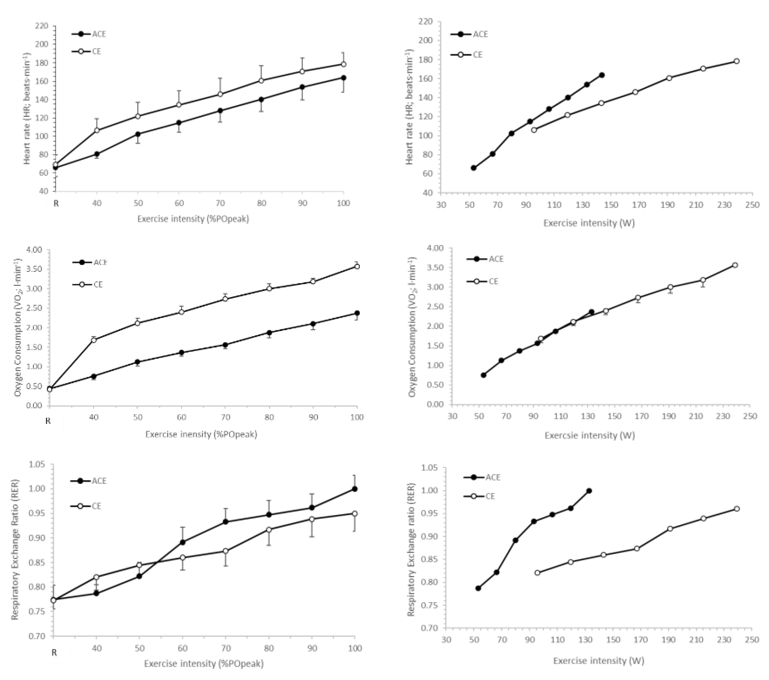
Figure 1. Heart rate (HR), oxygen uptake ( V ˙ O2) and respiratory exchange ratio (RER) responses to incremental arm crank (ACE) and cycle ergometry (CE). Data plotted in relation to %POpeak (left panel) and absolute power output (W) during exercise (right panel).
Ijerph 19 15311 g001
Figure 2. Carbohydrate and fat oxidation responses to incremental arm crank (ACE) and cycle ergometry (CE). Data plotted in relation to %POpeak (left panel) and absolute power output (W) during exercise (right panel). NB: Of the n = 13 participants, only n = 9 completed the final exercise stage at 100%POpeak; therefore, data for ACE at 100% POpeak and absolute power output of 133 W are for the remaining n = 9.
Ijerph 19 15311 g002
Table 1. Peak physiological responses for arm crank ergometry (ACE) and cycle ergometry (CE) in young, healthy males.
ACECEp
Mean ± SDMean ± SD
V ˙ O2 peak (L·min−1)2.73 ± 0.643.62 ± 0.46<0.001
V ˙ O2peak (mL·kg·−1min−1)36 ± 748 ± 5<0.001
V ˙ Epeak (L·min−1)100.4 ± 19.6120.1 ± 19.40.003
POpeak (W)133 ± 21239 ± 33<0.001
HRpeak (beats·min−1)170 ± 16182 ± 110.020
RPEC (Borg Scale)16 ± 318 ± 20.065
RPEL (Borg Scale)19 ± 119 ± 10.120
BLapeak (mmol·L−1)7.6 ± 1.99.0 ± 2.90.134
BLa+5 (mmol·L−1)8.0 ± 1.78.4 ± 3.00.179
RERpeak1.00 ± 0.100.96 ± 0.130.328
Note: V ˙ O2peak (L·min−1) = absolute peak oxygen uptake; V ˙ O2peak (mL·kg·−1min−1) = relative peak oxygen uptake; V ˙ Epeak (L·min−1) = peak minute ventilation; POpeak (W) = peak power output; HRpeak (beats·min−1) = peak heart rate; RPEC (Borg Scale) = peak rating of perceived exertion (central/cardiorespiratory effort); RPEL (Borg Scale) = peak rating of perceived exertion (local muscular effort); BLapeak (mmol·L−1) = peak blood lactate concentration, BLa+5 (mmol·L−1) = blood lactate concentration 5-min post-cessation of exercise; RERpeak = peak respiratory exchange ratio.
Table 2. Physiological responses (mean ± SD) for submaximal intensities (%POpeak) of arm crank ergometry (ACE) during 2- and 3-minute exercise stages in young, healthy males (n = 9).
Stage %POpeak p
40506070%POpeakStageInt.
HR2 min103 ± 22122 ± 19135 ± 22151 ± 22<0.050.5560.997
(beats·min−1)3 min107 ± 23125 ± 19138 ± 22152 ± 23
V ˙ O22 min0.65 ± 0.111.04 ± 0.301.34 ± 0.311.58 ± 0.33<0.050.9830.991
(L·min−1)3 min0.62 ± 0.111.07 ± 0.351.34 ± 0.331.58 ± 0.35
V ˙ CO22 min0.57 ± 0.160.94 ± 0.211.23 ± 0.251.43 ± 0.31<0.050.7730.919
(L·min−1)3 min0.57 ± 0.151.03 ± 0.221.23 ± 0.261.42 ± 0.41
RER2 min0.83 ± 0.030.88 ± 0.030.95 ± 0.030.96 ± 0.03<0.050.1450.774
3-min0.83 ± 0.030.92 ± 0.030.96 ± 0.040.95 ± 0.04
FOx2 min0.16 ± 0.040.20 ± 0.060.22 ± 0.080.31 ± 0.140.3240.9630.986
(g·min−1)3 min0.16 ± 0.040.17 ± 0.060.25 ± 0.100.33 ± 0.15
CHO2 min0.81 ± 0.781.49 ± 0.632.00 ± 0.822.20 ± 1.570.0030.6920.918
(g·min−1)3 min0.92 ± 0.961.88 ± 0.831.95 ± 1.022.15 ± 1.62
RPEC2 min7.2 ± 0.78.8 ± 1.010.6 ± 1.312.6 ± 1.1<0.050.1450.774
(Borg Scale)3 min7.2 ± 0.79.1 ± 1.110.2 ± 1.313.0 ± 1.6
RPEL2 min8.4 ± 1.211.0 ± 1.2)13.1 ± 0.814.3 ± 0.9<0.050.0880.968
(Borg Scale)3 min8.9 ± 1.112.0 ± 1.014.3 ± 0.814.8 ± 0.8
Note: HR (beats·min−1) = heart rate; V ˙ O2 (L·min−1) = absolute oxygen uptake; V ˙ CO2 (L·min−1) = absolute carbon dioxide production; RER = respiratory exchange ratio; FOx (g·min−1) = fat oxidation; CHO (g·min−1) = carbohydrate oxidation; RPEC (Borg Scale) = rating of perceived exertion (central/cardiorespiratory effort); RPEL (Borg Scale) = rating of perceived exertion (local muscular effort); p = p-value; %POpeak = exercise intensity main effect (40 vs. 50 vs. 60 vs. 70% POpeak); Stage = stage duration main effect (2-min vs. 3-min), Int = %POpeak × Stage interaction.
Publisher’s Note: MDPI stays neutral with regard to jurisdictional claims in published maps and institutional affiliations.