Article

Antibacterial Effect and Possible Mechanism of Salicylic Acid Microcapsules against Escherichia coli and Staphylococcus aureus

1
Department of Food Science and Technology, School of Perfume and Aroma Technology, Shanghai Institute of Technology, Shanghai 201418, China
2
Department of Food Science and Technology, School of Agriculture and Biology, Shanghai Jiao Tong University, Shanghai 200240, China
*
Authors to whom correspondence should be addressed.
Int. J. Environ. Res. Public Health 2022, 19(19), 12761; https://doi.org/10.3390/ijerph191912761
Submission received: 11 August 2022 / Revised: 1 October 2022 / Accepted: 3 October 2022 / Published: 6 October 2022
(This article belongs to the Special Issue Food Safety and Human Health)

Abstract

:
Microcapsules serve as a feasible formulation to load phenolic substances such as salicylic acid, a natural and safe antimicrobial agent. However, the antibacterial efficacy of salicylic acid microcapsules (SAMs) remains to be elucidated. Here, salicylic acid/β-cyclodextrin inclusion microcapsules were subjected to systematic antibacterial assays and preliminary antibacterial mechanism tests using Escherichia coli and Staphylococcus aureus as target organisms. It was found that the core-shell rhomboid-shaped SAMs had a smooth surface. SAMs exhibited a minimum inhibitory concentration (MIC) and a minimum bactericidal concentration (MBC) of 4 mg/mL against both bacteria. In the growth inhibition assay, 1/4 × MIC, 1/2 × MIC, and 1 × MIC of SAMs effectively retarded bacterial growth, and this effect was more prominent with the rise in the level of SAMs. Practically, SAMs possessed a rapid bactericidal effect at the 1 × MIC level with a reduction of more than 99.9% bacterial population within 10 min. A pronounced sterilization activity against E. coli and S. aureus was also observed when SAMs were embedded into hand sanitizers as antimicrobial agents. Moreover, exposure of both bacteria to SAMs resulted in the leakage of intracellular alkaline phosphatases and macromolecular substances (nucleic acids and proteins), which indicated the disruption of bacterial cell walls and cell membranes. In conclusion, SAMs were able to inactivate E. coli and S. aureus both in vitro and in situ, highlighting the promising utilization of this formulation for antimicrobial purposes in the area of food safety and public health.

1. Introduction

The growing demand for food safety has motivated the search for natural and safe antimicrobial substances to control pathogenic bacteria such as Staphylococcus aureus and Escherichia coli in the food industry. Salicylic acid derived from natural willow bark and other plants is a kind of safe agent which exerts a high potential for antimicrobial purposes during food processing and preservation [1]. A large number of scholars have focused on the antifungal efficiency of salicylic acid, since this natural compound can be produced by plants in response to microbial invasions [2,3]. Indeed, salicylic acid is effective in controlling postharvest fungal pathogens such as Penicillium expansum, Botrytis cinerea, Fusarium oxysporum, and Rhizopus stolonifer in fruit and vegetables [4,5,6]. Moreover, several studies have illustrated the inhibitory activity of starch- or chitosan-based films incorporated with salicylic acid as an active agent against bacterial strains such as S. aureus, E. coli, and Bacillus subtilis by a qualitative disc diffusion method [1,7]. To date, however, no quantitative studies (e.g., minimum inhibitory concentration tests and time-kill assays) have been conducted on the antibacterial activity of salicylic acid-based formulations. Moreover, the main disadvantage centers around low water solubility when salicylic acid is applied to foods. This makes further utilization of salicylic acid a bit challenging in the food industry.
Encapsulation serves as a strategy to counteract the low water solubility of phenolic substances, such as salicylic acid. This technology can entrap solid particles, liquid droplets, or gaseous compounds in wall materials, thus providing an effective approach for controlled release and protection of active compounds [8]. In the food and pharmaceutical industries, chitosan, sodium alginate, and β-cyclodextrin (β-CD) are commonly used as wall materials for encapsulating hydrophobic core substances [9,10,11]. Among these wall materials, β-CD is considered as an ideal one owing to its low cost, good thermal stability, and commercial development prospects [12]. Recently, molecular inclusion in β-CD complexes has been reported to be effective in masking the undesirable smell of essential oils and increasing their solubility [13].
Preliminary work in our group has demonstrated the successful preparation of salicylic acid/β-CD inclusion complexes and the resulting salicylic acid microcapsules (SAMs) exhibited appreciable water solubility. To our knowledge, there is currently no relevant study on the antibacterial activity of β-CD inclusion complexes containing salicylic acid, a safe and natural phenolic compound. Practically, it has been revealed that other phenolic substances can accumulate at the bacterial surface and disrupt cell membranes and cell walls [14]. Therefore, analysis of bacterial surface properties will contribute to a better understanding of the mode of action of SAMs. In this context, the aim of the current work was to elucidate the inhibitory activity of SAMs against S. aureus and E. coli via systematic antibacterial tests coupled with cell property analyses, which may help to broaden the application of salicylic acid in the food industry.

2. Materials and Methods

2.1. Materials

Nutrient agar (NA) and nutrient broth (NB) were purchased from Qingdao Hope Bio-Technology Co., Ltd., Qingdao, Shandong, China. Analytical grade ethanol was supplied by Shanghai Richjoint Chemical Reagents Co. Ltd., Shanghai, China. Salicylic acid, β-CD, sodium chloride, and other chemicals were obtained from Sinopharm Chemical Reagent Co., Ltd., Shanghai, China.

2.2. Test Strains

The target bacteria were Gram-negative E. coli ATCC 25922 and Gram-positive S. aureus ATCC 25923. Before testing, each strain was inoculated in NA at 37 °C for 24 h and then transferred to NB, followed by incubation at 37 °C for 24 h. Subsequently, bacterial suspensions were centrifuged (TGL-16M, Cence, Changsha, Hunan, China) at 2000 rpm for 3 min. The pellets were diluted in sterile saline to prepare bacterial suspensions with a final concentration of 5 × 10⁸ CFU/mL, which were used in subsequent experiments.

2.3. Preparation of SAMs

According to the Patent CN108210346B, salicylic acid/β-CD inclusion microcapsules were prepared [15]. This host-guest inclusion complex can be formed since the sugar structures of β-CD with hydrophobic cavities and hydrophilic exteriors allow fat-soluble salicylic acid to be encapsulated. Salicylic acid is stable in the inclusion complex due to the fact that its carboxyl group is located in the plane of the upper edge of β-CD molecule, and a weak hydrogen bond is formed between phenyl hydroxyl and glycoside oxygen [16]. A total of 7.5 g β-CD was dissolved in 100 mL distilled water (95 °C) to form a wall material solution. Salicylic acid (2.3 g) was dissolved in 0.4 mL ethanol at 20 °C and then slowly added to the wall material solution. The resulting mixture was continuously stirred at 500 rpm and 50 °C for 24 h to obtain the microcapsule solution. The solution was then cooled to 20 °C, filtrated, washed with deionized water at least five times, and dried at 50 °C for 2 h to prepare white SAM powders.
The effect of storage temperature on microcapsule stability was then assessed. The formulated SAMs were added to 20 mL NB in centrifuge tubes to reach final concentrations of 2 to 8 mg/mL. Tubes were then stored at different temperatures (−4 °C, 37 °C, and 40 °C) for 24 h. It was observed that all concentrations of SAMs retained transparent without precipitation or layer separation in NB.

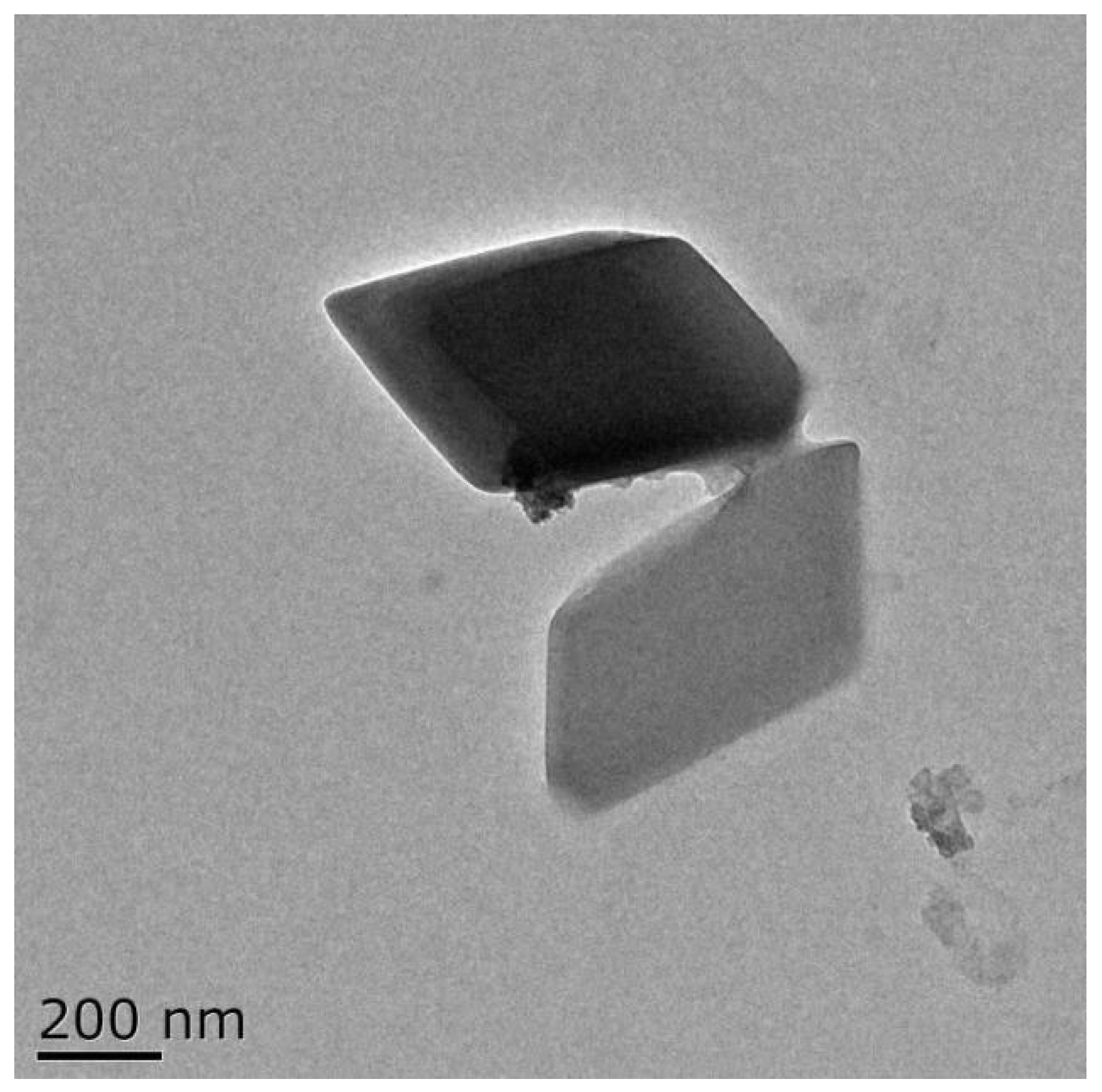
2.4. Characterization of Morphology

The morphological characteristics of SAMs were analyzed using a transmission electron microscope (TEM) (Tecnai G2 F20 S-TWIN, FEI, Hillsboro, USA). Briefly, SAM dispersions (1 mg/mL in distilled water) were prepared, dropped onto 200 meshes of carbon-coated copper grid, and then air-dried. TEM measurements were carried out at 200 kV at room temperature (25 °C).

2.5. Assessment of Loading Capacity

The loading capacity of SAMs was determined using UV-Vis spectroscopy (UV1800, Shimadzu, Kyoto, Japan) at a wavelength of 300 nm according to a modified version of methods proposed by Piletti et al. [11]. A standard calibration curve was obtained based on the absorbance of salicylic acid dilutions (10–30 μg/mL) in ethanol. Subsequently, 0.2 g of SAMs was added to 100 mL of absolute ethanol. After 24 h, the mixture was centrifuged at 4000 rpm for 5 min. The absorbance of the supernatant containing salicylic acid was measured, and the loading capacity of SAMs was calculated as follows:
Loading capacity = (the mount of encapsulated salicylic acid)/(the amount of microcapsules) × 100%

2.6. Determination of Minimum Inhibitory and Bactericidal Concentrations

Minimum inhibitory concentration (MIC) is an indicator of the antimicrobial strength of active components, while minimum bactericidal concentration (MBC) represents the minimum concentration required to kill 99.9% of the tested microorganisms [17]. Thus, lower MIC and MBC values correspond to stronger antibacterial and bactericidal abilities. The MIC and MBC values of SAMs against E. coli and S. aureus were determined through the two-fold serial dilution method [18]. Briefly, the prepared bacterial suspensions were added to NB, followed by the addition of SAMs to a final concentration of 2 to 16 mg/mL. Then, bacterial suspensions were incubated at 37 °C with shaking at 200 rpm for 24 h. The minimum concentration at which the visible growth of the target strain was completely inhibited was considered the MIC value. In contrast, the minimum concentration to completely kill the microorganisms was considered the MBC value. The experiments were repeated twice, with three replicates for each concentration.

2.7. Growth Inhibition Tests

The effect of SAMs on bacterial proliferation in E. coli and S. aureus was determined using UV-Vis spectroscopy at a wavelength of 600 nm [19]. Bacterial suspensions were added to NB supplemented with 0 ×, 1/4 ×, 1/2 ×, and 1 × MIC of SAMs. The growth of bacteria was determined after incubation at 37 °C with shaking at 200 rpm. The absorbance was determined every 2 h and the antibacterial dynamic curves were obtained from the data. All experiments were performed in triplicate.

2.8. Time-Kill Assays

The bactericidal effect of SAMs was evaluated using a time-kill assay [20]. Briefly, SAMs were added to different bacterial suspensions to the final level of 1 × MIC, which were then shaken at 200 rpm (37 °C). Bacterial colonies were determined after the bacterial suspensions were exposed to SAMs for 0, 0.25, 1, and 10 min by the plate count method. Sterile distilled water was used as a control group in a similar way. All experiments were performed in triplicate.

2.9. Antibacterial Efficacy of SAMs in Hand Sanitizers

Control hand sanitizers were obtained from the International School of Cosmetics at Shanghai Institute of Technology, China, with inactive ingredients including cocamidopropyl betaine, alkyl polyglucoside, cocamide DEA, 1,3-butylene glycol, glycerin, disodium EDTA, PEG-14M, and sodium chloride. SAM-based hand sanitizers were prepared with the addition of SAMs to the dose of 1/2 ×, 1 ×, and 2 × MIC, respectively. Briefly, either E. coli or S. aureus was incubated in NB at 37 °C overnight, and then adjusted to a bacterial concentration of 1 × 10⁷ CFU/mL. Approximately 1 g hand sanitizers were transferred to a conical flask containing 9 mL bacterial suspensions. Samples were vortexed for 0, 0.25, 1, and 10 min, respectively, and 2% lecithin was added as a neutralizer. Serial dilutions were spread on the surface of NA and then cultivated at 37 °C for 18–24 h. The viable counts were determined in triplicate for each decimal dilution.

2.10. Analysis of Cell Wall Permeability

The integrity of cell walls can be judged by alkaline phosphatase (AKP) activity, which was detected according to the method of Wang et al. [21] with slight modifications. The bacterial suspension was washed with PBS three times and then resuspended. Then, SAMs were added to achieve a final concentration of 1/2 × MIC and 1 × MIC. The samples were cultured at 37 °C for 0, 0.25, 1, and 10 min, and then 0.2 mL filtrates with 0.22 μm filter membrane were added to 1.8 mL of 0.1 mol/L Tris-HCl buffer (pH 8.0, containing 0.2 mg/mL 4-nitrophenyl phosphate disodium salt). Afterwards, mixtures were incubated at 25 °C for 30 min. The absorbance value at 410 nm was used to express the content of AKP in the solution. The blank control group was treated with sterile distilled water.

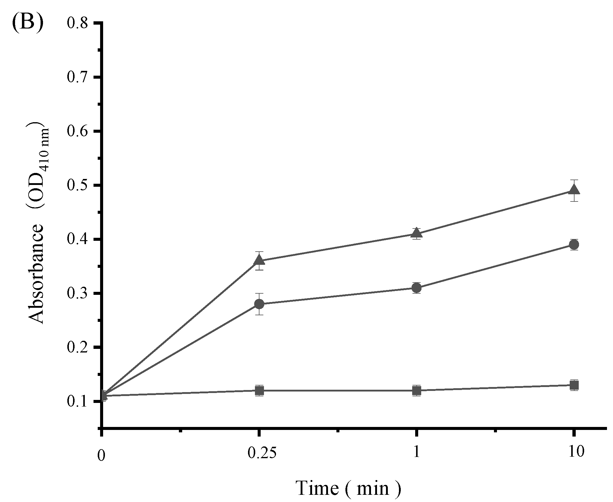
2.11. Measurement of Cell Membrane Permeability

Membrane permeability was determined by measuring the leakage of nucleic acids and proteins from the bacteria [22,23]. The bacterial suspension was washed with PBS three times, and exposed to SAMs dilutions with the final concentrations of 1/2 × MIC and 1 × MIC. After incubation at 37 °C and 120 rpm for 0, 0.25, 1, and 10 min, 5 mL of bacterial suspensions were removed and passed through a 0.22 μm filter. The content of nucleic acids in the bacterial suspensions was determined by the absorbance value of the filtrate at 260 nm. Similarly, the content of soluble proteins after filtration was determined by the Coomassie Blue Staining. The filtrates (1 mL) of all samples were thoroughly mixed with Coomassie brilliant blue (G-250) dye solutions (5 mL) for 2 min, and the absorbance of all solutions at 595 nm was measured. The blank control group was treated with sterile distilled water.

2.12. Data Analysis

Analysis of variance was performed using SAS 9.4 software. Duncan’s multiple comparison method was used to detect statistically significant differences at the p < 0.05 level.

3. Results and Discussion

3.1. TEM Morphology of SAMs

As shown in Figure 1, salicylic acid/β-CD inclusion microcapsules had an obvious core-shell rhomboid shape and a smooth surface with a diameter ranging from 0.5 to 0.6 μm. The clear β-CD shells around the salicylic acid sphere, with the wall thickness in the range of 40–60 nm, strongly confirmed the successful encapsulation of salicylic acid in β-CD. A similar result was reported by Anaya–Castro et al. [24], who found that clove essential oil could be embedded by β-CD to form a core-shell rhomboid structure.
The formation and shape of microcapsules are affected by many factors such as preparation methods, type of core and wall materials, and stirring speeds [25,26]. Similarly, other delivery systems such as emulsions with desirable characteristics can be produced by adjusting emulsifier type, continuous or dispersed phase concentration, or the emulsification process, etc [27,28]. Hence, control of fabrication process parameters is of great importance for the design of suitable delivery systems for food applications.

3.2. Loading Capacity of SAMs

Loading capacity has been widely used to determine the amount of encapsulated core materials [29]. In this work, the loading capacity of SAMs in β-CD was determined using a calibration curve based on the UV absorbance values. The loading capacity of SAMs was found to be 26.7% after the encapsulation processes. This showed that a low-molecular-weight organic acid such as salicylic acid was successfully encapsulated in β-CD using the host-guest inclusion complexation method. Similarly, cinnamon essential oil was embedded into a microcapsule with a high loading capacity of 30.2% in our previous work [30].

3.3. MIC and MBC Values of SAMs

The MIC and MBC tests were employed as a quantitative method to assess the antibacterial activity of SAMs. It was observed that SAMs exhibited both MIC and MBC values of 4 mg/mL against S. aureus and E. coli. In previous work, cinnamon essential oil displayed pronounced antibacterial performance against a variety of bacterial strains (e.g., S. aureus and E. coli), with MIC values ranging from 2.5 to 5 mg/mL, and MBC values ranging from 2.5 to 10 mg/mL [31]. It was thus indicated that SAMs in this work possessed excellent antibacterial properties comparable to cinnamon essential oil, whose potential for antibacterial applications in the food industry has been well documented. In this sense, future application of SAMs during food processing and preservation to ensure food safety can be anticipated.
It should be noted that the in vitro antibacterial activity of salicylic acid-based formulations has been only previously assessed qualitatively by a disc diffusion method [1,7]. For instance, chitosan films enriched with salicylic acid showed an inhibition zone diameter between 6 and 8 mm against S. aureus and E. coli [7]. Interestingly, our current work employed a quantitative approach to determine the bacteriostatic and bactericidal effects of SAMs, which enforces our knowledge on the antibacterial performance of salicylic acid on bacterial strains.

3.4. Growth Inhibition Activity of SAMs

In order to further explore the bacteriostatic activity of SAMs, dynamic growth curves of S. aureus and E. coli were determined under different concentrations of SAMs (0 ×, 1/4 ×, 1/2 ×, and 1 × MIC) (Figure 2). Both bacterial strains in the control group showed a rapid increase in OD600 values, reaching a steady state after 16 h for E. coli, and 20 h for S. aureus, respectively. Treatments with SAMs resulted in reduced bacterial growth, and this inhibitory effect was more pronounced with the rise in the concentration of SAMs. When SAMs were applied at the 1 × MIC level, a complete inhibition in bacterial growth was achieved, which confirmed the accuracy of the results derived from the MIC test. Notably, the bacteriostatic activity of SAMs was more prominent in S. aureus compared to that in E. coli. For example, the OD600 value of bacterial suspensions of S. aureus and E. coli was reduced by approximately 0.9 and 0.5, respectively, after exposure to 1/2 × MIC of SAMs for 24 h.
In agreement with the above mentioned observations, the growth of S. aureus and E. coli was retarded by 1/2 × MIC of ginger essential oil, and the decline in the OD600 value was more pronounced in S. aureus (Gram-positive) than E. coli (Gram-negative) [32]. The superior growth of E. coli under subinhibitory levels of SAMs and essential oils might be related to the thick outer lipopolysaccharide layer of Gram-negative bacteria, which provided protection against antibacterial agents [33,34]. Taken together, SAMs exerted good bacteriostatic activity in a dose-dependent manner, although their effect on Gram-positive and Gram-negative bacteria was different to some extent.

3.5. Time-Kill Activity of SAMs

A time-kill assay was performed using SAMs at the concentration of 1 × MIC to further assess its bactericidal activity. The bactericidal effects of SAMs were dependent on the duration of contact. As shown in Figure 3A, E. coli was gradually killed during exposure to SAMs, reaching a population reduction by approximately 4.5 log CFU/mL after 10 min. In the case of S. aureus, the bacterial population was eliminated by approximately 3.5 log CFU/mL within 1 min of contact with SAMs (Figure 3B). These results indicated that SAMs possessed rapid bactericidal effects against both S. aureus and E. coli.
In previous work, exposure to 4/5 × MIC of cinnamon essential oil for 2 h resulted in a reduction of approximately 99.9% population (i.e., 3-log population reduction) of S. aureus and E. coli [16]. Given the well-known antibacterial performance of cinnamon essential oil, it can be inferred that the rapid sterilization activity exerted by SAMs in this work was quite satisfactory. The bactericidal action of SAMs could be due to the effective interaction of salicylic acid in the microcapsules with bacteria in the media. Hence, SAMs possessed excellent bacteriostatic and bactericidal capacity, which highlighted the necessity of checking the application potential of SAMs in food, cosmeceutical, and pharmaceutical systems.

3.6. Antibacterial Efficacy of SAMs in Hand Sanitizers

As an effective way to prevent the spread of infectious disease, hand washing is attached with increasing importance [35]. Ethanol has been used as an active ingredient in hand sanitizers in food processing plants for a long time [36]. However, bacterial resistance to ethanol and other disinfectants has emerged as a potential food safety concern [37,38]. In this work, SAMs were exploited as an alternative to traditional hand sanitizers.
According to Figure 4, the populations of E. coli and S. aureus were rapidly and progressively reduced at all three tested concentrations. Also, SAMs-based hand sanitizers worked on bacterial strains in a dose-dependent manner. It was observed that the 1 × MIC level of SAMs was able to decrease at least 99% of the initial population (i.e., 2-log population reduction) of E. coli and S. aureus after 1 min, suggesting that SAMs were active for achieving bactericidal activity in hand sanitizers. Interestingly, a higher concentration (2 × MIC) of SAMs resulted in a reduction of 99.99% of the bacterial populations (i.e., 4-log population reduction) after 10 min. This result probably expands SAMs’ application as washing solutions in sanitizing products and environments.

3.7. Impact of SAMs on Bacterial Cell Wall Integrity

Bacterial AKP is normally located between cell walls and cell membranes. The activity of this enzyme will not be detected in extracellular environments unless the bacterial cell wall is disrupted [39]. Hence, cell wall damage can be interpreted by the leakage of intracellular AKP into bacterial cell suspensions. As presented in Figure 5, the OD410 value of extracellular AKP from both S. aureus (approximately 0.11) and E. coli (approximately 0.13) was maintained constant in the absence of SAMs. However, this value increased dramatically after S. aureus and E. coli were treated with SAMs. Furthermore, AKP activity from these two bacterial strains was enhanced along with an increase in SAMs concentration from 1/2 × MIC to 1 × MIC. For example, the OD410 value of extracellular AKP in S. aureus cell suspensions was increased from 0.11 to 0.39, and 0.11 to 0.49, respectively, in response to 1/2 × MIC and 1 × MIC of SAMs. It can thus be inferred that SAMs may act on the cell wall by disrupting its integrity, which results in the leakage of intracellular AKP into cell suspensions.
In agreement with the above mentioned findings, disruption of cell wall permeability contributed to enhanced extracellular AKP activity after exposure of bacteria to various antibacterial agents [39,40,41,42]. Zhao et al. [42] found that the activity of released AKP increased significantly after S. aureus was treated with 2 × MIC of microencapsulated dodecyl gallate with methyl-β-cyclodextrin for 6 h. Similarly, octopus scraps peptides-zinc chelates at the levels of 1/2 × MIC to 1 × MIC could damage the cell wall of E. coli, which resulted in the leakage of intracellular AKP [41]. These results indicated that disruption of cell wall integrity might be a key factor leading to bacterial death in the presence of antibacterial agents such as SAMs.

3.8. Impact of SAMs on Bacterial Cell Membrane Permeability

Maintenance of cell membrane structure is a prerequisite for bacterial growth and metabolic activity. When the bacterial cell membrane is disrupted, intracellular macromolecular substances (e.g., nucleic acids and proteins) tend to flow out [43,44]. Therefore, the release of cell constituents is a good indicator for assessing bacterial cell membrane integrity in response to antibacterial agents. In the current work, nucleic acids and proteins released from the cytoplasm were monitored by the detection of absorbance at 260 nm and 595 nm, respectively. As shown in Figure 6 and Figure 7, both cell constituents were released rapidly from S. aureus and E. coli into cell suspensions and their amounts increased multi-fold after treatment with increasing levels of SAMs. For instance, the OD260 value of E. coli increased significantly from 0.16 for the control to 1.27 and 2.45 in the presence of SAMs at the levels of 1/2 × MIC and 1 × MIC after 10 min, respectively (Figure 6). Moreover, there was a progressive release of proteins from S. aureus after exposure to SAMs for 0.25 min, followed by a steady state (Figure 7). These results clearly suggested that bacterial cell membrane integrity was compromised by SAMs, which could consequently result in cell death.
It has been reported that other phenolic substances can also cause disruption in the bacterial cell membrane. Cao et al. [45] found that epigallocatechin gallate could enhance the permeability of the inner membrane and outer membrane in E. coli and cause cell membrane depolarization. Similarly, Kombucha polyphenolic fraction disrupted the membrane integrity of Vibrio cholera as revealed by permeabilization assays, which might be related to the generation of intracellular reactive oxygen species [46]. Therefore, the action of salicylic acid and other phenolic substances on bacterial cell membranes may be due to their interactions with cell membrane proteins, which lead to the loss of chemiosmotic control and finally cell death [47].

4. Conclusions

The core-shell rhomboid-shaped SAMs possessed excellent antibacterial performance against both E. coli and S. aureus, possibly by acting on bacterial cell walls and cell membranes. Notably, a superior sterilization activity was achieved in 10 min with a reduction of more than 99.9% bacterial population. SAMs were also effective in inactivating E. coli and S. aureus when incorporated into hand sanitizers as active agents. These findings suggest that SAMs have the potential to be utilized as a feasible antibacterial formulation in food, cosmeceutical, and pharmaceutical industries to protect public health. In terms of food applications, the influence of environmental factors (e.g., heat, cold, acid, and salt) on the antibacterial activity of SAMs will be elucidated in future work to further explore the strengths of using this material during food manufacturing. Moreover, it would also be interesting to address the accumulative release of SAMs in the presence of the aforementioned environmental factors related to food processing and preservation.

Author Contributions

Conceptualization, S.H. and Y.W.; methodology, X.S. and R.L.; software, R.L. and Q.Z.; validation, R.L. and Q.Z.; formal analysis, S.H. and Y.W.; investigation, X.S. and R.L.; data curation, R.L. and Q.Z.; writing—original draft preparation, X.S.; writing—review and editing, S.H. and Y.W.; supervision, S.H. and Y.W.; funding acquisition, S.H. and Y.W. All authors have read and agreed to the published version of the manuscript.

Funding

This research was funded by the National Natural Science Foundation of China (No. 32001797), and the alliance plan of Shanghai Promotion Association of Tech-Transfer (SPAT) (No. LM201644, LM201528, and LM201328).

Institutional Review Board Statement

Not applicable.

Informed Consent Statement

Not applicable.

Data Availability Statement

All data are available in the article.

Conflicts of Interest

The authors declare no conflict of interest.

References

  1. Fang, Y.; Fu, J.; Tao, C.; Liu, P.; Cui, B. Mechanical properties and antibacterial activities of novel starch-based composite films incorporated with salicylic acid. Int. J. Biol. Macromol. 2020, 155, 1350–1358. [Google Scholar] [CrossRef] [PubMed]
  2. Asghari, M.; Aghdam, M.S. Impact of salicylic acid on post-harvest physiology of horticultural crops. Trends Food Sci. Technol. 2010, 21, 502–509. [Google Scholar] [CrossRef]
  3. Khan, N.; Bano, A.; Curá, J.A. Role of beneficial microorganisms and salicylic acid in improving rainfed agriculture and future food safety. Microorganisms 2020, 8, 1018. [Google Scholar] [CrossRef]
  4. Neto, A.C.; Luiz, C.; Maraschin, M.; Di Piero, R.M. Efficacy of salicylic acid to reduce Penicillium expansum inoculum and preserve apple fruits. Int. J. Food Microbiol. 2016, 221, 54–60. [Google Scholar] [CrossRef]
  5. Panahirad, S.; Zaare-Nahandi, F.; Safaralizadeh, R.; Alizadeh-Salteh, S. Postharvest control of Rhizopus stolonifer in peach (Prunus persica L. Batsch) fruits using salicylic acid. J. Food Saf. 2012, 32, 502–507. [Google Scholar] [CrossRef]
  6. Hussain, M.; Hamid, M.I.; Ghazanfar, M.U. Salicylic acid induced resistance in fruits to combat against postharvest pathogens: A review. Arch. Phytopathol. Plant Prot. 2015, 48, 34–42. [Google Scholar] [CrossRef]
  7. Hu, F.; Sun, T.; Xie, J.; Shao, Z. Functional properties of chitosan films with conjugated or incorporated salicylic acid. J. Mol. Struct. 2021, 1223, 129237. [Google Scholar] [CrossRef]
  8. Malaquias, L.F.B.; Sá-Barreto, L.C.L.; Freire, D.O.; Silva, I.C.R.; Karan, K.; Durig, T.; Lima, E.M.; Marreto, R.N.; Gelfuso, G.M.; Gratieri, T. Taste masking and rheology improvement of drug complexed with beta-cyclodextrin and hydroxypropyl-β-cyclodextrin by hot-melt extrusion. Carbohyd. Polym. 2018, 185, 19–26. [Google Scholar] [CrossRef]
  9. Arana-Sánchez, A.; Estarrón-Espinosa, M.; Obledo-Vázquez, E.N.; Padilla-Camberos, E.; Silva-Vázquez, R.; Lugo-Cervantes, E. Antimicrobial and antioxidant activities of Mexican oregano essential oils (Lippia graveolens H. B. K.) with different composition when microencapsulated in β-cyclodextrin. Lett. Appl. Microbiol. 2010, 50, 585–590. [Google Scholar] [CrossRef]
  10. Soliman, E.A.; El-Moghazy, A.Y.; El-Din, M.; Massoud, M.A. Microencapsulation of essential oils within alginate: Formulation and in vitro evaluation of antifungal activity. J. Enca. Adsor. Sci. 2013, 3, 48–55. [Google Scholar]
  11. Piletti, R.; Zanetti, M.; Jung, G.; De Mello, J.M.M.; Dalcanton, F.; Soares, C.; Riella, H.G.; Fiori, M.A. Microencapsulation of garlic oil by β-cyclodextrin as a thermal protection method for antibacterial action. Mat. Sci. Eng. C 2019, 94, 139–149. [Google Scholar] [CrossRef] [PubMed]
  12. Szente, L.; Szejtli, J. Cyclodextrins as food ingredients. Trends Food Sci. Technol. 2004, 15, 137–142. [Google Scholar] [CrossRef]
  13. Tao, F.; Hill, L.E.; Peng, Y.; Gomes, C.L. Synthesis and characterization of β-cyclodextrin inclusion complexes of thymol and thyme oil for antimicrobial delivery applications. LWT—Food Sci. Technol. 2014, 59, 247–255. [Google Scholar] [CrossRef]
  14. Wu, Y.; Bai, J.; Zhong, K.; Huang, Y.; Qi, H.; Jiang, Y.; Gao, H. Antibacterial activity and membrane-disruptive mechanism of 3-p-trans-coumaroyl-2-hydroxyquinic acid, a novel phenolic compound from pine needles of Cedrus deodara, against Staphylococcus aureus. Molecules 2016, 21, 1084. [Google Scholar] [CrossRef] [PubMed] [Green Version]
  15. Yang, X.; Song, X.; Ye, L.; Zhang, J.; Wang, L.; Sun, Y.; He, J. Composite wall material microcapsules loaded with salicylic acid and preparation method thereof. CN Patent 108210346 B, 23 June 2020. [Google Scholar]
  16. Belyakova, L.A.; Varvarin, A.M.; Lyashenko, D.Y.; Khora, O.V.; Oranskaya, E.I. Complexation in a β-cyclodextrin-salicylic acid system. Colloid J. 2007, 69, 546–551. [Google Scholar] [CrossRef]
  17. Cui, H.; Zhou, H.; Lin, L. The specific antibacterial effect of the Salvia oil nanoliposomes against Staphylococcus aureus biofilms on milk container. Food Control 2016, 61, 92–98. [Google Scholar] [CrossRef]
  18. Donsì, F.; Annunziata, M.; Sessa, M.; Ferrari, G. Nanoencapsulation of essential oils to enhance their antimicrobial activity in foods. LWT—Food Sci. Technol. 2011, 44, 1908–1914. [Google Scholar] [CrossRef]
  19. Li, K.; Guan, G.; Zhu, J.; Wu, H.; Sun, Q. Antibacterial activity and mechanism of a laccase-catalyzed chitosan–gallic acid derivative against Escherichia coli and Staphylococcus aureus. Food Control 2019, 96, 234–243. [Google Scholar] [CrossRef]
  20. Lee, D.S.; Je, J.Y. Gallic acid-grafted-chitosan inhibits foodborne pathogens by a membrane damage mechanism. J. Agric. Food Chem. 2013, 61, 6574–6579. [Google Scholar] [CrossRef]
  21. Wang, J.; Zhang, W.; Tang, C.; Xiao, J.; Xie, B.; Sun, Z. Synergistic effect of B-type oligomeric procyanidins from lotus seedpod in combination with water-soluble Poria cocos polysaccharides against E. coli and mechanism. J. Funct. Foods. 2018, 48, 134–143. [Google Scholar] [CrossRef]
  22. Lv, F.; Liang, H.; Yuan, Q.; Li, C. In vitro antimicrobial effects and mechanism of action of selected plant essential oil combinations against four food-related microorganisms. Food Res. Int. 2011, 44, 3057–3064. [Google Scholar] [CrossRef]
  23. Liu, G.; Ren, G.; Zhao, L.; Cheng, L.; Wang, C.; Sun, B. Antibacterial activity and mechanism of bifidocin A against Listeria monocytogenes. Food Control 2017, 73, 854–861. [Google Scholar] [CrossRef]
  24. Anaya-Castro, M.A.; Ayala-Zavala, J.F.; Muñoz-Castellanos, L.; Hernández-Ochoa, L.; Peydecastaing, J.; Durrieu, V. β-Cyclodextrin inclusion complexes containing clove (Eugenia caryophyllata) and Mexican oregano (Lippia berlandieri) essential oils: Preparation, physicochemical and antimicrobial characterization. Food Packag. Shelf Life 2017, 14, 96–101. [Google Scholar] [CrossRef] [Green Version]
  25. Santana, A.A.; Cano-Higuita, D.M.; De Oliveira, R.A.; Telis, V.R. Influence of different combinations of wall materials on the microencapsulation of jussara pulp (Euterpe edulis) by spray drying. Food Chem. 2016, 212, 1–9. [Google Scholar] [CrossRef] [PubMed]
  26. Su, Y.; Chen, Y.; Zhang, L.; Adhikari, B.; Xu, B.; Li, J.; Zheng, T. Synthesis and characterization of lotus seed protein-based curcumin microcapsules with enhanced solubility, stability, and sustained release. J. Sci. Food Agric. 2022, 102, 2220–2231. [Google Scholar] [CrossRef]
  27. Javad, S.; Gopirajah, R.; Rizvi, S.S. High internal phase oil-in-water emulsions stabilized by supercritical carbon dioxide extruded whey protein concentrate. Food Chem. 2022, 372, 131362. [Google Scholar] [CrossRef]
  28. Ghasemi, H.; Darjani, S.; Mazloomi, H.; Mozaffari, S. Preparation of stable multiple emulsions using food-grade emulsifiers: Evaluating the effects of emulsifier concentration, W/O phase ratio, and emulsification process. SN Appl. Sci. 2020, 2, 2002. [Google Scholar] [CrossRef]
  29. Yang, Z.; Fang, Y.; Ji, H. Controlled release and enhanced antibacterial activity of salicylic acid by hydrogen bonding with chitosan. Chin. J. Chem. Eng. 2016, 24, 421–426. [Google Scholar] [CrossRef]
  30. Song, X.; Xu, Y.; Wang, Y.; Chen, J.; Xiao, Y. Effects of cinnamon oil microcapsules on antioxidant activities and gut microbiota in mice. Food Sci. 2021, 42, 143–152. [Google Scholar]
  31. Oulkheir, S.; Aghrouch, M.; Mourabit, F.E.; Dalha, F.; Graich, H.; Amouch, F.; Ouzaid, K.; Moukale, A.; Chadli, S. Antibacterial activity of essential oils extracts from cinnamon, thyme, clove and geranium against a Gram negative and Gram positive pathogenic bacteria. J. Med. Plants Res. 2017, 3, 1–5. [Google Scholar]
  32. Wang, X.; Shen, Y.; Thakur, K.; Han, J.; Zhang, J.G.; Hu, F.; Wei, Z.J. Antibacterial activity and mechanism of ginger essential oil against Escherichia coli and Staphylococcus aureus. Molecules 2020, 25, 3955. [Google Scholar] [CrossRef] [PubMed]
  33. Bauza, B.; Carranza, H.; Pérez, C.; Sosa, Á.; López, R.; Velasco, O. Antimicrobial activity of ginger (Zingiber Officinale) and its application in food products. Food Rev. Int. 2019, 35, 407–426. [Google Scholar] [CrossRef]
  34. Hsouna, A.B.; Trigui, M.; Mansour, R.B.; Jarraya, R.M.; Damak, M.; Jaoua, S. Chemical composition, cytotoxicity effect and antimicrobial activity of Ceratonia siliqua essential oil with preservative effects against Listeria inoculated in minced beef meat. Int. J. Food Microbiol. 2011, 148, 66–72. [Google Scholar] [CrossRef]
  35. Momeni, S.S.; Tomlin, N.; Ruby, J.D. Isolation of Raoultella planticola from refillable antimicrobial liquid soap dispensers in a dental setting. J. Am. Dent. Assoc. 2015, 146, 241–245. [Google Scholar] [CrossRef] [PubMed] [Green Version]
  36. He, S.; Fong, K.; Wang, S.; Shi, X. Meat juice contributes to the stability of ethanol adaptation in Salmonella enterica serovar Enteritidis. Food Qual. Saf. 2021, 5, fyab017. [Google Scholar] [CrossRef]
  37. He, S.; Fong, K.; Wang, S.; Shi, X. Ethanol adaptation in foodborne bacterial pathogens. Crit. Rev. Food Sci. 2021, 61, 777–787. [Google Scholar] [CrossRef]
  38. He, S.; Zhan, Z.; Shi, C.; Wang, S.; Shi, X. Ethanol at subinhibitory concentrations enhances biofilm formation in Salmonella Enteritidis. Foods 2022, 11, 2237. [Google Scholar] [CrossRef]
  39. Cui, H.; Zhang, C.; Li, C.; Lin, L. Antimicrobial mechanism of clove oil on Listeria monocytogenes. Food Control 2018, 94, 140–146. [Google Scholar] [CrossRef]
  40. Lin, L.; Gu, Y.; Li, C.; Vittayapadung, S.; Cui, H. Antibacterial mechanism of ε-Poly-lysine against Listeria monocytogenes and its application on cheese. Food Control 2018, 91, 76–84. [Google Scholar] [CrossRef]
  41. Lin, Y.; Tang, X.; Xu, L.; Wang, S. Antibacterial properties and possible action mechanism of chelating peptides-zinc nanocomposite against Escherichia coli. Food Control 2019, 106, 106675. [Google Scholar] [CrossRef]
  42. Zhao, J.; Peng, T.; Liang, S.; Ma, M.; Deng, S. Antibacterial activity and action mechanism of microencapsulated dodecyl gallate with methyl-β-cyclodextrin. Food Control 2020, 109, 106953. [Google Scholar] [CrossRef]
  43. He, S.; Yang, Q.; Ren, X.; Zi, J.; Lu, S.; Wang, S. Antimicrobial efficiency of chitosan solutions and coatings incorporated with clove oil and/or ethylenediaminetetraacetate. J. Food Saf. 2014, 34, 345–352. [Google Scholar] [CrossRef]
  44. He, S.; Zhou, X.; Shi, C.; Shi, X. Ethanol adaptation induces direct protection and cross-protection against freezing stress in Salmonella enterica serovar Enteritidis. J. Appl. Microbiol. 2016, 120, 697–704. [Google Scholar] [CrossRef] [PubMed]
  45. Cao, Y.D.; Qian, H.L.; Feng, C.M.; Wang, T.; Guo, Z.Y.; Wu, X.K.; Zhang, S.H. Study on the mechanism of epigallocatechin gallate (EGCG) to the cell membrane of Escherichia coli. Sci. Adv. Mater. 2019, 11, 262–268. [Google Scholar] [CrossRef]
  46. Bhattacharya, D.; Ghosh, D.; Bhattacharya, S.; Sarkar, S.; Karmakar, P.; Koley, H.; Gachhui, R. Antibacterial activity of polyphenolic fraction of Kombucha against Vibrio cholerae: Targeting cell membrane. Lett. Appl. Microbiol. 2018, 66, 145–152. [Google Scholar] [CrossRef]
  47. Magdalena, E.S.; Agnieszka, N.; Agata, C. Plant extracts rich in polyphenols: Antibacterial agents and natural preservatives for meat and meat products. Crit. Rev. Food Sci. 2021, 61, 149–178. [Google Scholar]
Figure 1. TEM images of SAMs. Scale bars correspond to 200 nm.
Ijerph 19 12761 g001
Figure 2. Growth curve of E. coli (A) and S. aureus (B) during exposure to different concentrations of SAMs in NB at 37 °C for 24 h. The following concentrations of SAMs were used: control (□), 1/4 × MIC (◆), 1/2 × MIC (▲), and 1 × MIC (▼). NB, nutrient broth. Values are displayed as the mean ± standard deviation. Error bars show standard deviations, which have been plotted for all data points. Symbols without visible vertical bars suggest that the symbol size is larger than the standard deviation.
Ijerph 19 12761 g002
Figure 3. The population of E. coli (A) and S. aureus (B) during exposure to different concentrations of SAMs in NB at 37 °C for 10 min. The following concentrations of SAMs were used: control (□) and 1 × MIC (▽). NB, nutrient broth. Values are displayed as the mean ± standard deviation. Error bars show standard deviations, which have been plotted for all data points. Symbols without visible vertical bars suggest that the symbol size is larger than the standard deviation.
Ijerph 19 12761 g003
Figure 4. Inhibitory effect of hand sanitizers containing SAMs against E. coli (A) and S. aureus (B) during incubation at 37 °C for 10 min. The following concentrations of SAMs were used: control (■), 1/2 × MIC (●), 1 × MIC (▲), and 2 × MIC (▼). Values are displayed as the mean ± standard deviation.
Ijerph 19 12761 g004aIjerph 19 12761 g004b
Figure 5. Effect of SAMs on the amount of AKP leakage in E. coli (A) and S. aureus (B) during incubation at 37 °C for 10 min. The following concentrations of SAMs were used: control (■), 1/2 × MIC (●), and 1 × MIC (▲). Values are displayed as the mean ± standard deviation. Error bars show standard deviations, which have been plotted for all data points. Symbols without visible vertical bars suggest that the symbol size is larger than the standard deviation.
Ijerph 19 12761 g005aIjerph 19 12761 g005b
Figure 6. Effect of SAMs on the leakage of intracellular nucleic acids in E. coli (A) and S. aureus (B) during incubation at 37 °C for 10 min. The following concentrations of SAMs were used: control (■), 1/2 × MIC (●), and 1 × MIC (▲). Values are displayed as the mean ± standard deviation. Error bars show standard deviations, which have been plotted for all data points. Symbols without visible vertical bars suggest that the symbol size is larger than the standard deviation.
Ijerph 19 12761 g006
Figure 7. Effect of SAMs on the leakage of intracellular proteins in E. coli (A) and S. aureus (B) during incubation at 37 °C for 10 min. The following concentrations of SAMs were used: control (□), 1/2 × MIC (○), and 1 × MIC (△). Values are displayed as the mean ± standard deviation. Error bars show standard deviations, which have been plotted for all data points. Symbols without visible vertical bars suggest that the symbol size is larger than the standard deviation.
Ijerph 19 12761 g007aIjerph 19 12761 g007b
Publisher’s Note: MDPI stays neutral with regard to jurisdictional claims in published maps and institutional affiliations.

