Article

“Ghost”, a Well-Known but Not Fully Explained Echocardiographic Finding during Transvenous Lead Extraction: Clinical Significance

by
Dorota Nowosielecka
1,2,*,
Wojciech Jacheć
3,
Anna Polewczyk
4,5,
Łukasz Tułecki
2,
Paweł Stefańczyk
1 and
Andrzej Kutarski
6
1
Department of Cardiology, The Pope John Paul II Province Hospital, 22-400 Zamość, Poland
2
Department of Cardiac Surgery, The Pope John Paul II Province Hospital, 22-400 Zamość, Poland
3
2nd Department of Cardiology, Faculty of Medical Sciences, Silesian Medical University, 41-800 Zabrze, Poland
4
Institute of Medical Sciences, Jan Kochanowski University, 25-369 Kielce, Poland
5
Department of Cardiac Surgery, Świętokrzyskie Cardiology Center, 25-736 Kielce, Poland
6
Department of Cardiology, Medical University, 20-059 Lublin, Poland
*
Author to whom correspondence should be addressed.
Int. J. Environ. Res. Public Health 2022, 19(19), 12542; https://doi.org/10.3390/ijerph191912542
Submission received: 11 September 2022 / Revised: 25 September 2022 / Accepted: 26 September 2022 / Published: 1 October 2022

Abstract

:
“Ghosts” are fibrinous remnants that become visible during transvenous lead extraction (TLE). Methods: Data from transoesophageal echocardiography-guided TLE procedures performed in 1103 patients were analysed to identify predisposing risk factors for the development of so-called disappearing ghosts—flying ghosts (FG), or attached to the cardiovascular wall—stable ghosts (SG), and to find out whether the presence of ghosts affected patient prognosis after TLE. Results: Ghosts were detected in 44.67% of patients (FG 15.5%, SG 29.2%). The occurrence of ghosts was associated with patient age at first system implantation [FG (OR = 0.984; p = 0.019), SG (OR = 0.989; p = 0.030)], scar tissue around the lead (s) [FG (OR = 7.106; p < 0.001, OR = 1.372; p = 0.011), SG (OR = 1.940; p < 0.001)], adherence of the lead to the cardiovascular wall [FG (OR = 0.517; p = 0.034)] and the number of leads [SG (OR = 1.450; p < 0.002). The presence of ghosts had no impact on long-term survival after TLE in the whole study group [FG HR = 0.927, 95% CI (0.742–1.159); p = 0.505; SG HR = 0.845, 95% CI (0.638–1.132); p = 0.265]. Conclusions: The degree of growth and maturation of scar tissue surrounding the lead was the strongest factor leading to the development of both types of ghosts. The presence of either form of ghost did not affect long-term survival even after TLE indicated for infection.

1. Introduction

The constant interaction between intracardiac leads and the vein and heart structures results in a pathological build-up of the connective tissue around the leads. Lead-related endothelial trauma causes an inflammatory response of the vessel wall with subsequent scarring [1,2,3,4]. Fibrous connective tissue around the lead is well seen with transoesophageal echocardiography (TEE) or intracardiac echocardiography (ICE) [5]. It may have different forms such as floating connective tissue surrounding the lead, mobile thrombi on the lead, vegetation-like masses, lead thickening, adherence of the lead to the vein, cardiac wall, tricuspid apparatus or another lead [6,7,8,9,10,11,12]. Transvenous lead extraction (TLE) is a first-line strategy for the management of lead-related complications associated with cardiac-implantable electronic devices (CIED) [13,14,15]. The reactive fibrous capsule identified by ultrasounds may be a predictor of a more difficult and complicated TLE procedure. Routine post-operative TEE directed our attention to “landscape after battle” not only in search of procedure-related complications but also different forms of residual scar tissue after lead removal [5,8,16,17,18,19]. These tissue remnants have been referred to as “ghosts” [5,16,17,18,19,20,21,22,23,24,25,26,27]. The clinical significance of “ghosts” has been subject to considerable discussion [5,16,17,18,19], but there has been little agreement on their prognostic value, especially in patients with CIED infection.

2. Materials and Methods

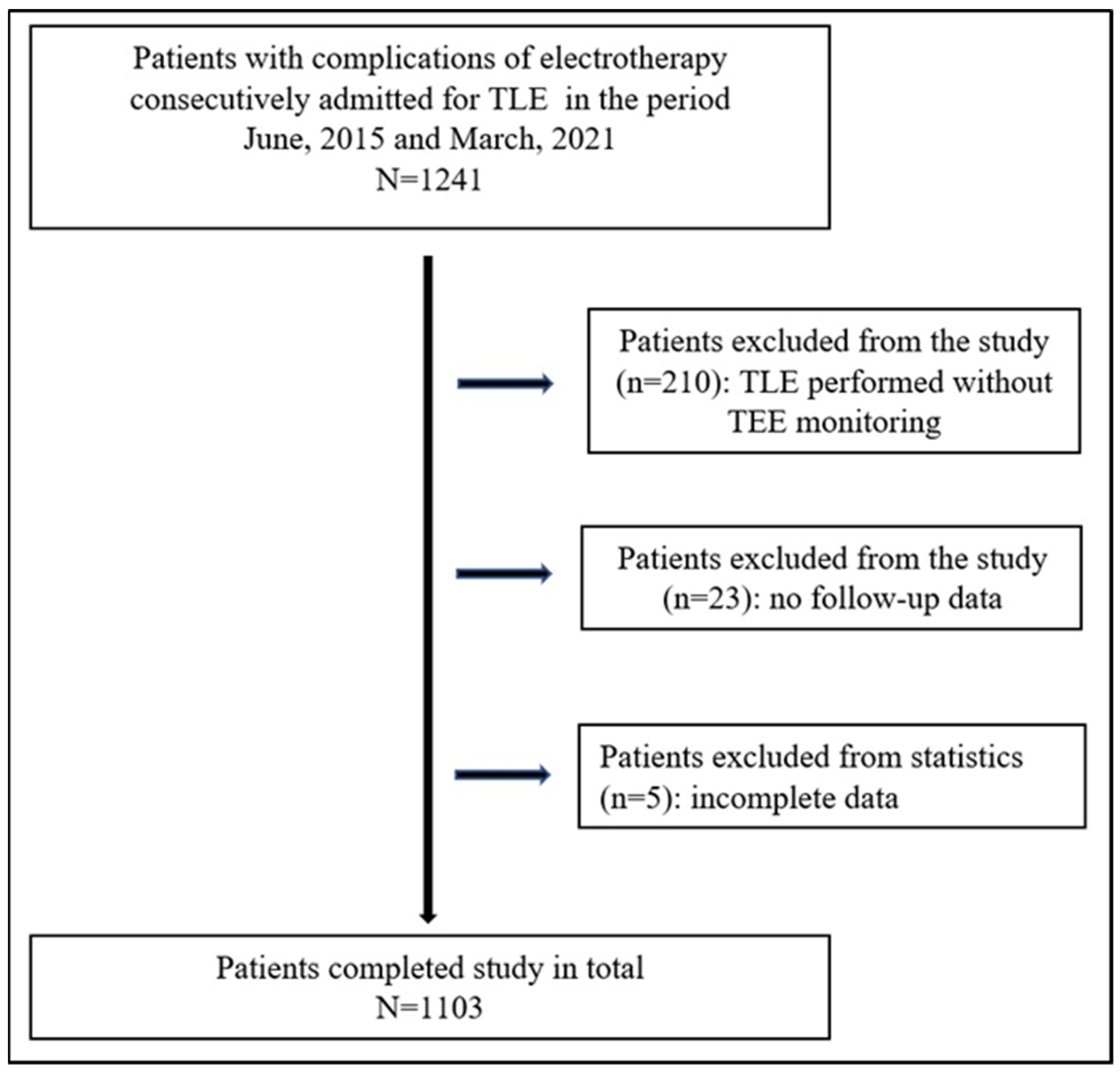
2.1. Study Population

This post hoc analysis used clinical data of 1103 patients who underwent TLE with complete TEE monitoring between June 2015 and March 2021 at one high-volume centre (Figure 1).

2.2. TLE Indications

Indications for TLE were: infectious (pocket infection, bacteremia with or without endocarditis, or any combination of the above) and non-infectious, i.e., mechanical damage of lead (electrical failure), dislodgement, extracardiac pacing, exit/entry block, perforation, upgrading, downgrading, avoidance of lead abandonment, threatening or potentially threatening lead (free ending, left heart, known lead-related TV dysfunction) and other (cancer, painful pocket, MRI and lead extraction to regain venous access in cases of complete venous occlusion including SVC syndrome).

2.3. TEE Monitoring of the Lead Extraction Procedure

TEE monitoring was performed using commercially available equipment (GE Vivid S 70 and Philips iE33) connected to a multiplane transducer (probes: 6VT-D, X7-2t Live 3D). All images were stored in a digital memory. In the preprocedural phase we evaluated lead position, extent of fibrous encapsulation, lead-to-lead adhesions, unaccounted-for masses on the leads as well as function of the tricuspid valve (TV) and the pericardium. During the intraprocedural phase TEE allowed monitoring of direct pulling on the cardiac structures, detachment and dislodgement of fibrous tissue or vegetation fragments, and separation of pericardial layers. The postprocedural phase included evaluation of TV function, remnants of masses removed during TLE and abnormal accumulation of fluid in the pericardial sac. Continuous TEE monitoring was described in detail in our earlier reports.

2.4. Definitions of Echocardiographic Phenomena

Lead fibrotic attachment to the cardiac wall was defined at TEE either as sleeve-like dense echoes involving the leads and extending to the surrounding structures (TV, atrial and ventricular wall) with loss of independent lead motion. The occurrence of such preoperative phenomena was described in our previous reports [6,7,8,16,28,29] and was not examined in this study. Vegetations identified with TEE before, during and after TLE (vegetation remnants) were not considered as ghosts and were subject to separate classification.
Stable ghosts (SG) were defined as new, post-extraction, floating masses visualized during intra- and post-extraction TEE monitoring. They were seen as echogenic, tubular masses with one ending fixed to venous or cardiac walls and structures, and with the second ending floating free (Figure 2), persisting until oesophageal probe removal [16].
Flying ghosts (FG) represent a dynamic phenomenon which may be observed while freeing the lead and cutting off the encapsulating fibrous tissue. When the dilatating sheath is moved down, the masses remain attached to the cardiovascular structures or are freed and after making several circular motions, flow into the TV, right ventricle (RV), thus disappearing from the TEE field of view. The phenomenon of FG was considered occurring when the described sequence of events was documented (Figure 3).
There was also a rare case of an FG that travelled to the systemic circulation through a patent foramen ovale (PFO) (Figure 4).
It should be underlined that scar tissue surrounding the lead is not visualized if located in the anonymous and subclavian veins. During mechanical lead dissection the encapsulation may be covered with a larger dilatating sheath and removed together with the lead (Figure 5).
Unlike mechanical systems, excimer laser sheaths utilise laser energy to vaporize fibrous tissue, thus decreasing ghost detection.

2.5. Lead Extraction Procedure

Lead extraction procedures were performed in a hybrid operating room or in an operating room, using mechanical systems such as polypropylene Byrd dilator sheaths (Cook® Medical, Leechburg, PA, USA), making use of the oblique cutting edge of the tip to dissect leads from fibrous sheaths that immobilized the intravascular and/or intracardiac segment of the lead [13,14,15]. Complete procedural, clinical success and complications of TLE were defined according to the HRS 2009 and 2017 guidelines and the 2018 EHRA expert consensus statement [13,14,15]. Major and minor complications were defined according to the 2018 EHRA Expert Consensus Statement on Lead Extraction [15].

2.6. Approval of the Bioethics Committee

The study was conducted according to the ethical guidelines of the Declaration of Helsinki and approved by the Bioethics Committee at the Regional Chamber of Physicians in Lublin no. 288/2018/KB/VII.

2.7. Statistical Analysis

The Shapiro–Wilk test was used to check whether continuous variables followed a normal distribution. Despite nonparametric distribution of some data (most continuous variables were normally distributed), for uniformity, all continuous variables are presented as the mean ± standard deviation. The categorical variables are presented as number and percentage. The significance of differences between groups (1,2,3) was determined using the nonparametric Chi2 test with Yates correction or the unpaired Mann–Whitney U test, as appropriate. Univariate and multivariable logistic regression was used to determine which parameters affected the occurrence of ghosts. The variables achieving p < 0.1 were included in the multivariable linear regression analysis. To determine the impact of FG and SG on long-term survival after TLE uni- and multivariable Cox regression analysis was used. The variables achieving p < 0.1 under univariable regression analysis were included in the multivariable regression model. To illustrate the impact of FG and SG on survival, the Kaplan–Meier survival curves were constructed and evaluated with log rank test. A p value of 0.05 or less was considered statistically significant. Statistical analyses were performed using Statistica version 13.3 (TIBCO Software Inc., Palo Alto, CA, USA).

3. Results

Baseline demographic and clinical characteristics of the study groups are summarized in Table 1.
TLE was indicated for systemic infection in 174 pts (15.78%), local (pocket) infection in 66 pts (5.98%) and non-infectious reasons in 863 cases (78.24%). Most patients (69.54%) had a pacemaker (any), whereas only 8.79% had a CRT-D device. Implant duration expressed as dwell time of the oldest lead in the patient before TLE was 120.5 months (Table S1). The rate of major complications was 2.45%, clinical success was obtained in 98.91% and procedural success in 95.74% of patients (Table S2).
Analysis of the clinical data (Table 1) indicated that only younger age at TLE and age at first system implantation increased the chances for the development of both types of ghosts. FG was more likely to develop in patients with non-infectious indications for extraction of a dual lead pacemaker system. Ghosts after TLE were more frequent in patients with multiple lead systems and numerous leads in the heart, similarly to patients with more CIED-related procedures before lead extraction. The most important factor explaining the presence of FG seem to be implant duration before TLE. In summary, the main factors for developing ghosts were the number of leads in the case of SG, and implant duration but not systemic infection as regards FG (Table S1).
Comparative analysis of TLE-related potential risk factors for major complications, procedure complexity and TLE efficacy (Table S2) showed that extraction of abandoned lead(s) predisposed to ghosts after TLE. The occurrence of any technical problem/difficulty, hemopericardium and TV damage during TLE were much more frequent in patients with FG. However, it seems to be a result of implant duration as well as the extent and maturation of scar tissue surrounding the leads (Tables S2 and S3).
Table S3 summarizes TEE findings before and after TLE with special focus on the presence of all forms of scar tissue. The last rows of the table compare mortality after TLE during short- and long-term follow-up depending on the type of ghost. Generally, patients with scar tissue surrounding the lead, blood clots on the lead, vegetation-like masses, lead thickening, true vegetations, lead adherence to any heart structure and lead-to-lead binding were much more prone to having both forms of ghosts, particularly FG. The more common presence of TLE-related severe TV damage (increased TR by 3 degrees and damage to tendinous chords) in patients with ghosts seemed to be secondary to implant duration or connective tissue growth and maturation.

3.1. Uni- and Multivariable Linear Regression Analysis

Univariable analysis showed that the factors predisposing to FG formation were older leads, the number of previous CIED-related procedures, lead-related scar tissue, the number of scars and lead-to-lead binding or adhesion to anatomical structures relating to the cardiovascular system (CVS). FG were less likely to develop in patients with older age at first CIED implantation and with lead-related infective endocarditis as indications for TLE. Multivariable regression analysis showed that any form of scar tissue around the lead and the number of scars were the main factors responsible for FG. Older age at first CIED implantation and the presence of strong lead-to-lead binding or adhesion to CVS structures decreased the probability of FG (Table 2).
Univariate analysis identified the following factors predicting the occurrence of SG after TLE: abandoned lead(s), the number of leads in the heart, scar tissue around the leads and the number of scars as well as lead-to-lead binding and adhesions to CVS structures. Older age at the time of implantation of the first CIED was associated with a lower probability of SG. Multivariable regression analysis showed that the factors increasing the likelihood of SG were the number of the leads in the heart and any form of lead-related scar tissue. Older age at first CIED implantation decreased the probability of SG (Table 2).

3.2. Survival Analysis

A total of 281 (25.48%) patients died during 1264 ± 644.7 (2–2466) days of follow-up.
As expected, survival in the group of patients who underwent TLE for infectious reasons was significantly worse compared to the group of patients who underwent TLE for non-infectious reasons. The 30-day mortality was 1.55% and was not related to the presence of both floating and stable ghosts. The log rank analysis of the survival curves of Kaplan–Meier did not show any influence of the presence of both forms of ghosts on long-term survival in the entire group of patients. Only the subgroup of patients with non-infectious indications for TLE showed statistically significantly better survival in whom the presence of stable ghosts after TLE was demonstrated (log rank p = 0.034) Table 3, Figure 6.
Multivariable Cox regression analysis confirmed the importance of the common factors that influence patient prognosis after TLE. In the entire group of patients mortality risk increased with older age (HR = 1.049; p < 0.001), higher NYHA functional class (HR = 1.462; p = 0.002), presence of any renal failure (HR = 1.550; p < 0.001), permanent atrial fibrillation (HR = 1.444; p = 0.004), higher Charlson comorbidity index (HR = 1.044; p = 0.012), resynchronisation therapy before TLE (HR = 1.072; p < 0.001), and systemic infection (HR = 1.894; p < 0.001). Women (HR = 0.621; p < 0.001) and patients with higher LVEF (HR = 0.987; p = 0.009) were less likely to die during follow-up.
In the infectious subgroup worse survival was associated with older age, higher NYHA class, presence of any renal failure and resynchronisation therapy before TLE. Prognosis appeared to be better in women and patients with higher LVEF.
In the non-infectious subgroup the prognostic factors were the same as in the entire group of patients (Table 4). Our analysis showed no impact of ghosts on long-term mortality after TLE both in the entire group of patients and in the two subgroups (infectious and non-infectious) (Table 4).
This result was confirmed mostly by the Kaplan-Meier survival curves and log rank analysis (p = 0.186 for the entire group) (Figure 6A). However, in the non-infectious subgroup, survival after TLE was significantly better in those with stable ghosts (p = 0.014) (Figure 6B), (Table S3).

4. Discussion

The most important finding of this study is that the presence of either form of ghost did not affect long-term survival after TLE indicated for device infection (Figure 6C,D).
Remnants of scar tissue anteriorly surrounding the lead before extraction were described in several case reports and named as “ghosts” [21,22,23,24,25,26,27]. Thereafter, a series of six reports were published [5,16,17,18,19], five of them addressed the importance of “stable ghosts” in the aspect of long-term mortality. The phenomenon was observed in 8% [19], 14% [18], 19% [17] and 60% [5] of post-operative TEE images. The reports on TEE monitoring during TLE procedures did not consider analysis of scar tissue remnants or the dynamic phenomenon of scar tissue cut free (mobilization) [28,29,30,31,32]. Out of the five large studies on “ghosts” Poterała et al. did not examine the influence of ghosts on long-term mortality [17], Narducci underlined that the presence of ghosts could be an independent predictor of mortality after TLE in patients with infections [18], Le Dolley indicated that ghosts were suggestive of device infection and seemed to be associated with the diagnosis of lead-related endocarditis [19]. Diemberger concluded that the presence of ghosts at post-TLE transoesophageal echocardiography and a closed CIED pocket were associated with a worse prognosis [20] but Caiati suggested that ghosts were mostly benign remnants of fibrotic lead capsule cut off during extraction [5]. A large sample of patients undergoing TEE during TLE combined with detailed description of various forms of scar tissue prompted us to perform this analysis. The present results show that the number of leads and implant duration, but not infectious indications, were the main predictors of ghost formation after TLE. Such TEE findings as scar tissue surrounding the lead, mobile thrombi, vegetation-like masses, lead thickening, lead adherence to heart structures and lead-to-lead binding were much more frequent in patients with both forms of ghosts, particularly with FG. The occurrence of ghosts had no negative prognostic significance. We described an ephemeral but not rare type of ghost—“flying ghost”—that can be observed during scrupulous TEE examinations in patients undergoing TLE.
Narducci showed that infective endocarditis and higher Charlson comorbidity score [18] were significant predictors of “ghosts”. However, we could not confirm this finding in our study. These differences can be explained in part by the sample size and the rate of systemic infection (64.1% versus 15.8%). In our study, the Charlson comorbidity score had no influence on the incidence of ghosts. TLE in patients with systemic infection carries a risk of septic pulmonary embolism which is related to the size of vegetations [19]. However, assessment of the extent of pulmonary vascular obstruction after TLE was not the goal of our study. According to the results in the present study it can be concluded that the amount of thrombotic material seen as travelling “ghosts” is irrelevant from a pulmonary haemodynamic point of view. This explains no impact on long-term survival.
We are aware that the long-term mortality of patients after TLE may have been influenced by other health-related factors such as the presence and extent of infection, comorbidities, the presence of chronic atrial fibrillation as well as the systemic immune-inflammation reaction [5,8,16,29,33,34]. However, we focused on the relevance of the connective tissue remnants released from the leads during the procedure. A phenomenon that raises disputes as to its prognostic significance.
TEE seems to be an appropriate tool for visualisation of scar tissue around the leads, since various forms of fibrosis were detected in 44% of the study patients. Furthermore, continuous monitoring of the extraction procedure permits visualisation of the fleeting nature of FG. Caiati suggests an even better sensitivity of ICE which can show the presence of SG in 60% of patients during TLE [5]. However, ICE is an invasive procedure involving insertion of an ICE tube. This remains in anatomical conflict with dilating sheaths and requires temporary withdrawal of the tube up to the inferior vena cava, thus limiting observation of mobile masses.
An additional aspect of scar tissue mobilisation during TLE, which is worth mentioning, is the presence of PFO and associated risk of crossed embolism. In our study, PFO was detected in 15 patients, however, only one of them had asymptomatic crossed embolism. The presence of CIED alone does not increase the risk of ischemic stroke in patients with PFO [35]. Paradoxical septic embolism can occur in the concomitant presence of CIED and PFO due to migration of thrombotic material during lead removal. A significant risk of brain emboli is associated with coexistence of vegetations and R-L leakage at any level of the right heart [36]. On the other hand, FG may be related to stroke during and after TLE [18,35]. It can be concluded that screening for PFO before TLE is justifiable. Studies on strategies of lead extraction in patients with a PFO, such as closure of the foramen ovale before TLE or temporary placement of filter devices for cerebral protection are warranted. Particular attention should be paid to patients with CIED infections and extensive build-up of scar tissue around the leads combined with a PFO meeting criteria for paradoxical embolism [7].

5. Conclusions

  • In approximately 30% of TLE procedures ghosts remain attached to the CVS wall (SG) but in around 15% of the extraction procedures freed ghosts (FG) travel spontaneously to the pulmonary vascular bed and disappear.
  • Younger patient age and the number of leads but not infectious indications are the factors predisposing to ghost formation during and after TLE.
  • The degree of growth and maturation of the connective tissue surrounding the lead before TLE is the strongest predictor of both types of ghosts.
  • The occurrence of ghosts is associated with complicated procedures, but it seems to be related to implant duration and scar growth.
  • The presence of both types of ghosts does not reduce survival after TLE.

Study Limitations

This is a single-center, observational, prospective study. TEE monitoring was mandatory. ICE was not the aim of the study. TLE was performed using mechanical systems without laser energy. We cannot interpolate our results on patients in whom laser energy is utilized. Histopathological analysis of “ghost” was not performed. Ghost formation in the anonymous veins was not explored by ultrasounds. The study did not diagnose asymptomatic pulmonary embolism using CT angiography.

Supplementary Materials

The following supporting information can be downloaded at: https://www.mdpi.com/article/10.3390/ijerph191912542/s1, Table S1. Indications for TLE, existing CIED and history of pacing. Table S2. TLE-related potential risk factors for major complications, procedure complexity and TLE efficacy and complications. Table S3. TEE findings during TLE with special focus on the presence all forms of scar tissue and mortality after TLE.

Author Contributions

D.N.—writing-original draft preparation and editing, data curation, corresponding author; W.J.—methodology, statistical study, results interpretation; A.P.—investigation, data curation, Ł.T.—date curation, P.S.—date curation, A.K.—supervision, writing-review. The authors are accountable for all aspects of the work in ensuring that questions related to the accuracy or integrity of any part of the work are appropriately investigated and resolved. All authors have read and agreed to the published version of the manuscript.

Funding

This research received no external funding.

Institutional Review Board Statement

The study was conducted in accordance with the Declaration of Helsinki (as revised in 2013). The study was approved by institutional Bioethics Committee at Regional Physicians Chamber in Lublin no. 288/2018/KB/VII.

Informed Consent Statement

Informed consent was taken from all the patients.

Data Availability Statement

Readers can access data supporting the conclusions of the study upon reasoned request to the authors.

Acknowledgments

We would like to thank all doctors participating in transvenous lead extraction procedures.

Conflicts of Interest

Authors declare no conflict of interest.

References

  1. Keiler, J.; Schulze, M.; Dreger, R.; Springer, A.; Öner, A.; Wree, A. Quantitative and Qualitative Assessment of Adhesive Thrombo-Fibrotic Lead Encapsulations (TFLE) of Pacemaker and ICD Leads in Arrhythmia Patients—A Post Mortem Study. Front. Cardiovasc. Med. 2020, 7, 602179. [Google Scholar] [CrossRef] [PubMed]
  2. Dvorak, P.; Novak, M.; Kamaryt, P.; Slana, B.; Lipoldova, J.; Dvorak, P. Histological findings around electrodes in pacemaker and implantable cardioverter-defibrillator patients: Comparison of steroid-eluting and non-steroid-eluting electrodes. Europace 2012, 14, 117–123. [Google Scholar] [CrossRef] [PubMed]
  3. Kozłowski, D.; Dubaniewicz, A.; Koźluk, E.; Grzybiak, M.; Krupa, W.; Kołodziej, P.; Pazdyga, A.; Adamowicz-Kornacka, M.; Walczak, E.; Walczak, F. The morphological conditions of the permanent pacemaker lead extraction. Folia Morphol. 2000, 59, 25–29. [Google Scholar]
  4. Kołodzińska, A.; Kutarski, A.; Koperski, Ł.; Grabowski, M.; Małecka, B.; Opolski, G. Differences in encapsulating lead tissue in patients who underwent transvenous lead removal. Europace 2012, 14, 994–1001. [Google Scholar] [CrossRef] [Green Version]
  5. Caiati, C.; Luzzi, G.; Pollice, P.; Favale, S.; Lepera, M.E. Novel Clinical Perspective on New Masses after Lead Extraction (Ghosts) by Means of Intracardiac Echocardiography. J. Clin. Med. 2020, 9, 2571. [Google Scholar] [CrossRef]
  6. Nowosielecka, D.; Polewczyk, A.; Jacheć, W.; Tułecki, Ł.; Tomków, K.; Stefańczyk, P.; Kleinrok, A.; Kutarski, A. A new approach to the continuous monitoring of transvenous lead extraction using transesophageal echocardiography—Analysis of 936 procedures. Echocardiography 2020, 37, 601–611. [Google Scholar] [CrossRef]
  7. Nowosielecka, D.; Polewczyk, A.; Jacheć, W.; Tułecki, Ł.; Kleinrok, A.; Kutarski, A. Echocardiographic findings in patients with cardiac implantable electronic devices-analysis of factors predisposing to lead-associated changes. Clin. Physiol. Funct. Imaging 2021, 41, 25–41. [Google Scholar] [CrossRef]
  8. Nowosielecka, D.; Jacheć, W.; Polewczyk, A.; Tułecki, Ł.; Kleinrok, A.; Kutarski, A. Prognostic Value of Preoperative Echocardiographic Findings in Patients Undergoing Transvenous Lead Extraction. Int. J. Environ. Res. Public Health 2021, 18, 1862. [Google Scholar] [CrossRef]
  9. Segreti, L.; Di Cori, A.; Soldati, E.; Zucchelli, G.; Viani, S.; Paperini, L.; De Lucia, R.; Coluccia, G.; Valsecchi, S.; Bongiorni, M.G. Major predictors of fibrous adherences in transvenous implantable cardioverter-defibrillator lead extraction. Heart Rhythm 2014, 11, 2196–2201. [Google Scholar] [CrossRef]
  10. Ho, G.; Bhatia, P.; Mehta, I.; Maus, T.; Khoche, S.; Pollema, T.; Pretorius, V.G. Prevalence and Short-Term Clinical Outcome of Mobile Thrombi Detected on Transvenous Leads in Patients Undergoing Lead Extraction. JACC Clin. Electrophysiol. 2019, 5, 657–664. [Google Scholar] [CrossRef]
  11. Beaser, A.D.; Aziz, Z.; Besser, S.A.; Jones, C.I.; Jameria, Z.; Kannan, A.; Upadhyay, G.A.; Broman, M.T.; Ozcan, C.; Tung, R.; et al. Characterization of Lead Adherence Using Intravascular Ultrasound to Assess Difficulty of Transvenous Lead Extraction. Circ. Arrhythm. Electrophysiol. 2020, 13, e007726. [Google Scholar] [CrossRef] [PubMed]
  12. Golzio, P.G.; Errigo, D.; Peyracchia, M.; Gallo, E.; Frea, S.; Castagno, D.; Budano, C.; Giustetto, C.; Rinaldi, M. Prevalence and prognosis of lead masses in patients with cardiac implantable electronic devices without infection. J. Cardiovasc. Med. 2019, 20, 372–378. [Google Scholar] [CrossRef] [PubMed]
  13. Wilkoff, B.L.; Love, C.J.; Byrd, C.L.; Bongiorni, M.G.; Carrillo, R.G.; Crossley, G.H.; Epstein, L.M.; Friedman, R.A.; Kennergren, C.E.; Mitkowski, P.; et al. Transvenous lead extraction: Heart Rhythm Society expert consensus on facilities, training, indications, and patient management: This document was endorsed by the American Heart Association (AHA). Heart Rhythm 2009, 6, 1085–1104. [Google Scholar] [CrossRef]
  14. Kusumoto, F.M.; Schoenfeld, M.H.; Wilkoff, B.L.; Berul, C.I.; Birgersdotter-Green, U.M.; Carrillo, R.; Cha, Y.M.; Clancy, J.; Deharo, J.C.; Ellenbogen, K.A.; et al. 2017 HRS expert consensus statement on cardiovascular implantable electronic device lead management and extraction. Heart Rhythm 2017, 14, e503–e551. [Google Scholar] [CrossRef] [PubMed] [Green Version]
  15. Bongiorni, M.G.; Burri, H.; Deharo, J.C.; Starck, C.; Kennergren, C.; Saghy, L.; Rao, A.; Tascini, C.; Lever, N.; Kutarski, A.; et al. 2018 EHRA expert consensus statement on lead extraction: Recommendations on definitions, endpoints, research trial design, and data collection requirements for clinical scientific studies and registries: Endorsed by APHRS/HRS/LAHRS. Europace 2018, 20, 1217. [Google Scholar] [CrossRef] [PubMed]
  16. Nowosielecka, D.; Jacheć, W.; Polewczyk, A.; Kleinrok, A.; Tułecki, Ł.; Kutarski, A. The prognostic value of transesophageal echocardiography after transvenous lead extraction: Landscape after battle. Cardiovasc. Diagn. Ther. 2021, 11, 394–410. [Google Scholar] [CrossRef]
  17. Poterała, M.; Kutarski, A.; Brzozowski, W.; Tomaszewski, M.; Gromadziński, L.; Tomaszewski, A. Echocardiographic assessment of residuals after transvenous intracardiac lead extraction. Int. J. Cardiovasc. Imaging 2020, 36, 423–430. [Google Scholar] [CrossRef]
  18. Narducci, M.L.; Di Monaco, A.; Pelargonio, G.; Leoncini, E.; Boccia, S.; Mollo, R.; Perna, F.; Bencardino, G.; Pennestrì, F.; Scoppettuolo, G.; et al. Presence of ‘ghosts’ and mortality after transvenous lead extraction. Europace 2017, 19, 432–440. [Google Scholar] [CrossRef] [Green Version]
  19. Le Dolley, Y.; Thuny, F.; Mancini, J.; Casalta, J.P.; Riberi, A.; Gouriet, F.; Bastard, E.; Ansaldi, S.; Franceschi, F.; Renard, S.; et al. Diagnosis of cardiac device-related infective endocarditis after device removal. JACC Cardiovasc. Imaging 2010, 3, 673–681. [Google Scholar] [CrossRef] [Green Version]
  20. Diemberger, I.; Biffi, M.; Lorenzetti, S.; Martignani, C.; Raffaelli, E.; Ziacchi, M.; Rapezzi, C.; Pacini, D.; Boriani, G. Predictors of long-term survival free from relapses after extraction of infected CIED. Europace 2018, 20, 1018–1027. [Google Scholar] [CrossRef]
  21. Alizadehasl, A.; Sarrafi Rad, N.; Pourafkari, L.; Haghjoo, M. Persistence of a pacemaker lead-like “ghost” 6 months after lead extraction. Echocardiography 2019, 36, 201–203. [Google Scholar] [CrossRef] [PubMed] [Green Version]
  22. Andreas, M.; Wiedemann, D.; Kocher, A.; Khazen, C. Materialization of ghosts: Severe intracardiac masses after pacemaker lead extraction requiring immediate surgical intervention. Heart Rhythm 2013, 10, 1826. [Google Scholar] [CrossRef] [PubMed]
  23. Jacobson, A.M.; Ailiani, R.G. Pseudoleads on Transesophageal Echocardiography. CASE Cardiovasc. Imaging Case Rep. 2018, 3, 35–38. [Google Scholar] [CrossRef] [PubMed]
  24. Pettemerides, V.; Macnab, A. Right atrial ghost following device extraction for infective endocarditis. Echo Res. Pract. 2019, 6, I3–I4. [Google Scholar] [CrossRef] [PubMed]
  25. Rizzello, V.; Dello Russo, A.; Casella, M.; Biddau, R. Residual fibrous tissue floating in the right atrium after percutaneous pacemaker lead extraction: An unusual complication early detected by intracardiac echocardiography. Int. J. Cardiol. 2008, 127, e67–e68. [Google Scholar] [CrossRef]
  26. Nazir, S.A.; Hudsmith, L.; Newton, J.D.; Betts, T.R. Chronic fibrous sheath mistaken for retained pacemaker product. Eur. J. Echocardiogr. 2009, 10, 343. [Google Scholar] [CrossRef] [Green Version]
  27. Kiuchi, K.; Fukuzawa, K.; Mori, S.; Nishii, T.; Matsumoto, K.; Ichibori, H.; Yamada, T. The details of an unusual “ghost” after transvenous lead extraction: Three-dimensional computed tomography analysis. J. Arrhythm. 2017, 33, 640–642. [Google Scholar] [CrossRef]
  28. Nowosielecka, D.; Jacheć, W.; Polewczyk, A.; Tułecki, Ł.; Tomkow, K.; Stefańczyk, P.; Tomaszewski, A.; Brzozowski, W.; Szcześniak-Stańczyk, D.; Kleinrok, A.; et al. Transesophageal Echocardiography as a Monitoring Tool During Transvenous Lead Extraction-Does It Improve Procedure Effectiveness? J. Clin. Med. 2020, 9, 1382. [Google Scholar] [CrossRef]
  29. Nowosielecka, D.; Polewczyk, A.; Jacheć, W.; Kleinrok, A.; Tułecki, Ł.; Kutarski, A. Transesophageal echocardiography for the monitoring of transvenous lead extraction. Kardiol. Pol. 2020, 78, 1206–1214. [Google Scholar] [CrossRef]
  30. Endo, Y.; O’Mara, J.E.; Weiner, S.; Han, J.; Goldberger, M.H.; Gordon, G.M.; Nanna, M.; Ferrick, K.J.; Gross, J.N. Clinical utility of intraprocedural transesophageal echocardiography during transvenous lead extraction. J. Am. Soc. Echocardiogr. 2008, 21, 861–867. [Google Scholar] [CrossRef]
  31. Hilberath, J.N.; Burrage, P.S.; Shernan, S.K.; Varelmann, D.J.; Wilusz, K.; Fox, J.A.; Eltzschig, H.K.; Epstein, L.M.; Nowak-Machen, M. Rescue transoesophageal echocardiography for refractory haemodynamic instability during transvenous lead extraction. Eur. Heart J. Cardiovasc. Imaging 2014, 15, 926–932. [Google Scholar] [CrossRef] [PubMed] [Green Version]
  32. Regoli, F.; D’Ambrosio, G.; Caputo, M.L.; Svab, S.; Conte, G.; Moccetti, T.; Klersy, C.; Cassina, T.; Demertzis, S.; Auricchio, A. New-onset pericardial effusion during transvenous lead extraction: Incidence, causative mechanisms, and associated factors. J. Interv. Card. Electrophysiol. 2018, 51, 253–261. [Google Scholar] [CrossRef] [PubMed]
  33. Hayıroğlu, M.İ.; Çınar, T.; Çinier, G.; Yüksel, G.; Pay, L.; Keskin, K.; Coşkun, C.; Ayan, G.; Çiçek, V.; Tekkeşin, A.İ. Prognostic value of serum albumin for long-term mortality in patients witch chamber permanent pacemakers. Biomark Med. 2022, 16, 341–348. [Google Scholar] [CrossRef] [PubMed]
  34. Hayıroğlu, M.İ.; Çınar, T.; Çinier, G.; Pay, L.; Yumurtaş, A.Ç.; Tezen, O.; Eren, S.; Kolak, Z.; Çetin, T.; Cicek, V.; et al. Evaluating systemic immune-inflammation index in patients with implantable cardioverter defibrillator for heart failure with reduced ejection fraction. Pacing Clin. Electrophysiol. 2022, 45, 188–195. [Google Scholar] [CrossRef] [PubMed]
  35. Agboola, K.M.; Lee, J.M.; Liu, X.; Novak, E.; Cuculich, P.S.; Cooper, D.H.; Noheria, A. Interaction of cardiac implantable electronic device and patent foramen ovale in ischemic stroke: A case-only study. Pacing Clin. Electrophysiol. 2019, 42, 341–348. [Google Scholar] [CrossRef]
  36. Lee, J.Z.; Agasthi, P.; Pasha, A.K.; Tarin, C.; Tseng, A.S.; Diehl, N.N.; Hodge, D.O.; DeSimone, C.V.; Killu, A.M.; Brady, P.A.; et al. Stroke in patients with cardiovascular implantable electronic device infection undergoing transvenous lead removal. Multicent. Study Heart Rhythm 2018, 15, 1593–1600. [Google Scholar] [CrossRef] [Green Version]
Figure 1. Flow chart of the recruitment process towards the study group.
Ijerph 19 12542 g001
Figure 2. SG (red arrows) in different areas of the cardiovascular system imaged by 2D and 3D TEE. (A) SG at the SVC orifice. (B) SG leaving the RAA. (C) SG in the SVC/RA. (D) Long SG stretched between the TV and SVC. (E) Large SG in the RA adhering to the septal leaflet. (F) SG adhering to the RV endocardium.
Ijerph 19 12542 g002
Figure 3. Concomitant occurrence of FG and SG and their fate imaged by 2D TEE (the sequences of the video and the behavior of the ghosts are represented “frame by frame”). (A) Ventricular lead (yellow arrows) dissected using the Byrd dilator sheath (green arrow). Scar tissue pushed in front of the catheter (red circle). The end piece of the lead adhered to the RA endocardium (blue arrow). (B) After lead extraction FG (red circle) in the RA and SG (blue circle) in the RV. (C) FG travelled in the bloodstream to the RV in the next heartbeat, whereas SG remained in the RA. (D) In the subsequent cardiac cycles FG „disappeared” in the pulmonary circulation, only SG remained in place.
Ijerph 19 12542 g003
Figure 4. Flying ghosts that travelled to the systemic circulation through the PFO imaged by 2D, 3D TEE (A) Coronary sinus lead extraction (yellow arrow) (CS). Encapsulating scar tissue (red arrow) dissected away from the pacing lead using the Byrd dilator sheath (green arrow). (B) Pieces of scar tissue (FG) (red arrow) freed during lead dissection. Pulling on the cardiac wall (yellow line) during extraction of the adherent lead. (C) FG, freed from the lead, floating in the RA. (D,E) The same FG passing through the PFO. (F) FG in the LA.
Ijerph 19 12542 g004
Figure 5. Examples of scar tissue surrounding the leads as seen after extraction (A,C,D,F) Examples of scar tissue surrounding the leads as seen after extraction (B) Calcification of the scar tissue. (E) Ossification of the scar tissue.
Ijerph 19 12542 g005
Figure 6. Kaplan–Meier probability of survival after TLE based on the presence of ghosts during or after TLE.
Ijerph 19 12542 g006
Table 1. Patient characteristics and comparison of potential patient-related risk factors for the occurrence of both forms of ghost.
Patient CharacteristicsFlying Ghosts (Ghosts Shifting Spontaneously to Pulmonary Vascular Bed)Stable Ghosts (Ghosts Remaining Attached to Cardiovascular Wall)Ghosts Absent during and after TLE
Group/number of patients1: N = 171 (15.50%)2: N = 322 (29.19%)3: N = 610 (55.31%)
Form of result presentationmean ± SD/n (%)
p (1 vs. 2)
mean ± SD/n (%)
p (2 vs. 3)
mean ± SD/n (%)
p (1 vs. 3)
Patient age during TLE [years]66.06 ± 16.20
p = 0.912
65.73 ± 15.29
p < 0.006
68.87 ± 13.38
p < 0.047
Patient age at first system implantation [years]53.53 ± 18.63
p = 0.044
56.21 ± 17.39
p = 0.028
59.37 ± 14.81
p < 0.001
Female70 (40.94)
p = 0.654
140 (43.48)
p = 0.260
232 (38.03)
p = 0.267
Etiology: ischemic heart disease109 (63.72)
p = 0.990
204 (63.35)
p = 0.421
404 (66.23)
p = 0.545
NYHA III & IV31 (18.13)
p = 0.799
54 (16.77)
p = 0.854
98 (16.07)
p = 0.592
LVEF average [%]48.43 ± 15.07
p = 0.610
49.04 ± 14.86
p = 0.335
47.93 ± 15.82
p = 0.819
LVEF category: significantly decreased (<30%)24 (14.04)
p = 0.714
40 (12.42)
p = 0.101
102 (16.72)
p = 0.468
Permanent atrial fibrillation39 (22.81)
p = 0.937
71 (22.05)
p = 0.499
148 (24.26)
p = 0.770
Congestive heart failure58 (33.92)
p = 0.085
84 (26.09)
p = 0.652
169 (27.71)
p = 0.137
Renal failure (any)34 (19.88)
p = 0.451
75 (23.19)
p = 0.313
162 (26.56)
p = 0.377
Charlson comorbidity index5.175 ±4.185
p = 0.710
4.708 ± 3.746
p = 0.099
5.090 ± 3.671
p = 0.434
TLE—transvenous lead extraction, SD—standard deviation, N number, NYHA—New York Heart Association functional class, LVEF—left ventricular ejection fraction.
Table 2. Factors that influenced ghost (flying or stable) occurrence during and after TLE, results of uni- and multivariable regression analysis.
Univariable RegressionMultivariable Regression
OR95% CIpOR95% CIp
Flying ghosts
Patient age at first system implantation
[by year]
0.9770.967–0.987<0.0010.9840.970–0.9970.019
Systemic infection0.5260.301–0.9200.0240.5390.289–1.0060.052
Number of CIED-related procedures before TLE1.2271.046–1.4390.0121.0810.832–1.4030.560
Dwell time of the oldest target lead in the patient before TLE [by year]1.0601.035–1.085<0.0010.9880.946–1.0330.607
Any form of scar tissue on the lead(s)10.345.625–19.02<0.0017.1063.424–14.75<0.001
Number of separate scars1.6851.457–1.949<0.0011.3721.076–1.7490.011
Lead adhesion to heart structures (any)2.8111.961–4.028<0.0010.5170.281–0.9530.034
Stable ghosts
Patient age at first system implantation
[by year]
0.9890.985–0.993<0.0010.9890.980–0.9990.030
Charlson comorbidity index0.9760.940–1.0130.197
Abandoned lead before TLE1.8831.207–2.9380.0051.1520.653–2.0320.623
Number of leads in the heart before TLE1.5541.280–1.887<0.0011.4501.141–1.8420.002
Number of CIED-related procedures before lead extraction1.1270.981–1.2960.0920.9400.786–1.1240.499
All forms of scar tissue on the lead(s)2.3041.708–3.106<0.0011.9401.293–2.909<0.001
Number of separate scars1.4241.260–1.609<0.0011.1470.939–1.4030.179
Lead adhesion to heart structures (any)1.6991.252–2.307<0.0011.0450.658–1.6600.851
TLE—transvenous lead extraction, CIED—cardiac implantable electronic device.
Table 3. Long term and 30-d survival after TLE.
Long Term and 30-Days Survival after TLEFlying Ghosts (Ghosts Shifting Spontaneously to Pulmonary Vascular Bed)Stable Ghosts (Ghosts Remaining Attached to Cardiovascular Wall)Ghosts Absent during and after TLE
Group/number of patients1: N = 1712: N = 3223: N = 610
Long-term survival of entire group of patients after TLE during mean 1264 ± 644.7 (2–2466) days follow-up. Log rank p for all model = 0.186
Follow-up (mean ± SD) [days]1048 ± 417.5
p < 0.001
1511 ± 588.0
p = 0.567
1544 ± 555.0
p < 0.001
Alive during follow-up (entire group)
(n, %)
143 (83.63)
Log rank test
p = 0.914
242 (75.16)
Log rank test
p = 0.121
437 (71.64)
Log rank test
p = 0.202
Died during follow-up (entire group)
(n, %)
28 (16.37)
Log rank test
p = 0.914
80 (24.84)
Log rank test
p = 0.121
173 (28.36)
Log rank test
p = 0.202
Long-term survival after TLE in the subgroup of non-infectious and infectious patients. Log rank p for all model = 0.036
Non-infectious; died/alive
(n, %)
19/131 (12.67)
Log rank test
p = 0.430
41/200 (17.01) Log rank test
p = 0.014
114/360 (24.05) Log rank test
p = 0.259
Infectious (all); died/alive
(n, %)
9/12 (42,86)
Chi2 (vs. non-infectious)
p = 0.001
39/42 (48.15)
Chi2 (vs. non-infectious)
p < 0.001
59/77 (43.38)
Chi2 (vs. non-infectious)
p < 0.001
Long-term survival after TLE in subgroup of patients with pocket infection. Log rank p for all model = 0.786
Local (pocket) infection; died/alive
(n, %)
2/4 (33.33)
Log rank test
p = 1.000
8/10 (44.44) Log rank test
p = 0.463
11/27 (28.95)
Log rank test
p = 0.917
Long term survival after TLE in subgroup of patients with systemic. Log rank p for all model = 0.948
Systemic infection; died/alive
(n, %)
7/8 (46.67)
Log rank test
p = 0.789
31/32 (49.21) Log rank test
p = 0.839
48/50 (48.98)
Log rank test
p = 0.812
30-days survival after TLE in entire group of patients
Entire group; died/alive
(n, %)
2/169 (1.17)
Chi2 (1 vs. 2) p = 0.660
7/315 (2.17)
Chi2 (2 vs. 32)
p = 0.747
10/600 (1.64)
Chi2 (1 vs. 3)
p = 0.929
30-days survival after TLE in subgroup of non-infectious patients
Non-infectious; died/alive
(n, %)
2/148 (1.33)
Chi2 (1 vs. 2)
p = 0.972
2/239 (0.83)
Chi2 (2 vs. 3)
p = 0.679
4/470 (0.84)
Chi2 (1 vs. 3)
p = 0.956
Infectious (all); died/alive
(n, %)
0/21 (0.00)
Chi2 (vs. non-infectious)
p = 0.581
5/76 (6.17)
Chi2 (vs. non-infectious)
p = 0.016
6/130 (4.41)
Chi2 (vs. non-infectious)
p = 0.012
30-days survival after TLE in subgroup of patients with pocket infection
Local (pocket) infection; died/alive
(n, %)
0/6 (0.00)0/18 (0.00)0/38 (0.00)
Long term survival after TLE in subgroup of patients with systemic infection
Systemic infection; died/alive
(n, %)
0/15 (0.00)
Chi2 (1 vs. 2)
p = 0.588
5/58 (7.94)
Chi2 (2 vs. 3)
p = 0.900
6/92 (6.12)
Chi2 (1 vs. 3)
p = 0.714
TLE—transvenous lead extraction, SD—standard deviation.
Table 4. Factors influencing long-term prognosis after TLE, results of uni- and multivariable Cox regression analysis.
Univariable Cox RegressionMultivariable Cox Regression
HR95%pHR95%p
All patients
Female gender0.4410.336–0.579<0.0010.6210.463–0.833<0.001
Patient age during TLE [by year]1.0491.038–1.061<0.0011.0401.025–1.054<0.001
NYHA FC class [by one]2.9142.410–3.523<0.0011.5261.197–1.946<0.001
LVEF [by 1p%]0.9660.958–0.973<0.0010.9870.981–0.9780.009
Renal failure (any)3.2692.587–4.131<0.0011.5991.243–2.0580.001
Ischemic heart disease1.4571.125–1.8870.0041.0140.758–1.3550.927
Permanent atrial fibrillation2.3721.866–3.015<0.0011.4431.122–1.8560.004
Charlson comorbidity index1.1421.111–1.174<0.0011.0441.010–1.0800.012
Flying ghosts (1st group)0.7950.537–1.1780.252
Stable ghosts (2nd group)0.8810.680–1.1410.336
Ghosts absent (3rd group)1.2230.962–1.5550.100
ICD presence before TLE1.1560.884–1.5120.292
Device type: CRTP/CRTD3.1252.382–4.100<0.0011.7021.248–2.322<0.001
Systemic infection2.8542.220–3.667<0.0011.8941.460–2.457<0.001
Isolated pocket infection1.2810.829–1.9790.265
Infectious patients
Female gender0.4850.301–0.7800.0030.5780.347–0.9640.036
Patient age during TLE [by year]1.0271.009–1.0450.0031.0371.014–1.0610.002
NYHA FC class [by one]2.2781.687–3.075<0.0011.6591.118–2.4620.012
LVEF [by 1p%]0.9690.957–0.981<0.0010.9880.972–1.0040.133
Renal failure (any)2.5121.729–3.649<0.0011.6861.136–2.5030.009
Ischemic heart disease0.9380.620–1.4210.764
Permanent atrial fibrillation1.6421.106–2.4380.0141.0100.657–1.5520.965
Charlson comorbidity index1.0961.047–1.147<0.0011.0250.972–1.0820.363
Flying ghosts (1st group)1.0890.548–2.1630.807
Stable ghosts (2nd group)1.0410.705–1.5360.841
Ghosts absent (3rd group)0.9400.645–1.3690.747
Device type: ICD1.0180.666–1.5560.935
Device type: CRTP/CRTD2.7831.861–4.161<0.0011.7661.087–2.8680.022
Isolated pocket infection0.5600.351–0.8940.0151.0830.581–2.0180.801
Vegetations presence1.6201.094–2.3990.0161.4050.835–2.3650.201
Non-infectious patients
Female gender0.4780.342–0.669<0.0010.6380.443–0.9190.016
Patient age during TLE [by year]1.0561.040–1.071<0.0011.0431.024–1.0620.000
NYHA FC class [by one]3.1402.439–4.043<0.0011.6191.165–2.2490.004
LVEF [by 1p%]0.9650.956–0.975<0.0010.9880.975–1.0000.048
Renal failure (any)3.3562.483–4.535<0.0011.4891.072–2.0680.017
Ischemic heart disease1.6021.151–2.2300.0050.8820.618–1.2580.488
Permanent atrial fibrillation2.8152.078–3.814<0.0011.6931.236–2.3200.001
Charlson comorbidity index1.1481.108–1.190<0.0011.0581.013–1.1050.011
Flying ghosts (1st group)0.8220.509–1.3300.425
Stable ghosts (2nd group)0.7210.508–1.0240.0681.1030.633–1.9230.728
Ghosts absent (3rd group)1.4281.043–1.9540.0261.3520.821–2.2260.236
Device type: ICD1.1620.820–1.6450.398
Device type: CRTP/CRTD2.7581.895–4.015<0.0011.6841.126–2.5200.011
TLE—transvenous lead extraction, NYHA FC—New York Heart Association functional class, LVEF—left ventricular ejection fraction, ICD—implantable cardioverter defibrillator, CRTP—cardiac resynchronisation therapy pacemaker, CRTD—cardiac resynchronisation therapy defibrillator.
Publisher’s Note: MDPI stays neutral with regard to jurisdictional claims in published maps and institutional affiliations.

