
The Evaluation of Orthotics in Reducing Hallux Valgus Angle in Patients with Hallux Valgus over a Twelve-Month Treatment

Abstract
1. Introduction
2. Materials and Methods
2.1. Study Design
2.2. Criteria
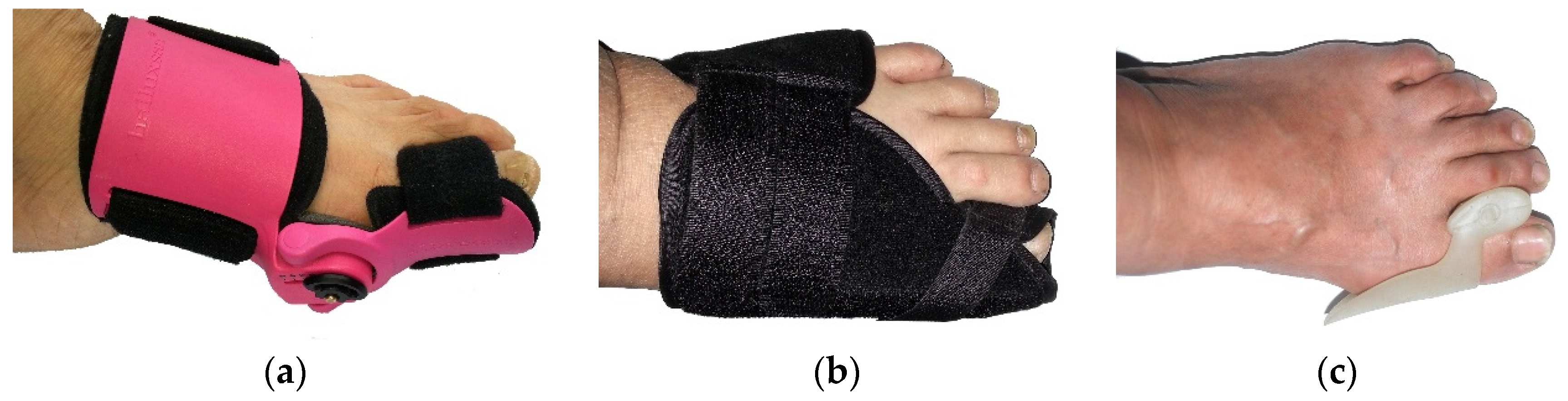
2.3. Orthotics
2.4. Procedure
2.5. Measuring Device
2.6. Measuring Method for Determining HVA Using the Measurement Device
- (a)
- Participants sat barefoot, keeping a straight back on a chair. The chair was adjusted to ensure that the participant’s lower leg was vertical and at 90° relative to the upper leg. The participant’s feet were placed flat on the floor without bearing body weight (Figure 3a).
- (b)
- A piece of A4 blank paper was placed underneath the foot of the participant. The new tool, called the right-angle wooden ruler, which was designed by the researcher and made by a carpenter, was used to keep the foot steady and to ensure that the heel and lateral foot shape were positioned at a 90° angle. The outside of the foot and the heel were placed as close as possible to the right-angle wooden ruler (Figure 3b).
- (c)
- Three prominent points on the foot were located: the metatarsal phalange joint, the interphalangeal joint, and the navicular. These three points were marked using a cross on the foot to be measured (Figure 3c).
- (d)
- The new measurement device was used to trace the outline of the foot from the heel to the end of the big toe (Figure 3d). The foot shape outline was traced on the A4 paper.
- (e)
- A set square was used to record each of three prominent points (the medial border of the soft tissue of the big toe, the ball of the big toe, and the medial border of the heel) on the A4 paper (Figure 3e).
- (f)
- A traced outline of the foot marked with the three prominent points was obtained on the A4 paper (Figure 3f).
- (g)
- A ruler was used to draw two tangent lines by connecting the most prominent points between the metatarsal phalange joint and the navicular, as well as between the metatarsal phalange joint and the phalanges joint. Two tangent lines were extended to meet each other (Figure 3g).
- (h)
- Finally, a protractor was used to record the HVA (Figure 3h).

2.7. Statistical Analysis
3. Results
4. Discussion
5. Conclusions
Author Contributions
Funding
Institutional Review Board Statement
Informed Consent Statement
Acknowledgments
Conflicts of Interest
References
- Al-Abdulwahab, S.S.; Al-Dosry, R.D. Hallux valgus and preferred shoe types among young healthy Saudi Arabian females. Ann. Saudi Med. 2000, 20, 319–321. [Google Scholar] [CrossRef]
- Bates, B.T.; Osternig, L.R.; Mason, B.; James, L.S. Foot orthotic devices to modify selected aspects of lower extremity mechanics. Am. J. Sport. Med. 1979, 7, 338–342. [Google Scholar] [CrossRef]
- Roddy, E.; Zhang, W.; Doherty, M. Prevalence and associations of hallux valgus in a primary care population. Arthritis Care Res. Off. J. Am. Coll. Rheumatol. 2008, 59, 857–862. [Google Scholar] [CrossRef]
- Cho, N.H.; Kim, S.; Kwon, D.J.; Kim, H.A. The prevalence of hallux valgus and its association with foot pain and function in a rural Korean community. J. Bone Joint Surg. Br. 2009, 91, 494–498. [Google Scholar] [CrossRef]
- Nix, S.; Smith, M.; Vicenzino, B. Prevalence of hallux valgus in the general population: A systematic review and meta-analysis. J. Foot Ankle Res. 2010, 3, 21. [Google Scholar] [CrossRef]
- Magnan, B.; Negri, S.; Maluta, T.; Dall’Oca, C.; Samaila, E. Minimally invasive distal first metatarsal osteotomy can be an option for recurrent hallux valgus. Foot Ankle Surg. 2019, 25, 332–339. [Google Scholar] [CrossRef]
- Menz, H.B.; Fotoohabadi, M.R.; Wee, E.; Spink, M.J. Validity of self-assessment of hallux valgus using the Manchester scale. BMC Musculoskelet. Disord. 2010, 11, 215. [Google Scholar] [CrossRef]
- Easley, M.E.; Trnka, H.J. Current concepts review: Hallux valgus part 1: Pathomechanics, clinical assessment, and nonoperative management. Foot Ankle Int. 2007, 28, 654–659. [Google Scholar] [CrossRef]
- Pique-Vidal, C.; Maled-Garcia, I.; Arabi-Moreno, J.; Vila, J. Radiographic angles in hallux valgus: Differences between measurements made manually and with a computerized program. Foot Ankle Int. 2006, 27, 175–180. [Google Scholar] [CrossRef]
- Wagner, P.; Ortiz, C.; Wagner, E. Rotational osteotomy for hallux valgus. A new technique for primary and revision cases. Tech. Foot Ankle Surg. 2017, 16, 3. [Google Scholar] [CrossRef]
- Nguyen, U.S.; Hillstrom, H.J.; Li, W.; Dufour, A.B.; Kiel, D.P.; Procter-Gray, E.; Gagnon, M.M.; Hannan, M.T. Factors associated with hallux valgus in a population-based study of older women and men: The MOBILIZE Boston Study. Osteoarthr. Cartil. 2010, 18, 41–46. [Google Scholar] [CrossRef]
- Okuda, R.; Kinoshita, M.; Yasuda, T.; Jotoku, T.; Kitano, N.; Shima, H. The shape of the lateral edge of the first metatarsal head as a risk factor for recurrence of hallux valgus. JBJS 2007, 89, 2163–2172. [Google Scholar] [CrossRef]
- Ferrari, J.; Higgins, J.P.T.; Prior, T.D. Interventions for treating hallux valgus (abductovalgus) and bunions. Cochrane Database Syst. Rev. 2004, 1, 1–61. [Google Scholar]
- Tang, S.F.; Chen, C.P.; Pan, J.L.; Chen, J.L.; Leong, C.P.; Chu, N.K. The effects of a new foot-toe orthosis in treating painful hallux valgus. Arch. Phys. Med. Rehabil. 2002, 83, 1792–1795. [Google Scholar] [CrossRef]
- Tehraninasr, A.; Saeedi, H.; Forogh, B.; Bahramizadeh, M.; Keyhani, M.R. Effects of insole with toe-separator and night splint on patients with painful hallux valgus: A comparative study. Prosthet. Orthot. Int. 2008, 32, 79–83. [Google Scholar] [CrossRef]
- Du Plessis, M.; Zipfel, B.; Brantingham, J.W.; Parkin-Smith, G.F.; Birdsey, P.; Globe, G.; Cassa, T.K. Manual and manipulative therapy compared to night splint for symptomatic hallux abducto valgus: An exploratory randomised clinical trial. Foot 2011, 21, 71–78. [Google Scholar] [CrossRef]
- Reina, M.; Lafuente, G.; Munuera, P.V. Effect of custom-made foot orthoses in female hallux valgus after one-year follow up. Prosthet. Orthot. Int. 2013, 37, 113–119. [Google Scholar] [CrossRef]
- Hurn, S.E.; Vicenzino, B.T.; Smith, M.D. Non-surgical treatment of hallux valgus: A current practice survey of Australian podiatrists. J. Foot Ankle Res. 2016, 9, 16. [Google Scholar] [CrossRef]
- Li, G.; Shen, J.; Smith, E.; Patel, C. Development of a Manual Measurement Device for Measuring Hallux Valgus Angle in Patients with Hallux Valgus. Int. J. Environ. Res. Public Health 2022, 19, 9108. [Google Scholar] [CrossRef]
- Chadchavalpanichaya, N.; Chueluecha, C. Effectiveness of hallux valgus strap: A prospective, randomized single-blinded controlled trial. Siriraj Med. J. 2017, 63, 42–46. [Google Scholar]
- Mirzashahi, B.; Ahmadifar, M.; Birjandi, M.; Pournia, Y. Comparison of designed slippers splints with the splints available on the market in the treatment of hallux valgus. Acta Med. Iran. 2012, 50, 107–112. [Google Scholar]
- Chadchavalpanichaya, N.; Prakotmongkol, V.; Polhan, N.; Rayothee, P.; Seng-Iad, S. Effectiveness of the custom-mold room temperature vulcanizing silicone toe separator on hallux valgus: A prospective, randomized single-blinded controlled trial. Prosthet. Orthot. Int. 2018, 42, 163–170. [Google Scholar] [CrossRef]


| Patients | N | Minimum | Maximum | Mean ± SD |
|---|---|---|---|---|
| Age (y) | 26 | 20 | 72 | 53.27 ± 12.72 |
| Height (cm) | 26 | 140 | 173 | 157.92 ± 6.23 |
| Weight (kg) | 26 | 48 | 90 | 62.00 ± 9.47 |
| BMI (kg/cm²) | 26 | 18.7 | 31.3 | 24.84 ± 3.39 |
| Moderate HV Treatment with Orthotic Type 1 | Moderate HV Treatment with Orthotic Type 2 | Mild HV Treatment with Orthotic Type 2 | Mild HV Treatment with Orthotic Type 3 | |||||||||
|---|---|---|---|---|---|---|---|---|---|---|---|---|
| HVA (°) Reduction | 95% (CI) | p- Value | HVA (°) Reduction | 95% (CI) * | p- Value | HVA (°) Reduction | 95% (CI) * | p- Value | HVA (°) Reduction | 95% (CI) * | p- Value | |
| Subjects | n = 12 | n = 8 | n = 15 | n = 7 | ||||||||
| Baseline (HVA) | 26.94 ± 4.83 | 17.06 ± 1.72 | 27.83 ± 4.52 | 18.31 ± 1.41 | ||||||||
| 0–3 Months | 2.17 ±3.31 | 0.07, 4.27 | 0.04 * | 0.30 ±2.37 | −1.01, 1.61 | 0.63 | 1.15 ±3.37 | −1.67, 3.97 | 0.37 | 1.23 ±0.96 | 0.34, 2.12 | 0.02 * |
| 0–6 Months | 3.45 ±3.93 | 0.81, 6.08 | 0.02 * | 0.77 ±2.56 | −0.64, 2.19 | 0.26 | 0.68 ±2.64 | −1.53, 2.88 | 0.49 | 1.16 ±1.14 | −0.26, 2.58 | 0.09 |
| 0–9 Months | 3.71 ±3.18 | 1.43, 5.99 | 0.01 * | 1.22 ±3.31 | −0.69, 3.14 | 0.19 | 0.84 ±2.33 | −1.31, 2.99 | 0.38 | 2.73 ±2.11 | −0.63, 6.08 | 0.08 |
| 0–12 Months | 5.05 ±5.15 | 1.37, 8.73 | 0.01 * | 1.20 ±3.16 | −0.71, 3.11 | 0.20 | 2.44 ±0.85 | 1.39, 3.49 | 0.00* | 3.08 ±2.36 | −0.68, 6.83 | 0.08 |
Publisher’s Note: MDPI stays neutral with regard to jurisdictional claims in published maps and institutional affiliations. |
© 2022 by the authors. Licensee MDPI, Basel, Switzerland. This article is an open access article distributed under the terms and conditions of the Creative Commons Attribution (CC BY) license (https://creativecommons.org/licenses/by/4.0/).
Share and Cite
Li, G.; Shen, J.; Smith, E.; Patel, C. The Evaluation of Orthotics in Reducing Hallux Valgus Angle in Patients with Hallux Valgus over a Twelve-Month Treatment. Int. J. Environ. Res. Public Health 2022, 19, 12531. https://doi.org/10.3390/ijerph191912531
Li G, Shen J, Smith E, Patel C. The Evaluation of Orthotics in Reducing Hallux Valgus Angle in Patients with Hallux Valgus over a Twelve-Month Treatment. International Journal of Environmental Research and Public Health. 2022; 19(19):12531. https://doi.org/10.3390/ijerph191912531
Chicago/Turabian StyleLi, Guoli, Jinsong Shen, Edward Smith, and Chetna Patel. 2022. "The Evaluation of Orthotics in Reducing Hallux Valgus Angle in Patients with Hallux Valgus over a Twelve-Month Treatment" International Journal of Environmental Research and Public Health 19, no. 19: 12531. https://doi.org/10.3390/ijerph191912531
APA StyleLi, G., Shen, J., Smith, E., & Patel, C. (2022). The Evaluation of Orthotics in Reducing Hallux Valgus Angle in Patients with Hallux Valgus over a Twelve-Month Treatment. International Journal of Environmental Research and Public Health, 19(19), 12531. https://doi.org/10.3390/ijerph191912531