Article

CTX-M-Producing Bacteria Isolated from a Highly Polluted River System in Portugal

1
CESAM and Biology Department, University of Aveiro, 3810-193 Aveiro, Portugal
2
Department of Life Sciences, University of Coimbra, 3000-456 Coimbra, Portugal
3
Department of Life Sciences and CFE, University of Coimbra, 3000-456 Coimbra, Portugal
*
Author to whom correspondence should be addressed.
These authors contributed equally to this work.
Int. J. Environ. Res. Public Health 2022, 19(19), 11858; https://doi.org/10.3390/ijerph191911858
Submission received: 18 July 2022 / Revised: 13 September 2022 / Accepted: 15 September 2022 / Published: 20 September 2022
(This article belongs to the Section Environmental Health)

Abstract

:
Enterobacteriaceae resistant to third-generation cephalosporins are a great concern for public health, as these are first-line drugs to treat infections. The production of carbapenemases and extended spectrum beta-lactamases (ESBLs) and/or the overexpression of AmpC β-lactamases are the main mechanisms of resistance to these antibiotics. Among the ESBLs, CTX-M β-lactamases are the most prevalent worldwide. Our aims were to determine the prevalence of cefotaxime-resistant Enterobacteriaceae along a heavily polluted river and characterize blaCTX-M carriers. River water was collected in 11 sites along the main course and tributaries, in two sampling moments. Water quality was evaluated and a collection of cefotaxime-resistant isolates was obtained. blaCTX-M carriers were characterized regarding phylogenetic affiliation, clonality, antibiotic susceptibility, gene diversity, and context. Water presented very low quality in all sites. From 147 cefotaxime-resistant isolates, 46% carried blaCTX-M and were affiliated with Escherichia, Klebsiella, Enterobacter, and Citrobacter. Molecular typing revealed clonal isolates in different sites and over the two years, suggesting survival of the strains in the river or continuous pollution inputs from the same sources. Eight variants of blaCTX-M were found, with blaCTX-M-15 being the most prevalent (52.5%). Sites with a lower water quality showed the highest resistance rates and prevalence of blaCTX-M, suggesting that river water may embody human health risks.

1. Introduction

The pollution of aquatic systems is a concerning issue of global proportions and it has been estimated that more than half of the water resources in the world are polluted [1,2]. Natural processes, such as precipitation and erosion, as well as anthropogenic impacts (urban, industrial, and agriculture origins) and intensive exploration of hydric resources, determine the surface water quality of these systems [3,4,5]. Rivers are among the most intensively human influenced ecosystems worldwide [6]. The accumulation of pollutants in water systems can lead to serious contamination problems with long-term effects on aquatic life and human health. Hence, in view of the diverse water usage purposes and management requirements, assessing water quality is crucial. Consequently, many indices and multivariate models have been proposed to measure surface water quality, none of which considers parameters related to antibiotic resistance [7,8,9].
Antibiotic-resistant bacteria (ARB) and antibiotic-resistant genes (ARGs) are a growing concern in surface waters [10,11,12]. Though naturally present in ecosystems, ARB and ARGs can enter aquatic environments through discharges of untreated water from different sources (e.g., agriculture and industry) and from discharges from wastewater treatment systems, which are considered a major source of these contaminants [10,11,12,13,14,15]. Mixtures of different types of pollutants reaching aquatic settings continuously (e.g., antibiotics, disinfectants, and metals) potentiate the transfer of ARGs between natural and incoming bacterial populations, unbalancing the ecosystem and propagating antibiotic resistance [10,11,12,16]. Hence, with the high levels of contaminants that occur in many rivers worldwide, these environmental compartments can act as hotspots for dispersion of ARGs and ARB.
Broad spectrum antibiotics such as third-generation cephalosporins are very relevant therapeutic agents, as they are the first-line drugs to treat infections caused by multi-resistant gram negative strains, including priority pathogens, such as some of those referred to as ESKAPE [17,18,19]. The consumption of these antibiotics in the European Union is preferentially in human medicine, although relevant amounts are used in veterinary or animal production [20]. Resistance to third-generation cephalosporins in gram negative bacteria is mediated mainly by extended spectrum β-lactamases (ESBLs), which confer resistance to all β-lactams, except to carbapenems and cephamycins, and are currently among the most globally dispersed antibiotic resistance mechanisms [17,21,22]. First identified in 1983 [23], these enzymes are now disseminated worldwide, mainly in gram negatives, showing a significant spread in every World Health Organization (WHO) geographic region [17,21,22,24]. CTX-M type β-lactamases are a huge success in terms of antibiotic resistance dispersion, currently being the most prevalent among ESBLs worldwide [25,26,27]. This dispersion is even more evident when considering only developed countries, reflected by the significant upward trend in Europe [25]. The blaCTX-M gene has been linked to highly successful plasmids and high-risk clones, and associated with mechanisms of co-resistance [17,26,28,29]. Even though the majority of β-lactamase genes have an unclear origin, the ancestor of the blaCTX-M gene is known to be present in the genome of species of the genus Kluyvera, belonging to the Enterobacteriaceae family [30,31,32,33]. There have been over 200 different variants of CTX-M enzymes detected so far (https://www.ncbi.nlm.nih.gov/pathogens/refgene/#blaCTX-M; last accessed 31 January 2022). These are divided into six groups: CTX-M-1, CTX-M-2, CTX-M-8, CTX-M-9, CTX-M-25, and KLU-like [21,26], where members of the same group have >94% amino acid identity and there is ≤90% identity across different groups [22]. Worldwide, the most common variants are CTX-M-14 and CTX-M-15, followed by CTX-M-1, CTX-M-2, and CTX-M-3 [25]. Though the vast majority of reports of ARB carrying CTX-M encoding genes refer to clinical isolates, CTX-M producers have been identified in all One Health compartments and interfaces [16,26].
Lis River (Central Portugal) is an example of a highly polluted aquatic system subjected to successive discharges of effluents over its course, which result in a decrease in water quality [34]. Three wastewater treatment plants (WWTPs) discharge their final effluents in this watercourse. Additionally, there are around 400,000 pigs in the Leiria region that produce waste equivalent to that of 1.2 million people [34]. Illegal discharges of piggery wastewater were reported to have a great impact on the water quality in the region, with values of fecal coliforms in the river reaching levels lower than the allowable limits for bathing waters [34,35]. Furthermore, urban effluents, untreated sewage, and agriculture also carry to the river degradable organics, nutrients, and pathogenic organisms [34]. Other contaminants from anthropogenic sources, such as metals, mainly Zn and Mn [36], and antibiotics, e.g., sulfamethoxazole, clarithromycin, and azithromycin [37], were also detected in the river. A recent study detected several clinically relevant Enterobacteriaceae resistant to carbapenems (last-resort antibiotics) in the Lis River, carrying clinically important ARGs and having been associated with hospital outbreaks worldwide, including Portugal [38,39]. Therefore, as it receives a variety of contaminants from different sources, this hydrographic system has the potential to serve as a reservoir and a reactor for the evolution and dispersion of ARB and ARGs. Thus, studies developed in this system can contribute to better understanding the role of rivers in the dispersion of resistance and, in general terms, the ecology of antibiotic resistance. Though poor water quality has been documented for decades in this hydrographic basin, including reports on serious ecological disasters in some particular sites [34,35], Lis River water usage in this region is quite important. For example, for irrigation, with agriculture as a key economic activity, or for leisure activities, as the river flows to the Atlantic ocean into busy beach areas. As such, this study had the following intentions: (1) to determine the prevalence of cefotaxime-resistant bacteria along the Lis River, in two consecutive years; (2) to determine the phylogenetic affiliation, clonality, and antibiotic susceptibility profiles of blaCTX-M carriers isolated from the Lis River; and (3) to characterize the diversity of blaCTX-M genes and their genomic context. For this purpose, water was collected along the Lis River from the spring to the mouth, in the river’s main course and tributaries, considering the location of probable contributors to water pollution in this hydrographic basin. Water quality and the prevalence of cefotaxime-resistant Enterobacteriaceae were determined. A collection of cefotaxime-resistant isolates was established. The blaCTX-M carriers were characterized regarding their phylogenetic affiliation, clonality, antibiotic resistance profiles, gene diversity, and context.

2. Materials and Methods

2.1. Study Area

The hydrographic basin of the Lis River is a coastal basin that comprises an area of approximately 850 km2 (Figure 1). The Lis River spring is located at an altitude of 500 m and the river flows into the Atlantic Ocean for a distance of approximately 40 km, with an annual average current of 2.69 m3/s. This hydrographic basin is entirely located on soils of the Western Mesocenozoic rim. The annual average temperature and precipitation in this area are 14.8 °C and 855 mm, respectively [34]. The surface water in the main course and its tributaries have a decades-long record of poor water quality owing to the contamination with domestic effluents, agricultural run-offs, and piggery wastewater [34,35,36,37,38,39].

2.2. Sampling, Water Quality Evaluation, and Cefotaxime-Resistant Bacteria Isolation

Water samples were collected in September of two consecutive years, 2018 and 2019, from the Lis River (Central Portugal). A total of fifteen sites were analysed, 11 distributed along the main river from the spring to the mouth (P1–P3, P6, P9, P12–P15) and 6 located in three of its affluents (P4, P5, P7, P8, P10, P11). For this selection, we considered urban and rural sites as the location of the wastewater treatment plants (three in the region) and the piggeries facilities, which are considered the main polluters in this hydrographic basin (Figure 1; [39]). Water was collected in sterile bottles and kept on ice for transportation. Physical/chemical parameters were measured three times on site, including pH, temperature, conductivity, and oxygen saturation, using a portable multi-log environmental meter (WTW, Germany). Additional water quality parameters were evaluated in triplicates by collecting 5 L samples, which were sent to a credited laboratory for further analysis, namely of nitrates (ion chromatography), biochemical oxygen demand (gauge), and phosphorus (spectrophotometry). The microbiological parameters evaluated included enterococci, faecal, and total coliforms, following standard procedures.
The National System of Information of Hydric Resources (SNIRH) criteria to classify superficial water courses according to quality features for multiple applications (https://snirh.apambiente.pt) were used for the categorization of each physicochemical and microbiological parameter into three levels of water quality: bad, fair, and good (Table S1A). The overall quality of the sampling site was determined according to the worst category attributed to at least one parameter. Water samples from each of the fifteen sites were filtrated in triplicate through 0.45 μm pore membranes (Pall Life Sciences) and placed on mFC agar supplemented with 4 μg/mL of cefotaxime (Sigma-Aldrich, St. Louis, MI, USA). To determine the proportion of cefotaxime-resistant bacteria, mFC agar without antibiotics was used. Plates were incubated at 37 °C and counts were performed after 24 h. Individual cefotaxime-resistant colonies were purified and stored in 20% glycerol at −80 °C.

2.3. Phylogenetic Affiliation

Individual colonies were used for amplification of a 16S rRNA gene fragment by PCR followed by sequence analysis. For that, a bacterial cell suspension of each isolate was prepared with 20 μL of dH2O. PCR amplification was carried out in a final volume of 25 μL containing 1 μL of cell suspension, 16.25 μL dH2O, 6.25 μL NZYTaq 2× Green Master Mix (2.5 mm MgCl2; 200 μm dNTPs; 1.25 U DNA polymerase) (NZYTech, Lisboa, Portugal), and 0.75 μL of each primer from a stock solution with a concentration of 10 μm. Conditions and primers (27F and 1492R) were used as described previously and detailed in Table S2. Positive and negative controls were included. The resulting amplicons were purified and Sanger sequenced at GATC, Konstanz, Germany. Nucleotide sequences were used for similarity searches against the GenBank database (https://www.ncbi.nlm.nih.gov/nucleotide/) using the BLASTn software (https://blast.ncbi.nlm.nih.gov/Blast.cgi). Shigella and Escherichia isolates were discriminated by culture in xylose lysine deoxycholate agar (XLD).

2.4. blaCTX-M Screening, Genetic Environment, and Molecular Typing

All purified isolates were screened by PCR for the presence of the blaCTX-M gene, using conditions and primers according to previous studies (Table S2). BOX-PCR and ERIC-PCR were used to assess clonality among all isolates carrying a blaCTX-M gene, with primers and conditions as presented in Table S2. The final volumes for the reactions were 25 μL, which included 1 μL of cell suspension, 16.25 μL dH2O, 6.25 μL NZYTaq 2× Green Master Mix (NZYTech, Lisbon, Portugal), and 2 μL of primer for BOX-PCR (from a solution at 10 μm) or 1 μL of each primer for ERIC-PCR (from a solution at 50 μm). Banding profiles were analysed with GelCompar II (Applied Maths, Saint-Martens-Latem, Belgium).
To determine the genetic context of the blaCTX-M gene, PCR was used with primers specific for sequences known to be associated with the blaCTX-M gene, namely ISEcp1 and IS26 for the upstream region of the gene, and orf477 and IS903 for the downstream region (Table S2), as previously described [40]. Positive controls were also used for every reaction. PCR products were then purified and sent for sequencing, and then assembled using BioEdit. Nucleotide and/or deduced amino acids sequences obtained were analysed against CARD (https://card.mcmaster.ca/analyze/blast), Genbank (https://blast.ncbi.nlm.nih.gov/Blast.cgi), and ISfinder (https://isfinder.biotoul.fr/blast.php) databases.

2.5. Antibiotic Susceptibility Testing of blaCTX-M Positives

Susceptibility to antibiotics was tested by the disc diffusion method on Mueller–Hinton agar according to the procedure established by the European Committee on Antimicrobial Susceptibility Testing (EUCAST v11.0; https://eucast.org/). All blaCTX-M positive isolates were tested against 16 antibiotics from 6 classes: beta-lactams (amoxicillin (AML, 10 μg), amoxicillin/clavulanic acid (AMC, 30 μg), piperacillin (PRL, 30 μg), piperacillin/tazobactam (TZP, 36 μg), ticarcillin (TIC, 75 μg), ticarcillin/clavulanic acid (TIM, 85 μg), cefotaxime (CTX, 5 μg), cefepime (FEP, 30 μg), ceftazidime (CAZ, 10 μg), aztreonam (ATM, 30 μg), and imipenem (IPM, 10 μg)), aminoglycosides (gentamicin (CN, 10 μg)), quinolones (ciprofloxacin (CIP, 5 μg)), tetracyclines (tetracycline (TE, 30 μg)), phenicols (chloramphenicol (C, 30 μg)), and the combination sulfamethoxazole/trimethoprim (SXT, 25 μg). After incubation at 37 °C for 18 h, isolates were classified as susceptible, resistant, or intermediate resistant. The quality control strain used was E. coli ATCC 25922.

2.6. Conjugation Assays

Conjugation assays were performed with the selected strains representing each blaCTX-M gene variant identified. Mating tests were performed with the broth culture conjugation method using the rifampicin-resistant E. coli CV601 as the recipient strain [41]. Briefly, donors and recipient strains were mixed at a ratio of 1:1 in broth culture and grown for 24 h at 37 °C without agitation. Transconjugants were selected on plate count agar (PCA) containing cefotaxime (4 μg/mL) and rifampicin (100 μg/mL) and confirmed by BOX-PCR and by the presence of the blaCTX-M gene. Susceptibility to antibiotics of the recipient strain and transconjugants was tested by the disc diffusion method on Mueller–Hinton agar, as described above.

3. Results

3.1. Water Quality Analysis and Cefotaxime-Resistant Bacteria Incidence

Water samples from all sites were classified as water with fair or poor quality, on both years (Figure 1, with P1 in 2018 representing the only sampling site with water samples classified with fair quality. In fact, the values registered for dissolved oxygen in all samples (except P1 in 2018) resulted in their classification as with poor water quality. Other parameters revealed the bad quality status of some sites such as phosphorus and nitrates, which were categorized as poor in nine and four sites, respectively, in at least one campaign (Figure 1). In some locations, the load of total and/or fecal coliforms increased in 2019, thus changing the categorization of these parameters from fair to bad (e.g., P7) or even from good to bad (e.g., P5). The highest number of poorly rated parameters was obtained at the P7 site in 2019 (six parameters).
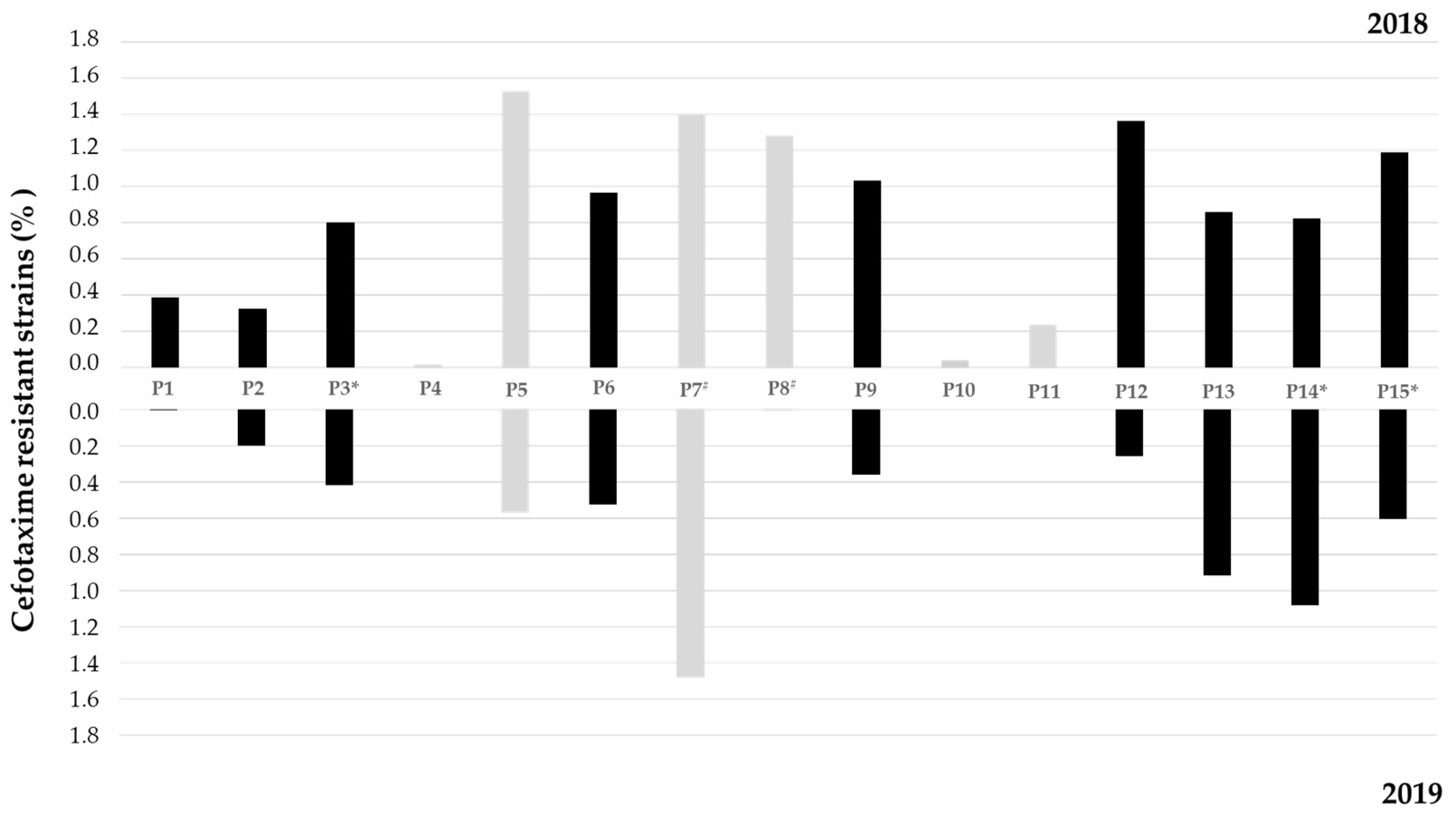
The prevalence of cefotaxime-resistant bacteria (CTXR) varied from 0.02 to 1.53% in 2018 and from 0 to 1.48% in 2019 (Figure 2), with the highest values observed in sites located in Lis River affluents (P5 and P7) in 2018. In 2018, the proportion of CTXR was higher than in 2019 in almost all sites and the occurrence of CTXR bacteria was registered in all sites. While in 2018, site P5 presented the highest proportion of CTXR, in 2019, it was observed in site P7. In 2019, no CTXR were detected in sites P4, P10, and P11, which also showed the lowest prevalence in 2018.

3.2. Phylogenetic Affiliation of Cefotaxime-Resistant Isolates

One hundred and forty-seven CTXR isolates were selected for further analysis, 91 isolated in 2018 and 56 in 2019. After 16S rRNA gene-based identification, a total of eight different genera were identified (Figure S1), five of which belonged to the Enterobacteriaceae family: Escherichia/Shigella, Klebsiella, Citrobacter, Enterobacter, and Pantoea. Shigella/Escherichia growth on xylose lysine deoxycholate agar (XLD) revealed that all isolates originated yellow colonies, confirming their affiliation to Escherichia. Non-Enterobacteriaceae genera were Acinetobacter, Aeromonas, and Pseudomonas (Figure S1). In 2018, all eight genera were found, whereas in the 2019 campaign, Pantoea isolates were not detected. Predominant genera varied between years, as shown in Figure S1. In 2018, Acinetobacter dominated, representing 35% of the total collection (n = 91), followed by Escherichia (32%), Klebsiella (12%), and Pseudomonas (10%). In 2019 (n = 56), Acinetobacter was represented by only one isolate, while Klebsiella and Escherichia were predominant (30% each). When comparing sampling sites (Figure S1), Escherichia was detected in 11 sites in 2018 and in 7 sites in 2019, and Klebsiella was detected in 5 sites in 2018 and 8 sites in 2019. Site P5 showed the highest diversity in 2018, with five genera isolated from this location, while in 2019, five different genera were retrieved from site P12 and four were retrieved from site P5.

3.3. Prevalence and Diversity of the blaCTX-M Gene among the Cefotaxime-Resistant Isolates

In 68 out of 147 isolates (46.3%), the presence of the blaCTX-M gene was detected, distributed in four different Enterobacteriaceae genera, namely, Escherichia (n = 39), Klebsiella (n = 24), Enterobacter (n = 3), and Citrobacter (n = 2). These isolates were obtained from almost all sampled sites along the river and affluents (excluding sites P4 and P10) and in both years, as shown in Figure 3 and Table 1. Based on BOX and ERIC PCR-based analysis, one representative of each typing profile was selected from each site and year, resulting in a collection of 54 isolates. The analysis of the deduced amino acid sequences revealed that blaCTX-M genes encoded seven different CTX-M variants, as shown in Table 1 and Figure 3.
Additionally, for 14 isolates (Table 1), it was not possible to determine the blaCTX-M encoding variant. The CTX-M-15 encoding gene was the most prevalent, being present in 21 of all blaCTX-M carriers (38.8%) (Table 1). The blaCTX-M-1, blaCTX-M-32, and blaCTX-M-65 genes were each present in 7.4% of the isolates. The blaCTX-M-3 and blaCTX-M-14 genes showed the lowest prevalence, each being present in only one isolate. All variants were found in Escherichia, except blaCTX-M-1, with blaCTX-M-15 prevailing with 10 out of 32 isolates (31%), and the less frequent being blaCTX-M-14 in only one isolate. In Klebsiella isolates, only blaCTX-M-15 was detected. blaCTX-M-3 was present in only one Enterobacter isolate. The single Citrobacter isolate carried a blaCTX-M-32 gene.
The analysis of the regions flanking the different blaCTX-M genes revealed the presence of 10 different genomic environments, as represented in Figure 4A–J. All shared the presence of the insertion sequence ISEcp1 in the upstream region of the gene, with variable distances to the gene (32 bp to 127 bp), mainly owing to the presence of different conserved regions previously reported [40,42,43] (Figure 4; Table 1). In the downstream region, orf477 and insertion sequence IS903 were identified, as previously reported [40,42,43].
In regard to the diversity of CTX-M encoding genes detected along the Lis River (Figure 3), blaCTX-M-15 is well distributed along the river, found in 11 of the 15 sites sampled. Site 5 showed the highest diversity, with four different variants detected, followed by sites 6, 7, 9, and 15, with three different CTX-M encoding genes detected. Klebsiella isolates carrying blaCTX-M-15 genes were detected only in two sites: 3 (urban area, downstream a WWTP) and 8 (in the vicinity of swine farms).

3.4. Antibiotic Susceptibility Testing of blaCTX-M Gene Carriers

Antibiotic susceptibility phenotypes were evaluated for all 54 isolates (Table 1). For analysis purposes, susceptibility profiles classified as intermediate were addressed as resistant. The overall results showed that isolates presented resistance levels below 60% to only four out of sixteen antibiotics, namely, piperacillin/tazobactam (38.8%), imipenem (3.7%), gentamicin (31.4%), and chloramphenicol (25.9%). Besides resistance to cefotaxime, the highest resistance rates were observed towards other beta-lactams, namely to cefepime, piperacillin, aztreonam, and ticarcillin, with 98% each, and 100% of the isolates were resistant to amoxicillin. Additionally, high resistance levels were observed for ciprofloxacin (79.6%), trimethoprim/sulfamethoxazole (72.2%), and tetracycline (62.9%).
Among Escherichia isolates, the lowest rates of resistance were observed towards imipenem (only 1 resistant), gentamicin (5 resistant isolates), and chloramphenicol (10 resistant isolates). Klebsiella isolates were highly susceptible to imipenem (1 resistant isolate), gentamicin (12 resistant isolates), tetracycline (9 resistant isolates), and chloramphenicol (3 resistant isolates).
Bacteria are considered multi-resistant when they are non-susceptible to antibiotics included in three or more classes [44]. Hence, a total of 79.6% (corresponding to 43 isolates) showed multi-resistant phenotypes. All Klebsiella isolates were multi-resistant, as well as the single Citrobacter isolate, while among the Escherichia isolates, 75% showed a multi-resistant phenotype (n = 24). On the other hand, none of the Enterobacter isolates showed multi-resistance. Three out of the 54 isolates showed resistance to all tested antibiotic classes.

3.5. Conjugation Assays

Conjugation assays were performed for 18 isolates, as indicated in Table 1, representing the different variants identified and in the different genera. Under the conditions tested, we obtained transconjugants resistant to cefotaxime and rifampicin from 9 out of the 17 donor strains (Table 2). The antibiotic susceptibility tests revealed that all transconjugants were resistant to amoxicillin; ticarcillin; piperacillin; aztreonam; ceftazidime; and, as expected, cefotaxime. None showed resistance to imipenem, gentamicin, or chloramphenicol. Two presented a multi-resistance phenotype (E23T carrying a blaCTX-M-27 and K1T carrying a blaCTX-M-15).

4. Discussion

Rivers can act as reservoirs of multi-resistant bacteria, as they receive contaminants from all sources like WWTPs, industrial effluents, agricultural activities, hospital sewage, or animal production effluents [10,11,12,16]. All of these examples of contamination have been reported in the Lis River [34,35,36,37,38,45], rendering it of particular interest to the spread and evolution of antibiotic resistance in these ecosystems.
When analyzing the water microbiological, physical, and chemical data, it was clear that the Lis River presents very poor water quality from the spring to the mouth, and in its affluents, as has been reported in previous studies [34,35,45]. For instance, sampling site P7, located in one of the Lis River affluents, globally presented the worst values observed in both years. The fact that this site is near several pig farms suggests a significant contribution from these sources of contamination. Swine slurry is a mixture of pig feces and urine with wastewater, and sometimes precipitation, which contains mainly suspended solids, nitrogen, phosphorous, and potassium [46]. Phosphorous levels were high across the river, but site P7 presented the highest values on both years. Furthermore, phosphorus levels in sites P3, P14, and P15 showed some concerning values in both years, which can relate to the location of these sampling sites downstream WWTPs.
In terms of abundance of Enterobacteriaceae resistant to cefotaxime along the river, site P7 also had the highest value among all sites. Besides the pollutants mentioned above, animal-production-derived effluents may contain different metals, antibiotics, antibiotic-resistant bacteria, and ARGs [47,48]. Moreover, it has been shown that ARGs from piggeries effluents can travel far by entering river courses, thus adding an additional public health risk [48]. In contrast, site P1 represents the spring of the Lis River, showing a small number of isolates resistant to cefotaxime, consistent with lower levels of pollution at this site. Focusing on the sites located in the river’s main course (P1, P2, P3, P6, P9, P12, P13, P14, and P15), it is noticeable that the percentage of cefotaxime-resistant Enterobacteriaceae displayed a tendency to increase along the river course, probably owing to an additive effect of different sources of contamination.
In 2019, all isolates retrieved from sites P7 and P8 carried the blaCTX-M gene. Both sites are set at the Ribeira dos Milagres affluent, which is the part of the Lis River most affected by pollution [34]. In fact, the presence of the blaCTX-M gene has been associated with high levels of anthropogenic influence [40].
Escherichia and Klebsiella together represented 93% of blaCTX-M carriers. These two genera include clinically relevant pathogens, and in this case, are particularly critical owing to their ESBL-producing and multi-resistance traits. Therefore, their frequent occurrence in the Lis River is alarming, as these waters can easily reach the population. Risks to human health arise from the fact that the water from the Lis River is frequently used for irrigation, fishing, and leisure activities, facilitating the contact of humans with resistant bacteria [49]. Previous studies confirmed the transfer of antibiotic-resistant bacteria from irrigation water to vegetables that are consumed raw [41]. Moreover, the transfer of cefotaxime-resistant bacteria from water to humans during leisure activities has been confirmed [50]. Infections caused by resistant strains from these groups of bacteria are growing every year across all continents [17,25]. They are often found in clinical environments (i.e., hospitals) and it is becoming harder to respond to these infections, thus there is an urgent need for new antibiotics for dealing with infections caused by members of these groups, according to the World Health Organization [51].
When analyzing clonality among our collection, we noticed that, even though most of the isolates had unique profiles, a few isolates collected in different sites presented identical profiles. This may indicate that these strains can survive in this environment for some period, being transported along the river. If their genetic determinants of resistance are located in mobile elements, their persistence may promote gene transfer to other bacteria. Furthermore, clonal isolates were also collected in different years, suggesting that they may derive from a persistent contamination source. However, further studies are needed to confirm this hypothesis.
The blaCTX-M genes are well known for being associated with other antibiotic resistance genes. This derives from the fact that blaCTX-M genes are often located in conjugative plasmids that harbor resistance genes to fluoroquinolones, aminoglycosides, and sulfonamides [28]. Keeping this in mind, it is not surprising that a total of 79.6% of the isolates were classified as multi-resistant. Despite the low rate of resistance to imipenem (4%), it is still very concerning. Carbapenems are used as last resource antibiotics to treat Enterobacteriaceae infections [20], and their increased prescription is promoted by the growing prevalence of ESBL producers. Ultimately, this may result in higher numbers of carbapenemase-producing Enterobacteriaceae infections. Worryingly, a recent study showed that the Lis River is in fact contaminated with carbapenem-resistant Enterobacteriaceae [38,39], carrying plasmids that harbour different genetic determinants of resistance, identical to those already described in clinical settings, namely, blaNDM, blaKPC, and blaGES. Furthermore, through qPCR analysis, blaCTX-M was detected in all sampling sites and showed a significant increase in the overall gene abundance over one year [39].
In contrast, high resistance rates to tetracycline and trimethoprim/sulfamethoxazole (63% and 72%, respectively) are not surprising as these are antibiotics commonly used in animal production, a common practice in this region. Furthermore, sulfamethoxazole was one of the main antibiotics detected in the Lis River water by Paiga and co-authors [37].
Though a strategy based on culture-dependent methods can be seen as a limitation, to characterize in detail these resistance mechanisms, this approach is fundamental and quite relevant in epidemiological terms. Higher blaCTX-M gene diversity was observed in sites located in urban areas (e.g., P5 and P6) or in the proximity of pig farms (e.g., P7 and P9). Interestingly, the majority of Klebsiella isolates carrying blaCTX-M genes were detected in sites located in urban areas (P3, P5, and P6), next to WWTPs (P3 and P14), or in the proximity of pig farms (P8 and P9). All CTX-M variants identified in this study have been previously identified in Portugal in ESBL producers isolated not only in clinical, but also in environmental samples, such as, for example, from companion [52,53] and wild animals [54], retail meat [55], and wastewater-treated effluents or rivers [14,15,40,56,57]. Most variants were also previously found in rivers in Portugal [40], although, to the best of our knowledge, this is the first report showing the presence of blaCTX-M-65 in a Portuguese river. This variant has been identified worldwide in various sources such as food-related products, wild and companion animals, or aquatic systems [58,59,60], but less commonly in European countries. A recent study in Portugal reported the presence of E. coli isolates retrieved from retail meat carrying blaCTX-M-65 [55]. In fact, the genomic context was identical to that characterized in CTX-M-65-producing E. coli isolates obtained in this study; that is, with blaCTX-M-65 flanked by ISEcp1 (upstream) and IS903 (downstream) [55].
In this study, blaCTX-M-15 stood out as the most prevalent gene variant, detected in 52.5% of isolates. The blaCTX-M-15 was identified for the first time in 1980s and, since then, has become the most prevalent blaCTX-M gene variant worldwide, in both hospitals and the environment [40,61,62,63,64]. It was also the main variant found in Portuguese aquatic systems [25,39] and WWTPs, including in treated effluents [14,15,57].
The results obtained here can be interpreted as a global alert for aquatic systems of similar characteristics. The Lis hydrographic basin gathers several features relevant for the study of antibiotic resistance dissemination in aquatic systems. On one hand, surface water use is quite relevant in this region, with agriculture being a key economic activity. On the other hand, this hydrographic basin encompasses an area of both urban and rural characteristics. Several anthropogenic impacts are identified along the river’s main course and its tributaries, with wastewater treatment plants and pig farms potentially constituting major contributors to the global low water quality that has been reported here in the last decades [34,35,36,37,38,39]. Thus, information is needed to determine the main sources of contamination and thus implement adequate management plans. For instance, the data provided here can give an indication of the efficiency of wastewater treatment, thus helping to support necessary changes. Informed measures are generally more economically sustainable because they are more effective.
The overall results highlight the relevance of similar studies to the global knowledge on the prevalence and dispersion of antibiotic resistance in correlation with water pollution and anthropogenic impacts. Water scarcity and growing worldwide population has brought attention to the need to provide more water through alternative approaches such as, for example, desalination and water reuse, as well as for water quality improvement in global water resources [2]. Water resource management plans and policy decisions usually envision water quality evaluation based on a few parameters, but do not consider antibiotic-resistant bacteria and antibiotic resistance determinants, despite their public health relevance. Hence, our work also brings attention to the need to establish prompt municipal warning systems that include monitoring of water pollutants (including parameters related to antibiotic resistance), identifying the main contributors to decreases in water quality, and following water management plans.

5. Conclusions

As expected, the data from this work expose the high levels of pollution in the Lis River, and suggest that pig farms represent a major source of pollution in this region. However, agricultural run-offs, wastewater from hospitals, and WWTPs should also be considered. Water samples from highly polluted sampling sites also demonstrated a higher prevalence of CTX-M producers of high clinical relevance and that exhibit multi-resistance phenotypes. Altogether, these findings constitute concerning factors to natural environments as well as public health. Further research is necessary to evaluate the actual size of the risk to public health posed by the different uses that are given to this water system. For instance, agricultural products irrigated with this water or aquatic animals used for human consumption should be evaluated, as well as recreational areas, by inspecting not only water, but also sediments and beach sand. Additionally, these studies could be complemented with data obtained through culture-independent strategies.

Supplementary Materials

The following supporting information can be downloaded at: https://www.mdpi.com/article/10.3390/ijerph191911858/s1, Table S1: Physical, chemical, and microbiological parameters measured to classify water quality status as poor, fair, or good (A), determined in 2018 and 2019 sampling campaigns (B) and overall classification. The classification considers parameters and maximum recommended values established by the Portuguese government (adapted from https://snirh.apambiente.pt/snirh/_dadossintese/qualidadeanuario/boletim/tabela_classes.php); Table S2. Primers used in this study; Figure S1. Genus-level affiliation of isolates retrieved from each site (1 to 15) in 2018 (A) and 2019 (B), with the number of isolates for each genus represented in bars. References [39,42,65,66,67,68] are cited in the supplementary materials.

Author Contributions

M.T.: Conception, Methodology, Data Curation, Formal Analysis, Visualization, Investigation, Writing—Original Draft Preparation, Supervision; J.L.: Investigation, Formal Analysis, Writing—Reviewing and Editing; P.T.: Investigation, Writing—Reviewing and Editing; I.H.: Conception, Methodology, Data Curation, Resources, Writing—Reviewing and Editing, Supervision, Funding Acquisition. All authors have read and agreed to the published version of the manuscript.

Funding

We acknowledge financial support from FCT/MCTES through national funds to CESAM (UIDP/50017/2020 + UIDB/50017/2020 + LA/P/0094/2020), CFE (UIDB/04004/2020), and individual grants to Pedro Teixeira (SFRH/BD/132046/2017) and Marta Tacão (CEECIND/00977/2020).

Institutional Review Board Statement

Not applicable.

Informed Consent Statement

Not applicable.

Data Availability Statement

Not applicable.

Conflicts of Interest

The authors declare no conflict of interest.

References

  1. Saravanan, A.; Kumar, P.S.; Jeevanantham, S.; Karishma, S.; Tajsabreen, B.; Yaashikaa, P.R.; Reshma, B. Effective water/wastewater treatment methodologies for toxic pollutants removal: Processes and applications towards sustainable development. Chemosphere 2021, 280, 130595. [Google Scholar] [CrossRef] [PubMed]
  2. United Nations. The United Nations World Water Development Report 2021: Valuing Water; UNESCO: Paris, France, 2021. [Google Scholar]
  3. Schwarzenbach, R.P.; Escher, B.I.; Fenner, K.; Hofstetter, T.B.; Johnson, C.A.; Von Gunten, U.; Wehrli, B. The challenge of micropollutants in aquatic systems. Science 2006, 313, 1072–1077. [Google Scholar] [CrossRef] [PubMed]
  4. Carpenter, S.R.; Caraco, N.F.; Correll, D.L.; Howarth, R.W.; Sharpley, A.N.; Smith, V.H. Nonpoint pollution of surface waters with phosphorus and nitrogen. Ecol. Appl. 1998, 8, 559–568. [Google Scholar] [CrossRef]
  5. Loos, R.; Gawlik, B.M.; Locoro, G.; Rimaviciute, E.; Contini, S.; Bidoglio, G. EU-wide survey of polar organic persistent pollutants in European river waters. Environ. Pollut. 2009, 157, 561–568. [Google Scholar] [CrossRef] [PubMed]
  6. Tejerina-Garro, F.L.; Maldonado, M.; Ibañez, C.; Pont, D.; Roset, N.; Oberdorff, T. Effects of natural and anthropogenic environmental changes on riverine fish assemblages: A framework for ecological assessment of rivers. Braz. Arch. Biol. Technol. 2005, 48, 91–108. [Google Scholar] [CrossRef]
  7. Gad, M.; Elsayed, S.; Moghanm, F.S.; Almarshadi, M.H.; Alshammari, A.S.; Khedher, K.M.; Eid, E.M.; Hussein, H. Combining Water Quality Indices and Multivariate Modeling to Assess Surface Water Quality in the Northern Nile Delta, Egypt. Water 2020, 12, 2142. [Google Scholar] [CrossRef]
  8. Gad, M.; Saleh, A.H.; Hussein, H.; Farouk, M.; Elsayed, S. Appraisal of Surface Water Quality of Nile River Using Water Quality Indices, Spectral Signature and Multivariate Modeling. Water 2022, 14, 1131. [Google Scholar] [CrossRef]
  9. Yan, T.; Shen, S.-L.; Zhou, A. Indices and models of surface water quality assessment: Review and perspectives. Environ. Pollut. 2022, 308, 119611. [Google Scholar] [CrossRef]
  10. Marti, E.; Variatza, E.; Balcazar, J.L. The role of aquatic ecosystems as reservoirs of antibiotic resistance. Trends Microbiol. 2014, 22, 36–41. [Google Scholar] [CrossRef]
  11. Taylor, N.G.H.; Verner-Jeffreys, D.W.; Baker-Austin, C. Aquatic systems: Maintaining, mixing and mobilising antimicrobial resistance? Trends Ecol. Evol. 2011, 26, 278–284. [Google Scholar] [CrossRef]
  12. Wellington, E.M.H.; Boxall, A.B.A.; Cross, P.; Feil, E.J.; Gaze, W.H.; Hawkey, P.M.; Johnson-Rollings, A.S.; Jones, D.L.; Lee, N.M.; Otten, W.; et al. The role of the natural environment in the emergence of antibiotic resistance in gram-negative bactéria. Lancet Infect. Dis. 2013, 13, 155–165. [Google Scholar] [CrossRef]
  13. Rizzo, L.; Manaia, C.; Merlin, C.; Schwartz, T.; Dagot, C.; Ploy, M.C.; Michael, I.; Fatta-Kassinos, D. Urban wastewater treatment plants as hotspots for antibiotic resistant bacteria and genes spread into the environment: A review. Sci. Total Environ. 2013, 447, 345–360. [Google Scholar] [CrossRef] [PubMed]
  14. Silva, I.; Tacão, M.; Tavares, R.D.S.; Miranda, R.; Araújo, S.; Manaia, C.M.; Henriques, I. Fate of cefotaxime-resistant Enterobacteriaceae and ESBL-producers over a full-scale wastewater treatment process with UV disinfection. Sci. Total Environ. 2018, 639, 1028–1037. [Google Scholar] [CrossRef] [PubMed]
  15. Tavares, R.D.S.; Tacão, M.; Figueiredo, A.S.; Duarte, A.S.; Esposito, F.; Lincopan, N.; Manaia, C.M.; Henriques, I. Genotypic and phenotypic traits of blaCTX-M-carrying Escherichia coli strains from an UV-C-treated wastewater effluent. Water Res. 2020, 184, 116079. [Google Scholar] [CrossRef] [PubMed]
  16. Lupo, A.; Coyne, S.; Berendonk, T.U. Origin and evolution of antibiotic resistance: The common mechanisms of emergence and spread in water bodies. Front. Microbiol. 2012, 3, 18. [Google Scholar] [CrossRef] [PubMed]
  17. Adler, A.; Katz, D.E.; Marchaim, D. The continuing plague of Extended-Spectrum β-lactamase-producing Enterobacteriaceae infections. Infect. Dis. Clin. N. Am. 2020, 30, 347–375. [Google Scholar] [CrossRef]
  18. Chong, Y.; Shimoda, S.; Shimono, N. Current epidemiology, genetic evolution and clinical impact of Extended-Spectrum β-lactamase-producing Escherichia coli and Klebsiella pneumonia. Infect. Genet. Evol. 2018, 61, 185–188. [Google Scholar] [CrossRef]
  19. Rice, L.B. Federal funding for the study of antimicrobial resistance in nosocomial pathogens: No ESKAPE. J. Infect. Dis. 2008, 197, 1079–1081. [Google Scholar] [CrossRef]
  20. European Centre for Disease Prevention and Control (ECDC); European Food Safety Authority (EFSA); European Medicines Agency (EMA). ECDC/EFSA/EMA second joint report on the integrated analysis of the consumption of antimicrobial agents and occurrence of antimicrobial resistance in bacteria from humans and food-producing animals: Joint Interagency Antimicrobial Consumption and Resistance Analysis (JIACRA) Report. EFSA J. 2017, 15, e04872. [Google Scholar]
  21. Bush, K. Past and present perspectives on β-lactamases. Antimicrob. Agents Chemother. 2018, 62, e01076-18. [Google Scholar] [CrossRef]
  22. Bush, K.; Bradford, P.A. Epidemiology of β-lactamase-producing pathogens. Clin. Microbiol. Rev. 2020, 33, e00047-19. [Google Scholar] [CrossRef] [PubMed]
  23. Knothe, H.; Shah, P.; Krcmery, V.; Antal, M.; Mitsuhashi, S. Transferable resistance to cefotaxime, cefoxitin, cefamandole and cefuroxime in clinical isolates of Klebsiella pneumoniae and Serratia marcescens. Infection 1983, 11, 315–317. [Google Scholar] [CrossRef] [PubMed]
  24. Ojer-Usoz, E.; González, D.; García-Jalón, I.; Vitas, A.I. High dissemination of Extended-Spectrum β-lactamase-producing Enterobacteriaceae in effluents from wastewater treatment plants. Water Res. 2014, 56, 37–47. [Google Scholar] [CrossRef] [PubMed]
  25. Bevan, E.R.; Jones, A.M.; Hawkey, P.M. Global epidemiology of CTX-M β-lactamases: Temporal and geographical shifts in genotype. J. Antimicrob. Chemother. 2017, 72, 2145–2155. [Google Scholar] [CrossRef]
  26. Cantón, R.; González-Alba, J.M.; Galán, J.C. CTX-M enzymes: Origin and diffusion. Front. Microbiol. 2012, 3, 110. [Google Scholar] [CrossRef]
  27. Hussain, H.I.; Aqib, A.I.; Seleem, M.N.; Shabbir, M.A.; Hao, H.; Iqbal, Z.; Kulyar, M.F.; Zaheer, T.; Li, K. Genetic basis of molecular mechanisms in β-lactam resistant gram-negative bacteria. Microb. Pathog. 2021, 158, 105040. [Google Scholar] [CrossRef]
  28. Tacão, M.; Moura, A.; Correia, A.; Henriques, I. Co-resistance to different classes of antibiotics among ESBL-producers from aquatic systems. Water Res. 2014, 48, 100–107. [Google Scholar] [CrossRef]
  29. Toleman, M.A.; Bennett, P.M.; Walsh, T.R. ISCR elements: Novel gene-capturing systems of the 21st Century? Microbiol. Mol. Biol. Rev. 2006, 70, 296–316. [Google Scholar] [CrossRef]
  30. Decousser, J.W.; Poirel, L.; Nordmann, P. Characterization of a chromosomally encoded Extended-Spectrum class A beta-lactamase from Kluyvera cryocrescens. Antimicrob. Agents Chemother. 2001, 45, 3595–3598. [Google Scholar] [CrossRef]
  31. Humeniuk, C.; Arlet, G.; Gautier, V.; Grimont, P.; Labia, R.; Philippon, A. Beta-lactamases of Kluyvera ascorbata; probable progenitors of some plasmid-encoded CTX-M types. Antimicrob. Agents Chemother. 2002, 46, 3045–3049. [Google Scholar] [CrossRef]
  32. Poirel, L.; Kampfer, P.; Nordmann, P. Chromosome-encoded Ambler class A beta-lactamase of Kluyvera georgiana; a probable progenitor of a subgroup of CTX-M extended-spectrum beta-lactamases. Antimicrob. Agents Chemother. 2002, 46, 4038–4040. [Google Scholar] [CrossRef] [PubMed] [Green Version]
  33. Rodriguez, M.M.; Power, P.; Radice, M.; Vay, C.; Famiglietti, A.; Galleni, M.; Ayala, J.A.; Gutkind, G. Chromosome-encoded CTX-M-3 from Kluyvera ascorbata: A possible origin of plasmid-borne CTX-M-1-derived cefotaximases. Antimicrob. Agents Chemother. 2004, 48, 4895–4897. [Google Scholar] [CrossRef] [PubMed]
  34. Vieira, J.; Fonseca, A.; Vilar, V.J.P.; Boaventura, R.A.R.; Botelho, C.M.S. Water quality in Lis river; Portugal. Environ. Monit. Assess. 2012, 184, 7125–7140. [Google Scholar] [CrossRef] [PubMed]
  35. Vieira, J.; Fonseca, A.; Vilar, V.J.P.; Boaventura, R.A.R.; Botelho, C.M.S. Water quality modelling of Lis River; Portugal. Environ. Sci. Pollut. Res. 2013, 20, 508–524. [Google Scholar] [CrossRef] [PubMed]
  36. Vieira, J.S.; Botelho, C.M.S.; Boaventura, R.A.R. Trace metal fractionation by the sequential extraction method in sediments from the Lis River (Portugal). Soil Sediment Contam. Int. J. 2009, 18, 102–119. [Google Scholar] [CrossRef]
  37. Paíga, P.; Santos, L.H.M.L.M.; Ramos, S.; Jorge, S.; Silva, J.G.; Delerue-Matos, C. Presence of pharmaceuticals in the Lis river (Portugal): Sources; fate and seasonal variation. Sci. Total Environ. 2016, 573, 164–177. [Google Scholar] [CrossRef]
  38. Teixeira, P.; Tacão, M.; Pureza, L.; Gonçalves, J.; Silva, A.; Cruz-Schneider, M.P.; Henriques, I. Occurrence of carbapenemase-producing Enterobacteriaceae in a Portuguese river: blaNDM; blaKPC and blaGES among the detected genes. Environ. Pollut. 2020, 260, 113913. [Google Scholar] [CrossRef]
  39. Teixeira, P.; Tacão, M.; Henriques, I. Occurrence and distribution of Carbapenem-resistant Enterobacterales and carbapenemase genes along a highly polluted hydrographic basin. Environ. Pollut. 2022, 300, 118958. [Google Scholar] [CrossRef]
  40. Tacão, M.; Correia, A.; Henriques, I. Resistance to broad-spectrum antibiotics in aquatic systems: Anthropogenic activities modulate the dissemination of blaCTX-M-like genes. Appl. Environ. Microbiol. 2012, 78, 4134–4140. [Google Scholar] [CrossRef]
  41. Araújo, S.; Silva, I.; Tacão, M.; Patinha, C.; Alves, A.; Henriques, I. Characterization of antibiotic resistant and pathogenic Escherichia coli in irrigation water and vegetables in household farms. Int. J. Food Microbiol. 2017, 257, 192–200. [Google Scholar] [CrossRef]
  42. Eckert, C.; Gautier, V.; Arlet, G. DNA sequence analysis of the genetic environment of various blaCTX-M genes. J. Antimicrob. Chemother. 2006, 57, 14–23. [Google Scholar] [CrossRef]
  43. Lartigue, M.F.; Poirel, L.; Nordmann, P. Diversity of genetic environment of blaCTX-M genes. FEMS Microbiol. Lett. 2004, 234, 201–207. [Google Scholar] [CrossRef]
  44. Magiorakos, A.-P.; Srinivasan, A.; Carey, R.B.; Carmeli, Y.; Falagas, M.E.; Giske, C.G.; Harbarth, S.; Hindler, J.F.; Kahlmeter, G.; Olsson-Liljequist, B.; et al. Multidrug-resistant; extensively drug-resistant and pandrug-resistant bacteria: An international expert proposal for interim standard definitions for acquired resistance. Clin. Microbiol. Infect. 2012, 18, 268–281. [Google Scholar] [CrossRef]
  45. Fonseca, A.; Botelho, C.; Boaventura, R.A.R.; Vilar, V.J.P. Integrated hydrological and water quality model for river management: A case study on Lena River. Sci. Total Environ. 2014, 485–486, 474–489. [Google Scholar] [CrossRef]
  46. Girard, M.; Nikiema, J.; Brzezinski, R.; Buelna, G.; Heitz, M. A review of the environmental pollution originating from the piggery industry and of the available mitigation technologies: Towards the simultaneous biofiltration of swine slurry and methane. Can. J. Civ. Eng. 2009, 36, 1946–1957. [Google Scholar] [CrossRef]
  47. Han, B.; Yang, F.; Tian, X.; Mu, M.; Zhang, K. Tracking antibiotic resistance gene transfer at all seasons from swine waste to receiving environments. Ecotoxicol. Environ. Saf. 2021, 219, 112335. [Google Scholar] [CrossRef]
  48. Yang, Y.; Liu, Z.; Xing, S.; Liao, X. The correlation between antibiotic resistance gene abundance and microbial community resistance in pig farm wastewater and surrounding rivers. Ecotoxicol. Environ. Saf. 2019, 182, 109452. [Google Scholar] [CrossRef]
  49. Gonçalves, J.M.; Ferreira, S.; Nunes, M.; Eugénio, R.; Amador, P.; Filipe, O.; Duarte, I.M.; Teixeira, M.; Vasconcelos, T.; Oliveira, F.; et al. Developing irrigation management at district scale based on water monitoring: Study on Lis valley, Portugal. AgriEngineering 2020, 2, 78–95. [Google Scholar] [CrossRef]
  50. Leonard, A.F.C.; Zhang, L.; Balfour, A.J.; Garside, R.; Hawkey, P.M.; Murray, A.K.; Ukoumunne, O.C.; Gaze, W.H. Exposure to and colonisation by antibiotic-resistant E coli in UK coastal water users: Environmental surveillance; exposure assessment; and epidemiological study (Beach Bum Survey). Environ. Int. 2018, 114, 326–333. [Google Scholar] [CrossRef]
  51. World Health Organization. Global Priority List of Antibiotic-Resistant Bacteria to Guide Research; Discovery; and Development of New Antibiotics; WHO: Geneva, Switzerland, 2017. [Google Scholar]
  52. Carvalho, I.; Alonso, C.A.; Silva, V.; Pimenta, P.; Cunha, R.; Martins, C.; Igrejas, G.; Torres, C.; Poeta, P. Extended-Spectrum Beta-Lactamase-producing Klebsiella pneumoniae isolated from healthy and sick dogs in Portugal (2020). Microb. Drug Resist. 2020, 26, 709–715. [Google Scholar] [CrossRef]
  53. Carvalho, I.; Safia Chenouf, N.; Cunha, R.; Martins, C.; Pimenta, P.; Pereira, A.R.; Martínez-Álvarez, S.; Ramos, S.; Silva, V.; Igrejas, G.; et al. Antimicrobial resistance genes and diversity of clones among ESBL- and acquired AmpC-producing Escherichia coli isolated from fecal samples of healthy and sick cats in Portugal. Antibiotics 2021, 10, 262. [Google Scholar] [CrossRef]
  54. Stedt, J.; Bonnedahl, J.; Hernandez, J.; Waldenström, J.; McMahon, B.J.; Tolf, C.; Olsen, B.; Drobni, M. Carriage of CTX-M type extended spectrum β-lactamases (ESBLs) in gulls across Europe. Acta Vet. Scand. 2015, 57, 74. [Google Scholar] [CrossRef]
  55. Leão, C.; Clemente, L.; Moura, L.; Seyfarth, A.M.; Hansen, I.M.; Hendriksen, R.S.; Amaro, A. Emergence and clonal spread of CTX-M-65-producing Escherichia coli from retail meat in Portugal. Front. Microbiol. 2021, 12, 653595. [Google Scholar] [CrossRef]
  56. Calhau, V.; Mendes, C.; Pena, A.; Mendonça, N.; Silva, G.J. Virulence and plasmidic resistance determinants of Escherichia coli isolated from municipal and hospital wastewater treatment plants. J. Water Health 2015, 13, 311–318. [Google Scholar] [CrossRef]
  57. Mesquita, E.; Ribeiro, R.; Silva, C.J.C.; Alves, R.; Baptista, R.; Condinho, S.; Rosa, M.J.; Perdigão, J.; Caneiras, C.; Duarte, A. An update on wastewater multi-resistant bacteria: Identification of clinical pathogens such as Escherichia coli O25b:H4-B2-ST131-producing CTX-M-15 ESBL and KPC-3 carbapenemase-producing Klebsiella oxytoca. Microorganisms 2021, 9, 576. [Google Scholar] [CrossRef]
  58. Furlan, J.P.; Lopes, R.; Gonzalez, I.H.L.; Ramos, P.L.; Stehling, E.G. Comparative analysis of multidrug resistance plasmids and genetic background of CTX-M-producing Escherichia coli recovered from captive wild animals. Appl. Microbiol. Biotechnol. 2020, 104, 6707–6717. [Google Scholar] [CrossRef]
  59. Palmeira, J.D.; Cunha, M.V.; Carvalho, J.; Ferreira, H.; Fonseca, C.; Torres, R.T. Emergence and spread of cephalosporinases in Wildlife: A review. Animals 2021, 11, 1765. [Google Scholar] [CrossRef]
  60. Sun, Y.; Zeng, Z.; Chen, S.; Ma, J.; He, L.; Liu, Y.; Deng, Y.; Lei, T.; Zhao, J.; Liu, J.-H. High prevalence of blaCTX-M extended-spectrum B-lactamase genes in Escherichia coli isolates from pets and emergence of CTX-M-64 in China. Clin. Microbiol. Infect. 2010, 16, 1475–1481. [Google Scholar] [CrossRef]
  61. Girlich, D.; Bonnin, R.A.; Naas, T. Occurrence and diversity of CTX-M-producing Escherichia coli from the Seine River. Front. Microbiol. 2020, 11, 603578. [Google Scholar] [CrossRef]
  62. Hassen, B.; Abbassi, M.S.; Benlabidi, S.; Ruiz-Ripa, L.; Mama, O.M.; Ibrahim, C.; Hassen, A.; Hammami, S.; Torres, C. Genetic characterization of ESBL-producing Escherichia coli and Klebsiella pneumoniae isolated from wastewater and river water in Tunisia: Predominance of CTX-M-15 and high genetic diversity. Environ. Sci. Pollut. Res. 2020, 27, 44368–44377. [Google Scholar] [CrossRef]
  63. Kittinger, C.; Lipp, M.; Folli, B.; Kirschner, A.; Baumert, R.; Galler, H.; Grisold, A.J.; Luxner, J.; Weissenbacher, M.; Farnleitner, A.; et al. Enterobacteriaceae isolated from the River Danube: Antibiotic resistances; with a focus on the presence of ESBL and carbapenemases. PLoS ONE 2016, 11, 1–17. [Google Scholar] [CrossRef] [Green Version]
  64. Lopes, R.; Fuentes-Castillo, D.; Fontana, H.; Rodrigues, L.; Dantas, K.; Cerdeira, L.; Henriques, I.; Lincopan, N. Endophytic lifestyle of global clones of extended-spectrum b-lactamase producing priority pathogens in fresh vegetables: A Trojan horse strategy favoring human colonization? mSystems 2021, 6, e01125-20. [Google Scholar] [CrossRef]
  65. Lane, D.J. 16S/23S rRNA Sequencing. In Nucleic Acid Techniques in Bacterial Systematic; Stackebrandt, E., Goodfellow, M., Eds.; John Wiley and Sons: New York, BY, USA, 1991; pp. 115–175. [Google Scholar]
  66. Henriques, I.S.; Fonseca, F.; Alves, A.; Saavedra, M.J.; Correia, A. Occurrence and diversity of integrons and β-lactamase genes among ampicillin-resistant isolates from estuarine waters. Res. Microbiol. 2006, 157, 938–947. [Google Scholar] [CrossRef]
  67. Versalovic, J.; Koeuth, T.; Lupski, R. Distribution of repetitive DNA sequences in eubacteria and application to fingerprinting of bacterial genomes. Nucleic Acids Res. 1991, 19, 6823–6831. [Google Scholar] [CrossRef]
  68. Saladin, M.; Cao, V.T.; Lambert, T.; Donay, J.L.; Herrmann, J.L.; Ould-Hocine, Z.; Verdet, C.; Delisle, F.; Philippon, A.; Arlet, G. Diversity of CTX-M beta-lactamases and their promoter regions from Enterobacteriaceae isolated in three Parisian hospitals. FEMS Microbiol. Lett. 2002, 209, 161–168. [Google Scholar]
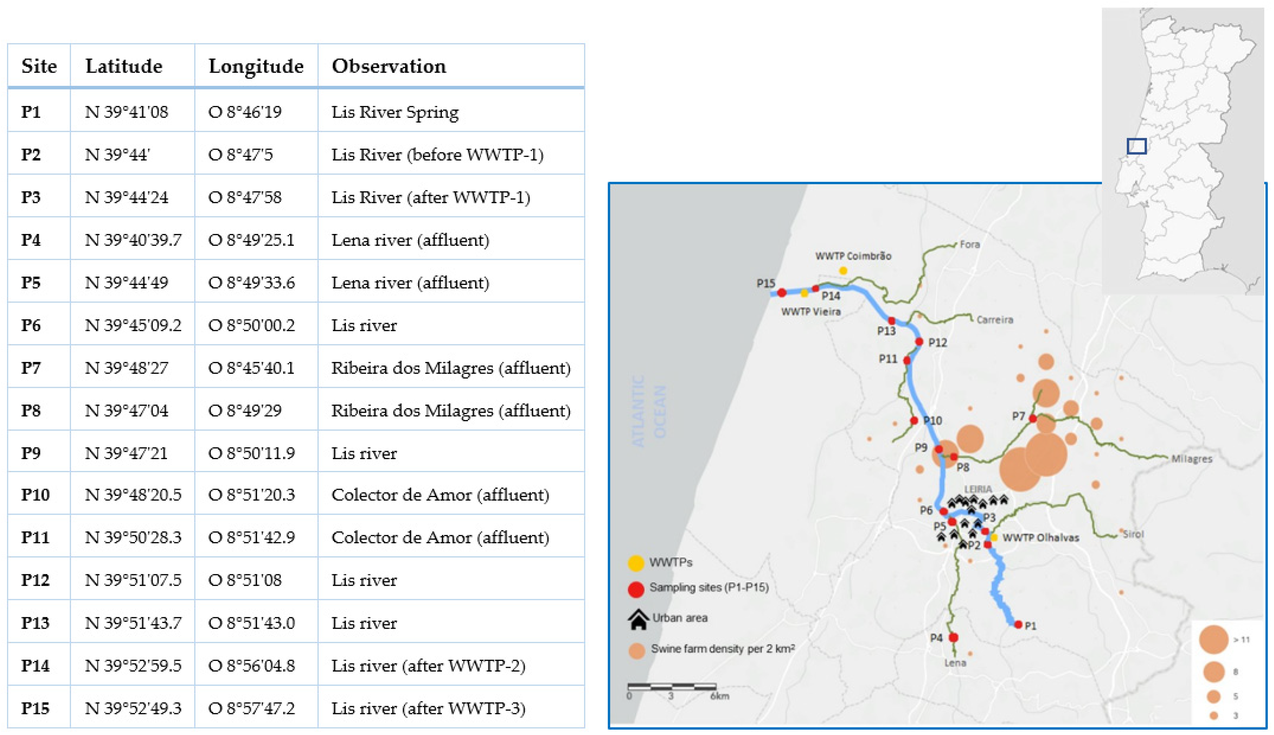
Figure 1. Sampling sites (P1–P15), coordinates, and other information and map showing sites (red dots) located in the Lis River main course (P1–P3, P6, P9, P12–P15) and tributaries (P4, P5, P7, P8, P10). WWTPs are indicated using yellow dots and the density of swine farms is in orange (from Teixeira et al., 2022 [39]).
Ijerph 19 11858 g001
Figure 2. Proportion of cefotaxime-resistant bacteria determined in all sampling sites located in the Lis River from spring (P1) to mouth (P15) (P1–P3, P6, P9, P12–P15; black) and three affluents (P4/P5, P7/P8, and P10/P11; light grey); Sites located downstream WWTP and in the vicinities of pig farms are indicated by * and #, respectively.
Ijerph 19 11858 g002
Figure 3. CTX-M encoding genes detected in the Lis riverine system. Black dots for Escherichia isolates, red for Klebsiella, and blue for Citrobacter or Enterobacter. Sites located downstream WWTP and in the vicinities of pig farms are indicated by * and #, respectively.
Ijerph 19 11858 g003
Figure 4. Genomic environments for the variants of the blaCTX-M gene found in the isolates. Ten different environments were observed, and they are represented from (AJ).
Ijerph 19 11858 g004
Table 1. Description of blaCTX-M carriers isolated in the Lis River concerning isolation year and location, identification, antibiotic susceptibility profiles (black—resistant, grey—intermediate resistance, white—susceptible), and blaCTX-M variant detected and genomic context.
AffiliationCampaign YearSiteStrain aAntibiotic Susceptibility b blaCTX-M variantcGenetic Environment of blaCTX-M
AMLAMCPRLTZPTICTIMATMCAZCTXFEPIPMCNCIPTECSXT ISEcp1IS26orf477IS903In Figure 3
Escherichia20181E1 nd+ +nd
1E2 * −1+ + A
5E3 * −32+ + H
5E4 −65+ +J
7E5 * −1+ + A
8E6 −1+ + A
8E7 * −15+ + D
9E8 * −65+ +J
9E9 nd+ + nd
11E10 * −14+ +C
13E11 nd+ + nd
13E12 −15+ + E
15E13 * −15+ + D
1E14 nd+ nd
1E15 −55+ + I
2E16 −65+ +J
3E17 −15+ + D
3E18 −15+ + D
5E19 * −32+ + H
6E20 −15+++ F
20192E21 −15+ + D
5E22 * −27+ +G
6E23 * −27+ +G
6E24 * −55+ + I
7E25 nd+ nd
7E26 −32+ + H
7E27 −15+ + D
8E28 −15+ + D
9E29 −27+ +G
15E30 −15+ + D
15E31 * −65+ +J
15E32 −1+ + A
Klebsiella20185K1 * −15+ + D
8K2 nd+ + nd
8K3 −15+ + D
9K4 −15+ + D
14K5 −15+ + D
14K6 * −15+ + D
20193K7 −15+ + D
3K8 * −15+ + D
3K9 nd+ + nd
3K10 −15+ + D
5K11 −15+ + D
6K12 nd+ + nd
9K13 nd+ + nd
9K14 nd+ + nd
12K15 −15+++ F
13K16 nd+ + nd
14K17 nd+ + nd
14K18 −15+ + D
Citrobacter201812C1 nd + nd
201912C2 * −3+ + B
12C3 * nd+ + nd
Enterobacter 13C4 * −32+ + H
a AML—amoxicillin, AMC—amoxicillin/clavulanic acid, PRL—piperacillin, TZP—piperacillin/tazobactam, TIC—ticarcillin, TIM—ticarcillin/clavulanic acid, ATM—aztreonam, CAZ—ceftazidime, CTX—cefotaxime, FEP—cefepime, IPM—imipenem, CN—gentamicin, CIP—ciprofloxacin, TE—tetracycline, C—chloramphenicol, SXT—sulfamethoxazole/trimethoprim; b strains used in conjugation assays are marked with *; c nd: not determined.
Table 2. Antibiotic susceptibility profiles of donor and transconjugant strains carrying blaCTX-M genes and recipient strain E. coli CV601 (black—resistant, grey—intermediate resistance, white—susceptible).
Antibiotic Susceptibility
Donor/Transconjugant Strains and blaCTX-M GeneAMLAMCPRLTZPTICTIMATMCAZCTXFEPIPMCNCIPTECSXT
Escherichia E3:: blaCTX-M-32E3D
E3T
Escherichia E5:: blaCTX-M-1E5
E5T
Escherichia E19:: blaCTX-M-32E19
E19T
Escherichia E23:: blaCTX-M-27E23
E23T
Escherichia E24:: blaCTX-M-55E24
E24T
Klebsiella K1:: blaCTX-M-15K1
K1T
Klebsiella K6:: blaCTX-M-15K6
K6T
Citrobacter C3:: blaCTX-M-ndC3
C3T
Citrobacter C4:: blaCTX-M-32C4
C4T
Recipient strain E. coli CV601-
AML—amoxicillin, AMC—amoxicillin/clavulanic acid, PRL—piperacillin, TZP—piperacillin/tazobactam, TIC—ticarcillin, TIM—ticarcillin/clavulanic acid, ATM—aztreonam, CAZ—ceftazidime, CTX—cefotaxime, FEP—cefepime, IPM—imipenem, CN—gentamicin, CIP—ciprofloxacin, TE—tetracycline, C—chloramphenicol, SXT—sulfamethoxazole/trimethoprim.
Publisher’s Note: MDPI stays neutral with regard to jurisdictional claims in published maps and institutional affiliations.