Review

The Impact of Training on the Loss of Cardiorespiratory Fitness in Aging Masters Endurance Athletes

by
Johannes Burtscher
1,2,
Barbara Strasser
3,
Martin Burtscher
4,* and
Gregoire P. Millet
1,2
1
Department of Biomedical Sciences, University of Lausanne, CH-1015 Lausanne, Switzerland
2
Institute of Sport Sciences, University of Lausanne, CH-1015 Lausanne, Switzerland
3
Medical Faculty, Sigmund Freud Private University, A-1020 Vienna, Austria
4
Department of Sport Science, University of Innsbruck, A-6020 Innsbruck, Austria
*
Author to whom correspondence should be addressed.
Int. J. Environ. Res. Public Health 2022, 19(17), 11050; https://doi.org/10.3390/ijerph191711050
Submission received: 2 August 2022 / Revised: 31 August 2022 / Accepted: 1 September 2022 / Published: 3 September 2022
(This article belongs to the Special Issue Exercise and Physical Fitness)

Abstract

:
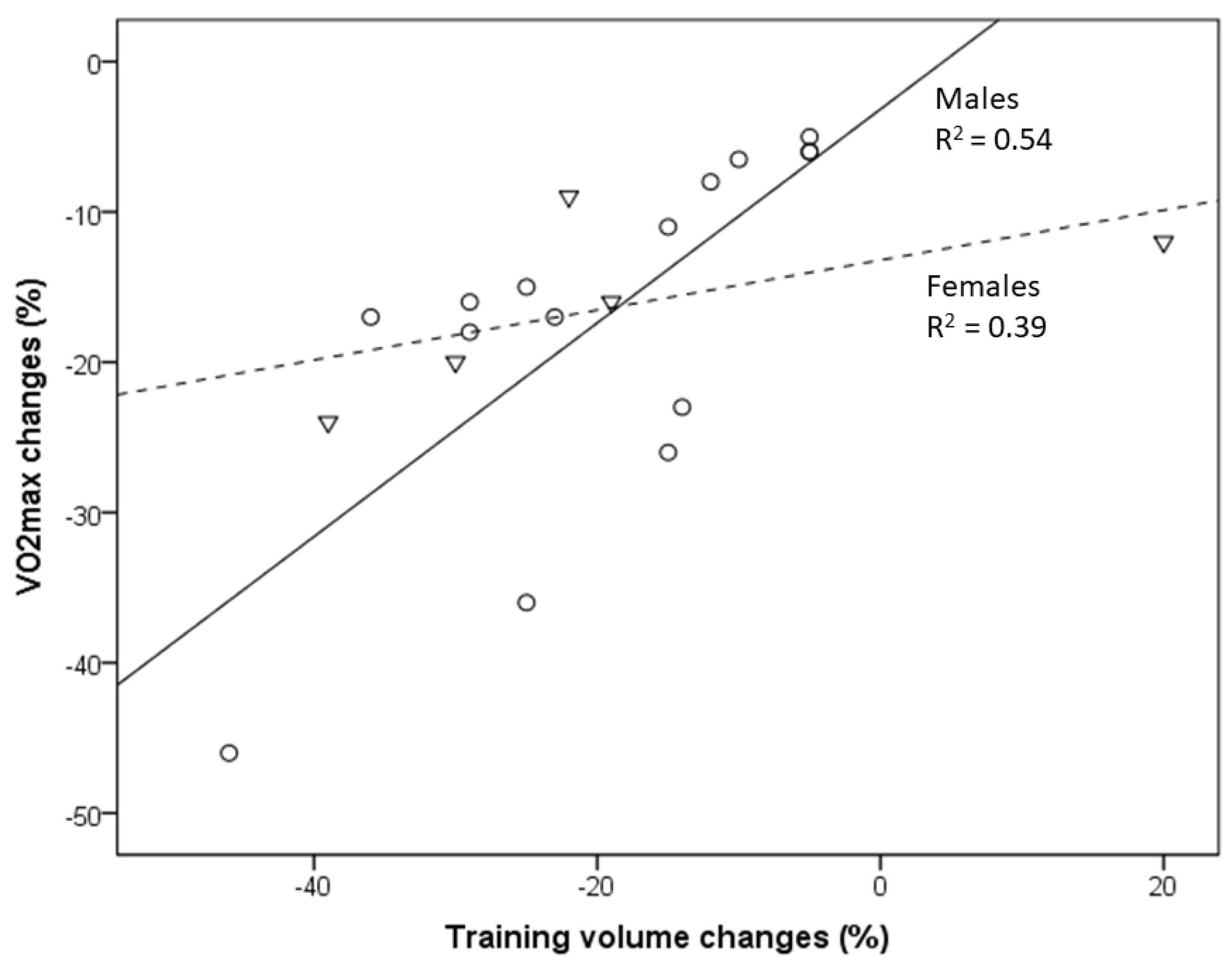
Elite masters endurance athletes are considered models of optimal healthy aging due to the maintenance of high cardiorespiratory fitness (CRF) until old age. Whereas a drop in VO2max in masters athletes has been broadly investigated, the modifying impact of training still remains a matter of debate. Longitudinal observations in masters endurance athletes demonstrated VO2max declines between −5% and −46% per decade that were closely related to changes in training volume. Here, using regression analyses, we show that 54% and 39% of the variance in observed VO2max decline in male and female athletes, respectively is explained by changes in training volume. An almost linear VO2max decrease was observed in studies on young and older athletes, as well as non-athletes, starting a few days after training cessation, with a decline of as much as −20% after 12 weeks. Besides a decline in stroke volume and cardiac output, training cessation was accompanied by considerable reductions in citrate synthase and succinate dehydrogenase activity (reduction in mitochondrial content and oxidative capacity). This reduction could largely be rescued within similar time periods of training (re)uptake. It is evident that training reduction or cessation leads to a considerably accelerated VO2max drop, as compared to the gradual aging-related VO2max decline, which can rapidly nullify many of the benefits of preceding long-term training efforts.

1. Introduction

Highly functional older individuals in general [1] and elite masters endurance athletes in particular are considered models of optimal healthy aging [2], which is characterized by the maintenance of a high level of cardiorespiratory fitness (CRF) in older age. CRF can be assessed by individual maximal aerobic power (VO2max) [3,4]. Ample evidence attests a close association between CRF and endurance exercise performance with longevity [5,6,7,8]. Conversely, a decline in CRF is related to reduced endurance performance and an elevated risk of morbidity and mortality [9,10]. A landmark study by Saltin and colleagues [11] convincingly demonstrated a rapid loss of CRF when healthy and physically active individuals became physically inactive (i.e., 20 days of bed rest). A relatively fast recovery and an increase in VO2max above baseline was observed during a subsequent 8-week training period [11]. Accordingly, high CRF, even if acquired and maintained through regular exercise training for many years (i.e., a typical condition for masters athletes), rapidly drops when training is ceased, e.g., due to injury or illness, jeopardizing the hard-earned exercise benefits [12]. Whereas the aging-related decline in VO2max has been comprehensively evaluated in general and endurance-trained populations [6,13,14], the importance of maintaining appropriate training loads and, in particular, the consequences of short- and long-term training reduction/cessation and those of training (re)uptake in masters endurance athletes have been much less investigated. However, regular and appropriate training stimuli are of utmost importance to improve or maintain muscular performance and CRF [11,12]. Thus, here, we aim to evaluate and summarize longitudinal observations on VO2max changes related to training variations of masters endurance athletes in the long and short term.

2. Materials and Methods

The present narrative review is reported following the IMRAD (introduction, methods, results, discussion) format [15]. A comprehensive search and screening strategy was used to identify relevant literature. PubMed and Web of Science were searched using combinations of terms covering the topics of training-related VO2max changes in masters endurance (primarily runners) athletes: (masters OR master OR endurance athletes OR elite runners) AND (VO2max OR aerobic power OR aerobic capacity) AND (humans) AND (aging OR older) AND (training). Studies that assessed VO2max changes longitudinally and reported associated changes in at least one parameter of training load were considered eligible for inclusion. We focused on studies that followed participants over several time points and/or evaluated participants of different age groups.
In addition, we performed a literature search on studies reporting effects of short-term (up to 12 weeks) training cessation or training (re)uptake (beginning with training or training reuptake after training cessation) on VO2max and associated physiological changes. Because such data on masters athletes are scarce, a representative range of studies including young and older healthy subjects (initial VO2max > 25 mL/min/kg) was considered, with a focus on studies that followed participants over several time points.
Data are presented descriptively. Multiple linear regression (stepwise variable selection) was applied to evaluate the predictive importance of independent variables with respect to VO2max changes. Training volume, age, sex, the observation period (in years) and baseline VO2max (at the beginning of the longitudinal observation) were considered as independent variables.

3. Results

Results of six long-term studies that satisfied our inclusion criteria on masters athletes reporting longitudinal changes in VO2max with aging and related changes of training characteristics are shown in Table 1.
The number of the predominantly male participants in the various age groups ranged between 6 and 34. The age of participants after follow-up varied between 46.5 and 82.8 years, and the observation periods ranged between 6 and 22 years (Table 1). The VO2max (mL/min/kg) decline per decade ranged from −5% to −46% per decade and was closely related to the changes in training volume (running, km/week) (Table 1, Figure 1).
Studies reporting training intensities (running, min/km) indicate that training volume reductions parallel intensity reductions, except one of the female groups of a study by Eskurza et al., who increased training volume but reduced training intensity during follow-up [16]. Male masters who maintained near-normal training volume (not more than 10% reduction or maintenance of high training volume) showed a VO2max decline of between −5% and −6.5% per decade [17,18,19,20,21].
Table 1. Longitudinal changes in VO2max with aging and related changes of training characteristics in masters endurance athletes. Positive values of the training intensity measure (min/km) indicate a decrease in intensity.
ReferenceN
m (Males)
f (Females)
Observation
Period (Years)
Age (Years)
Post
VO2max (mL/min/kg)
Pre vs. Post (% Change)
Change Per Decade
Training
V: Volume, km/Week
I: Intensity, min/km
Pre vs. Post (% Change)
Eskurza et al., 2002 [16]6 f661.045.2 ± 2.1 vs. 42.1 ± 2.1 (−7%)
−12%
V: 38.0 vs. 45.8 (+20)
I: 5.3 vs. 5.6 (+5%)
Eskuzra et al., 2002 [16]10 f656.050.0 ± 2.2 vs. 43.8 ± 2.2 (−12%)
−20%
V: 62.0 vs. 42.0 (−32%)
I: 5.3 vs. 5.6 (+5%)
Hawkins et al., 2001 [21]31 m953.558.7 ± 1.7 vs. 50.4 ± 1.5 (−14%)
−16%
V: 61.8 vs. 43.6 (−29%)
Hawkins et al., 2001 [21]34 m862.253.4 ± 1.4 vs. 46.2 ± 1.4 (−13%)
−17%
V: 56.2 vs. 43.3 (−23%)
Hawkins et al., 2001 [21]13 m971.146.2 ± 2.5 vs. 36.4 ± 2.6 (−21%)
−23%
V: 43.8 vs. 37.5 (−14%)
Hawkins et al., 2001 [21]8 m782.841.5 ± 3.1 vs. 28.4 ± 2.7 (−32%)
−46%
V: 49.4 vs. 26.7 (−46%)
Hawkins et al., 2001 [21]24 f851.248.7 ± 1.6 vs. 45.2 ± 1.2 (−7%)
−9%
V: 55.1 vs. 37.7 (−22%)
Hawkins et al., 2001 [21]16 f858.346.7 ± 1.3 vs. 40.8 ± 1.8 (−13%)
−16%
V: 39.4 vs. 31.8 (−19%)
Hawkins et al., 2001 [21]9 f873.239.4 ± 1.6 vs. 31.8 ± 2.8 (−19%)
−24%
V: 43.6 vs. 26.6 (−39%)
Pollock et al.
1997 [19]
9 m9.260.4 ± 8.555.4 ± 8.7 vs. 52.1 ± 6.8 (−6%)
−6.5%
V: 61 vs. 55 (−10%)
I: 4.9 vs. 5.2 (+6%)
Pollock et al.
1997 [19]
9m1070.4 ± 8.552.1 ± 6.8 vs. 43.2 ± 6.3 (−17%)
−17%
V: 55 vs. 35 (−36%)
I: 5.2 vs. 5.9 (+14%)
Pollock et al.
1997 [19]
10 m1059.5 ± 10.354.2 ± 7.7 vs. 50.0 ± 6.9 (−8%)
−8%
V: 49 vs. 38 (−12%)
I: 4.9 vs. 5.4 (+10%)
Pollock et al.
1997 [19]
10 m1069.8 ± 10.250.0 ± 6.9 vs. 40.8 ± 9.5 (−18%)
−18%
V: 38 vs. 27 (−29%)
I: 5.4 vs. 6.3 (+17%)
Katzel et al.
2001 [20]
7 m8.77051.3 ± 2.4 vs. 48.6 ± 1.8 (−5%)
−6%
highly trained
(no essential change)
Katzel et al.
2001 [20]
21 m8.77149.8 ± 1.1 vs. 38.2 ± 0.9 (−23%)
−26%
moderately trained (volume and intensity reduction)
Katzel et al.
2001 [20]
12 m8.77449.4 ± 2.2 vs. 33.8 ± 1.8 (−32%)
−36%
not trained (rather sedentary)
Rogers et al.
1990 [18]
15 m86254.0 ± 1.7 vs. 51.8 ± 1.8 (−4%)
−5%
highly trained
Trappe et al. 1996 [17]10 m2246.568.8 vs. 59.2 (−14%)
−6%
highly trained
Trappe et al.
1996 [17]
18 m2246.564.1 vs. 48.9 (−24%)
−11%
moderately trained
Trappe et al. 1996 [17]15 m2246.570.7 vs. 46.7 (−34%)
−15%
not trained
Those who reduced training volume by between 11% and 20% or were moderately trained showed a VO2max decline of between −8% and −26% per decade [17,18,19,20,21], and those with training volume reductions of more than 20% or who became almost sedentary had a VO2max decline of between −15% and −46% per decade [17,18,19,20,21]. Eskurza et al. and Hawkins et al. reported data on female athletes, also indicating a training-dependent loss of VO2max [16,21]. Regression analysis including male athletes [17,18,19,20,21] revealed a close association between reported VO2max reductions and related changes in training volumes with aging. Fifty-four percent of the variance in the observed VO2max decline was explained by training-volume changes (Figure 1), and this percentage increased to 70% when the age of the athletes was considered. No other variables improved the explanation of the VO2max decline. Within the groups of females [16,21], 39% of the variance in VO2max change was explained by changes in the training volume (Figure 1).
Figure 1. Relationship between VO2max decline and the reduction in training volume with aging of masters athletes (from data presented in Table 1) [16,17,18,19,20,21]. Circles indicate males, and triangles indicate females. Changes in the training volume explain 54% and 39% of the variance in VO2max changes in male and female masters athletes, respectively.
Ijerph 19 11050 g001
Few data on training cessation or training (re)uptake responses (physiological characteristics) in older masters are available. Thus, results of studies on young and older endurance athletes and healthy individuals reporting short-term changes in VO2max and related physiological changes due to training cessation or training (re)uptake were considered for this analysis (Table 2).
Despite some variations, an almost linear decrease in VO2max up to about −20% from a few days to 12 weeks of training cessation was demonstrated [22,23,24,25,28,29,30,32,34,35,36,38] (Table 2 and Figure 2). Based on the few studies on females [24,25], no important sex difference could be derived. However, when training cessation is associated with bed rest, a marked decline in the VO2max (e.g., −16.5%) may occur after only 3 days, which seems to be more pronounced in well-trained compared to rather sedentary individuals [37]. With training (re)uptake, VO2max again increases considerably in healthy young and older individuals, but this increase was larger during a 12-week training period in older male subjects [31] and one female masters athlete [12] (29.3% and 31.2%) when compared to young individuals (15.4%) (Table 2 and Figure 2). The observed decrease in VO2max was primarily associated with a reduction in cardiac output (decrease in stroke volume with even slight increases in maximal heart rate), especially shortly after training cessation, but a diminished arteriovenous oxygen difference became increasingly important with increasing duration of training cessation (Table 2 and Figure 2). In addition, training cessation was accompanied by relatively large and increasing reductions in citrate synthase and succinate dehydrogenase activity (up to about −40%) in 12 weeks. Citrate synthase is a commonly used marker for mitochondrial content and succinate dehydrogenase activity, providing information about the capacity of mitochondrial complex II, a protein complex contributing to oxidative phosphorylation, as well as ATP production and cellular energy supply. A reduction in these two markers indicates a reduced cellular aerobic capacity based on reduced mitochondrial content and/or efficiency. With training (re)uptake in older individuals, continuously increasing cardiac output with training duration contributed more importantly to the VO2max improvement than the arteriovenous–oxygen differences (Table 2). This is in contrast to young individuals, who showed a larger improvement of the arteriovenous–oxygen difference, especially during the first weeks of training (Table 2).

4. Discussion

The presented findings from longitudinal studies highlight the importance of continuing training activities with respect to VO2max changes of masters athletes in the long and short term. Based on studies including young and older athletes, as well as non-athletes, a considerable percentage of age-related VO2max decline (per decade) seems to occur rapidly (e.g., within 12 weeks) after training cessation. However, this effect can be largely restored by appropriate training (re)uptake. Changes in cardiac output (more importantly) and arteriovenous oxygen difference accompanied by reduced levels and activity of mitochondrial enzymes (e.g., citrate synthase and succinate dehydrogenase) are associated with training cessation or training (re)uptake interventions.

4.1. VO2max Decline in the Long Term: Effects of Training in Masters Athletes

Masters endurance athletes represent an excellent model of healthy aging that is relevant for the evaluation of consequences of changing in training habit on VO2max. Although VO2max seems to inexorably decrease with aging, even in life-long runners, the potential modulatory capacity of short- and long-term training variations remains a matter of debate. In healthy sedentary adults of both sexes, VO2max declines by about 10% per decade after the age of 25–30 years and slightly more at older ages (e.g., older than 60–70 years). Overall, this rate seems not to differ much in endurance athletes [6]. Longitudinal data from a healthy population indicate an exacerbated VO2max reduction with aging; whereas the reduction was about 8% in the fourth decade (over 10 years), it amounted 23% in the seventh decade and was more pronounced in males than females [13]. Whereas higher physical activity levels were associated with higher VO2max values across all ages and in both sexes, physical activity (quartiles) did not change the slope of the VO2max drop [13]. However, this may not hold true for (at least male) masters endurance athletes, who are able to maintain high training volumes (and likely intensity) until old age (Table 1, Figure 1). The presented data derived from longitudinal studies are in line with cross-sectional studies [39]. Cross-sectional data from 203 men (male endurance athletes and untrained subjects, 20–90 years) indicate that the weekly training volume constitutes a significant positive predictor of age-related changes in aerobic capacity [40]. Other cross-sectional data of masters athletes (20 power and 19 endurance athletes, 37–90 years) recently reported a peak aerobic and anaerobic power decline of around 7–14% per decade, with no difference between athletic disciplines or sexes [41]. Because exercise tests were stopped when heart rates exceeded the age-predicted maximal heart rate by 10 bpm and due to the lack of information on training characteristics, assessments of potential training effects on the observed VO2max decline are not possible [41]. Similarly, another cross-sectional study suggests that active older men (n = 146) lose maximal aerobic power at a rate of about 12%, and active older women (n = 82) lose maximal aerobic power at a rate of about 8% per decade between the ages of 40 and 80 years [42]. However, the VO2max decline becomes considerably steeper in masters athletes older than 80 years old, even in the absence of major illnesses or orthopedic issues [43]. Again, aging and training effects cannot be readily disentangled in that study due to the large variations regarding years of training (15.2 ± 9.7) and weekly training milage (33.5 ±19.3) [42].
In a comparison of American road running events, it was recently shown that the sex gap in performance decreases with increased age [44]. In the present study, the association between VO2max decline and changes in training volume (and intensity) is less pronounced in female compared to male endurance athletes [16,21]. This may partly be related to the lack of studies evaluating female athletes but also to the outlier of one female group studied by Eskurza et al. [16]. Nevertheless, a somewhat lower trainability of aerobic capacity in women may contribute to those differences [45].
Longitudinal changes in VO2max in cross-country skiers (age 58.7 ± 2.3 years) who continuously competed and reported unchanged training patterns amounted to only −4.1 ± 3.7 % per decade [46], which fits well with longitudinal data from male masters athletes (runners) presented in the present study, with a decrease in VO2max of between −5% and −6.5% per decade shown in those who continued to engage in regular vigorous endurance exercise, training and competitions [17,18,19,20]. The exaggerated VO2max decline in masters athletes who reduced training volumes (Figure 1) can be readily explained from both training scientific and practical perspectives [47,48,49]. Training characteristics (e.g., training volume, intensity and frequency) are undisputedly important modifiers of VO2max in both young and older athletes [50]. Reductions in volume and/or intensity of exercise training and the resulting decline in VO2max and performance in aging endurance athletes may be caused by various factors, such as decreasing motivation to train and/or compete, musculoskeletal injuries, pain and/or aging-related diseases [2,6,51,52,53], sometimes even associated with excessive training and competition activities [54]. Consequently, the aging-related CRF decrease, especially in male masters athletes, will remain small in those who are able to avoid factors provoking considerable training reductions. In summary, male masters athletes who can maintain their training volume (and intensity) can minimize their VO2max decline to about −5% to −6.5% per decade [17,18,19,20], but those values increase to up to −26% in those who reduce their training to a moderate level and can even be as high as −46% per decade in those who become sedentary [17,18,19,20,21].

4.2. Is There a Potential Role for Body Composition Explaining the VO2max Decline in Masters Athletes?

The effects of regular exercise are closely related to body composition and are therefore difficult to disentangle. Aging-related training reductions or cessation are often associated with changes in body composition. Longitudinal and cross-sectional data [55] indicate a significant contribution of lean body mass (LBM) alterations to VO2max changes, an effect that seems to be more pronounced in males than in females. Accordingly, a recent study demonstrated that lifelong aerobic exercise attenuated the age-related loss of quadricep muscle volume by about 50% in men but not in women, whereas adipose tissue infiltration into muscle was attenuated by about 50% in both sexes [56]. Higher training intensity throughout life provided increased protection against adipose tissue infiltration into muscle. The aerobic qualities of skeletal muscle among these lifelong exercisers likely contributed to the strong muscle mass-to-VO2max relationship. In line with these finding, no association between appendicular muscle mass and age was found in highly active older adults of both sexes [57]. The significant difference in muscle mass between exercising and sedentary individuals clearly illustrates that levels of physical activity play a considerable role in the phenotype of aging muscle. A recent study provided novel data on body composition in masters track and field athletes [58]. The authors concluded that loss of skeletal muscle mass and changes in bioimpedance phase angle (a raw parameter of cellular function and an indicator of tissue hydration and nutritional status [59]) are important contributors to the age-related reduction in anaerobic power, even in athletes who maintain high levels of physical activity in old age, indicating a deterioration of muscle quality in old age. However, it is also possible that a favorable body composition and nutritional conditions are prerequisites to participate in masters sport. Therefore, future research employing longitudinal study designs is needed, with the aim of determining which of these measures alone or in combination optimally predict individuals’ fitness and general health markers.

4.3. VO2max Decline in the Short Term: Effects of Training Cessation and Training (Re)Uptake

The VO2max decline in endurance-trained individuals usually does not occur gradually over decades but rather consists of a “slow component” related to aging per se and a “rapid component” due to training reduction/cessation. As demonstrated in the presented results, an almost linear VO2max decrease or increase was observed during a 12-week period of training cessation or training (re)uptake, respectively, in healthy young and older individuals [12,24,28,31]. Compared to the −5% to −6.5% VO2max decline within a decade in male masters athletes [17,18,19,20], VO2max steeply decreased as a result of training cessation, even in young endurance-trained (the majority of participants included in this study were male) athletes by −7% after only 12 days and dropped further to −18% after 12 weeks of training cessation [22]. Similar observations have been published for young competitive female runners, for whom 5 days of training cessation did not yield significant changes, whereas 10 days resulted in a 7.6% drop, which was linked to reduced running performance after 15 days [24].
However, VO2max rapidly improves after a few weeks of training (re)uptake after cessation or when training starts from a sedentary status. VO2max improvements occurred more rapidly during the first 3 weeks (12% vs. 8.4%) in young compared to older healthy men, but after 12 weeks, the increases were even higher in the older compared to younger men (15.4% vs. 29.3%) [31], at least in the selected studies considered here (Figure 2). A 31% VO2max increase was reported in a female masters athlete after only 4 weeks of training (re)uptake following trauma-related training cessation [12]. A meta-analysis demonstrated a 16.3% VO2max improvement in previously sedentary older adults (>60 years) of both sexes after taking up an aerobic exercise training program; the effects increased with longer training duration (i.e., >16–20 weeks) [60]. VO2max increases of 9–13% were observed after 8 weeks of high-intensity interval training (HIIT) in men and women of a broad age range (20 to 70+), without significant differences between age groups [61]. These findings should encourage masters athletes who become sedentary for any reason to take up training again as soon as possible. In summary, a VO2max loss corresponding to aging-related decline of at least one aging decade can be restored by appropriate training (re)uptake within a couple of weeks.

4.4. Aging-Related VO2max Loss despite Maintenance of High Training Volume and Intensity: The Slow Component

The age-related decline in endurance performance and its physiological determinants are largely mediated by a reduction in the volume and intensity of training commonly observed in masters endurance athletes [47]. However, as demonstrated, a VO2max loss of between −5% and −6.5% per decade occurred even in those who continued to engage in regular vigorous endurance exercise training and competitions [17,18,19,20]. The question arises as to which aging effects that are largely independent of training habits are responsible for this decline. In contrast to the decrease in lung function [62], both cross-sectional and longitudinal studies indicate that the decreases in maximal heart rate and stroke volume are the major contributors to the observed decline in VO2max in masters endurance athletes [40,47,48,63,64]. Alteration of the intrinsic heart rate may primarily account for the reduction in maximal heart rate, but diminished chronotropic responsiveness to β-adrenergic stimulation probably adds to this reduction [8,65]. In addition, lower ejection fractions at maximal exercise volume have been reported in both older sedentary and older endurance-trained athletes compared with their young peers, which is apparently related to reduced β-adrenergic responsiveness [66]. Whereas left ventricular filling pressure and arteriovenous oxygen difference remain almost unchanged at peak exercise, chronotropic and inotropic reserve (and probably Frank–Starling reserve) seem to decline even with healthy aging [67]. Whereas longitudinal and radial contributions to stroke volume do not seem to differ between groups of varying ages and training status, differences in longitudinal pumping were observed between young sedentary and old highly trained athletes compared to old sedentary individuals; the incidence of left ventricular atrioventricular plane displacement was higher in the former groups [68]. Moreover, slight macrovascular and microvascular dysfunctions accompanied by stiffening of central elastic arteries and impaired peripheral endothelial function are aging-related effects that negatively affect blood flow and oxygen delivery to exercising skeletal muscles [69]. Although endothelial function may be preserved in the leg muscles of life-long physically active people due to elevated systemic nitric oxide bioavailability, this did not restore the aging-related decline in hyperemic response [70,71]. No or only negligible impairments were reported in masters athletes with regard to muscle fiber morphology and capillary supply of the muscle tissue [72].

4.5. Aging-Related VO2max Decrease or Increase with Training Cessation or (Re)Uptake: The Fast Component

Age-related VO2max decline is determined by multiple factors, including reductions in blood volume, maximal heart rate and cardiac output; increased stiffening of the arterial walls; exercise-induced arterial hypoxemia; and diminished peripheral oxygen extraction. However, both loss and improvement of VO2max due to recent changes in exercise training seem to be primary consequences of changes (decrease or increase, respectively) in maximal cardiac output [11,22,31]. In a study on young male athletes, within the first 3 weeks, training cessation elicited a rapid response that involved a decrease in oxygen delivery due to reduced maximal cardiac output, followed by an increasing contribution of elevated arteriovenous oxygen difference [22] (Table 2). Maximal cardiac output seems to largely be a consequence of the reduction in plasma volume related limiting ventricular filling and maximal stroke volume [28,73]. The reduced stroke volume is incompletely compensated by a slight increase in maximal heart rate [28,73]. Whereas the arteriovenous oxygen difference may more rapidly recover in young and healthy males during training (re)uptake, increasing maximal cardiac output seems to contribute more importantly in older males [31]. Short-term training in older men and women results in rapid elevation of plasma volume and associated increases in cardiac output and VO2max [74]. This expansion of plasma volume following short-term exercise training (e.g., 2–4 weeks) has been documented in both cross-sectional and longitudinal studies [75,76]. Thermal and non-thermal components have been suggested to contribute to the elevation of plasma levels of electrolytes and proteins [75,76].
As shown in the study by Coyle et al., muscle capillarization did not markedly change over the 12-week training cessation (but remained higher than in the sedentary subjects); thus, the authors suggested that the decreasing arteriovenous oxygen difference could be attributed to a loss of mitochondria and/or mitochondrial function [22]. However, as muscle mitochondrial respiration is submaximal at VO2max, this likely is not the cause of VO2max impairment after short-term training cessation [77,78]. Calculation of the arteriovenous oxygen difference using the Fick equation suggests its reduction may result from elevated mixed venous oxygen content caused by distribution deficits of blood flow, i.e., reduced blood flow within working skeletal muscles. Skeletal muscle blood flow is diminished during dynamic exercise in older and rather sedentary individuals, resulting from a reduced vascular conductance (impaired functional sympatholysis), but improves with exercise training [79,80]. A demonstrated improvement in sympatholysis resulting from short-term exercise training was modulated by training intensity and was mediated by a nitric-oxide-dependent mechanism [81]. Moreover, elevated sympathetic vasoconstrictor responsiveness during exercise was demonstrated in older men but blunted leg vasodilator responsiveness in older women [80]. As indicated by prospective training studies, muscle sympathetic nerve activity is reduced after exercise training [82].
Thus, changes in vascular conductance due to short-term training cessation and training (re)uptake may be involved in the difference in arteriovenous oxygen response to VO2max changes, whereas long-term training reduction/cessation may lead to more profound remodeling of capillarization and the loss of mitochondrial content and/or efficiency.
Angiogenesis occurs as a consequence of appropriate exercise stimuli in order to increase oxygen diffusion and to improve the removal of metabolites within the contracting muscles [83]. Aging-related changes in the ultrastructure of the endothelium and the associated impairment of microcirculation are associated with a reduced CRF, but the adaptability of microcirculation to exercise stimuli seems to be maintained in old age [84,85]. Aging-related mitochondrial dysfunctions [86] and associated oxidative stress [87] are well-established hallmarks of aging. Conversely, exercise has been shown to preserve mitochondrial health in skeletal muscle [88,89,90] and slow down aging-related deterioration of antioxidative defense systems [91].
Despite the scarcity of longitudinal studies on the CRF of masters athlete, the available data indicate that reduced oxygen delivery to working muscles, mainly due to diminished cardiac output (and possibly also due to maldistribution of cardiac output), plays a major role until late middle age, and a decline in skeletal muscle oxidative capacity, at least partly due to mitochondrial dysfunction, may become increasingly important in older age (i.e., above 70 years) [92].
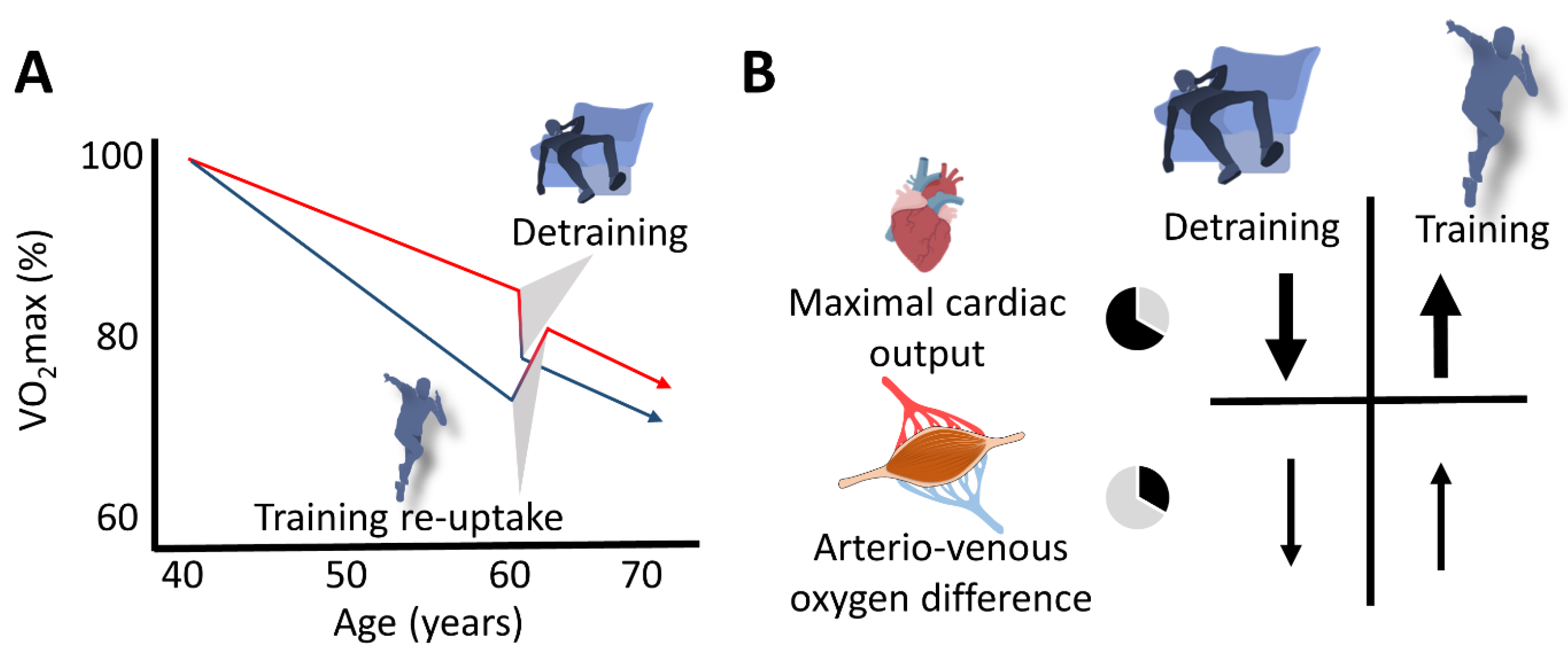
Fast (training cessation or (re)uptake) and slow (aging per se) components potentially modulating the VO2max decline in masters endurance athletes are schematically depicted in Figure 3.

5. How to Support the Maintenance of Sufficient Training Stimuli in Aging Masters Athletes

Training characteristics (e.g., training volume, intensity and frequency) are undisputedly important modifiers of VO2max in both young and older athletes. The ability to maintain a high level of exercise-training stimulus with aging seems to represent the most important measure with respect to limiting the rate of decline in VO2max and related endurance performance [93]. Training cessation and the associated CRF decrease may often be sequelae of disease, injuries [94] and/or motivational changes across the athletic lifespan [95]. Here, we do not focus on these aspects but solely consider physiological consequences of training cessation or training (re)uptake with respect to the aging-related CRF decline.
The decline in VO2max is considerably mediated by a reduction in exercise “stimuli”. Exercise stimuli are characterized by several components, including exercise-training intensity, session duration and training frequency. Several suggestions regarding effective training methods specifically for masters runners have been published [96]. Similar to younger endurance athletes [97], a combination of continuous moderate-intensity training (MIT) and high-intensity interval training (HIIT) generally appears to be efficient for masters runners. Running energy cost is largely determined by peripheral adaptations (capillarization and mitochondrial density) mainly induced by training volume/mileage at low intensity [98,99], which may explain why masters runners with several years of practice are known to be more economical than their younger counterparts [100,101]. The rate of capillarization induced by the same training program appears similar between young (22 years) and old (69 years) individuals [102]. Therefore, such low-intensity training may be less important for masters. In contrast, HIIT is associated with a well-established elevation of activity of several mitochondrial enzymes, mitochondrial biogenesis and increased levels of respiratory control [103], as first demonstrated by the pioneering work of Holloszy [104]. Various types of high-intensity training (e.g., HIIT and sprint interval training (SIT)) have since been linked to more specific mitochondrial adaptations, including changes in the phosphorylation state of mitochondria-related signaling proteins (CaMKII or AMPK), alterations in gene expression of regulators of mitochondrial function and biogenesis (e.g., PPAR; PGC-1α) or mitochondrial protein synthesis rates [105]. Moreover, the activity of numerous mitochondrial enzymes (including the mentioned citrate synthase and succinate dehydrogenase) and oxidative phosphorylation capacity are likely enhanced.
Overall, it appears that masters profit most from performing a majority of their training at high intensity (e.g., at severe or supramaximal intensities) when compared to their younger counterparts. However, this assumption can be challenged for several reasons. First, high training volume at low intensity is necessary to maintain the benefits induced by HIIT [106], with an excessive amount of HIIT sessions possibly being detrimental for mitochondrial biogenesis [107]. Secondly, the “classical” type of HIIT (e.g., 2–3 min intervals at 100% vVO2max) may not be optimal for older individuals due to slower VO2 kinetics in association with aging [108]. Whereas the time constant of the primary phase for exercise at moderate intensity averaged 50–70 s in healthy old (age 60–80 years) individuals, it amounted to only ∼30 s in young (20–30 years) healthy populations [109]. Slower muscle oxygen delivery with aging has direct consequences with respect to the prescription of HIIT, as the speed of VO2 kinetics is important to determine the interval duration (i.e., fast kinetics allow for short intervals, whereas slower kinetics require longer work intervals) [110]. Masters athletes are recommended to perform HIIT with longer interval durations (>5 min in the severe-intensity domain, i.e., ~90% vVO2max) than for younger athletes (∼3 min at vVO2max). Thirdly, intermittent submaximal (e.g., 15 s runs with hard bouts at 90 or 100% of vVO2max and an average velocity equal to 85%) sessions of varying amplitudes were also effective in masters runners, as the duration spent at VO2max was up to 14 min [111]. This type of session leads to considerable stimulation of the cardiovascular system combined with velocities sustainable by older individuals but high enough to stimulate neuromuscular functions. Fourthly, the need for appropriate training stimuli to counteract age-induced loss of muscle mass and strength (i.e., sarcopenia) is well documented [112]. There is no doubt that exercise should be considered a cornerstone in the treatment of such skeletal muscle wasting. Beyond the clear benefits of resistance exercise training, which significantly improves muscle mass and strength in older persons, aerobic low-intensity exercise training attenuates sarcopenia (and cachexia) [113]. Finally, the risk of increased injuries due to high-intensity running during HIIT is not negligible [114], particularly in older runners with higher rates of knee cartilage damage [115].
Altogether, reductions in volume and/or intensity of exercise training and the resulting decline in VO2max and performance in aging endurance athletes can be explained by various factors, such as decreasing motivation to train and/or compete, musculoskeletal injuries, pain and/or aging-related diseases. Appropriate training modifications and specific psychological and medical support are important tools to keep training breaks reasonably short and avoid substantial decreases in VO2max and performance. For instance, considerable reductions in pre-competition training volume for 2–3 weeks (tapering) are known to not compromise VO2max and even improve performance. With regard to regular training intensities, a combination of high-volume training at low intensity (≥75% of overall training volume) with low-volume training at threshold and high-intensity interval training (≤20%) appears to be an appropriate method to optimize endurance training adaptations in most middle- and long-distance runners [116].
In summary, the controversy with respect to volume versus intensity components [117,118] of exercise training is also of interest for older athletes. Despite their age-related decline in VO2max relative to economy, we believe that the main principles of “polarized training” remain valid in masters athletes. However, some adaptations are required (e.g., submaximal short intermittent and/or long-interval HIIT or the addition of resistance training). Furthermore, innovative training methods based on the use of systemic [119] or localized (i.e., blood flow restriction; BFR) hypoxia are relevant complements to “traditional” exercise training in older athletes. Such approaches represent potential therapeutic solutions to attain exercise benefits; for example, continuous hypoxic training induces similar cardiovascular adaptations with lower walking velocity and mechanical load compared to similar exercises in normoxia [120,121]. BFR counteracts sarcopenia and maintains muscle mass/strength with lower loads [122]. These hypoxic methods are therefore particularly well-suited for load-compromised individuals, for example, masters endurance athletes recovering from injury [123].
Finally, delayed alterations in body composition and healthy dietary choices may crucially contribute to the impressive performance of masters athletes. A reduced prevalence of sarcopenia and superior appendicular skeletal muscle mass in old age was observed in former Tokyo 1964 Olympic athletes, especially among those who continued their exercise habits with high exercise intensity [124]. In addition, the maintenance of sufficient energy availability (>30 kcal/kg LBM/day) and the ingestion of more protein-based foods (≥30 g per meal) is required to prevent the development of sarcopenia in aging athletes. This is particularly important during periods of increased training volume or, vice versa, during periods of immobilization. However, protein ingestion not only supports muscle protein synthesis but also provides amino acids, which are essential for an athlete’s recovery and adaptation. The main finding of a recent study of masters marathon runners was that protein intake per LBM, both during the tapering period and the race, was highly related to race performance, acute race-induced changes in body composition (especially of LBM) and selected metabolic and muscle-damage-related blood markers [125]. Thus, it can be assumed that the effect of protein intake during the preparation period and the race may be of even greater importance in aging endurance athletes than the amount of carbohydrate intake. Although there is little evidence that masters athletes metabolize dietary protein differently than younger athletes, endurance athletes should focus on a balanced distribution of moderate protein-containing, nutrient-dense meals throughout the day and consume as much as 0.5 g/kg immediately after exercise to replenish amino acid oxidative losses. [126]

6. Limitations

At least three major limitations have to be considered with regard to this review: (1) the number of longitudinal studies with precise details on training characteristics was low, (2) female masters athletes were scarcely studied and (3) mechanisms responsible for VO2max changes in masters athletes of both sexes remain to be elucidated. Furthermore, findings on the (re)uptake of training are predominantly derived from non-masters athletes.

7. Conclusions

Training reduction or cessation leads to an accelerated VO2max decline, as compared to the gradual aging-related VO2max decrease. This can rapidly nullify many of the benefits of preceding long-term training efforts. Conversely, resuming exercise training has the potential to quickly restore the entire or at least parts of the lost VO2max, exercise performance and health status. Interesting case studies are available to support the assumption that regular training or a return to exercise are effective for maintaining a high level of cardiovascular fitness. For example, an elite marathon runner (ex-Olympian athlete who retired from running at 32 years old with a best performance of 2:13:59 and retired from running for a 16-year period) who ran a marathon in 2:30:15 at the age of 59 [127]. Another such examples is a 70-year-old male runner with a marathon performance of 2:54:23 [128].
Despite the pronounced effects of training cessation and reuptake in the general population and masters athletes, as well as the substantial public health consequences, longitudinal studies are scarce, and the mechanistic underpinnings are largely unexplored. Future well-designed studies evaluating the time course of CRF alterations related to various training modifications and associated changes in risks and benefits with respect to the health of aging endurance athletes are urgently needed and will provide information with respect to how to expand health spans of aging athletes and non-athlete individuals.

Author Contributions

Conceptualization, M.B. and J.B.; literature review and discussion, J.B., B.S., M.B. and G.P.M.; writing and proofreading, J.B., B.S. and M.B. All authors have read and agreed to the published version of the manuscript.

Funding

This research received no external funding.

Institutional Review Board Statement

Not applicable.

Informed Consent Statement

Not applicable.

Data Availability Statement

Not applicable.

Conflicts of Interest

The authors declare no conflict of interest.

References

  1. Calbet, J.A.L. The biological and psychosocial aspects of successful aging in high functional elders: A longitudinal study. Scand. J. Med. Sci. Sports 2019, 29 (Suppl. S1), 5–6. [Google Scholar] [CrossRef] [PubMed]
  2. Tanaka, H. Aging of Competitive Athletes. Gerontology 2017, 63, 488–494. [Google Scholar] [CrossRef] [PubMed]
  3. Burtscher, J.; Ruedl, G.; Posch, M.; Greier, K.; Burtscher, M. The upper limit of cardiorespiratory fitness associated with longevity: An update. AIMS Public Health 2019, 6, 225–228. [Google Scholar] [CrossRef]
  4. Valenzuela, P.L.; Maffiuletti, N.A.; Joyner, M.J.; Lucia, A.; Lepers, R. Lifelong Endurance Exercise as a Countermeasure Against Age-Related V˙O2max Decline: Physiological Overview and Insights from Masters Athletes. Sports Med. 2020, 50, 703–716. [Google Scholar] [CrossRef]
  5. Burtscher, J.; Burtscher, M. Run for your life: Tweaking the weekly physical activity volume for longevity. Br. J. Sports Med. 2019, 54, 759–760. [Google Scholar] [CrossRef]
  6. Tanaka, H.; Seals, D.R. Endurance exercise performance in Masters athletes: Age-associated changes and underlying physiological mechanisms. J. Physiol. 2008, 586, 55–63. [Google Scholar] [CrossRef] [PubMed]
  7. Clausen, J.S.; Marott, J.L.; Holtermann, A.; Gyntelberg, F.; Jensen, M.T. Midlife Cardiorespiratory Fitness and the Long-Term Risk of Mortality: 46 Years of Follow-Up. J. Am. Coll. Cardiol. 2018, 72, 987–995. [Google Scholar] [CrossRef]
  8. Strasser, B.; Burtscher, M. Survival of the fittest VO2 max, a key predictor of longevity. Front. Biosci. 2018, 23, 1505–1516. [Google Scholar] [CrossRef]
  9. Lazarus, N.R.; Harridge, S.D.R. Declining performance of master athletes: Silhouettes of the trajectory of healthy human ageing? J. Physiol. 2017, 595, 2941–2948. [Google Scholar] [CrossRef]
  10. Kondamudi, N.; Mehta, A.; Thangada, N.D.; Pandey, A. Physical Activity and Cardiorespiratory Fitness: Vital Signs for Cardiovascular Risk Assessment. Curr. Cardiol. Rep. 2021, 23, 172. [Google Scholar] [CrossRef]
  11. Saltin, B.; Blomqvist, G.; Mitchell, J.H.; Johnson, R.L.; Wildenthal, K.; Chapman, C.B. Response to exercise after bed rest and after training. Circulation 1968, 38, VII1–VII78. [Google Scholar] [PubMed]
  12. Nichols, J.F.; Robinson, D.; Douglass, D.; Anthony, J. Retraining of a competitive master athlete following traumatic injury: A case study. Med. Sci. Sports Exerc. 2000, 32, 1037–1042. [Google Scholar] [CrossRef] [PubMed]
  13. Fleg, J.L.; Morrell, C.H.; Bos, A.G.; Brant, L.J.; Talbot, L.A.; Wright, J.G.; Lakatta, E.G. Accelerated Longitudinal Decline of Aerobic Capacity in Healthy Older Adults. Circulation 2005, 112, 674–682. [Google Scholar] [CrossRef] [PubMed]
  14. Pimentel, A.E.; Gentile, C.L.; Tanaka, H.; Seals, D.R.; Gates, P.E. Greater rate of decline in maximal aerobic capacity with age in endurance-trained than in sedentary men. J. Appl. Physiol. 2003, 94, 2406–2413. [Google Scholar] [CrossRef]
  15. Ferrari, R. Writing narrative style literature reviews. Med. Writ. 2015, 24, 230–235. [Google Scholar] [CrossRef]
  16. Eskurza, I.; Donato, A.J.; Moreau, K.L.; Seals, D.R.; Tanaka, H. Changes in maximal aerobic capacity with age in endurance-trained women: 7-yr follow-up. J. Appl. Physiol. 2002, 92, 2303–2308. [Google Scholar] [CrossRef] [PubMed]
  17. Trappe, S.W.; Costill, D.L.; Vukovich, M.; Jones, J.; Melham, T. Aging among elite distance runners: A 22-yr longitudinal study. J. Appl. Physiol. 1996, 80, 285–290. [Google Scholar] [CrossRef] [PubMed]
  18. Rogers, M.A.; Hagberg, J.M.; Martin, W.H.; Ehsani, A.A.; Holloszy, J.O. Decline in VO2max with aging in master athletes and sedentary men. J. Appl. Physiol. 1990, 68, 2195–2199. [Google Scholar] [CrossRef]
  19. Pollock, M.L.; Mengelkoch, L.J.; Graves, J.E.; Lowenthal, D.T.; Limacher, M.C.; Foster, C.; Wilmore, J.H. Twenty-year follow-up of aerobic power and body composition of older track athletes. J. Appl. Physiol. 1997, 82, 1508–1516. [Google Scholar] [CrossRef]
  20. Katzel, L.I.; Sorkin, J.D.; Fleg, J.L. A Comparison of Longitudinal Changes in Aerobic Fitness in Older Endurance Athletes and Sedentary Men. J. Am. Geriatr. Soc. 2001, 49, 1657–1664. [Google Scholar] [CrossRef]
  21. Hawkins, S.A.; Marcell, T.J.; Jaque, S.V.; Wiswell, R.A. A longitudinal assessment of change in &OV0312;O2max and maximal heart rate in master athletes. Med. Sci. Sports Exerc. 2001, 33, 1744–1750. [Google Scholar] [CrossRef] [PubMed]
  22. Coyle, E.F.; Martin, W.H.; Sinacore, D.R.; Joyner, M.J.; Hagberg, J.M.; Holloszy, J.O. Time course of loss of adaptations after stopping prolonged intense endurance training. J. Appl. Physiol. 1984, 57, 1857–1864. [Google Scholar] [CrossRef] [PubMed]
  23. Cullinane, E.M.; Sady, S.P.; Vadeboncoeur, L.; Burke, M.; Thompson, P.D. Cardiac size and VO2max do not decrease after short-term exercise cessation. Med. Sci. Sports Exerc. 1986, 18, 420–424. [Google Scholar] [CrossRef] [PubMed]
  24. Doherty, R.A.; Neary, J.P.; Bhambhani, Y.N.; Wenger, H.A. Fifteen-day cessation of training on selected physiological and performance variables in women runners. J. Strength Cond. Res. 2003, 17, 599–607. [Google Scholar] [CrossRef] [PubMed]
  25. Drinkwater, B.L.; Horvath, S.M. Detraining effects on young women. Med. Sci. Sports 1972, 4, 91–95. [Google Scholar] [CrossRef]
  26. Giada, F.; Bertaglia, E.; De Piccoli, B.; Franceschi, M.; Sartori, F.; Raviele, A.; Pascotto, P. Cardiovascular adaptations to endurance training and detraining in young and older athletes. Int. J. Cardiol. 1998, 65, 149–155. [Google Scholar] [CrossRef]
  27. Heath, G.W.; Gavin, J.R.; Hinderliter, J.M.; Hagberg, J.M.; Bloomfield, S.A.; Holloszy, J.O. Effects of exercise and lack of exercise on glucose tolerance and insulin sensitivity. J. Appl. Physiol. 1983, 55, 512–517. [Google Scholar] [CrossRef]
  28. Houmard, J.A.; Hortobágyi, T.; Johns, R.A.; Bruno, N.J.; Nute, C.C.; Shinebarger, M.H.; Welborn, J.W. Effect of Short-Term Training Cessation on Performance Measures in Distance Runners. Int. J. Sports Med. 1992, 13, 572–576. [Google Scholar] [CrossRef]
  29. Katzel, L.I.; Busby-Whitehead, M.J.; Hagberg, J.M.; Fleg, J.L. Abnormal exercise electrocardiograms in master athletes after three months of deconditioning. J. Am. Geriatr. Soc. 1997, 45, 744–746. [Google Scholar] [CrossRef]
  30. Martin, W.H., 3rd; Coyle, E.F.; Bloomfield, S.A.; Ehsani, A.A. Effects of physical deconditioning after intense endurance training on left ventricular dimensions and stroke volume. J. Am. Coll. Cardiol. 1986, 7, 982–989. [Google Scholar] [CrossRef] [Green Version]
  31. Murias, J.M.; Kowalchuk, J.M.; Paterson, D.H. Time course and mechanisms of adaptations in cardiorespiratory fitness with endurance training in older and young men. J. Appl. Physiol. 2010, 108, 621–627. [Google Scholar] [CrossRef] [PubMed]
  32. Pavlik, G.; Bachl, N.; Wollein, W.; Lángfy, G.; Prokop, L. Resting Echocardiographic Parameters After Cessation of Regular Endurance Training. Int. J. Sports Med. 1986, 7, 226–231. [Google Scholar] [CrossRef] [PubMed]
  33. Prior, S.J.; Goldberg, A.P.; Ortmeyer, H.K.; Chin, E.R.; Chen, D.; Blumenthal, J.B.; Ryan, A.S. Increased Skeletal Muscle Capillarization Independently Enhances Insulin Sensitivity in Older Adults After Exercise Training and Detraining. Diabetes 2015, 64, 3386–3395. [Google Scholar] [CrossRef] [PubMed]
  34. Ready, A.E.; Quinney, H.A. Alterations in anaerobic threshold as the result of endurance training and detraining. Med. Sci. Sports Exerc. 1982, 14, 292–296. [Google Scholar] [CrossRef] [PubMed]
  35. Schulman, S.P.; Fleg, J.L.; Goldberg, A.P.; Busby-Whitehead, J.; Hagberg, J.M.; O’Connor, F.C.; Gerstenblith, G.; Becker, L.C.; Katzel, L.I.; Lakatta, L.E.; et al. Continuum of Cardiovascular Performance Across a Broad Range of Fitness Levels in Healthy Older Men. Circulation 1996, 94, 359–367. [Google Scholar] [CrossRef] [PubMed]
  36. Sinacore, D.R.; Coyle, E.F.; Hagberg, J.M.; Holloszy, J.O. Histochemical and Physiological Correlates of Training- and Detraining-Induced Changes in the Recovery From a Fatigue Test. Phys. Ther. 1993, 73, 661–667. [Google Scholar] [CrossRef]
  37. Smorawiński, J.; Nazar, K.; Kaciuba-Uscilko, H.; Kamińska, E.; Cybulski, G.; Kodrzycka, A.; Bicz, B.; Greenleaf, J.E. Effects of 3-day bed rest on physiological responses to graded exercise in athletes and sedentary men. J. Appl. Physiol. 2001, 91, 249–257. [Google Scholar] [CrossRef]
  38. Houston, M.E.; Bentzen, H.; Larsen, H. Interrelationships between skeletal muscle adaptations and performance as studied by detraining and retraining. Acta Physiol. Scand. 1979, 105, 163–170. [Google Scholar] [CrossRef]
  39. Ganse, B.; Kleerekoper, A.; Knobe, M.; Hildebrand, F.; Degens, H. Longitudinal trends in master track and field performance throughout the aging process: 83,209 results from Sweden in 16 athletics disciplines. GeroScience 2020, 42, 1609–1620. [Google Scholar] [CrossRef]
  40. Kusy, K.; Zieliński, J. Aerobic capacity in speed-power athletes aged 20-90 years vs endurance runners and untrained participants. Scand. J. Med. Sci. Sports 2014, 24, 68–79. [Google Scholar] [CrossRef]
  41. Bagley, L.; McPhee, J.S.; Ganse, B.; Müller, K.; Korhonen, M.T.; Rittweger, J.; Degens, H. Similar relative decline in aerobic and anaerobic power with age in endurance and power master athletes of both sexes. Scand. J. Med. Sci. Sport. 2019, 29, 791–799. [Google Scholar] [CrossRef] [PubMed]
  42. Wiswell, R.A.; Hawkins, S.A.; Jaque, S.V.; Hyslop, D.; Constantino, N.; Tarpenning, K.; Marcell, T.; Schroeder, E.T. Relationship between physiological loss, performance decrement, and age in master athletes. J. Gerontol. Ser. A Biol. Sci. Med. Sci. 2001, 56, M618–M626. [Google Scholar] [CrossRef] [PubMed]
  43. Ganse, B.; Drey, M.; Hildebrand, F.; Knobe, M.; Degens, H. Performance Declines Are Accelerated in the Oldest-Old Track and Field Athletes 80 to 94 Years of Age. Rejuvenation Res. 2020, 24, 20–27. [Google Scholar] [CrossRef] [PubMed]
  44. Sousa, C.V.; Da Silva Aguiar, S.; Rosemann, T.; Nikolaidis, P.T.; Knechtle, B. American Masters Road Running Records—The Performance Gap Between Female and Male Age Group Runners from 5 Km to 6 Days Running. Int. J. Environ. Res. Public Health 2019, 16, 2310. [Google Scholar] [CrossRef] [PubMed]
  45. Diaz-Canestro, C.; Montero, D. Sex Dimorphism of VO2max Trainability: A Systematic Review and Meta-analysis. Sports Med. 2019, 49, 1949–1956. [Google Scholar] [CrossRef] [PubMed]
  46. Grimsmo, J.; Arnesen, H.; Maehlum, S.; Mæhlum, S. Changes in cardiorespiratory function in different groups of former and still active male cross-country skiers: A 28-30-year follow-up study. Scand. J. Med. Sci. Sports 2010, 20, e151–e161. [Google Scholar] [CrossRef]
  47. Reaburn, P.; Dascombe, B. Endurance performance in masters athletes. Eur. Rev. Aging Phys. Act. 2008, 5, 31–42. [Google Scholar] [CrossRef]
  48. Ogawa, T.; Spina, R.J.; Martin, W.H.; Kohrt, W.M.; Schechtman, K.B.; Holloszy, J.O.; Ehsani, A.A. Effects of aging, sex, and physical training on cardiovascular responses to exercise. Circulation 1992, 86, 494–503. [Google Scholar] [CrossRef]
  49. Burtscher, M. Exercise Limitations by the Oxygen Delivery and Utilization Systems in Aging and Disease: Coordinated Adaptation and Deadaptation of the Lung-Heart Muscle Axis—A Mini-Review. Gerontology 2013, 59, 289–296. [Google Scholar] [CrossRef]
  50. Kohrt, W.M.; Malley, M.T.; Coggan, A.R.; Spina, R.J.; Ogawa, T.; Ehsani, A.A.; Bourey, R.E.; Martin, W.H.; Holloszy, J.O. Effects of gender, age, and fitness level on response of VO2max to training in 60–71 yr olds. J. Appl. Physiol. 1991, 71, 2004–2011. [Google Scholar] [CrossRef]
  51. Tayrose, G.A.; Beutel, B.G.; Cardone, D.A.; Sherman, O.H. The Masters Athlete: A Review of Current Exercise and Treatment Recommendations. Sports Health 2015, 7, 270–276. [Google Scholar] [CrossRef] [PubMed]
  52. Wilke, J.; Vogel, O.; Vogt, L. Why Are You Running and Does It Hurt? Pain, Motivations and Beliefs about Injury Prevention among Participants of a Large-Scale Public Running Event. Int. J. Environ. Res. Public Heath 2019, 16, 3766. [Google Scholar] [CrossRef] [PubMed]
  53. Malchrowicz-Mośko, E.; Gravelle, F.; Dąbrowska, A.; León-Guereño, P. Do Years of Running Experience Influence the Motivations of Amateur Marathon Athletes? Int. J. Environ. Res. Public Health 2020, 17, 585. [Google Scholar] [CrossRef] [PubMed]
  54. Burtscher, J.; Vanderriele, P.-E.; Legrand, M.; Predel, H.-G.; Niebauer, J.; O’Keefe, J.H.; Millet, G.P.; Burtscher, M. Could Repeated Cardio-Renal Injury Trigger Late Cardiovascular Sequelae in Extreme Endurance Athletes? Sports Med. 2022, 1–16. [Google Scholar] [CrossRef] [PubMed]
  55. Hawkins, S.A.; Wiswell, R.A. Rate and mechanism of maximal oxygen consumption decline with aging: Implications for exercise training. Sports Med. 2003, 33, 877–888. [Google Scholar] [CrossRef]
  56. Chambers, T.L.; Burnett, T.R.; Raue, U.; Lee, G.A.; Finch, W.H.; Graham, B.M.; Trappe, T.A.; Trappe, S. Skeletal muscle size, function, and adiposity with lifelong aerobic exercise. J. Appl. Physiol. 2020, 128, 368–378. [Google Scholar] [CrossRef]
  57. Pollock, R.D.; Carter, S.; Velloso, C.P.; Duggal, N.A.; Lord, J.M.; Lazarus, N.R.; Harridge, S.D.R. An investigation into the relationship between age and physiological function in highly active older adults. J. Physiol. 2015, 593, 657–680. [Google Scholar] [CrossRef]
  58. Alvero-Cruz, J.R.; Brikis, M.; Chilibeck, P.; Frings-Meuthen, P.; Guzmán, J.F.V.; Mittag, U.; Michely, S.; Mulder, E.; Tanaka, H.; Tank, J.; et al. Age-Related Decline in Vertical Jumping Performance in Masters Track and Field Athletes: Concomitant Influence of Body Composition. Front. Physiol. 2021, 12, 643649. [Google Scholar] [CrossRef]
  59. Campa, F.; Toselli, S.; Mazzilli, M.; Gobbo, L.A.; Coratella, G. Assessment of Body Composition in Athletes: A Narrative Review of Available Methods with Special Reference to Quantitative and Qualitative Bioimpedance Analysis. Nutrients 2021, 13, 1620. [Google Scholar] [CrossRef]
  60. Huang, G.; Gibson, C.A.; Tran, Z.V.; Osness, W.H. Controlled Endurance Exercise Training and VO2max Changes in Older Adults: A Meta-Analysis. Prev. Cardiol. 2005, 8, 217–225. [Google Scholar] [CrossRef]
  61. Støren, O.; Helgerud, J.; Sæbø, M.; Støa, E.M.; Bratland-Sanda, S.; Unhjem, R.J.; Hoff, J.; Wang, E. The Effect of Age on the VO2max Response to High-Intensity Interval Training. Med. Sci. Sports Exerc. 2017, 49, 78–85. [Google Scholar] [CrossRef] [PubMed]
  62. Burtscher, J.; Millet, G.P.; Gatterer, H.; Vonbank, K.; Burtscher, M. Does Regular Physical Activity Mitigate the Age-Associated Decline in Pulmonary Function? Sports Med. 2022, 52, 963–970. [Google Scholar] [CrossRef]
  63. Rivera, A.M.; Pels, A.E.; Sady, S.P.; Sady, M.A.; Cullinane, E.M.; Thompson, P.D. Physiological factors associated with the lower maximal oxygen consumption of master runners. J. Appl. Physiol. 1989, 66, 949–954. [Google Scholar] [CrossRef] [PubMed]
  64. Tanaka, H.; Monahan, K.D.; Seals, D.R. Age-predicted maximal heart rate revisited. J. Am. Coll. Cardiol. 2001, 37, 153–156. [Google Scholar] [CrossRef]
  65. Christou, D.D.; Seals, D.R. Decreased maximal heart rate with aging is related to reduced β-adrenergic responsiveness but is largely explained by a reduction in intrinsic heart rate. J. Appl. Physiol. 2008, 105, 24–29. [Google Scholar] [CrossRef]
  66. Schulman, S.P.; Lakatta, E.; Fleg, J.L.; Lakatta, L.; Becker, L.C.; Gerstenblith, G. Age-related decline in left ventricular filling at rest and exercise. Am. J. Physiol. Circ. Physiol. 1992, 263, H1932–H1938. [Google Scholar] [CrossRef]
  67. Pandey, A.; Kraus, W.E.; Brubaker, P.H.; Kitzman, D.W. Healthy Aging and Cardiovascular Function: Invasive Hemodynamics During Rest and Exercise in 104 Healthy Volunteers. JACC Heart Fail. 2019, 8, 111–121. [Google Scholar] [CrossRef]
  68. Steding-Ehrenborg, K.; Boushel, R.C.; Calbet, J.A.; Åkeson, P.; Mortensen, S.P. Left ventricular atrioventricular plane displacement is preserved with lifelong endurance training and is the main determinant of maximal cardiac output. J. Physiol. 2015, 593, 5157–5166. [Google Scholar] [CrossRef]
  69. Seals, D.R.; Moreau, K.L.; Gates, P.E.; Eskurza, I. Modulatory influences on ageing of the vasculature in healthy humans. Exp. Gerontol. 2006, 41, 501–507. [Google Scholar] [CrossRef]
  70. Nyberg, M.; Blackwell, J.R.; Damsgaard, R.; Jones, A.M.; Hellsten, Y.; Mortensen, S. Lifelong physical activity prevents an age-related reduction in arterial and skeletal muscle nitric oxide bioavailability in humans. J. Physiol. 2012, 590, 5361–5370. [Google Scholar] [CrossRef] [Green Version]
  71. Calbet, J.A.L. Ageing, exercise and cardiovascular health: Good and bad news. J. Physiol. 2012, 590, 5265–5266. [Google Scholar] [CrossRef] [PubMed]
  72. McKendry, J.; Joanisse, S.; Baig, S.; Liu, B.; Parise, G.; Greig, C.; Breen, L. Superior Aerobic Capacity and Indices of Skeletal Muscle Morphology in Chronically Trained Master Endurance Athletes Compared With Untrained Older Adults. J. Gerontol. Ser. A 2020, 75, 1079–1088. [Google Scholar] [CrossRef]
  73. Coyle, E.F.; Hemmert, M.K.; Coggan, A.R. Effects of detraining on cardiovascular responses to exercise: Role of blood volume. J. Appl. Physiol. 1986, 60, 95–99. [Google Scholar] [CrossRef]
  74. Petrella, R.J.; Cunningham, D.A.; Paterson, D.H. Effects of 5-Day Exercise Training in Elderly Subjects on Resting Left Ventricular Diastolic Function and VO2max. Can. J. Appl. Physiol. 1997, 22, 37–47. [Google Scholar] [CrossRef] [PubMed]
  75. Convertino, V.A. Blood volume: Its adaptation to endurance training. Med. Sci. Sports Exerc. 1991, 23, 1338–1348. [Google Scholar] [CrossRef]
  76. Sawka, M.N.; Convertino, V.A.; Eichner, E.R.; Schnieder, S.M.; Young, A.J. Blood volume: Importance and adaptations to exercise training, environmental stresses, and trauma/sickness. Med. Sci. Sports Exerc. 2000, 32, 332–348. [Google Scholar] [CrossRef] [PubMed]
  77. Boushel, R.; Gnaiger, E.; Calbet, J.A.; Gonzalez-Alonso, J.; Wright-Paradis, C.; Sondergaard, H.; Ara, I.; Helge, J.W.; Saltin, B. Muscle mitochondrial capacity exceeds maximal oxygen delivery in humans. Mitochondrion 2011, 11, 303–307. [Google Scholar] [CrossRef] [PubMed]
  78. Boushel, R.; Gnaiger, E.; Larsen, F.J.; Helge, J.W.; González-Alonso, J.; Ara, I.; Munch-Andersen, T.; van Hall, G.; Søndergaard, H.; Saltin, B.; et al. Maintained peak leg and pulmonary VO2 despite substantial reduction in muscle mitochondrial capacity. Scand. J. Med. Sci. Sports 2015, 25 (Suppl. S4), 135–143. [Google Scholar] [CrossRef] [PubMed]
  79. Hearon, C.M.; DiNenno, F.A. Regulation of skeletal muscle blood flow during exercise in ageing humans. J. Physiol. 2016, 594, 2261–2273. [Google Scholar] [CrossRef]
  80. Koch, D.W.; Newcomer, S.C.; Proctor, D.N. Blood Flow to Exercising Limbs Varies With Age, Gender, and Training Status. Can. J. Appl. Physiol. 2005, 30, 554–575. [Google Scholar] [CrossRef]
  81. Jendzjowsky, N.G.; DeLorey, D.S. Short-term exercise training enhances functional sympatholysis through a nitric oxide-dependent mechanism. J. Physiol. 2013, 591, 1535–1549. [Google Scholar] [CrossRef] [PubMed]
  82. DeLorey, D.S. Sympathetic vasoconstriction in skeletal muscle: Modulatory effects of aging, exercise training, and sex. Appl. Physiol. Nutr. Metab. 2021, 46, 1437–1447. [Google Scholar] [CrossRef] [PubMed]
  83. Hoier, B.; Hellsten, Y. Exercise-Induced Capillary Growth in Human Skeletal Muscle and the Dynamics of VEGF. Microcirculation 2014, 21, 301–314. [Google Scholar] [CrossRef] [PubMed]
  84. Degens, H. Age-Related Changes in the Microcirculation of Skeletal Muscle. Adv. Exp. Med. Biol. 1998, 454, 343–348. [Google Scholar] [CrossRef]
  85. DeLorey, D.S.; Paterson, D.H.; Kowalchuk, J.M. Effects of ageing on muscle O2 utilization and muscle oxygenation during the transition to moderate-intensity exercise. Appl. Physiol. Nutr. Metab. 2007, 32, 1251–1262. [Google Scholar] [CrossRef]
  86. Sun, N.; Youle, R.J.; Finkel, T. The Mitochondrial Basis of Aging. Mol. Cell 2016, 61, 654–666. [Google Scholar] [CrossRef]
  87. Chan, D.C. Mitochondria: Dynamic Organelles in Disease, Aging, and Development. Cell 2006, 125, 1241–1252. [Google Scholar] [CrossRef]
  88. Hood, D.A.; Memme, J.M.; Oliveira, A.N.; Triolo, M. Maintenance of Skeletal Muscle Mitochondria in Health, Exercise, and Aging. Annu. Rev. Physiol. 2019, 81, 19–41. [Google Scholar] [CrossRef]
  89. Huertas, J.R.; Casuso, R.A.; Agustín, P.H.; Cogliati, S. Stay Fit, Stay Young: Mitochondria in Movement: The Role of Exercise in the New Mitochondrial Paradigm. Oxidative Med. Cell. Longev. 2019, 2019, 7058350. [Google Scholar] [CrossRef]
  90. Granata, C.; Caruana, N.J.; Botella, J.; Jamnick, N.A.; Huynh, K.; Kuang, J.; Janssen, H.A.; Reljic, B.; Mellett, N.A.; Laskowski, A. Multi-omics reveal intricate network of mitochondrial adaptations to training in human skeletal muscle. bioRxiv 2021. [Google Scholar] [CrossRef]
  91. Bouzid, M.A.; Filaire, E.; Matran, R.; Robin, S.; Fabre, C. Lifelong Voluntary Exercise Modulates Age-Related Changes in Oxidative Stress. Inter. J. Sports Med. 2018, 39, 21–28. [Google Scholar] [CrossRef] [PubMed]
  92. Betik, A.C.; Hepple, R.T. Determinants of VO2 max decline with aging: An integrated perspective. Appl. Physiol. Nutr. Metab. 2008, 33, 130–140. [Google Scholar] [CrossRef] [PubMed]
  93. Lepers, R.; Stapley, P.J. Master Athletes Are Extending the Limits of Human Endurance. Front. Physiol. 2016, 7, 613. [Google Scholar] [CrossRef]
  94. Mckean, K.A.; Manson, N.A.; Stanish, W.D. Musculoskeletal Injury in the Masters Runners. Clin. J. Sport Med. 2006, 16, 149–154. [Google Scholar] [CrossRef]
  95. Medic, N.; Starkes, J.L.; Young, B. Examining relative age effects on performance achievement and participation rates in Masters athletes. J. Sports Sci. 2007, 25, 1377–1384. [Google Scholar] [CrossRef] [PubMed]
  96. Pugliese, L.; Porcelli, S.; Vezzoli, A.; La Torre, A.; Serpiello, F.R.; Pavei, G.; Marzorati, M. Different Training Modalities Improve Energy Cost and Performance in Master Runners. Front. Physiol. 2018, 9, 21. [Google Scholar] [CrossRef] [PubMed]
  97. Stöggl, T.; Sperlich, B. Polarized training has greater impact on key endurance variables than threshold, high intensity, or high volume training. Front. Physiol. 2014, 5, 33. [Google Scholar] [CrossRef] [PubMed]
  98. Hawley, J.A.; Bishop, D.J. High-intensity exercise training—too much of a good thing? Nat. Rev. Endocrinol. 2021, 17, 385–386. [Google Scholar] [CrossRef]
  99. Daussin, F.N.; Zoll, J.; Dufour, S.P.; Ponsot, E.; Lonsdorfer-Wolf, E.; Doutreleau, S.; Mettauer, B.; Piquard, F.; Geny, B.; Richard, R. Effect of interval versus continuous training on cardiorespiratory and mitochondrial functions: Relationship to aerobic performance improvements in sedentary subjects. Am. J. Physiol. Integr. Comp. Physiol. 2008, 295, R264–R272. [Google Scholar] [CrossRef]
  100. Valenzuela, P.L.; Santos-Lozano, A.; Lucia, A. More on the Record-Breaking Performance in a 70-Year-Old Marathoner. N. Engl. J. Med. 2019, 381, 293–294. [Google Scholar] [CrossRef]
  101. Pantoja, P.D.; Morin, J.B.; Peyré-Tartaruga, L.A.; Brisswalter, J. Running Energy Cost and Spring-Mass Behavior in Young versus Older Trained Athletes. Med. Sci. Sports Exerc. 2016, 48, 1779–1786. [Google Scholar] [CrossRef] [PubMed]
  102. Murias, J.M.; Kowalchuk, J.M.; Ritchie, D.; Hepple, R.T.; Doherty, T.J.; Paterson, D.H. Adaptations in Capillarization and Citrate Synthase Activity in Response to Endurance Training in Older and Young Men. J. Gerontol. Ser. A Biol. Sci. Med. Sci. 2011, 66, 957–964. [Google Scholar] [CrossRef] [PubMed]
  103. Granata, C.; Jamnick, N.A.; Bishop, D.J. Principles of Exercise Prescription, and How They Influence Exercise-Induced Changes of Transcription Factors and Other Regulators of Mitochondrial Biogenesis. Sports Med. 2018, 48, 1541–1559. [Google Scholar] [CrossRef] [PubMed]
  104. Holloszy, J.O. Biochemical adaptations in muscle. Effects of exercise on mitochondrial oxygen uptake and respiratory enzyme activity in skeletal muscle. J. Biol. Chem. 1967, 242, 2278–2282. [Google Scholar] [CrossRef]
  105. MacInnis, M.J.; Gibala, M.J. Physiological adaptations to interval training and the role of exercise intensity. J. Physiol. 2017, 595, 2915–2930. [Google Scholar] [CrossRef]
  106. Granata, C.; Oliveira, R.S.F.; Little, J.P.; Renner, K.; Bishop, D.J. Mitochondrial adaptations to high-volume exercise training are rapidly reversed after a reduction in training volume in human skeletal muscle. FASEB J. 2016, 30, 3413–3423. [Google Scholar] [CrossRef]
  107. Granata, C.; Oliveira, R.S.F.; Little, J.P.; Bishop, D.J. Forty high-intensity interval training sessions blunt exercise-induced changes in the nuclear protein content of PGC-1α and p53 in human skeletal muscle. Am. J. Physiol. Endocrinol. Metab. 2020, 318, E224–E236. [Google Scholar] [CrossRef]
  108. Poole, D.C.; Jones, A.M. Oxygen Uptake Kinetics. Compr. Physiol. 2012, 2, 933–996. [Google Scholar] [CrossRef]
  109. Babcock, M.A.; Paterson, D.H.; Cunningham, D.A.; Dickinson, J.R. Exercise on-transient gas exchange kinetics are slowed as a function of age. Med. Sci. Sports Exerc. 1994, 26, 440–446. [Google Scholar] [CrossRef]
  110. Millet, G.P.; Candau, R.; Fattori, P.; Bignet, F.; Varray, A. VO2 responses to different intermittent runs at velocity associated with VO2max. Can. J. Appl. Physiol. 2003, 28, 410–423. [Google Scholar] [CrossRef]
  111. Billat, V.L.; Slawinksi, J.; Bocquet, V.; Chassaing, P.; Demarle, A.; Koralsztein, J.P. Very Short (15 s–15 s) Interval-Training Around the Critical Velocity Allows Middle-Aged Runners to Maintain VO2 max for 14 minutes. Int. J. Sports Med. 2001, 22, 201–208. [Google Scholar] [CrossRef] [PubMed]
  112. Bowen, T.S.; Schuler, G.; Adams, V. Skeletal muscle wasting in cachexia and sarcopenia: Molecular pathophysiology and impact of exercise training. J. Cachexia Sarcopenia Muscle 2015, 6, 197–207. [Google Scholar] [CrossRef] [PubMed]
  113. Borst, S.E. Interventions for sarcopenia and muscle weakness in older people. Age Ageing 2004, 33, 548–555. [Google Scholar] [CrossRef] [PubMed]
  114. Rynecki, N.D.; Siracuse, B.L.; Ippolito, J.A.; Beebe, K.S. Injuries sustained during high intensity interval training: Are modern fitness trends contributing to increased injury rates? J. Sports Med. Phys. Fit. 2019, 59, 1206–1212. [Google Scholar] [CrossRef] [PubMed]
  115. Mor, A.; Grijota, M.; Nørgaard, M.; Munthe, J.; Lind, M.; Déruaz, A.; Pedersen, A.B. Trends in arthroscopy-documented cartilage injuries of the knee and repair procedures among 15-60-year-old patients. Scand. J. Med. Sci. Sports 2015, 25, e400–e407. [Google Scholar] [CrossRef]
  116. Campos, Y.; Casado, A.; Vieira, J.G.; Guimarães, M.; Sant’Ana, L.; Leitão, L.; da Silva, S.F.; de Azevedo, P.H.S.M.; Vianna, J.; Domínguez, R. Training-intensity Distribution on Middle- and Long-distance Runners: A Systematic Review. Int. J. Sports Med. 2021, 43, 305–316. [Google Scholar] [CrossRef] [PubMed]
  117. Bishop, D.J.; Botella, J.; Granata, C. CrossTalk opposing view: Exercise training volume is more important than training intensity to promote increases in mitochondrial content. J. Physiol. 2019, 597, 4115–4118. [Google Scholar] [CrossRef]
  118. MacInnis, M.J.; Skelly, L.E.; Gibala, M.J. CrossTalk proposal: Exercise training intensity is more important than volume to promote increases in human skeletal muscle mitochondrial content. J. Physiol. 2019, 597, 4111–4113. [Google Scholar] [CrossRef]
  119. Burtscher, J.; Mallet, R.T.; Pialoux, V.; Millet, G.P.; Burtscher, M. Adaptive Responses to Hypoxia and/or Hyperoxia in Humans. Antioxid. Redox Signal. 2022. [Google Scholar] [CrossRef]
  120. Menéndez, A.F.; Saudan, G.; Sperisen, L.; Hans, D.; Saubade, M.; Millet, G.P.; Malatesta, D. Effects of Short-Term Normobaric Hypoxic Walking Training on Energetics and Mechanics of Gait in Adults with Obesity. Obesity 2018, 26, 819–827. [Google Scholar] [CrossRef] [Green Version]
  121. Girard, O.; Malatesta, D.; Millet, G.P. Walking in Hypoxia: An Efficient Treatment to Lessen Mechanical Constraints and Improve Health in Obese Individuals? Front. Physiol. 2017, 8, 73. [Google Scholar] [CrossRef] [PubMed]
  122. Libardi, C.A.; Chacon-Mikahil, M.P.; Cavaglieri, C.R.; Tricoli, V.; Roschel, H.; Vechin, F.C.; Conceicao, M.S.; Ugrinowitsch, C. Effect of concurrent training with blood flow restriction in the elderly. Int. J. Sports Med. 2015, 36, 395–399. [Google Scholar] [CrossRef] [PubMed]
  123. Millet, G.P.; Debevec, T.; Brocherie, F.; Malatesta, D.; Girard, O. Therapeutic Use of Exercising in Hypoxia: Promises and Limitations. Front. Physiol. 2016, 7, 224. [Google Scholar] [CrossRef] [PubMed]
  124. Tanaka, T.; Kawahara, T.; Aono, H.; Yamada, S.; Ishizuka, S.; Takahashi, K.; Iijima, K. A comparison of sarcopenia prevalence between former Tokyo 1964 Olympic athletes and general community-dwelling older adults. J. Cachex. Sarcopenia Muscle 2021, 12, 339–349. [Google Scholar] [CrossRef] [PubMed]
  125. Methenitis, S.; Mouratidis, A.; Manga, K.; Chalari, E.; Feidantsis, K.; Arnaoutis, G.; Arailoudi-Alexiadou, X.; Skepastianos, P.; Hatzitolios, A.; Mourouglakis, A.; et al. The importance of protein intake in master marathon runners. Nutrition 2021, 86, 111154. [Google Scholar] [CrossRef]
  126. Moore, D.R. Protein Requirements for Master Athletes: Just Older Versions of Their Younger Selves. Sports Med. 2021, 51, 13–30. [Google Scholar] [CrossRef]
  127. Lepers, R.; Bontemps, B.; Louis, J. Physiological Profile of a 59-Year-Old Male World Record Holder Marathoner. Med. Sci. Sports Exerc. 2020, 52, 623–626. [Google Scholar] [CrossRef]
  128. Robinson, A.T.; Watso, J.C.; Babcock, M.C.; Joyner, M.J.; Farquhar, W.B. Record-Breaking Performance in a 70-Year-Old Marathoner. N. Engl. J. Med. 2019, 380, 1485–1486. [Google Scholar] [CrossRef]
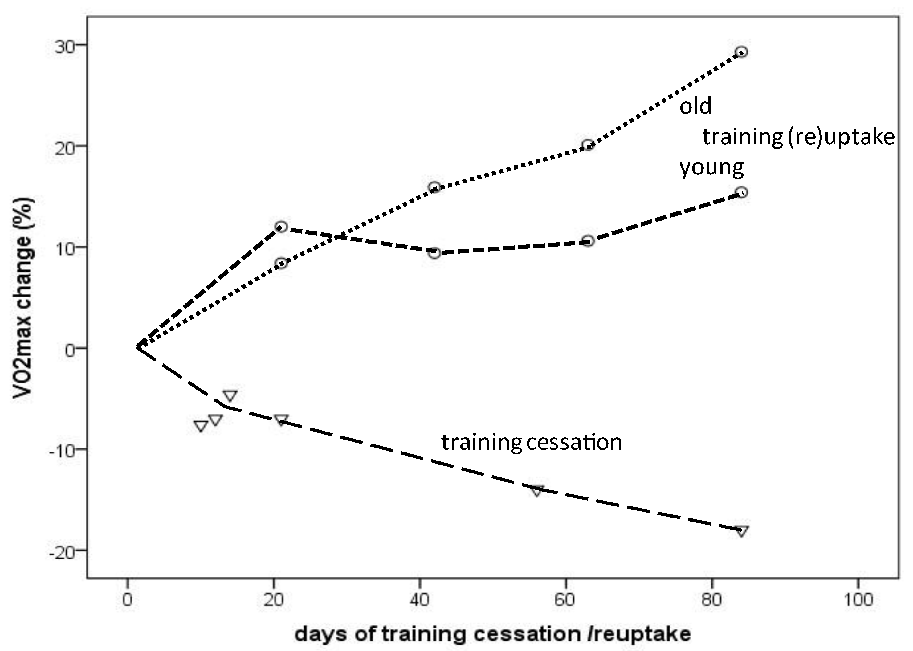
Figure 2. VO2max change depending on the amount (days) of training cessation (triangles) or training (re)uptake (circles: dotted line indicates older individuals; dashed line indicates young individuals) (from selected longitudinal data [22,24,28,31] presented in Table 2).
Ijerph 19 11050 g002
Figure 3. Schematic representation of the slow (aging) and the fast (changes in training habits) components of aging in masters endurance athletes. (A) The red line represents the aging-related decline in VO2max of continuously active endurance athletes, and the lower (blue) line represents that of masters athletes who terminated their competitive activities at the age of 40 years. Detraining rapidly enhances the rate of VO2max decline in active athletes, and recovery of VO2max can rapidly be achieved after (re)uptake of training. (B) Decline in maximal cardiac output represents the main explanatory mechanism for both the loss and restoration of VO2max.
Ijerph 19 11050 g003
Table 2. Effects of short-term (up to 12 weeks) training cessation and training (re)uptake on VO2max and related changes of physiological parameters (cardiorespiratory parameters are maximal values).
ReferenceN
m (Males)
f (Females)
Duration (Days) of
Training Cessation (ce)
Training (Re)Uptake (re)
Age (Years)VO2max (mL/min/kg)
Pre vs. Post
(% Change)
Changes in Related
Physiological Parameters (% Change)
Coyle et al.,
1984 [22]
6 m
1 f
12 (ce)29.1 ± 3.262.1 ± 3.3 vs. 57.7 ± 2.6
(−7%)
heart rate (+4%)
stroke volume (−10%)
cardiac output (−7%)
arteriovenous O2diff (+0.4%)
oxygen pulse (−11%)
citrate synthase (−17.1%)
succinate dehydrogenase (−18.5%)
Coyle et al.,
1984 [22]
6 m
1 f
21 (ce)29.1 ± 3.262.1 ± 3.3 vs. 59.7 ± 3.1
(−7%)
heart rate (+4%)
stroke volume (−11%)
cardiac output (−8%)
arteriovenous O2diff (+2%)
oxygen pulse (−11%)
citrate synthase (−23.7%)
succinate dehydrogenase (−23.9%)
Coyle et al.,
1984 [22]
6 m
1 f
56 (ce)29.1 ± 3.262.1 ± 3.3 vs. 53.2 ± 2.1
(−14%)
heart rate (+6%)
stroke volume (−14%)
cardiac output (−9%)
arteriovenous O2diff
(−4%)
oxygen pulse (−19%)
citrate synthase (−40.6%)
succinate dehydrogenase (−38.4%)
Coyle et al.,
1984 [22]
6 m
1 f
84 (ce)29.1 ± 3.262.1 ± 3.3 vs. 50.8 ± 1.9
(−18%)
heart rate (+5%)
stroke volume (−13%)
cardiac output (−10%)
arteriovenous O2diff
(−7%)
oxygen pulse (−20%)
citrate synthase (−39.6%)
succinate dehydrogenase (−32.5%)
Cullinane et al., 1986 [23]15 m10 (ce)28.2 ± 5.661.3 ± 6.2 vs. 61.2 ± 5.6 (−1.6%)heart rate (+5%)
ventilation (+1.5%)
stroke volume (−2.6%)
Doherty et al., 2003 [24]7 f10 (ce)21.0 ± 2.649.8 ± 1.3 vs. 46.0 ± 1.3
(−7.6%)
heart rate (+1.5%)
stroke volume (−1%)
cardiac output (−0.5%)
arteriovenous O2diff
(−7%)
Drinkwater and Horwath, 1972 [25]7 f90 (ce)14-1747.8 ± 1.8 vs. 40.4 ± 1.0
(−15.4%)
heart rate (+1.5%)
ventilation (−10.3%)
Giada et al., 1998 [26]12 m60 (ce)55 ± 543 ± 7 vs. 36 ± 7
(−16.3%)
Giada et al., 199812 m60 (ce)24 ± 659 ± 10 vs. 49 ± 9
(−16.9%)
Heath et al., 1983 [27]6 m
2 f
10 (ce)28 ± 358.6 ± 2.2 vs. 57.6 ± 2.1 (−1.7%)
Houmard et al., 1992 [28]9 m
3 f
14 (ce)20.1 ± 1.461.6 ± 2.2 vs. 58.7 ± 1.8
(−4.6%)
heart rate (+4.7%)
plasma volume (−5.1%)
citrate synthase (−25.3%)
Katzel et al., 1997 [29]10 m90 (ce)59 ± 850 ± 5 (−11 to −20%)
Martin et al., 1986 [30]5 m 1 f42 (ce)26 ± 162.7 ± 4.0 (−6.5%), 21 days;
(−20.3%), 56 days
stroke volume
21 days (−10%)
56 days (−17%)
Murias et al., 2010 [31]8 m21 (re)68.0 ± 7.028.3 ± 7.1 vs. 30.7 ± 6.0
(+8.4%)
heart rate (−3.5%)
stroke volume (+6.8%)
cardiac output (+7%)
arteriovenous O2diff
(+3.7%)
Murias et al., 2010 [31]8 m21 (re)23.0 ± 5.048.0 ± 6.1 vs. 53.8 ± 7.6
(+12%)
heart rate (−2.1%)
stroke volume (+5.5%)
cardiac output (+3.1%)
arteriovenous O2diff
(+7.5%)
Murias et al., 2010 [31]8 m42 (re)68.0 ± 7.028.3 ± 7.1 vs. 32.8 ± 7.6
(+15.9%)
heart rate (−2.1%)
stroke volume (+9.1%)
cardiac output (+11.3%)
arteriovenous O2diff
(+5.2%)
Murias et al., 2010 [31]8 m42 (re)23.0 ± 5.048.0 ± 6.1 vs. 52.5 ± 6.4%
(+9.4%)
heart rate (−2.1%)
stroke volume (+7.9%)
cardiac output (+5.4%)
arteriovenous O2diff
(+4.8%)
Murias et al., 2010 [31]8 m63 (re)68.0 ± 7.028.3 ± 7.1 vs. 34.0 ± 5.8
(+20.1%)
heart rate (−1.4%)
stroke volume (+15.2%)
cardiac output (+17.9%)
arteriovenous O2diff
(+3.7%)
Murias et al., 2010 [31]8 m63 (re)23.0 ± 5.048.0 ± 6.1 vs. 53.1 ± 6.5%
(+10.6%)
heart rate (−2.1%)
stroke volume (+12.6%)
cardiac output (+10.4%)
arteriovenous O2diff
(+0.7%)
Murias et al., 2010 [31]8 m84 (re)68.0 ± 7.028.3 ± 7.1 vs. 36.6 ± 6.5
(+29.3%)
heart rate (+0.7%)
stroke volume (+14.8%)
cardiac output (+20.8%)
arteriovenous O2diff
(+8.8%)
Murias et al., 2010 [31]8 m84 (re)23.0 ± 5.048.0 ± 6.1 vs. 55.4 ± 5.5%
(+15.4%)
heart rate (−1.1%)
stroke volume (+10.9%)
cardiac output (+9.7%)
arteriovenous O2diff
(+6.8%)
Nichols et al., 2000 [12]1 f14 (re)49.442.0 vs. 48.1
(+14.5%)
heart rate (+1.6%)
Nichols et al., 2000 [12]1 f28 (re)49.442.0 vs. 55.1
(+31.2%)
heart rate (−2.7%)
Pavlik et al., 1986 [32]42 m60 (ce)22.9 ± 0.772.2 vs. 67.0 (30 days) vs. 62.5 (45 days)
(−7% and −13%);
no further decrease after 45 days
Prior et al., 2015 [33]7 m
5 f
14 (ce)65 ± 331.2 ± 2.3 vs. 29.3 ± 1.9
(−6%)
citrate synthase
(−28.6%)
Ready and Quinney, 1982 [34]12 m63 (ce)25.0 ± 3.664.2 ± 9.5 vs. 59.3 ± 6.4, (3 weeks)
57.5 ± 6.4 (6 weeks) 57.3 ± 8.8 (9 weeks)
(−10.7%)
Schulman et al., 1996 [35]8 m84 (ce)59.6 ± 349.9 ± 1.9 vs. 42.0 ± 2.2
(−15.8%)
heart rate (+4.1%)
cardiac index (−10.5%)
stroke volume index
(−14.2%)
Sinacore et al., 1993 [36]5 m
1 f
84 (ce)29 ± 1061.3 ± 7 vs. 50.8 ± 7
(−17.1%)
Smorawinski et al., 2001 [37]10 m3 (ce, bedrest)20.3 ± 1.954.8 ± 2.1 (−16.5%)blood lactate
(−8% to −20%)
Publisher’s Note: MDPI stays neutral with regard to jurisdictional claims in published maps and institutional affiliations.