Article

Lack of Associations between Environmental Exposures and Environmental Enteric Dysfunction among 18-Month-Old Children in Rural Malawi

1
Center for Child, Adolescent and Maternal Health Research, Faculty of Medicine and Health Technology, Tampere University, 33014 Tampere, Finland
2
Department of Paediatrics, Tampere University Hospital, 33521 Tampere, Finland
3
School of Public Health & Family Medicine, Kamuzu University of Health Sciences, Private Bag 360, Chichiri, Blantyre 3, Malawi
4
Fimlab Laboratories, Pirkanmaa Hospital District, 33521 Tampere, Finland
*
Author to whom correspondence should be addressed.
Int. J. Environ. Res. Public Health 2022, 19(17), 10891; https://doi.org/10.3390/ijerph191710891
Submission received: 24 June 2022 / Revised: 25 August 2022 / Accepted: 28 August 2022 / Published: 1 September 2022
(This article belongs to the Section Children's Health)

Abstract

:
Environmental enteric dysfunction (EED) is common and contributes to linear growth faltering (stunting) and mortality among children in low-resource settings. A few studies on the environmental causes of EED have been conducted but the exact exposures that cause or predispose children to EED are context-specific and not clear. This study aimed to assess associations between selected environmental exposures and EED markers among 620 18-month-old children. This was a secondary analysis of data from Malawian children who participated in a randomized controlled trial (iLiNS-DYAD, registered at clinicaltrials.gov as NCT01239693) from birth to 18 months of age. Data on environmental exposures, including drinking water source, sanitation, exposure to animals, housing materials, season, residential area, and food insecurity were collected at enrolment. Biomarkers of EED included concentrations of calprotectin, regenerating 1B protein (REG1B), and alpha-1-antitrypsin from stool samples to assess intestinal inflammation, repair, and permeability, respectively. We performed bivariate and multivariable analyses to assess associations between environmental exposures and EED biomarkers. Adjusting for possible confounders, we did not find associations between the selected environmental exposures and the three biomarkers. These results do not provide support for our hypothesis that the studied adverse environmental exposures are associated with increased concentrations of children’s EED markers in rural Malawi.

1. Introduction

Environmental enteric dysfunction (EED) is a common condition among children in low-income settings. It is characterized by increased intestinal permeability, intestinal inflammation, and impaired barrier function [1,2]. EED typically starts in infancy and continues into adulthood [1,3]. Growing evidence has demonstrated that EED has a strong association with stunting which contributes to more than one million deaths among children under 5 years old every year [4,5].
As the name implies, EED is considered environmentally caused [1,3], but the exact factors in the environment resulting in this condition are not well understood. Typically, these factors include unsafe drinking water, poor sanitary facilities, exposure to animals, and season [1]. Several studies have demonstrated that unsafe drinking water and sanitation, exposure to chickens, cows, and goats, and the rainy season may be risk factors for EED [6,7,8,9,10,11]. However, an intervention addressing water, sanitation, and hygiene (WASH) do not have a positive impact on reducing EED in rural Zimbabwe [12]. Consequently, there are inconsistent data about the association between environmental factors and EED. Additionally, other factors including geographical context, food insecurity, and housing materials contribute to stunting, which is strongly associated with EED, but their associations with EED are poorly understood [5,13,14,15,16,17]. Evidence that these potential environmental factors are associated with EED concomitantly is lacking especially in young children.
To assess the associations of EED with environmental factors concomitantly, we considered other factors as covariates which have been associated with intestinal health [18,19]. In particular, this included breastfeeding, dietary supplementation with small-quantity lipid-based nutrient supplements (SQ-LNS) and antibiotic use, which were reported to be associated with gut microbiome [20,21,22]. Our study aimed to investigate whether there would be associations between these exposures and EED in 18-month-old rural Malawian children. As biomarkers of EED, we used fecal concentrations of calprotectin, regenerating 1B protein (REG1B), and alpha-1-antitrypsin, indicating inflammation, cellular repair, and permeability in the gastrointestinal tract [23,24,25,26]. We hypothesized that exposure to adverse environmental conditions would be associated with increased fecal concentrations of these three biomarkers among rural Malawian children.

2. Materials and Methods

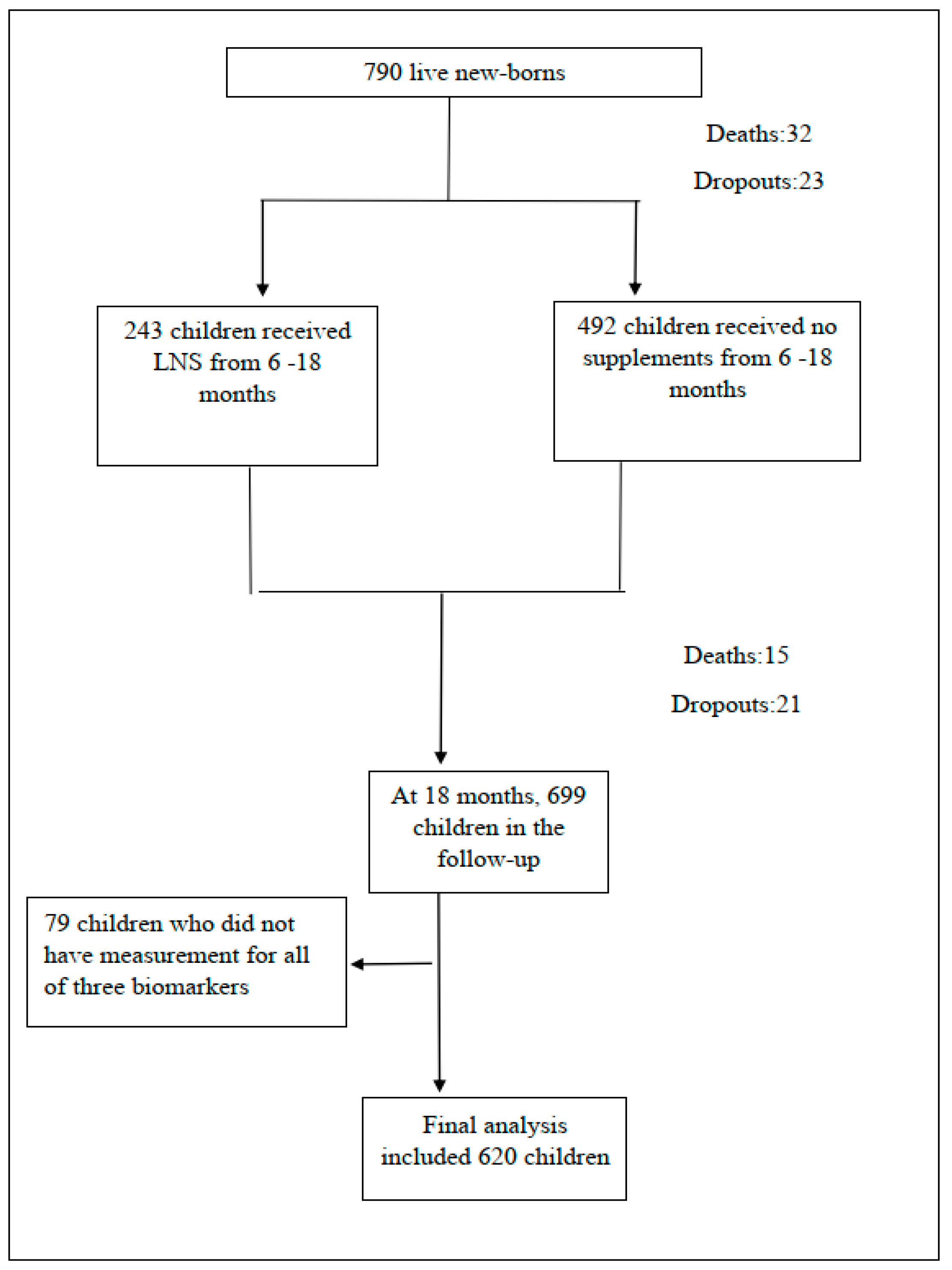
2.1. Study Participants

The present study was a secondary analysis of data from a randomized controlled trial (iLiNS-DYAD trial) on the effects of lipid-based nutrient supplements on pregnant women and infants in Mangochi district, Malawi [27]. In brief, pregnant women with less than 20 completed weeks gestation were enrolled and randomized to three different groups in which they received different nutrient supplements: iron and folic acid (IFA), multiple micronutrients (MMN), or SQ-LNS. The current analysis included newborns who were followed-up until 18 months of age. Infants whose mothers had received SQ-LNS during pregnancy received 20 g/day SQ-LNS from 6 to 18 months and other infants received no supplements. The iLiNS-DYAD trial was prospectively registered at clinicaltrials.gov as NCT01239693.
Main efficacy analyses from the trial data do not support a hypothesis that provision of SQ-LNS to pregnant women and their children would increase the infants’ birth weight or growth by 18 months of age [27,28]. In the current ancillary study to the trial, we analyzed the associations between selected environmental exposures and intestinal health among the participating children at 18 months of age.
Before enrolment, all pregnant women gave a signed informed consent for participation and another consent was given before enrolment of their children. The original trial was approved by the ethics committees of College of Medicine in Malawi and Pirkanmaa Hospital District in Finland.

2.2. Environmental Exposures

We selected the following environmental exposures for this analysis based on our previous study [19]: drinking water source (well, lake and river vs. piped water and borehole); sanitation (regular pit latrine and none vs. water closet and ventilated improved pit latrine); exposure to chickens, goats or cows, representing exposure to feces of these animals possibly causing intestinal infection [29,30]; wall material of the main house (unburnt brick, straw, grass or mud vs. burnt brick); roofing material of the main house (grass and other vs. iron sheets or tiles); season when children’s stool samples were collected (rainy in December–March and hot-dry in August–November vs. cold-dry in April–July); residential area (Malindi, Mangochi, and Lungwena); and food insecurity score (food secure, mildly food insecure, moderately food insecure, and severely food insecure), indicating if food intake or eating patterns were disrupted due to physical or economic conditions in the household in the past month [31,32].
Research assistants collected information on the environmental exposures using questionnaires when mothers were enrolled. In addition, the data on exposure to animals were recorded using questionnaires a few days before the collection of children’s stool samples at 18 months of age.

2.3. Outcomes

We included concentrations of calprotectin, REG1B, and alpha-1-antitrypsin in stools at 18 months as intestinal health outcomes in this analysis. These biomarkers of EED have been validated for measuring intestinal health in pediatric populations [12,23,33]. Increase in concentrations of these biomarkers reflected increased inflammation, repair, or permeability in gastrointestinal tract, respectively [24,34].
Research assistants collected stool samples from children during home visits and stored the samples in cryovial tubes at −20 °C in local laboratories. Within 48 h, these samples were transported to a central laboratory and stored at −80 °C before they were shipped to Tampere University in Finland for laboratory analysis. If the child had diarrhea at home visit, the stool collection from these children was postponed for 2 weeks. To measure fecal concentrations of these three biomarkers, we used commercial enzyme-linked immunosorbent assay (ELISA) kits for calprotectin (Hycult Biotech, Uden, The Netherlands), REG1B (TECHLAB, Blacksburg, VA, USA), and alpha-1-antitrypsin (PromoCell GmbH, Heidelberg, Germany). We chose these kits based on their suitability and sensitivity [34,35,36]. These kits provided lower limit of detection (LOD) which was 16 µg/g for calprotectin, 6.25 µg/g for REG1B, and 1.8 mg/dL for alpha-1-antitrypsin. Briefly, we thawed and weighed around 100 mg of stools for calprotectin or alpha-1-antitrypsin, and around 50 mg for REG1B. A dilution factor of 1:50 was used for calprotectin, 1:10,000 for REG1B and 1:250 for alpha-1-antitrypsin. The final concentration was calculated based on the respective standard curve plotted with the absorbance versus different concentrations of standards.

2.4. Other Variables

Based on previous research, we considered the following variables as possible covariates in our analyses [19,20,21,22,37,38]: maternal age, maternal HIV infection status (infected/not infected), maternal educational achievement in years, child sex, weight-for-length z-score (WLZ) and length-for-age z-score (LAZ) at 18 months, continued breastfeeding (yes/no), and number of days of antibiotic use between birth and 18 months of age (above/below median days) in children.
We obtained information on maternal age and educational achievement using questionnaires at enrollment and data on HIV infection status using a whole-blood antibody rapid diagnosis test (Alere Determine™ HIV-1/2, Abbott Alere, Matsudo, Japan). We collected data on child sex from health records at birth, and data on continued breastfeeding by questionnaires at 18 months. Length and weight at 18 months were assessed using a length board (Harpenden Infantometer, Holtain Limited, Crosswell, UK) and an infant weighing scale (SECA 735). WLZ and LAZ were calculated based on World Health Organization (WHO) Child Growth Standards [39]. Data on antibiotic use were extracted from medical records from birth until 18 months.

2.5. Data Analysis

We performed data analyses using Stata software version 15.0 (StataCorp, College Station, TX, USA). We described continuous variables using mean (standard deviation) and categorical variables using percentage.
For the original values of EED biomarker concentrations lower than LOD, we did not repeat measurements due to limited amounts of stool samples. We replaced these values with a half of LOD, which was recommended to be used by WHO GEMS/Food-EURO workshop [40]. Due to skewed distribution of biomarker concentrations, we used log-transformed values as outcomes in the models. We used Pearson’s correlation coefficients to estimate the correlation between these EED biomarkers. The correlation was considered strong with coefficient >0.5; moderate with coefficient >0.3 but <0.5; and weak with coefficient >0.1 but <0.3 [41]. We assessed the association between environmental exposures and EED biomarkers by bivariate analysis and multivariable analysis using linear regression. For multivariable analysis, our models were adjusted for all environmental exposures and the following background characteristics: maternal age, HIV infection status, educational achievement, child sex, WLZ and LAZ at 18 months, continued breastfeeding, antibiotic use, and dietary intervention. For hypothesis testing, a p-value < 0.05 was deemed statistically significant. Considering possible effect medication by the child’s sex, we included sex *environmental exposures variables in multivariable models for EED biomarkers.
We checked multicollinearity by a variance inflation factor (VIF) tool and removed environmental exposures from multivariable models if they contributed to multicollinearity. The exclusion criteria included a VIF-value of more than 1 and at the same time a change in any p-value from statistically significant to non-significant when the respective exposure was removed from the multivariable model. To assess the robustness of our multivariable models, we performed a sensitivity analysis in which missing data in independent variables were imputed by chained equations with 20 imputed datasets.

3. Results

Of the 790 live-born infants enrolled in the study, 47 (6%) died and 44 (6%) dropped out from the follow-up by 18 months of age. Another 79 children were excluded because of missing biomarker data, leaving data from 620 children in the analysis (Figure 1).
There were no differences in anthropometric indicators at 18 months of age between the included and excluded children, but children whose data were excluded from the analysis had mothers with higher mean educational achievement (5 years vs. 4 years) and a lower proportion of continued breastfeeding than those included in the analysis (83% vs. 93%). The participants’ mean WLZ and LAZ at 18 months were −0.2 and −1.7, respectively; the prevalence of wasting and stunting was 4% and 37%, respectively (Table 1).
The median (interquartile range) concentrations of calprotectin, REG1B, and alpha-1-antitrypsin at 18 months were 111 µg/g (51, 293), 38 µg/g (7, 162), and 4.9 mg/dL (2.6, 7.9), respectively (Figure 2). There was a strong positive correlation between calprotectin and alpha-1-antitryspsin level, and weak positive correlations between calprotectin and REG1B and between REG1B and alpha-1-antitrypsin (Table S1).
In the bivariate analysis, most of the selected environmental exposures were not associated with concentrations of EED biomarkers (Table 2). As an exception, there was an association between the season of stool sample collection and the stool’s calprotectin and alpha-1-antitrypsin concentration and between the type of sanitary facility in the participants’ home and calprotectin concentration. The children’s stool biomarker concentrations were on average higher in rainy season or hot-dry season than in cold-dry season and lower in households with no sanitary facility or a regular pit latrine than in households with a water closet or a ventilated improved pit latrine (Table 2).
Roofing material used as one of environmental exposures, was excluded from the multivariable analysis due to multicollinearity (Table S2). The analyses of adjustment for covariates did not change the association between the season of stool sample collection and the stool’s calprotectin concentration but removed that between the season and alpha-1-antitrypsin concentration (Table 3). The adjusted analyses also indicated an association between the participants’ residential area and REG1B concentration, with participants living in Mangochi having a lower concentration on average than those in Lungwena (Table 3).
Results from the sensitivity analysis on the association between EED biomarkers and environmental exposures with imputed data were consistent with those in the multivariable analysis (Table S3).
For covariates earlier associated with gut microbiome, increased antibiotic use was associated with lower concentrations of calprotectin (p = 0.002) and alpha-1-antitrypsin (p = 0.009). There were no associations between continued breastfeeding or dietary supplementation with SQ-LNS and any of these biomarkers (p > 0.05, Table S4).
Inclusion of sex *environmental exposures variables in multivariable models for EED biomarkers did not show any indication of sex differences in associations between the studied environmental exposures and EED biomarkers.

4. Discussion

Our analysis explored the association between multiple environmental exposures and EED. In a sample of 620 rural Malawian 18-month-old children, after adjusting for potential confounders, we found no associations between most of the selected environmental exposures and fecal concentrations of calprotectin, REG1B, and alpha-1-antitrypsin which represented intestinal inflammation, repair, and permeability, respectively. We only observed two associations between the season and calprotectin concentration and between residential area and REG1B concentration.
Even though these two associations were found in our samples, they were less likely to be indications of similar associations between these two environmental exposures and EED in our target population which our samples were drawn from. This may be explained by the correlations between EED biomarkers and the consistency between different biomarkers. Our three biomarkers were correlated with each other, which has been indicated by a previous study in Bangladesh [34]. However, the season and the residential area were not consistently associated with the other two markers. Moreover, considering the complexity of the pathways of EED [42], it is likely that the consistency of associations between different biomarkers and the same environmental exposure is needed, but we did not see the associations between the season or the residential area and other two biomarkers. Therefore, it is more likely that this sample finding of associations between the season or the residential area and one single EED biomarker is spurious.
There are several factors related to the study design and implementation that could theoretically introduce biases in the results. These factors include number of excluded children, selection of covariates, utility of EED biomarkers, missing data, and reduction in sample size due to the selection of analysis models. However, the excluded children had on average similar characteristics compared with those included children. We included covariates that had been considered appropriate in previous studies such as breastfeeding and antibiotic use [20,21,43,44,45,46,47,48,49] to minimize the possible bias [50] in multivariable analysis. For biomarkers, calprotectin, REG1B, and alpha-1-antitrypsin have been widely used to study typical features of EED [34,51]. We also confirmed the robustness of our results using multiple imputations for missing data in the sensitivity analysis with multiple linear regression models [52]. Therefore, we believe that our sample findings are valid and representative of the target population.
The lack of association between EED and drinking water source, sanitation, or exposure to animals in rural Malawian children partially contradicts previous results from other study sites [6,7,8,9]. Similar to ours, the study in Peru provided no interventions to the living environment and used fecal alpha-1-antitrypsin as one of EED biomarkers. However, different features such as higher median concentration of alpha-1-antitrypsin at 18 months (0.35 mg/g vs. 0.049 mg/g), the use of myeloperoxidase and neopterin as markers of intestinal inflammation, multiple measurements for markers, and different classifications for exposures [9], might contribute to different findings from Peru. The MAL-ED study across multiple sites and two studies in Bangladesh measured environmental exposures using a joint index for drinking water source, sanitation facilities, and other socioeconomic factors or created an EED score by combining EED markers [6,7,8], while we considered each environmental exposure as a separate element and used each EED marker as the respective outcome. These differences in exposure and outcome definitions make it difficult to draw a more generic conclusion on the association between drinking water source, sanitation, exposure to animals and EED markers among children in low-income settings.
Elevated food insecurity in rural Malawi might contribute to worsening EED when there are insufficient food and drinking water in the dry season, according to the speculations from a study on the effects of zinc and albendazole treatments on EED [14]. In our sample, we did not see this association. Regarding housing materials, roofing material was excluded from the analysis due to multicollinearity and the adjusted model included only the wall material. Earlier studies have indicated a weak association with an intestinal problem (diarrhea) and housing materials [53], but we did not see an association of housing materials with EED in the Malawian context.
In our study, antibiotic use was inversely associated with calprotectin and alpha-1-antitrypsin concentrations in stools. This result is consistent with findings of a previous study from the iLiNS-DYAD trial, documenting an inverse relationship between antibiotic use and microbiota maturity and diversity that were associated with our three EED biomarkers [18,19].
Notably, improved latrine was associated with higher concentration of calprotectin compared to regular pit latrine and no latrine in bivariate analysis. A longitudinal study in Peru [9] provided similar results that improved sanitation facilities were associated with higher concentration of myeloperoxidase (a marker of intestinal inflammation). This may be explained by the location of latrines with unproper management of fecal sludge [9,54,55,56]. Higher level of intestinal inflammation may be due to more exposure to pathogens in soil surrounding the main house which was close to the improved latrine [56,57]. The proportion of households having improved latrine only accounted for 10% in our study. Therefore, it is possible that this proportion is too small to capture differences in concentrations of EED biomarkers between children living with different types of latrines in adjusted models.

5. Conclusions

Our results suggest that the selected environmental exposures are not associated with EED markers of children in the rural Malawian setting. These findings contribute to scientific evidence on the possible environmental causes of young children’s intestinal conditions such as EED in Sub-Saharan Africa.

Supplementary Materials

The following supporting information can be downloaded at: https://www.mdpi.com/article/10.3390/ijerph191710891/s1, Table S1: Pearson’s correlations between biomarkers of intestinal health; Table S2: Values of VIF for environmental exposures; Table S3: Associations between environmental exposures and intestinal biomarkers in multivariable analysis after imputation; Table S4: Associations between breastfeeding, antibiotic use, and dietary intervention and intestinal biomarkers in multivariable analysis.

Author Contributions

Conceptualization: P.A., U.A., L.H., Y.-M.F. and Z.L; methodology: P.A., U.A., L.H., Y.-M.F. and Z.L.; formal analysis: Z.L.; data curation: L.H.; writing—original draft preparation: Z.L.; writing—review and editing: P.A., U.A., L.H., Y.-M.F., K.M., H.H., D.C. and C.C.; supervision: P.A., U.A. and Y.-M.F.; project administration: P.A. and U.A. All authors have read and agreed to the published version of the manuscript.

Funding

This research was funded by the Finnish Funding Agency for Technology and Innovation, the Foundation for Paediatric Research in Finland, and the Competitive State Research Financing of the Expert Responsibility area of Tampere University Hospital [9M004]. The original trial was funded by a grant to the University of California, Davis from the Bill & Melinda Gates Foundation [OPP49817] and the funding from the Office of Health, Infectious Diseases, and Nutrition, Bureau for Global Health, U.S. Agency for International Development (USAID) under terms of Cooperative Agreement No. AID-OAA-A-12-00005, through the Food and Nutrition Technical Assistance III Project (FANTA), managed by FHI 360.

Institutional Review Board Statement

The study was conducted in accordance with the Declaration of Helsinki, and approved by Ethics Committee of College of Medicine in Malawi and Ethics Committee of Pirkanmaa Hospital District in Finland (protocol code: R10157 and date of approval: 10 September 2015). The original trial was registered at clinicaltrials.gov as NCT01239693.

Informed Consent Statement

Informed consent was obtained from all subjects involved in the study.

Data Availability Statement

The data will be available from the authors upon reasonable request.

Acknowledgments

The authors thank William A. Petri, Department of Medicine at the University of Virginia, USA, who donated REG1B kits free of charge. The authors also thank Eini Eskola, laboratory analyst of Faculty of Medicine and Health Technology at Tampere University, Finland, who contributed to the measurement of calprotectin concentrations.

Conflicts of Interest

The authors declare no conflict of interest.

References

  1. Crane, R.J.; Jones, K.D.; Berkley, J.A. Environmental enteric dysfunction: An overview. Food Nutr. Bull. 2015, 36, S76–S87. [Google Scholar] [CrossRef] [PubMed]
  2. Hodges, P.; Tembo, M.; Kelly, P. Intestinal Biopsies for the Evaluation of Environmental Enteropathy and Environmental Enteric Dysfunction. J. Infect. Dis. 2021, 224, S856–S863. [Google Scholar] [CrossRef] [PubMed]
  3. Sullivan, P.B. Environmental Enteric Dysfunction: Reeemergence of an Old Disease. J. Infect. Dis. 2021, 224, S873–S875. [Google Scholar] [CrossRef]
  4. Myatt, M.; Khara, T.; Schoenbuchner, S.; Pietzsch, S.; Dolan, C.; Lelijveld, N.; Briend, A. Children who are both wasted and stunted are also underweight and have a high risk of death: A descriptive epidemiology of multiple anthropometric deficits using data from 51 countries. Arch. Public Health 2018, 76, 28. [Google Scholar] [CrossRef]
  5. Morais, M.B.; Silva, G.A.P.D. Environmental enteric dysfunction and growth. J. Pediatr. 2019, 95, 85–94. [Google Scholar] [CrossRef]
  6. George, C.M.; Oldja, L.; Biswas, S.K.; Perin, J.; Lee, G.O.; Ahmed, S.; Haque, R.; Sack, R.B.; Parvin, T.; Azmi, I.J.; et al. Fecal Markers of Environmental Enteropathy are Associated with Animal Exposure and Caregiver Hygiene in Bangladesh. Am. J. Trop. Med. Hyg. 2015, 93, 269–275. [Google Scholar] [CrossRef]
  7. Campbell, R.K.; Schulze, K.J.; Shaikh, S.; Mehra, S.; Ali, H.; Wu, L.; Raqib, R.; Baker, S.; Labrique, A.; West, K.P., Jr.; et al. Biomarkers of Environmental Enteric Dysfunction Among Children in Rural Bangladesh. J. Pediatr. Gastroenterol. Nutr. 2017, 65, 40–46. [Google Scholar] [CrossRef]
  8. McCormick, B.J.J.; Lee, G.O.; Seidman, J.C.; Haque, R.; Mondal, D.; Quetz, J.; Lima, A.A.M.; Babji, S.; Kang, G.; Shrestha, S.K.; et al. Dynamics and Trends in Fecal Biomarkers of Gut Function in Children from 1–24 Months in the MAL-ED Study. Am. J. Trop. Med. Hyg. 2017, 96, 465–472. [Google Scholar] [CrossRef] [PubMed]
  9. Exum, N.G.; Lee, G.O.; Olórtegui, M.P.; Yori, P.P.; Salas, M.S.; Trigoso, D.R.; Colston, J.M.; Schwab, K.J.; McCormick, B.J.J.; Kosek, M.N. A Longitudinal Study of Household Water, Sanitation, and Hygiene Characteristics and Environmental Enteropathy Markers in Children Less than 24 Months in Iquitos, Peru. Am. J. Trop. Med. Hyg. 2018, 98, 995–1004. [Google Scholar] [CrossRef]
  10. Lin, A.; Arnold, B.F.; Afreen, S.; Goto, R.; Huda, T.M.N.; Haque, R.; Raqib, R.; Unicomb, L.; Ahmed, T.; Colford, J.M.; et al. Household environmental conditions are associated with enteropathy and impaired growth in rural Bangladesh. Am. J. Trop. Med. Hyg. 2013, 89, 130–137. [Google Scholar] [CrossRef] [Green Version]
  11. Crane, R.J.; Parker, E.; Fleming, S.; Gwela, A.; Gumbi, W.; Ngoi, J.M.; de Laurent, Z.R.; Nyatichi, E.; Ngari, M.; Wambua, J.; et al. Cessation of exclusive breastfeeding and seasonality, but not small intestinal bacterial overgrowth, are associated with environmental enteric dysfunction: A birth cohort study amongst infants in rural Kenya. EClinicalMedicine 2022, 47, 101403. [Google Scholar] [CrossRef] [PubMed]
  12. Gough, E.K.; Moulton, L.H.; Mutasa, K.; Ntozini, R.; Stoltzfus, R.J.; Majo, F.D.; Smith, L.E.; Panic, G.; Giallourou, N.; Jamell, M.; et al. Effects of improved water, sanitation, and hygiene and improved complementary feeding on environmental enteric dysfunction in children in rural Zimbabwe: A cluster-randomized controlled trial. PLoS Negl. Trop. Dis. 2020, 14, e0007963. [Google Scholar] [CrossRef] [PubMed]
  13. Nuñez, P.A.; Fernández, M.S.; Turjanski, P.; Pérez, A.; Rivero, M.R.; De Angelo, C.; Salomón, O.D.; Cueto, G. Substantial reduction in child stunting is differentially associated to geographical and socioeconomic disparities in Misiones Province, Argentina. Trop. Med. Int. Health 2020, 25, 874–885. [Google Scholar] [CrossRef] [PubMed]
  14. Ryan, K.N.; Stephenson, K.B.; Trehan, I.; Shulman, R.J.; Thakwalakwa, C.; Murray, E.; Maleta, K.; Manary, M.J. Zinc or albendazole attenuates the progression of environmental enteropathy: A randomized controlled trial. Clin. Gastroenterol. Hepatol. 2014, 12, 1507–1513. [Google Scholar] [CrossRef]
  15. Belayneh, M.; Loha, E.; Lindtjørn, B. Seasonal Variation of Household Food Insecurity and Household Dietary Diversity on Wasting and Stunting among Young Children in A Drought Prone Area in South Ethiopia: A Cohort Study. Ecol. Food Nutr. 2021, 60, 44–69. [Google Scholar] [CrossRef] [PubMed]
  16. Uwiringiyimana, V.; Osei, F.; Amer, S.; Veldkamp, A. Bayesian geostatistical modelling of stunting in Rwanda: Risk factors and spatially explicit residual stunting burden. BMC Public Health 2022, 22, 159. [Google Scholar] [CrossRef]
  17. Tusting, L.S.; Gething, P.W.; Gibson, H.S.; Greenwood, B.; Knudsen, J.; Lindsay, S.W.; Bhatt, S. Housing and child health in sub-Saharan Africa: A cross-sectional analysis. PLoS Med. 2020, 17, e1003055. [Google Scholar] [CrossRef]
  18. Kortekangas, E.; Fan, Y.M.; Chaima, D.; Lehto, K.M.; Malamba-Banda, C.; Matchado, A.; Chingwanda, C.; Liu, Z.; Ashorn, U.; Cheung, Y.B.; et al. Associations between Gut Microbiota and Intestinal Inflammation, Permeability and Damage in Young Malawian Children. J. Trop. Pediatr. 2022, 68, fmac012. [Google Scholar] [CrossRef]
  19. Kortekangas, E.; Kamng’ona, A.W.; Fan, Y.M.; Cheung, Y.B.; Ashorn, U.; Matchado, A.; Poelman, B.; Maleta, K.; Dewey, K.G.; Ashorn, P. Environmental exposures and child and maternal gut microbiota in rural Malawi. Paediatr. Perinat. Epidemiol. 2020, 34, 161–170. [Google Scholar] [CrossRef]
  20. Kim, H.; Sitarik, A.R.; Woodcroft, K.; Johnson, C.C.; Zoratti, E. Birth Mode, Breastfeeding, Pet Exposure, and Antibiotic Use: Associations with the Gut Microbiome and Sensitization in Children. Curr. Allergy Asthma Rep. 2019, 19, 22. [Google Scholar] [CrossRef]
  21. McDonnell, L.; Gilkes, A.; Ashworth, M.; Rowland, V.; Harries, T.H.; Armstrong, D.; White, P. Association between antibiotics and gut microbiome dysbiosis in children: Systematic review and meta-analysis. Gut Microbes 2021, 13, 1870402. [Google Scholar] [CrossRef] [PubMed]
  22. Kamng’ona, A.W.; Young, R.; Arnold, C.D.; Patson, N.; Jorgensen, J.M.; Kortekangas, E.; Chaima, D.; Malamba, C.; Ashorn, U.; Cheung, Y.B.; et al. Provision of Lipid-Based Nutrient Supplements to Mothers During Pregnancy and 6 Months Postpartum and to Their Infants from 6 to 18 Months Promotes Infant Gut Microbiota Diversity at 18 Months of Age but Not Microbiota Maturation in a Rural Malawian Setting: Secondary Outcomes of a Randomized Trial. J. Nutr. 2020, 150, 918–928. [Google Scholar]
  23. Kolho, K.L.; Alfthan, H. Concentration of fecal calprotectin in 11,255 children aged 0–18 years. Scand. J. Gastroenterol. 2020, 55, 1024–1027. [Google Scholar] [CrossRef] [PubMed]
  24. Costa, F.; Mumolo, M.G.; Bellini, M.; Romano, M.R.; Ceccarelli, L.; Arpe, P.; Sterpi, C.; Marchi, S.; Maltinti, G. Role of faecal calprotectin as non-invasive marker of intestinal inflammation. Dig. Liver Dis. 2003, 35, 642–647. [Google Scholar] [CrossRef]
  25. Guerrant, R.L.; Leite, A.M.; Pinkerton, R.; Medeiros, P.H.; Cavalcante, P.A.; DeBoer, M.; Kosek, M.; Duggan, C.; Gewirtz, A.; Kagan, J.C.; et al. Biomarkers of Environmental Enteropathy, Inflammation, Stunting, and Impaired Growth in Children in Northeast Brazil. PLoS ONE 2016, 11, e0158772. [Google Scholar] [CrossRef]
  26. Kosek, M.; Haque, R.; Lima, A.; Babji, S.; Shrestha, S.; Qureshi, S.; Amidou, S.; Mduma, E.; Lee, G.; Yori, P.P.; et al. Fecal markers of intestinal inflammation and permeability associated with the subsequent acquisition of linear growth deficits in infants. Am. J. Trop. Med. Hyg. 2013, 88, 390–396. [Google Scholar] [CrossRef]
  27. Ashorn, P.; Alho, L.; Ashorn, U.; Cheung, Y.B.; Dewey, K.G.; Harjunmaa, U.; Lartey, A.; Nkhoma, M.; Phiri, N.; Phuka, J.; et al. The impact of lipid-based nutrient supplement provision to pregnant women on newborn size in rural Malawi: A randomized controlled trial. Am. J. Clin. Nutr. 2015, 101, 387–397. [Google Scholar] [CrossRef]
  28. Ashorn, P.; Alho, L.; Ashorn, U.; Cheung, Y.B.; Dewey, K.G.; Gondwe, A.; Harjunmaa, U.; Lartey, A.; Phiri, N.; Phiri, T.E.; et al. Supplementation of Maternal Diets during Pregnancy and for 6 Months Postpartum and Infant Diets Thereafter with Small-Quantity Lipid-Based Nutrient Supplements Does Not Promote Child Growth by 18 Months of Age in Rural Malawi: A Randomized Controlled Trial. J. Nutr. 2015, 145, 1345–1353. [Google Scholar] [CrossRef]
  29. Penakalapati, G.; Swarthout, J.; Delahoy, M.J.; McAliley, L.; Wodnik, B.; Levy, K.; Freeman, M.C. Exposure to Animal Feces and Human Health: A Systematic Review and Proposed Research Priorities. Environ. Sci. Technol. 2017, 51, 11537–11552. [Google Scholar] [CrossRef]
  30. Headey, D.; Nguyen, P.; Kim, S.; Rawat, R.; Ruel, M.; Menon, P. Is Exposure to Animal Feces Harmful to Child Nutrition and Health Outcomes? A Multicountry Observational Analysis. Am. J. Trop. Med. Hyg. 2017, 96, 961–969. [Google Scholar] [CrossRef] [PubMed]
  31. Coates, J.; Swindale, A.; Bilinsky, P. Household Food Insecurity Access Scale (HFIAS) for Measurement of Food Access: Indicator Guide V3; FHI 360/FANTA: Washington, DC, USA, 2007. [Google Scholar]
  32. Deitchler, M.; Ballard, T.; Swindale, A.; Coates, J. Introducing a Simple Measure of Household Hunger for Cross-Cultural Use; FANTA2: Washington, DC, USA, 2011. [Google Scholar]
  33. Lin, A.; Ali, S.; Arnold, B.F.; Rahman, M.Z.; Alauddin, M.; Grembi, J.; Mertens, A.N.; Famida, S.L.; Akther, S.; Hossen, M.S.; et al. Effects of Water, Sanitation, Handwashing, and Nutritional Interventions on Environmental Enteric Dysfunction in Young Children: A Cluster-randomized, Controlled Trial in Rural Bangladesh. Clin. Infect. Dis. 2020, 70, 738–747. [Google Scholar] [CrossRef] [PubMed]
  34. Naylor, C.; Lu, M.; Haque, R.; Mondal, D.; Buonomo, E.; Nayak, U.; Mychaleckyj, J.C.; Kirkpatrick, B.; Colgate, R.; Carmolli, M.; et al. Environmental Enteropathy, Oral Vaccine Failure and Growth Faltering in Infants in Bangladesh. EBioMedicine 2015, 2, 1759–1766. [Google Scholar] [CrossRef] [PubMed]
  35. Asgarshirazi, M.; Shariat, M.; Nayeri, F.; Dalili, H.; Abdollahi, A. Comparison of Fecal Calprotectin in Exclusively Breastfed and Formula or Mixed Fed Infants in the First Six Months of Life. Acta Med. Iran. 2017, 55, 53–58. [Google Scholar] [PubMed]
  36. Peterson, K.M.; Buss, J.; Easley, R.; Yang, Z.; Korpe, P.S.; Niu, F.; Ma, J.Z.; Olortegui, M.P.; Haque, R.; Kosek, M.N.; et al. REG1B as a predictor of childhood stunting in Bangladesh and Peru. Am. J. Clin. Nutr. 2013, 97, 1129–1133. [Google Scholar] [CrossRef] [PubMed]
  37. Savino, F.; Castagno, E.; Calabrese, R.; Viola, S.; Oggero, R.; Miniero, R. High faecal calprotectin levels in healthy, exclusively breast-fed infants. Neonatology 2010, 97, 299–304. [Google Scholar] [CrossRef]
  38. Lankelma, J.M.; Cranendonk, D.R.; Belzer, C.; de Vos, A.F.; de Vos, W.M.; van der Poll, T.; Wiersinga, W.J. Antibiotic-induced gut microbiota disruption during human endotoxemia: A randomised controlled study. Gut 2017, 66, 1623–1630. [Google Scholar] [CrossRef]
  39. WHO Multicentre Growth Reference Study Group. WHO Child Growth Standards based on length/height, weight and age. Acta Paediatr. Suppl. 2006, 450, 76–85. [Google Scholar]
  40. European Food Safety Authority. Management of left-censored data in dietary exposure assessment of chemical substances. EFSA J. 2010, 8, 1557. [Google Scholar]
  41. Cohen, J. Statistical Power Analysis for the Behavioral Sciences, 2nd ed.; Lawrence Erlbaum Associates: Hillsdale, NJ, USA, 1988. [Google Scholar]
  42. Church, J.A.; Parker, E.P.; Kosek, M.N.; Kang, G.; Grassly, N.C.; Kelly, P.; Prendergast, A.J. Exploring the relationship between environmental enteric dysfunction and oral vaccine responses. Future Microbiol. 2018, 13, 1055–1070. [Google Scholar] [CrossRef]
  43. Miyake, H.; Lee, C.; Chusilp, S.; Bhalla, M.; Li, B.; Pitino, M.; Seo, S.; O’Connor, D.L.; Pierro, A. Human breast milk exosomes attenuate intestinal damage. Pediatr. Surg. Int. 2020, 36, 155–163. [Google Scholar] [CrossRef]
  44. Lange, K.; Buerger, M.; Stallmach, A.; Bruns, T. Effects of Antibiotics on Gut Microbiota. Dig Dis. 2016, 34, 260–268. [Google Scholar] [CrossRef] [PubMed]
  45. Bandsma, R.H.J.; Sadiq, K.; Bhutta, Z.A. Persistent diarrhoea: Current knowledge and novel concepts. Paediatr. Int. Child Health 2019, 39, 41–47. [Google Scholar] [CrossRef] [PubMed]
  46. Nickles, M.A.; Hasan, A.; Shakhbazova, A.; Wright, S.; Chambers, C.J.; Sivamani, R.K. Alternative Treatment Approaches to Small Intestinal Bacterial Overgrowth: A Systematic Review. J. Altern. Complement. Med. 2021, 27, 108–119. [Google Scholar] [CrossRef]
  47. Candido, F.G.; Valente, F.X.; Grześkowiak, Ł.M.; Moreira, A.P.B.; Rocha, D.M.U.P.; Alfenas, R.C.G. Impact of dietary fat on gut microbiota and low-grade systemic inflammation: Mechanisms and clinical implications on obesity. Int. J. Food Sci. Nutr. 2018, 69, 125–143. [Google Scholar] [CrossRef]
  48. Woodruff, C.; Fabacher, D.; Latham, C. Fecal alpha 1-antitrypsin and infant feeding. J. Pediatr. 1985, 106, 228–232. [Google Scholar] [CrossRef]
  49. Lewis, J.D.; Chen, E.Z.; Baldassano, R.N.; Otley, A.R.; Griffiths, A.M.; Lee, D.; Bittinger, K.; Bailey, A.; Friedman, E.S.; Hoffmann, C.; et al. Inflammation, Antibiotics, and Diet as Environmental Stressors of the Gut Microbiome in Pediatric Crohn’s Disease. Cell Host Microbe 2017, 22, 247. [Google Scholar] [CrossRef] [PubMed]
  50. Marill, K.A. Advanced statistics: Linear regression, part II: Multiple linear regression. Acad. Emerg. Med. 2004, 11, 94–102. [Google Scholar] [CrossRef] [PubMed]
  51. Fahim, S.M.; Das, S.; Gazi, M.A.; Alam, M.A.; Hasan, M.M.; Hossain, M.S.; Mahfuz, M.; Rahman, M.M.; Haque, R.; Sarker, S.A.; et al. Helicobacter pylori infection is associated with fecal biomarkers of environmental enteric dysfunction but not with the nutritional status of children living in Bangladesh. PLoS Negl. Trop. Dis. 2020, 14, e0008243. [Google Scholar] [CrossRef]
  52. Sterne, J.A.; White, I.R.; Carlin, J.B.; Spratt, M.; Royston, P.; Kenward, M.G.; Wood, A.M.; Carpenter, J.R. Multiple imputation for missing data in epidemiological and clinical research: Potential and pitfalls. BMJ 2009, 338, b2393. [Google Scholar] [CrossRef]
  53. Yaya, S.; Hudani, A.; Udenigwe, O.; Shah, V.; Ekholuenetale, M.; Bishwajit, G. Improving Water, Sanitation and Hygiene Practices, and Housing Quality to Prevent Diarrhea among Under-Five Children in Nigeria. Trop. Med. Infect. Dis. 2018, 3, 41. [Google Scholar] [CrossRef]
  54. Jenkins, M.W.; Cumming, O.; Cairncross, S. Pit latrine emptying behavior and demand for sanitation services in Dar Es Salaam, Tanzania. Int. J. Environ. Res. Public Health 2015, 12, 2588–2611. [Google Scholar] [CrossRef] [PubMed]
  55. Holm, R.; Tembo, J.M.; Thole, B. A comparative study of fecal sludge management in Malawi and Zambia: Status, challenges and opportunities in pit latrine emptying. Afr. J. Environ. Sci. Tech. 2015, 9, 783–792. [Google Scholar] [CrossRef]
  56. Blackett, C.I.; Hawkins, P.; Heymans, C. The Missing Link in Sanitation Service Delivery: A Review of Fecal Sludge Management in 12 Cities; World Bank Group: Washington, DC, USA, 2014. [Google Scholar] [CrossRef]
  57. Morita, T.; Perin, J.; Oldja, L.; Biswas, S.; Sack, R.B.; Ahmed, S.; Haque, R.; Bhuiyan, N.A.; Parvin, T.; Bhuyian, S.I.; et al. Mouthing of Soil Contaminated Objects is Associated with Environmental Enteropathy in Young Children. Trop. Med. Int. Health 2017, 22, 670–678. [Google Scholar] [CrossRef] [PubMed] [Green Version]
Figure 1. Flow chart of participants.
Ijerph 19 10891 g001
Figure 2. The distribution of concentrations of fecal calprotectin, REG1B, and alpha-1-antitrypsin at 18 months of age.
Ijerph 19 10891 g002
Table 1. Characteristics of participants included and excluded .
CharacteristicIncluded
N = 620
Excluded
N = 170
p-Value
Maternal age, years25 (6)24 (6)0.077
Mother, HIV infected a77 (12%)17 (10%)0.588
Educational achievement, years4 (3)5 (4)<0.001 **
Child sex, boys299 (48%)73 (46%)0.595
WLZ at 18 months−0.2 (1.0)−0.1 (0.9)0.606
WLZ at 18 months < −2, wasting22 (4%)4 (0%)0.250
LAZ at 18 months−1.7 (1.1)−1.6 (1.1)0.600
LAZ at 18months < −2, stunting226 (37%)18 (29%)0.267
Continued breastfeeding by 18 months537 (93%)40 (83%)0.045 *
Number of days of antibiotic use between birth and 18 months 8 (18) 13 (20)0.135
Values were mean (SD) or N(%). p value was obtained from student t test for continuous variables, and Fisher’s exact test for categorical variables. * p < 0.05, ** p < 0.01. a HIV, human immunodeficiency virus, LAZ, length-for-age z-score, WLZ, weight-for-length z-score.
Table 2. Associations between environmental exposures and intestinal biomarkers in bivariate analysis .
Environmental ExposuresCalprotectin§ REG1BAlpha-1-antitrypsin
Coef. (95%CI)p-ValueCoef. (95%CI)p-ValueCoef. (95%CI)p-Value
Well, lake and river as drinking water source, 543/620 (vs. piped water and borehole)−0.25 (−0.54, 0.05) 0.100−0.03 (−0.48, 0.42) 0.8990.03 (−0.20, 0.27)0.785
Regular pit latrine and none, 563/620 (vs. improved latrine)−0.32 (−0.64, −0.01)0.045 *−0.11 (−0.59, 0.38)0.665−0.16 (−0.42, 0.09)0.205
Exposure to chickens, 303/620 (vs. not exposure)−0.00 (−0.19, 0.18) 0.9770.04 (−0.24, 0.32)0.7930.04 (−0.11, 0.19)0.604
Exposure to goats, 147/620 (vs. not exposure)−0.01 (−0.23, 0.20) 0.916−0.20 (−0.53, 0.13)0.2420.00 (−0.17, 0.18)0.974
Exposure to cows, 16/620 (vs. not exposure)0.31 (−0.27, 0.88) 0.2950.56 (−0.32, 1.45)0.2120.06 (−0.40, 0.52)0.802
Poor quality wall of the main house, 391/620 (vs. burnt brick)−0.19 (−0.38, 0.00)0.0510.11(−0.18, 0.40)0.455−0.14 (−0.29, 0.01)0.069
Poor quality roofing material of the main house, 498/620 (vs. iron sheets or tiles)−0.03 (−0.26, 0.20)0.818−0.08 (−0.44, 0.27) 0.649−0.13 (−0.31, 0.06)0.171
Season (vs. Cold-dry)
Rainy, 216/6200.45 (0.23, 0.67)<0.001 **−0.06 (−0.41, 0.29)0.7500.20 (0.01, 0.38)0.034 *
Hot-dry, 218/6200.43 (0.21, 0.66)<0.001 **−0.02 (−0.37, 0.33)0.9230.13 (−0.05, 0.31)0.154
Residential area (vs. Lungwena)
Malindi, 117/6200.23 (−0.01, 0.47)0.066−0.05 (−0.43, 0.32) 0.7890.10 (−0.09, 0.30)0.297
Mangochi 171/6200.09 (−0.12, 0.31)0.386−0.28 (−0.60, 0.05)0.1000.13 (−0.04, 0.30)0.144
Food insecurity (vs. food secure)
Mildly food insecure, 76/613−0.09 (−0.44, 0.26)0.6180.16 (−0.38, 0.69)0.562−0.06 (−0.33, 0.22)0.684
Moderately food insecure, 196/613−0.07 (−0.36, 0.21)0.6060.05 (−0.38, 0.48)0.813−0.14 (−0.36, 0.09)0.229
Severely food insecure, 243/613−0.04 (−0.32, 0.23)0.750−0.18 (−0.60, 0.24)0.403−0.02 (−0.23, 0.20)0.862
Results were from simple linear regression models. * p-value < 0.05. ** p-value < 0.01. § REG1B, regenerating 1B protein.
Table 3. Associations between environmental exposures and intestinal biomarkers in multivariable analysis .
Environmental ExposuresCalprotectin§ REG1BAlpha-1-antitrypsin
Coef. (95%CI)p-ValueCoef. (95%CI)p-ValueCoef. (95%CI)p-Value
N = 558N = 558N = 558
Well, lake and river as drinking water source, 543/620 (vs. piped water and borehole)−0.14 (−0.45, 0.17)0.379−0.06 (−0.54, 0.41)0.7930.10 (−0.15, 0.34)0.431
Regular pit latrine and none, 563/620 (vs. improved latrine)−0.22 (−0.56, 0.13)0.216−0.10 (−0.64, 0.43)0.708−0.05 (−0.32, 0.23)0.730
Exposure to chicken, 303/620 (vs. not exposure)−0.02 (−0.22, 0.18)0.8580.06 (−0.25, 0.37)0.7030.02 (−0.14, 0.18)0.806
Exposure to goats, 147/620 (vs. not exposure)−0.00 (−0.23, 0.23)0.996−0.22 (−0.58, 0.14)0.2280.02 (−0.17, 0.20)0.847
Exposure to cows, 16/620 (vs. not exposure)0.24 (−0.35, 0.82)0.4250.64 (−0.27, 1.54)0.168−0.03 (−0.49, 0.43)0.904
Poor quality wall material of the main house, 391/620 (vs. burnt brick)−0.10 (−0.31, 0.11)0.3670.22 (−0.11, 0.55)0.195−0.05 (−0.22, 0.11)0.525
Season (vs. Cold-dry)
Rainy, 216/6200.39 (0.13, 0.65)0.003 **−0.07 (−0.47, 0.32)0.7150.16 (−0.05, 0.36)0.133
Hot-dry, 218/6200.37 (0.12, 0.62)0.004 **0.04 (−0.35, 0.43)0.8460.10 (−0.10, 0.30)0.315
Residential area (vs. Lungwena)
Malindi, 117/6200.02 (−0.26, 0.31)0.868−0.10 (−0.54, 0.35)0.6700.04 (−0.18, 0.27)0.698
Mangochi, 171/620−0.08 (−0.34, 0.18)0.547−0.45 (−0.86, −0.04)0.031 *0.02 (−0.19, 0.23)0.873
Food insecurity (vs. food secure)
Mildly food insecure, 76/6130.02 (−0.37, 0.42)0.9100.27 (−0.34, 0.88)0.388−0.01 (−0.32, 0.30)0.940
Moderately food insecure, 196/613−0.01 (−0.33, 0.32)0.9700.05 (−0.45, 0.56)0.834−0.08 (−0.34, 0.18)0.548
Severely food insecure, 243/6130.08 (−0.24, 0.40)0.612−0.13 (−0.63, 0.36)0.5950.10 (−0.15, 0.35)0.430
Results were from multivariable regression models adjusted for other environmental exposures from bivariate analysis, maternal age, educational achievement, HIV infection status, child sex, WLZ and LAZ at 18 months, continued breastfeeding status, antibiotic use and dietary intervention. * p-value < 0.05, ** p-value < 0.01. § REG1B, regenerating 1B protein.
Publisher’s Note: MDPI stays neutral with regard to jurisdictional claims in published maps and institutional affiliations.

