Review

Progress in the Preparation and Modification of Zinc Ferrites Used for the Photocatalytic Degradation of Organic Pollutants

1
Faculty of Maritime and Transportation, Ningbo University, Ningbo 315211, China
2
College of Metrology & Measurement Engineering, China Jiliang University, Hangzhou 310018, China
*
Authors to whom correspondence should be addressed.
Int. J. Environ. Res. Public Health 2022, 19(17), 10710; https://doi.org/10.3390/ijerph191710710
Submission received: 22 July 2022 / Revised: 19 August 2022 / Accepted: 25 August 2022 / Published: 28 August 2022

Abstract

:
Zinc ferrite is a type of photocatalytic material with high physicochemical stability, narrow band gap, high carrier separation efficiency, high porosity, and paramagnetism, which makes it easy to recover. Thus, zinc ferrite is widely used as a photocatalyst in water treatment. In this paper, the preparation principles as well as the advantages and disadvantages of typical methods used to prepare zinc ferrite including hydrothermal, co-precipitation, sol-gel, and other novel methods such as biosynthesis have been summarized. Modification methods such as elemental doping, composite formation, and morphological modification have been highlighted. Using these modification methods, the catalytic activity of zinc ferrite toward the photocatalytic degradation of organic pollutants in water has been enhanced. Biosynthesis is regarded as a promising preparation method that uses biological materials instead of chemical materials to achieve the large-scale preparation of zinc ferrite using low cost, energy efficient, and environmentally friendly processes. Meanwhile, the combination of multiple modification techniques to enhance the photocatalytic performance of zinc ferrite will be an important research trend in the future.

Graphical Abstract

1. Introduction

The rapid development of industrialization and urbanization over the past few decades, which is due to the rapid growth of the global population, has led to an increase in energy consumption and a series of environmental issues. Wastewater discharged during industrial processes generally contains complex compositions including organic dyes (carmine, rhodamine B (RhB), methyl blue (MB)), phenols (phenol, bisphenol A), pesticides (dimethyl dichlorovinyl phosphate, dichloro diphenyl trichloroethane, 4-chlorophenol), antibiotics (tetracycline hydrochloride, terramycin), etc. Such chemical substances have strong water solubility, good photostability, and thermal stability, and are difficult to degrade naturally. Thus, the direct discharge of such wastewater will lead to changes in the water color, inhibition in plant growth, human cancer, etc., which are threatening aquatic ecosystems and human beings [1]. For example, basic dyes can irritate the skin and induce cancer [2], phenol can cause cardiovascular disease [3], and tetracycline can destroy the respiration and digestion of soil microorganisms [4]. In order to meet the environmental standards, wastewater must be further treated with pollutant degradation materials in order to remove the harmful pollutants present. Various wastewater treatment methods have been developed including adsorption [5,6], coagulation [7,8], sedimentation [9,10], membrane technologies [11,12], advanced oxidation process [13,14], and biological approaches [15,16].
As one of the most advanced oxidation processes, photocatalysis is currently at the forefront of laboratory research. The principle of photocatalysis is that the photocatalyst produces redox capacity under solar light irradiation to achieve the purpose of degrading pollutants [17,18], reducing CO2 [19], or splitting water to produce hydrogen [20,21]. Semiconductors, which are low in cost and high in photocatalytic efficiency, are usually used as catalysts in photocatalytic oxidation reactions. At present, the nanoscaled semiconductor photocatalyst, which has a unique size, surface, grain boundary, and quantum tunneling effects, is one of the most active research areas in photocatalysis. Nanomaterials have several advantages over traditional pollutant degradation materials including good solubility, large specific surface area, abundant active sites, short particle diffusion distances, and good adsorption properties [22]. The active sites in the catalyst can be fully utilized due to the excellent dispersion in pollutant solutions and the high adsorption capacity of nanomaterials. These properties provide an efficient optimization method for carrier separation and charge transfer behavior inside the catalyst [23]. However, there are problems associated with nanomaterials such as facile agglomeration and difficulty in recycling. The unique magnetic properties of spinel ferrite make it an extremely useful nanomaterial for various applications such as medical devices [24,25,26], electronic components [27], and sewage treatment [28,29].
Spinel ferrite has a face-centered cubic crystal structure, which belongs to the Fd-3m space group. Its general formula is MFe2O4, where M represents a divalent metal ion (Co, Zn, Mg, Ni, Ba, and Mn, etc.) [30]. Figure 1 shows that spinel ferrite is classified into normal spinel, inverse spinel, and complex spinel based on its crystal structure [31]. There are eight molecular units in the cubic cell of spinel ferrite, namely M8Fe16O32. Normally, there are 96 voids between the 32 oxygen anions in the cubic dense array of the spinel crystals. There are eight divalent cations in the 64 tetrahedral A sites, while Fe3+ occupies half of the 32 octahedral B sites. Inverse spinel structures have Fe3+ ions occupying the tetrahedral sites and other metal cations occupying the octahedral sites [32,33,34]. The complex spinel structure consists of M2+ and Fe3+ randomly occupying tetrahedral and octahedral sites with the structural formula (M2+1−λFe3+λ) (M2+λFe3+2−λ) O2−4, where λ indicates the degree of inversion [35,36]. When the structure changes, metal ions begin to occupy the inversion site and the magnetic behavior changes to ferromagnetism. This cation rearrangement results in the formation of two magnetic sublattices, which further leads to changes in the magnetization value of zinc ferrite (ZnFe2O4) [37].
ZnFe2O4 usually has a normal spinel structure with strong chemical bonds, tight atomic stacking, and good stability. In the past, the crystal structure of ZnFe2O4 at the nanoscale was thought to be a complex spinel structure (i.e., (Zn2+1−λFe3+λ) (Zn2+λFe3+2−λ) O2−4 [37,38]). In recent years, a crystal structure that is composed of a single-phase mixture of normal spinel and inverse spinel crystals has also been proposed [39,40].
As with all spinel ferrites, ZnFe2O4 shows high photocatalytic activity, and is easy to prepare and recover, while Zn2+ greatly improves its photosensitive properties, gas-sensitive properties, wave absorption properties, and antioxidant capacity [41,42,43]. ZnFe2O4 has a Curie temperature of 375 °C and a Néel temperature of −263 °C [44,45]. Consequently, ZnFe2O4 is stable and exhibits excellent paramagnetic properties in conventional preparation processes (e.g., hydrothermal synthesis, usually below 250 °C) and working conditions (room temperature, 20 °C). In addition, ZnFe2O4 has good photochemical stability, high porosity, and narrow band gap (1.9 eV), which makes it a good candidate as a photocatalyst [46,47]. ZnFe2O4 is now widely used in organic dye degradation [48], heavy metal recovery [49], photoelectric fuel cell [50], antibiotic degradation [51], etc., and has good reusability [52]. ZnFe2O4 can be extracted from industrial waste with low cost, which is conducive to its massive production [53].
ZnFe2O4 is a typical N-type iron-based semiconductor, which has free electron conduction bands with higher energy and a full electron valence band with a definite band gap. When the photon energy is greater than the semiconductor’s band gap, the electrons move from the valence band to the conductive band (CB) of the semiconductor, while the generated holes remain in the valence band (VB), forming electron–hole pairs (see Equation (1)). The excited electron can either recombine with a hole (see Equation (2)) or react with an adsorbed species (see Equation (3)) [54]. Hydroxyl radicals can be produced by photocatalytic oxidation (see Equation (4)). Organic pollutants react with hydroxyl radicals to generate intermediates (see Equation (5)), which in turn reacts with hydroxyl radicals to form mineral salts, carbon dioxide, and water (see Equation (6)), while superoxide radicals can be formed by the electrons in the conduction band reacting with oxygen-containing molecules (see Equation (7)).
ZnFe2O4 + hv → eCB + h+VB + ZnFe2O4
eCB + h+VB → heat(recombination)
h+VB + Organic pollutants → [intermediate] *
h+VB + OH → OH
Organic pollutants + OH → [intermediate] *
[intermediate] * + OH → mineral salts + CO2↑ + H2O
eCB + O2 → O2
A large group of studies have been conducted to enhance the optical properties of ZnFe2O4 nanomaterials because the photochemical activity of simple stacked of ZnFe2O4 is limited. ZnFe2O4 and its modified samples have been successfully prepared by hydrothermal processes, the coprecipitation technique, biosynthesis, and other methods. For example, ZnFe2O4, Zn0.5Cu0.5Fe2O4, ZnFe1.9Bi0.1O4, ZnFe1.95Ce0.05O4, Zn0.5Cu0.5Fe1.9Bi0.1O4 and Zn0.5Cu0.5Fe1.95Ce0.05O4 were synthesized by the solution combustion route for the photocatalytic removal of bisphenol A in wastewater [55]. ZnFe2O4/Fe2O3 hollow nanotubes were prepared for the degradation of ciprofloxacin [56].
Based on a literature search utilizing the ScienceDirect database with the keywords “zinc ferrite” and “photocatalysis”, 951 articles were found that had been published during last decade, 2012–2021, with most of the articles published in journals that are in the fields of Materials Science, Chemical Engineering, and Environmental Science (Figure 2a). Figure 2b shows the numbers of articles regarding different methods of preparing ZnFe2O4 and their variation over the last decades. In this paper, the basic properties of ZnFe2O4, the common preparation methods as well as their advantages and disadvantages and the research progress in modification were reviewed. By summarizing the current development and issues in ZnFe2O4 research, this mini-review aims to provides a reference for ZnFe2O4 photocatalytic technology in preparation and modification.

2. Synthesis of ZnFe2O4

The morphology, size, and structure of the photocatalyst particles can be affected by the synthesis method used, which in turn affects their catalytic performance, adsorption properties, etc. The bottom-up method (e.g., sol-gel, coprecipitation) generates nanoparticles by stacking atoms on top of each other and the resulting materials are homogeneous and free from impurities. The top-down method such as ball milling decomposes large particles of the photocatalytic material into small particles, but it has many limitations including impure products, uneven particles, crystal defects, uneven distribution, and surface structure defects.
ZnFe2O4 nanoparticles have been synthesized using grinding, combustion, ceramic, hydrothermal, solvothermal, co-precipitation, sol-gel, and biosynthesis methods. The following sections provide detailed descriptions of the morphologies and physical properties of ZnFe2O4 prepared using these methods.

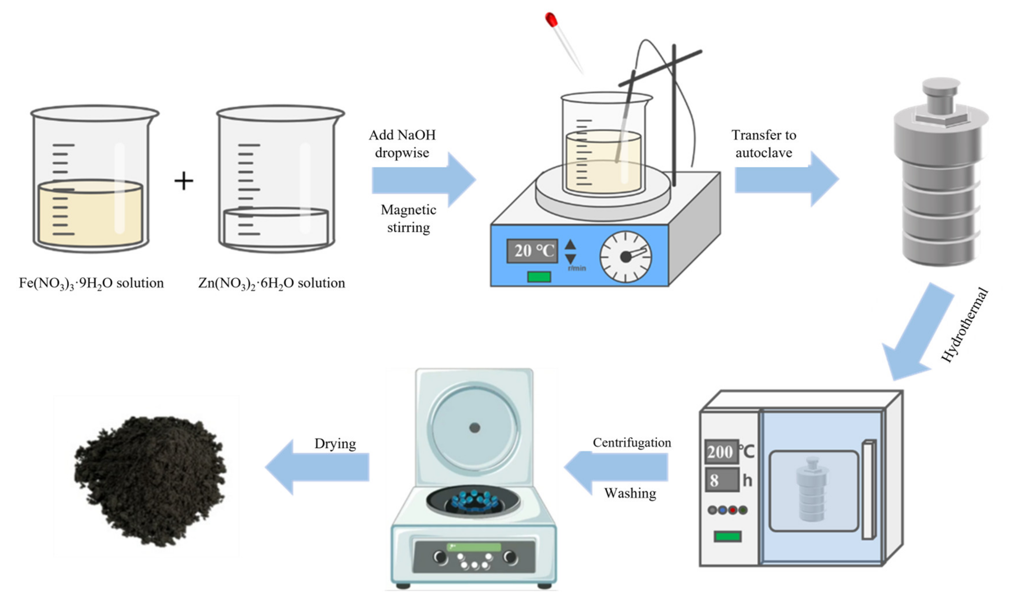
2.1. Hydrothermal Synthesis/Solvothermal Process

The hydrothermal synthesis involves dissolving a 1:2 ratio of Zn2+ and Fe3+ in water, followed by the dropwise addition of ethylene glycol or ethanol with stirring to ensure that the solution is well-mixed. The resulting mixture is then placed into an autoclave and heated under high pressure to dissolve and recrystallize the precursor(s). The purity, size, saturation magnetization value, and photocatalytic activity of the product can be controlled by varying the concentration of the reactants, the hydrothermal temperature, reaction time, pH conditions, amount of surfactant, etc. In spite of its potential for the large-scale synthesis of ferrite, the hydrothermal synthesis has disadvantages including long reaction times, expensive autoclave equipment, and difficult to observe crystal growth. Because several materials used in hydrothermal synthesis are insoluble in water but readily soluble in organic solvents, the solvothermal method has been developed. The solvothermal method follows the same steps as hydrothermal synthesis with the exception that the precursor is dissolved in a non-aqueous organic solvent rather than water. In the solvothermal method, the solvent strongly affects the properties of the materials and the choice of an unsuitable solvent may even degrade the properties of the resulting materials. A schematic representation of the hydrothermal synthesis of ZnFe2O4 is shown in Figure 3.
Zhu et al. [57] prepared ZnFe2O4 by the hydrothermal method with the hydrothermal reaction temperature of 160, 180, and 200 °C, respectively. The averaged crystal size of ZnFe2O4 increased from 7.3 to 8.2 nm, the saturation magnetization value increased from 15.05 to 24.56 emu/g, and the crystal density increased upon increasing the hydrothermal temperature (Figure 4). It has also been found by Zhu et al. [57] that the magnetization value of the catalysts synthesized by hydrothermal synthesis was about six times that of the catalysts synthesized by the ceramic method, because part of the Fe3+ in the materials synthesized by hydrothermal synthesis occupied the A site and formed the complex spinel structure. A comparison by Lestari et al. [58] showed that the saturated magnetization value of ZnFe2O4 synthesized by the hydrothermal method was ~10 times higher than that of the sol-gel method.
Kurian et al. [59] synthesized ZnFe2O4 utilizing both a hydrothermal synthesis and solvothermal process using ethylene glycol and a small amount of polyethylene glycol as solvents. The hydrothermal method produced ZnFe2O4 with a particle size of 10.8 nm and crystallite size of 6.95 nm, whereas the solvothermal method produced a spherical porous structure composed of multiple 5–20 nm crystallites with a particle size of 87.3 nm. Not only did the nanoparticles prepared using the solvothermal method have a lower band gap of 0.3 eV than those prepared via the hydrothermal synthesis, but they also exhibited a saturation magnetization value of 57.76 emu/g, which was 7.6 times greater. Yoo et al. [60] found that the pure phase ZnFe2O4 was prepared by increasing in pH from 10 to 14 and reducing the hydrothermal reaction time from 12 to 5 h. Based on this, the filling pressure was controlled to obtain ZnFe2O4 nanoparticles of different diameters. When 70 mL of the reaction solutions was filled to the 100 mL reaction vessel, the average particle size was only 6 nm, which was much smaller than the critical size of 15–18 nm. At this size, the nanoparticles were transformed from the multi-domain to single-domain, which resulted in superparamagnetic behavior. Remanence was absent after the magnetic field was removed.

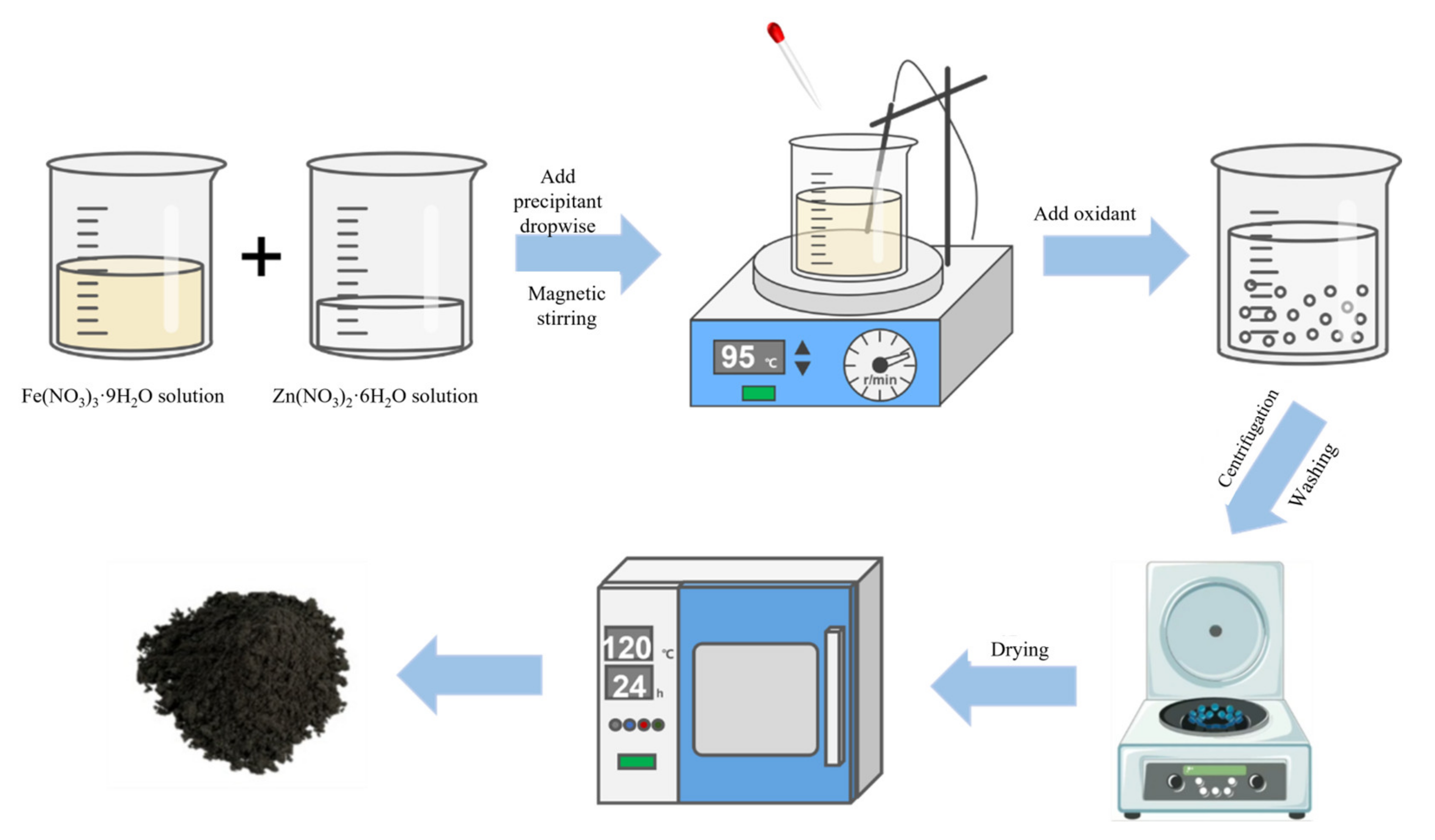
2.2. Co-Precipitation Technique
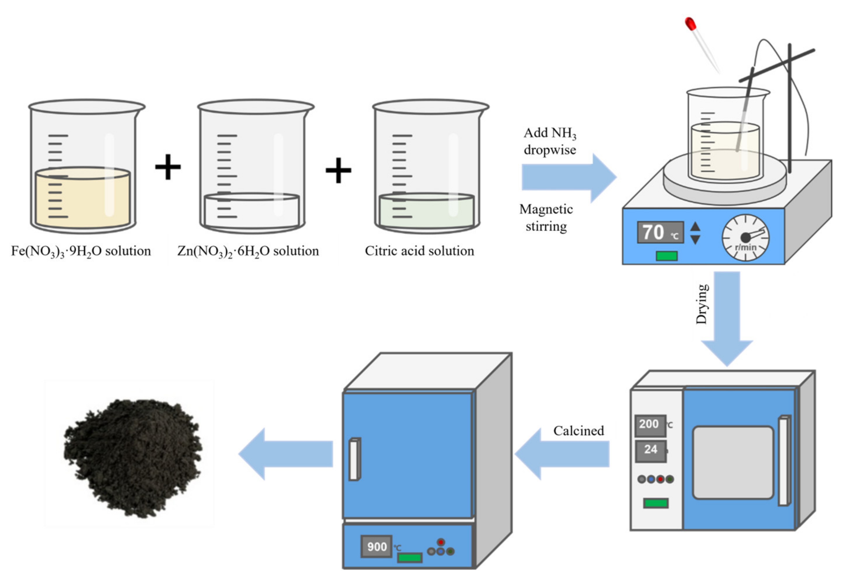
As for the co-precipitation technique, soluble metal ions (such as sulfate, nitrate, and chloride) are co-precipitated with a precipitant such as NaOH, oxalate, or carbonate in a solvent and the precipitate is then dried and sintered under an appropriate atmosphere and temperature to obtain crystalline oxides. The co-precipitation technique is suitable for large-scale production since it is simple and economic. However, there are disadvantages including the agglomeration of the final products, pH-dependent reaction, and dropwise addition of the precipitant. Figure 5 shows the general steps of the co-precipitation technique used for the synthesis of ZnFe2O4.
Nguyen et al. [61] showed that the purity of ZnFe2O4 obtained from the co-precipitation technique was significantly affected by the pH conditions; the higher the pH, the purer the product. At a pH of 6, α-Fe2O3 was produced due to insufficient Zn2+, and the precipitate was not pure. High purity ZnFe2O4 products were obtained upon increasing the pH to 13. ZnFe2O4 with particle diameters of 4 and 6 nm were prepared by Hoque et al. [38] by a co-precipitation technique. Using low-temperature spin kinetics and frequency dependence of the magnetization rate, it was demonstrated that ZnFe2O4 materials with these particle diameters are superparamagnetic.

2.3. Sol–Gel Method

The sol-gel method is a wet chemical method used to prepare ferrite nanoparticles. In this method, a solution containing precursors, usually metal chlorides and metal alkyl oxides, is first converted into gels by hydrolysis, condensation, and polymerization. The gel was further dried and calcined to finally obtain the desired crystalline oxides. The sol-gel method requires low reaction temperatures, no special equipment, and produces a uniform distribution of phases and particles in the multi-component system with controlled size and morphology, but the nanoparticles need to be heat treated to remove the impurities to improve the final purity of the materials. Figure 6 shows a schematic representation of the general steps used in the sol-gel synthesis of ZnFe2O4.
Somvanshi et al. [62] prepared ZnFe2O4 with a specific surface area of 11.534 m2/g using the sol-gel self-combustion method. Sonia et al. [63] found that ZnFe2O4 prepared using the sol-gel method had better homogeneity but slightly lower stability when compared to the material prepared via the co-precipitation technique. Prasad et al. [64] used citrate gel to synthesize ZnFe2O4 with an average size of 22 nm and saturation magnetization value of 12.87 emu/g. Kumar et al. [65] studied the effect of the calcination temperature on the material properties; an increase in the calcination temperature from 550 to 900 °C led to a decrease in the saturation magnetization value from 6.84 to 4.26 emu/g.

2.4. Biosynthesis Method

The features of simplicity, low cost, and low waste of biosynthesis make it an emerging synthetic method. It has been successfully demonstrated that nanoparticles comprised of metals and their oxides can be produced using alternative biological compounds such as the extracts from plant roots and leaves, fruit peels, seeds, biomass waste, sugar, and bacteria [66]. Vitamins, proteins, and mineral salts present in biomaterials provide nanoparticles with excellent compatibility and can also act as capping and reducing agents to enhance their stability and reduction rate [67]. Due to the biodiversity of plants, the preparation of nanoparticles via biosynthesis has a promising future.
Balasubramanian et al. [68] synthesized ZnFe2O4 via a biosynthesis method. The flower extract of nyctanthes arbor-tristis and a mixed solution of iron nitrate and zinc nitrate were thoroughly mixed and precipitated. The precipitate was then dried and calcined to obtain pure ZnFe2O4 nanoparticles. The uncalcined nanoparticles had a particle size of 4.5 nm, specific surface area of 200 m2/g, and pore size of 2.8 nm, which were 0.375, 4, and 2.92 times higher than those obtained using a calcination temperature of 800 °C, respectively. In the RhB degradation experiments, the ZnFe2O4 synthesized in this study was able to degrade 85–100% of the contaminant within 30 min. Surendra et al. [69] prepared a ZnFe2O4 nano sample with Jatropha extract assisted combustion, which had the morphology of porous flakes and agglomeration. Due to the small particle size and active sites in the starting material, which can remove up to 98% of malachite green under UV irradiation, it exhibited good photocatalytic activity. Rahmani et al. [70] dissolved metal nitrate precursors in a solution of Simbang Darah leaf extract. The hydrothermal synthesis was carried out and 10–80 nm spherical clusters of ZnFe2O4 were prepared, which were able to degrade 99.66% of direct red 81 dye after 2 h of solar irradiation. Tatarchuk et al. [71] used quince fruit extract to dissolve metal nitrate, where the obtained solution was heated for 30 min to evaporate. The formed gel ignited by itself and burned slowly with large amounts of gases. The resulting ferrite powder was ground and washed twice with distilled water to prepare paramagnetic CoxZn1−xFe2O4 (where x = 0.0 … 1.0 with step 0.2). The photodegradation rates of organic pollutants by ZnFe2O4 with different preparation methods are shown in Table 1.

2.5. Other Synthesis Methods

In addition to the above-mentioned methods, the methods used to prepare ZnFe2O4 also include microwave, ball milling, vapor deposition, acoustic chemical, pulsed laser, ceramic, and spray pyrolysis methods. Listed in Table 2 are the preparation principles, advantages, and disadvantages of these methods.

2.5.1. Microwave-Assisted Method

The microwave-assisted method is a method that uses microwave to provide a thermally uniform nucleation environment for materials [79,80]. Microwave assisted synthesis technology has the characteristics of a fast heating rate, simple operation, short reaction time, high energy utilization efficiency, low cost, and high yield. However, this method requires complex equipment, which needs different reactors to apply different pressures, and needs long reactor operation times.
The ternary dual Z-scheme composite g-C3N4/ZnFe2O4/Bi2S3 was synthesized by Sarkar et al. [81] through a simple microwave-assisted process. Through the experiment, the best working condition was determined as the oxidant peroxymonosulfate (PMS) reached 1 g/L, the g-C3N4/ZnFe2O4/Bi2S3 (10 wt%) dose was 0.25 g/L, the concentration of 2,4,6-trichlorophenol (TCP) was 20 mg/L, and the degradation rate of TCP increased to 98.9% (Figure 7a–c). A dose of g-C3N4/ZnFe2O4/Bi2S3 (10 wt%) exceeding 0.25 g/L resulted in a reduced degradation rate of TCP due to catalyst particle agglomeration and increased solution turbidity. Increasing the pollutant concentration to 500 mg/L reduces the degradation efficiency to 60% due to the fact that, at larger TCP concentrations, more intermediates and by-products are produced, which consume free radicals and thus reduce the effective activity of the system (Figure 7d). In the reusability test, the TCP degradation rate decreased to 81.5% after five cycles, which was due to the leaching of Zn, Fe, and Bi ions from the catalyst after each cycle (Figure 7e).
Tamaddon et al. [82] prepared novel magnetically separable ZnFe2O4/methylcellulose spherical-shaped nanoparticles by microwave-assisted co-precipitation for the degradation of metronidazole (MNZ) under UV lamp irradiation. The surface morphologies of these spherical particles in the ZnFe2O4@methylcellulose were smooth, uniform, and compact, while the particles were loosely aggregated. The removal efficiency of MNZ was 92.65% in the synthetic samples and 71.12% in the real samples. The removal efficiency decreased with an increasing dose of the model pollutant due to the photocatalyst surface saturation at higher concentrations. Similar conclusions were also obtained by Sarkar et al. [81].

2.5.2. Ball Milling Method

Ball milling is a process in which a mixture of metal salts is ground either dry or wet so that the compound is repeatedly broken and reconstituted. Ball milling has the advantages of low cost, simple operation, no use of toxic and expensive solutions, prevention of particle agglomeration, etc., and has the potential for large-scale production. The product purity of the ball milling method is affected by the grinding time. It takes tens of hundreds of hours to obtain a pure product, and there is a risk of material contamination during milling. Zhang et al. [83] obtained pure ZnFe2O4 by microwave-assisted ball milling for 30 h. The saturation magnetization value reached 82.23 emu/g, which was much higher than that obtained by high-energy ball milling (30 emu/g) [84], hydrothermal synthesis (12 emu/g) [85], and the solvothermal method (24.32 emu/g) [86].

2.5.3. Sonochemical Method

The sonochemical method is a well-defined method for nanoparticle preparation that uses ultrasound as the driving force to accelerate the chemical reaction and increase product yield. When ultrasound is transmitted through a solution of precursors, the high energy of the sound ultrasound field is concentrated and causes acoustic cavitation in the solution. Accompanied by the collapse of the cavitation bubble, the energy is released in a very small space, so that the liquid environment produces an unusually high temperature to increase the speed of the chemical reaction. The method has the advantages of low energy consumption, high reaction rate, uniform product size, simple operation, and high economic benefit. However, there are also some disadvantages such as low magnetic moment and poor crystallinity. Patil et al. [87] prepared ZnFe2O4 with superparamagnetic behavior and a saturation magnetization of 5.321 emu/g by the sonochemical method. Raja et al. [88] synthesized ZnFe2O4/CeO2 nanocomposites by the sonochemical method for the degradation of methyl orange (MO) in visible light. Compared to the pure ZnFe2O4, the ZnFe2O4/CeO2 (1:1) nanocomposite exhibited a higher specific surface area and pore volume, thus providing more active sites for reactant molecules and reached a higher MO degradation efficiency.

3. Modification of ZnFe2O4-Based Photocatalysts

Since the band gap of ZnFe2O4 was only 1.9 eV, the photogenerated electron-hole pairs quickly recombined, thus decreasing its photocatalytic activity. To improve its photochemical performance, three ways of catalyst modification have been proposed: (1) Elemental doping, which introduces a second or even a third element; (2) composite modification, which compounds ZnFe2O4 nanoparticles with materials that have high activity and a low carrier recombination rate (e.g., metal oxides, metal sulfides, and carbon-based materials); and (3) morphological modification, which optimizes the nanostructure by turning the simple stacked low-dimensional materials into high-dimensional materials to increase the specific surface area, reaction sites, and carrier transport efficiency while decreasing the possibility of carrier recombination.

3.1. Elemental Doping

The photochemical properties of ZnFe2O4 can be enhanced upon doping into the gaps in the crystal structure of ZnFe2O4: Zn2+ at the A sites, Fe3+ at the B sites or O sites. Table 3 shows some of the metal ion doping for ZnFe2O4 and it is clear that the type, sites, and amount of doping affect the band gap and, in turn, the photocatalytic activity of ZnFe2O4. By using this modification method, it is possible to change the size of the cells in the material as well as increase the specific surface area and number of active sites. This can have a positive effect on its photochemical, electrical, and magnetic properties.
Musa et al. [95] prepared nanofibers comprised of pure ZnFe2O4 and Al-doped ZnFe2O4 (0.5% Al) using a combination of the electrospinning method and calcination. The diameter of the pure ZnFe2O4 nanofibers remained unchanged upon increasing the calcination temperature, but the diameter of the Al-doped ZnFe2O4 nanofibers decreased significantly, which proved that the Al ions had been successfully doped into the ZnFe2O4 crystal structure and changed the crystal structure and microscopic properties of the resulting samples (Figure 8a). On one hand, Al doping promoted the reversal of the cation distribution within the material and improved the magnetization value of the material, thus enhancing the recovery performance of the ZnFe2O4 photocatalyst; on the other hand, Al doping introduced an impurity energy level and inhibited the carrier complex. Al doping reduces the UV-Vis diffuse reflectance spectroscopy (DRS) absorbance of ZnFe2O4 (Figure 8b), and the energy band gap of the doped material was reduced to 1.7 eV (Figure 8c), and the percentage of RhB degradation increased from 25.66% to 38.83% after 60 min of visible light irradiation (Figure 8d).
Reyes-Rodríguez et al. [96] obtained Zn1−xMgxFe2O4 samples with an average diameter of 15.5 nm by doping Mg2+ into the ZnFe2O4 crystal structure using the sol-gel method. The saturation magnetization value of Zn1−xMgxFe2O4 first increased and then decreased with an increasing Mg2+ doping concentration, reaching a maximum value of 44.85 emu/g at x = 0.5. Son et al. [89] replaced Zn2+ with Ni2+ and the resulting Zn1−xNixFe2O4 crystal structure was transformed into an inverse spinel structure. The oxygen vacancies increased, and the material band gap was reduced to a minimum value of 1.72 eV with an increasing Ni2+ doping ratio, thus improving the photocatalytic performance.
Peymani-Motlagh et al. [97] prepared Co0.5Zn0.5Fe1.9R0.1O4 (R = Gd, Pr, Sm) upon introducing Gd3+, Pr3+, and Sm3+ into the B sites, and the photocatalytic activities of the observed MO degradation were in the order of: Gd3+ > Pr3+ > Sm3+. Borhan et al. [98] prepared ZnFe2−xCrxO4 (x = 0, 0.25, 0.5, 1.0, 1.5, 2.0) using the sol-gel method. The saturation magnetization value decreased from 4.35 to 3.25 emu/g upon increasing the chromium content.
Li et al. [99] prepared a carbon-nitrogen doped ZnFe2O4 yolk-shell structure by adding dopamine as a carbon and nitrogen source. The carbon and nitrogen ions replaced the oxygen anions, and the energy band position was slightly negatively shifted, which expanded the absorption range of visible light. The internal void of the yolk-shell structure could produce a stronger scattering of incident light. The internal void of the yolk-shell structure could also produce a stronger scattering of incident light. The degradation rate of the yolk-shell structured ZnFe2O4 was 1.73 times higher than that of undoped ZnFe2O4. The yolk-shell structured ZnFe2O4, after doping with C and N, exhibited a 1.3-fold higher photocatalytic performance than that of the broken structure after doping with C and N.

3.2. Composite Modification

Since 46% of the complete energy in sunlight is situated in the visible light region, ZnFe2O4 nanoparticles with narrow band gaps (1.9  eV) could be more adequate than traditional semiconductors such as TiO2 (3.2  eV) and ZnO (3.2  eV) in employing solar energy to photocatalytically degrade contaminants. However, few semiconductor photocatalysts with their band edge potentials are suitable on their own. Combining ZnFe2O4 with other semiconductors, metals, and carbon materials is necessary for effective catalyst applications. The key requirement for new photocatalysts is to optimize the band gap so that the electron complexation rate is negligible and to make the CB edge more negative and the VB edge more positive. That is, to make the semiconductor highly reducible for electrons in the CB (+0.5 to −1.5 V vs. normal hydrogen electrode (NHE)) and highly oxidizable for holes in the VB (+1.0 to +3.5 V vs. NHE) (Figure 9).

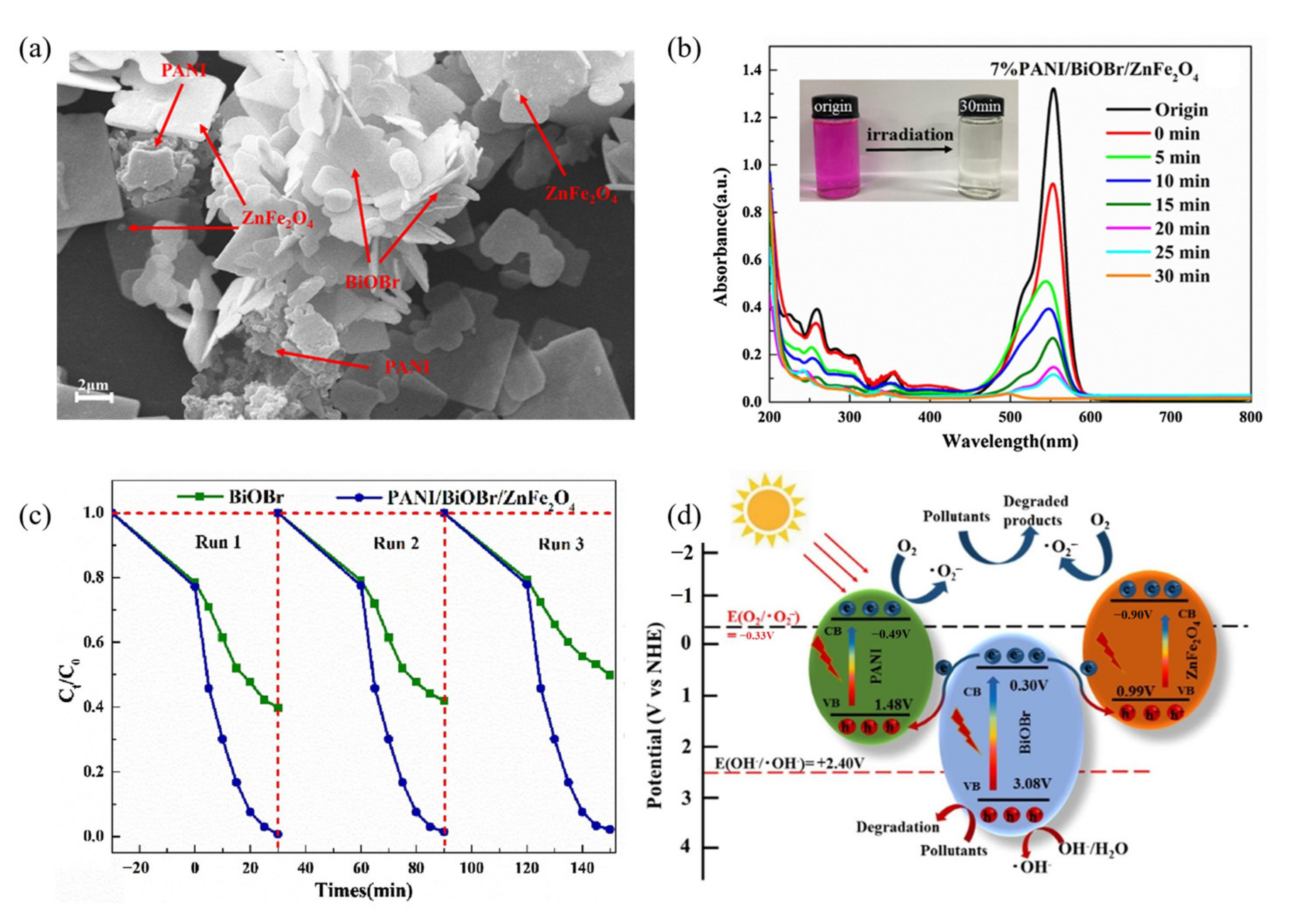
3.2.1. Composites of ZnFe2O4 and Conducting Polymers

Commonly used conducting polymers include polyacetylene (PCE), polypyrrole (PPY), polyindole (PBE), and polyaniline (PANI), and their copolymers, which are simple and inexpensive to prepare. The combination of ZnFe2O4 with conducting polymers not only reduced the material agglomeration, but also increased the number of active sites, shortened the carrier diffusion pathway, and improved the carrier mobility.
Using the in situ polymerization method, Das et al. [101] prepared a series of polypyrrole (PPY) sensitized zinc ferrite/graphitic carbon nitride (ZFCN) heterojunction (ZFCN@10PPY, ZFCN@20PPY, and ZFCN@30PPY) nanocomposites. High resolution transmission electron microscope images showed that the edges of the ZnFe2O4 and g-C3N4 (graphitic carbon nitride, CN) lattice were interconnected (Figure 10a), indicating that the two components were not simply deposited, but formed a heterogeneous structure. The nitrogen adsorption-desorption isotherms and pore size distribution, as shown in Figure 10b,c, indicate that the specific surface area of ZFCN@20PPY was higher when compared with the CN, PPY, and ZFCN, and the pores that formed were narrower. Figure 10d shows that ZFCN@20PPY had a very low luminescence spectral intensity, confirming that the low band gap polypyrrole can act as a photosensitizer to provide additional electrons for ZnFe2O4/CN. The photocatalytic mechanism of the material is shown in Figure 10e, in which the photoexcited electrons could easily migrate from PPY to the CB of CN, and then further transfer to the CB of ZnFe2O4, and the photogenerated electrons captured reactive oxygen species such as superoxide and hydroxyl groups on the ZnFe2O4 surface to degrade the pollutants.
Zhang et al. [102] successfully prepared a dual Z-scheme heterojunction photocatalyst PANI/BiOBr/ZnFe2O4 (Figure 11a) using the hydrothermal method. The lone pairs of electrons on the outer nitrogen atoms of PANI can enhance the adsorption of pollutant molecules and the BiOBr flower-like structure improves the oxidation ability of the composite. By forming heterojunctions, the visible response range is widened to include the entire visible spectroscopy. A slight blue shift was observed as the irradiation time increased, caused by de-ethylation and the breakdown of the conjugate structure of RhB as the intensity of the characteristic peak at 554 nm decreased (Figure 11b). The reaction solution also turned from magenta to colorless. The results showed that the PANI/BiOBr/ZnFe2O4 composites exhibited a higher stability than that in RhB degradation (Figure 11c). When exposed to visible light, pure BiOBr showed a degradation rate of 50.26%, compared to the previous of 60.25%. However, RhB degradation by the PANI/BiOBr/ZnFe2O4 composite material was not significantly affected, decreasing from 99.26% to 97.86%. As shown in Figure 11d, a dual Z-scheme reaction mechanism based on the results of fluorescence photoluminescence (PL) spectroscopy, free radical capture experiments, ESR spin-trap with DMPO technique, calculation using the horizontal axis intercept of the Mott-Schottky plots has been proposed. PANI (−0.49 V vs. NHE) and ZnFe2O4 (−0.90 V vs. NHE) accumulate photogenerated electrons with strong reducing abilities, and these electrons can easily reduce O2 to O2. In addition to directly oxidizing pollutants, photogenic holes (3.08 V vs. NHE) that accumulated in the VB of BiOBr can also oxidize H2O/OH to radical OH.

3.2.2. ZnFe2O4 Composite Using Metal Oxides

Combining ZnFe2O4 with some metal oxides with strong oxidation ability, high carrier separation efficiency, and an ability to inhibit electron-hole recombination can improve its performance. Wang et al. [103] prepared S-type heterojunction ZnFe2O4/SnFe2O4 photocatalysts using a one-step solvothermal method. The experimental results showed that a moderate amount of SnFe2O4 (5%) promoted carrier separation and enabled the material to achieve the best visible light absorption efficiency, but an excess of SnFe2O4 led to a wider band gap and hindered the photogenerated carrier capability (Figure 12a). The 5% SnFe2O4/ZnFe2O4 PL intensity was the lowest in all materials (Figure 12b), transient photocurrent was 17.5 × 10⁻⁸ A, about pure ZnFe2O4 (10 × 10−8 A), and 1.75 times that of pure SnFe2O4 (1 × 10⁻⁸ A) (Figure 12c). It was further proved that an appropriate amount of SnFe2O4 was favorable for carrier separation and charge transfer behavior in nano-heterojunction materials. The SnFe2O4 composite doubled the saturation magnetization strength of ZnFe2O4 and enabled the composite to be separated from the aqueous solution quickly and easily (Figure 12d).
Al-Shwaiman et al. [104] prepared CeO2/ZnFe2O4 using a simple chemical co-precipitation technique. The specific surface area of CeO2/ZnFe2O4 was 126.99 m2/g and the mesopore pore size was 2.96 nm, which was 1.46 and 1.16 times that of pure ZnFe2O4 and 1.31 and 1.50 times that of CeO2, respectively. During the degradation of MB, the photocatalytic efficiency of the composites was 1.57 times that of pure ZnFe2O4 and 2.75 times that of CeO2, which verified the improvement in the photocatalytic efficiency observed upon increasing the specific surface area and mesopore size.

3.2.3. ZnFe2O4 Composite Using Metal Sulfides

The presence of elemental sulfur promotes the electron transfer rate of metal sulfides in redox reactions, which makes the conductivity of metal sulfides better than that of the metal oxides. In addition, metal sulfides also have the advantages of good mechanical stability, high thermal stability, easy synthesis, and low cost. It is important to note that metal sulfides exhibit serious photocorrosion, photochemical instability, and other disadvantages, which greatly affect their stability. The combination of metal sulfides with ZnFe2O4 can induce the separation of the photogenerated carriers and inhibit their recombination, which improves the photochemical performance of ZnFe2O4.
Akshhayya et al. [105] prepared ZnFe2O4/SnS2 using an acoustochemically-assisted method and the synergistic effect of the materials resulted in a lower PL spectral intensity in the composites when compared to ZnFe2O4 and SnS2, a lower electron-hole pair recombination rate, and the introduction of defects in the lattice structure of the composites, which increased the transient photocurrent and further improved the electron mobility. Singh et al. [106] prepared CdS-modified ZnFe2O4 nanocubes using a hydrothermal synthesis. Modification using CdS nanoparticles significantly reduced the PL intensity and improved the electron-hole separation. The nanocubes comprised of the ZnFe2O4 and CdS nanoparticles exhibited a synergistic effect to widen the PL band gap and improved the UV-Visible absorption range.

3.2.4. ZnFe2O4 Composites Using Carbon-Based Materials

Carbon materials such as graphene and graphitic carbon nitride have the advantages of a large specific surface area, good dispersion, strong adsorption ability, high electron mobility, and stable performance. Different kinds of carbon materials combined with ZnFe2O4 can effectively improve the photochemical properties of ZnFe2O4 [107].
Jun et al. [108] prepared a unique poly hybrid by depositing lanthanum oxide carbonate (La2CO5) and ZnFe2O4 on reduced graphene oxide (LaZF@rGO) using a hydrothermal-precipitation method. Compared with the pure ZnFe2O4, the band gap of the composite was reduced by 0.44 eV (Figure 13a). The activities of the photocatalytic materials in different proportions were arranged as follows: 1:1 LaZF@rGO (0.0612 min−1) > ZF@rGO (0.0314 min−1) > 2:1 LaZF@rGO (0.0218 min−1) > 4:1 LaZF@rGO (0.0105 min−1) > 5:1 LaZF@rGO (0.0074 min−1) (Figure 13b). Figure 13c shows the inhibition effect of different free radical scavengers on the reaction, and the inhibition effect of benzoquinone (BQ) was the most obvious. It can be seen that the addition of the catalyst can generate superoxide radicals under light. The magnetization value of LaZF@rGO was 24.59 emu/g, which was lower than that of ZnFe2O4 (41.90 emu/g), but still sufficient for its magnetic separation from aqueous solutions (Figure 13d). As shown in Figure 13e, the electrons excited by La2CO5 at the CB were transferred to the CB of the ZnFe2O4 through the rGO surface. The VB holes of La2CO5 migrate to the VB of ZnFe2O4 with the help of rGO. The rGO acts as a medium for electron-hole transport, thereby inhibiting the recombination of electron-hole pairs.
Baynosa et al. [109] prepared ZnFe2O4/rGO using a one-step hydrothermal synthesis and found that the interaction between the ZnFe2O4 nanoparticles and graphene flakes limited the crystallization of ZnFe2O4, so the addition of acetate acts as a protective agent to promote the stable generation of nanocrystals. The specific surface area of the ZnFe2O4/rGO composite after loading the nanoparticles onto graphene flakes was as high as 141.3 m2/g, which was 1.67-flod higher than the surface area of the rGO-ZnFe2O4 hybrid prepared using a hydrothermal synthesis reported by Wang et al. [110]. Gao et al. [111] synthesized C-ZnFe2O4 by the microwave induction method, which could absorb 90% of the incident waves and had good pumping properties.

3.3. Morphological Modification

As the morphology of ZnFe2O4 change, its carrier transport efficiency, surface area, active sites, and light absorption capacity change, which affect its photochemical properties. Recently, many researchers have explored the different morphologies of ZnFe2O4 and their photochemical properties. According to the extent to which the spatial dimension of the nanomaterials is constrained by the nanometer size, ZnFe2O4 can be classified as zero-, one-, two-, or three-dimensional. Zero-dimensional nanomaterials mainly include nanoparticles and nanoclusters, which are the basis of all morphological modifications and usually have advantages such as a large specific surface area and abundant active sites, but also have disadvantages such as facile agglomeration and poor light absorption. The current modifications of zero-dimensional ZnFe2O4 are mainly divided into two types: (1) Adjusting the microstructure of the ZnFe2O4 nanoparticles by adjusting the preparation conditions such as solvent, temperature, precipitant, and pH; and (2) assembling ZnFe2O4 onto other high-performance or high-dimensional photocatalysts to improve the photocatalytic performance. Figure 14 shows the SEM images of ZnFe2O4 with different morphologies.

3.3.1. One-Dimensional ZnFe2O4

One-dimensional nanomaterials include nanowires, nanotubes, and nanorods. The wire structure can reduce the agglomeration of spherical particles, expose more active sites, and improve the radial electron transport efficiency. Ghazkoob et al. [112] prepared ZnFe2O4 nanowires and ZnFe2O4 nanowire/BiVO4 composites with an average length of 6.3 μm and an average diameter of 38 nm using AC pulse electrodeposition and hydrothermal synthesis, respectively. The nanowire structure increased the contact area between the catalyst and reaction solution and improved the photocatalytic efficiency, but the magnetic nanowires of a single material aggregated and produced curling, and the adjacent magnetic moments were not parallel in the curling mode, which led to the reduction in the nanowire saturation magnetization value. The BiVO4 coating effectively reduced the aggregation and curling among the ZnFe2O4 nanowires and improved the magnetization value of the material.
Zhang et al. [116] synthesized nanotubes composed of ZnFe2O4 nanotubes with a diameters of 5~10 nm using carbon nanotubes as a template (Figure 15a). The nanotube structure not only increased the specific surface area, but also the saturation magnetization value up to 45.4 emu/g. Liu et al. [113] developed a method to fabricate hollow oxides on electrospun nanofiber templates using the atomic layer deposition of oxides, and the resulting products not only retained the nanofiber structure between the tubes, but also appeared to have hollow structures in their bodies (Figure 15b). The extra-long one-dimensional nanofibers provided good radial transport channels for carriers and increased the ability of charge trapping by the catalyst surface, thus facilitating the redox reaction. The hollow structure shortened the carrier transport distance from the interior to the surface, increased the amount of light scattering within the fiber, and improved the light absorption capacity.

3.3.2. Two-Dimensional ZnFe2O4

Two-dimensional nanomaterials are only available as nanofilms with a high specific surface area, low flat-band dot positions, large charge layer widths, and short carrier diffusion lengths, which make them ideal for developing photocatalysts. Zhu et al. [117] prepared new porous ultrathin ZnFe2O4 nanofilms with a different number of layers using the Langmuir–Blodgett (LB) method on ZnFe2O4 that was close to the monolayer, closely aligned at a surface pressure of 40 mN/m, and a deposition rate of 1 mm/min. Figure 16a,b shows that the single layer ZnFe2O4 (LB-ZFO-1) had the highest corresponding SPV signal of 4.87 μV compared to the multi-layer ZnFe2O4. The quantum efficiency of the single layer was 1.7 times that of the five layer (LB-ZFO-5) and 2.6 times that of the nine layer (LB-ZFO-9). It was proven that the monolayer ZnFe2O4 thin film had better efficiency of the photogenerated carrier separation and charge transfer behavior. Both monolayer and multilayer ZnFe2O4 films in the 500, 1000 and 2500 Hz frequency measurements of the Mott–Schottky figure showed a positive slope of the linear area, indicating that the preparation of the ZnFe2O4 film was indeed an n-type semiconductor (Figure 16c). Comparing the capacitive properties of the TiO2 porous films with ZnFe2O4, they found that both the monolayer and multilayer films had capacitive properties, which led to the degradation of the photocatalytic properties of the films (Figure 16d).
Chen et al. [118] prepared translucent ZnFe2O4 films on MgAl2O4 substrates using pulsed laser deposition with a Curie temperature close to 327 °C. Luo et al. [119] prepared ZnFe2O4/Fe3+-TiO2 heterojunction composite films using lithography-assisted chemical solution deposition techniques. The film with a 5 μm thickness had a surface contact angle as low as 3° in the MB solution and was super-hydrophilic. With the recycling of the material, the film was gradually soaked, and the time to degrade the pollutants of the same concentration decreased from 24 min to 19 min.
Erusappan et al. [120] synthesized ZnFe2O4/BN two-dimensional heterostructure nanocomposites with different wt% of boron nitride (BN) by the solvothermal method. The incorporation of 9.3% BN over the ZnFe2O4 catalysts showed the best synergistic effect between Zn and BN, which enhanced the charge separation and electron transport efficiency. The degradation rate of dyes (CR) (Figure 17a) and antibiotics (Figure 17b) reached 0.00982 and 0.01019 min−1, respectively, showing the best proof. Almost no activity loss was found (Figure 17c,d) after four cycles of reuse, indicating good photostability.

3.3.3. Three-Dimensional ZnFe2O4

There are a wide variety of three-dimensional nanomaterials, which are usually low-dimensional nanomaterials that have been modified or combined to form three-dimensional composite nanomaterials such as nanowire arrays [121], hollow spheres [122], and frameworks [123,124]. Three-dimensional nanomaterials can effectively use the physical structure to expose more active sites and improve light absorption.
Chen et al. [114] constructed a NiS-modified CuO@ZnFe2O4 hierarchical nanoarray structure photocatalyst that was composed of CuO nanowires as the core and ultrathin ZnFe2O4 nanosheets as the shell, as shown in Figure 18. The nanowire array structure not only had large gap channels, a large specific surface area, and many active sites, but also had a high incident light capture capability. Meanwhile, the layered structure allowed the charge to be transferred outward from the inner part of the catalyst layer-by-layer, which significantly reduced the charge complexation rate.
Hollow structures such as hollow spheres and hollow skeletons have the advantages of low density, good dispersion, large specific surface area, short carrier transfer pathway, adjustable photorefractive index, strong light scattering ability, and high plasticity, which make them a promising research direction. Hollow structures are synthesized using hard-template, soft-template, and self-template methods.
Zhang et al. [125] synthesized double-shell Ag/AgCl/polydopamine (PDA)/ZnFe2O4 nanocubes using hollow ZnFe2O4 nanospheres (G-ZnFe2O4) prepared via the hydrothermal and self-etching process with PDA used as the template and reducing agent. The PDA template was dissolved in KOH solution to form double-shelled Ag/AgCl/ZnFe2O4 hollow nanocubes (DAGZNs). Due to the dissolution of the PDA, the specific surface area of DAGZNs increased from 43.90 (before the removal of the template) to 79.73 m2/g, and the pore size expanded from 2.69 to 6.84 nm (Figure 19a,b). Figure 19c indicates that the PL intensity of the DAGZN photocatalyst was significantly higher than that of the bulk material, and that its high photocatalytic activity may be due to its double-shell hollow structure. The transient photocurrent in Figure 19d shows that the photocurrent response of DAGZNs was stronger than that of the Ag/AgCl/PDA/G-ZnFe2O4 nanoparticles, which further demonstrates the advantage of the hollow multi-shell structure. Figure 19e shows a schematic representation of the light scattering effect in the hollow double-shell structure and the possible response mechanism of the photocatalytic performance of DAGZNs under visible light irradiation proposed by the authors based on their results. The external cubic structure and internal hollow sphere structure increased the number of channels for light scattering inside the material, and these channels could effectively enhance the light absorption, and therefore the photocatalytic activity.
Cao et al. [115] synthesized a novel hollow porous cube ZnFe2O4 via calcination using a metal-organic skeleton (Prussian blue (PB)) as a precursor. During calcination, the organic matter in the precursor slowly decomposed, in which the gas diffused outward to create pores in the ZnFe2O4 oxide layer. The structure could increase the amount of light scattered and improve the photocatalytic activity.

3.4. Pollutant Degradation Performance of Photocatalysts Prepared Using Different Modification Methods

ZnFe2O4 has a narrow band gap of 1.9 eV, which allows the material to easily generate electron-hole pairs that sequentially produce oxygen-containing substances. Therefore, ZnFe2O4 nanoparticles are widely used in the study of the removal of organic pollutants such as organic dyes, phenols, antibiotics, etc.

3.4.1. Organic Dyes

Organic dyes are typical pollutants in wastewater from the textile industry, which is one major source of industrial wastewater pollution [126]. Organic dyes can be divided into cationic dyes (RhB, MB), anionic dyes (methylene orange (MO), (Congo red (CR)), and disperse dyes according to the ionic charge they dissociated into in the aqueous medium. In the photocatalytic degradation of organic dyes, the dye molecules are first adsorbed on the surface of the photocatalyst and then participate in the redox reaction. Chahar et al. [127] fabricated CoxZn1−xFe2O4 (x = 0.0, 0.1, 0.2, 0.3, 0.4 and 0.5) for MB degradation. The results showed that Co0.5Zn0.5Fe2O4 had the highest MB degradation efficiency (77%), which was distinctly higher than that of the undoped ZnFe2O4. Shakil [128] prepared (CdxZn1−xCo0.25Fe1.75O4; x = 0, 0.25, 0.5 and 0.75) for the removal of MO under visible light. Based on the PL spectroscopy analysis, the substitution of Co and Cd resulted in a decrease in PL intensity, which led to reduced photogenerated electron–hole pair recombination thus increases the photocatalytic efficiency. Meanwhile, the composite also showed good reusability and stability.

3.4.2. Phenols

Phenolic compounds are widely found in various industrial processes such as oil refining, the petrochemical industry, the pharmaceutical industry, and resin manufacturing [129]. Absorption combined with incineration is one of the most common method of removing phenolic compounds from aqueous solutions. However, the incineration of phenolic compounds will emit gaseous pollutants such as dioxins, which effect both the environment and human health [130]. Photocatalytic degradation of phenolic compounds is a much more environmentally friendly technique. Mady et al. [131] fabricated aγ-MnO2@ZnFe2O4/rGO nanohybrids for phenol degradation using a one-pot hydrothermal self-assembly method. The nanohybrid had a specific surface area as high as 376.88 m2/g, which provided a large number of adsorption/active sites. The catalytic efficiency of the γ-MnO2@ZnFe2O4/rGO nanohybrid was the highest (100%) compared to the other composites including δ-MnO2@ZnFe2O4, γ-MnO2@rGO, and ZnFe2O4@rGO. This high efficiency may be attributed to the high surface area of the nanohybrid compared to the other composites and the ability of both MnO2 and ZnFe2O4 to activate PMS through an electron transfer mechanism to produce sulfate radicals.

3.4.3. Antibiotics

Antibiotics usually come from pharmaceutical industries, which produce pharmaceuticals and personal care products. The direct discharge of wastewater containing untreated antibiotics will endanger human and animal health by bio-accumulating and bio-magnifying into the food chain, increasing antimicrobial resistance and inhibiting the growth of bacteria that are beneficial to the eco-system [132]. Dhiman et al. [133] synthesized Ni1−xZnxFe2O4 (x = 0, 0.1, 0.3, and 0.5) for levofloxacin degradation using the solution combustion method. Ni0.1Zn0.9Fe2O4 showed the highest degradation efficiency of levofloxacin (96.8%) within 90 min under visible light irradiation. O2− was regarded as the main active species in the photocatalysis reaction. Levofloxacin was degraded into low molecular weight compounds through four pathways including de-piperazinylation, decarboxylation, quinolone moiety transfer, and piperazine ring oxidation.
Table 4 lists some of the results obtained in recent studies on the photocatalytic performance of ZnFe2O4-based composites, which are prepared by various modification methods in pollutant degradation. It can be seen that most of the ZnFe2O4-based composites showed high photocatalytic activity within a short reaction time. A few studies have also shown a good degradation effect on actual wastewater such as pharmaceutical wastewater.

4. Conclusions

ZnFe2O4 is a photocatalyst that exhibits high stability, high carrier separation efficiency, and easy recovery due to its spinel structure, narrow band gap, and special magnetic properties. Commonly used ZnFe2O4 synthesis methods include the hydrothermal/solvothermal method as well as the co-precipitation, sol-gel, and biosynthesis methods. Recently, various catalyst modification methods have been studied such as elemental doping, composite modification, and morphological modification to improve the photocatalytic activity and reusability of ZnFe2O4. Great progress has been made in ZnFe2O4 photocatalyst research, but many challenges still remain. Therefore, it is necessary to conduct further research on ZnFe2O4 in the field of photocatalysis, focusing on the following aspects:
(1)
Developing innovative preparation methods. Hydrothermal, co-precipitation, and sol–gel methods are relatively mature, but they are expensive and energy-intensive. Expanding the biosynthesis concept to each preparation method and the development of green methods could reduce the particle size of the nanoparticles and improve their active sites at the same time, with low cost, simple operation, and no chemical pollution. It is possible to improve the biosynthesis methods by studying trace minerals or enzymes, and further research into the synthesis mechanisms can be conducted based on the enzymes and minerals present in plants.
(2)
Expanding materials in a modified way. In order to better suit the practical applications, a variety of methods such as doping and composite modification may be used to improve the performance and stability of single ZnFe2O4 photocatalysts. By doping different elements with defects or combining them with strong redox-capable nanomaterials, heterojunctions can be formed to adjust the material band gap, improve carrier separation, and reduce carrier recombination, thereby improving the photochemical performance.
(3)
There is a need for further investigation into the light absorption mechanisms of different morphologies. At present, the research on the morphological modification of ZnFe2O4 is relatively limited, which mainly focuses on one-dimensional modification. Some studies have shown that the selection of different dimensional modifications leads to an exponential increase in the light absorption capacity. An in-depth understanding of the light absorption and photocatalytic mechanisms of multidimensional materials is needed. Research can be conducted on a number of excellent three-dimensional structures such as hollow multi-shell structures, nanosheet arrays, etc.
ZnFe2O4 photocatalysts have received increasing research attention in recent years. With the efforts of researchers and the advancement of preparation and characterization techniques, it is believed that ZnFe2O4 photocatalysts can be widely utilized in industrial applications in the near future.

Author Contributions

J.Z. and Y.Z. conceived the idea for this review article; Z.C., J.Z. and X.F. prepared a draft of the paper with J.Z. and Y.Z.; Y.Z. and Y.Y. reviewed and edited the manuscript; Z.C. and S.W. reviewed and finalized the paper. All authors have read and agreed to the published version of the manuscript.

Funding

This research was supported by the Natural Science Foundation of Zhejiang Province (grant no. LY20E060003) and the National Natural Science Foundation of China (grant no. 51976094).

Institutional Review Board Statement

Not applicable.

Informed Consent Statement

Not applicable.

Data Availability Statement

Not applicable.

Acknowledgments

The authors are very grateful to the anonymous reviewers for their valuable suggestions and comments.

Conflicts of Interest

The authors declare no conflict of interest.

References

  1. Mishra, A.; Kumari, M.; Swati; Kumar, R.; Iqbal, K.; Thakur, I.S. Persistent organic pollutants in the environment: Risk assessment, hazards, and mitigation strategies. Bioresour. Technol. Rep. 2022, 19, 101143. [Google Scholar] [CrossRef]
  2. Liu, H.; Ren, X.; Chen, L. Synthesis and characterization of magnetic metal–organic framework for the adsorptive removal of Rhodamine B from aqueous solution. J. Ind. Eng. Chem. 2016, 34, 278–285. [Google Scholar] [CrossRef]
  3. Sellaoui, L.; Kehili, M.; Lima, E.C.; Thue, P.S.; Bonilla-Petriciolet, A.; Lamine, A.B.; Dotto, G.L.; Erto, A. Adsorption of phenol on microwave-assisted activated carbons: Modelling and interpretation. J. Mol. Liq. 2019, 274, 309–314. [Google Scholar] [CrossRef]
  4. Gao, Y.; Li, Y.; Zhang, L.; Huang, H.; Hu, J.; Shah, S.M.; Su, X. Adsorption and removal of tetracycline antibiotics from aqueous solution by graphene oxide. J. Colloid Interface Sci. 2012, 368, 540–546. [Google Scholar] [CrossRef]
  5. Ramutshatsha-Makhwedzha, D.; Mavhungu, A.; Moropeng, M.L.; Mbaya, R. Activated carbon derived from waste orange and lemon peels for the adsorption of methyl orange and methylene blue dyes from wastewater. Heliyon 2022, 8, e09930. [Google Scholar] [CrossRef]
  6. Garcia-Rosero, H.; Romero-Cano, L.A.; Aguilar-Aguilar, A.; Bailon-Garcia, E.; Carvalho, A.P.; Perez-Cadenas, A.F.; Carrasco-Marin, F. Adsorption and thermal degradation of Atenolol using carbon materials: Towards an advanced and sustainable drinking water treatment. J. Water Process Eng. 2022, 49, 102987. [Google Scholar] [CrossRef]
  7. Rao, T.; Ma, X.; Yang, Q.; Cheng, S.; Ren, G.; Wu, Z.; Sires, I. Upgrading the peroxi-coagulation treatment of complex water matrices using a magnetically assembled mZVI/DSA anode: Insights into the importance of ClO radical. Chemosphere 2022, 303 Pt 1, 134948. [Google Scholar] [CrossRef]
  8. Sayin, F.E.; Karatas, O.; Ozbay, I.; Gengec, E.; Khataee, A. Treatment of real printing and packaging wastewater by combination of coagulation with Fenton and photo-Fenton processes. Chemosphere 2022, 306, 135539. [Google Scholar] [CrossRef]
  9. Wang, Y.; Jin, X.; Zhuo, N.; Zhu, G.; Cai, Z. Interaction-sedimentation strategy for highly efficient removal of refractory humic substances in biologically treated wastewater effluent: From mechanistic investigation to full-scale application. J. Hazard. Mater. 2021, 418, 126145. [Google Scholar] [CrossRef]
  10. Rebosura, M., Jr.; Salehin, S.; Pikaar, I.; Keller, J.; Sharma, K.; Yuan, Z. The impact of primary sedimentation on the use of iron-rich drinking water sludge on the urban wastewater system. J. Hazard. Mater. 2021, 402, 124051. [Google Scholar] [CrossRef]
  11. Xu, Y.; Zhu, Y.; Chen, Z.; Zhu, J.; Chen, G. A Comprehensive Review on Forward Osmosis Water Treatment: Recent Advances and Prospects of Membranes and Draw Solutes. Int. J. Environ. Res. Public Health 2022, 19, 8215. [Google Scholar] [CrossRef] [PubMed]
  12. Qiu, Y.B.; Ren, L.F.; Shao, J.H.; Xia, L.; Zhao, Y. An integrated separation technology for high fluoride-containing wastewater treatment: Fluoride removal, membrane fouling behavior and control. J. Clean. Prod. 2022, 349, 131225. [Google Scholar] [CrossRef]
  13. Liu, T.; Yao, B.; Luo, Z.; Li, W.; Li, C.; Ye, Z.; Gong, X.; Yang, J.; Zhou, Y. Applications and influencing factors of the biochar-persulfate based advanced oxidation processes for the remediation of groundwater and soil contaminated with organic compounds. Sci. Total Environ. 2022, 836, 155421. [Google Scholar] [CrossRef] [PubMed]
  14. Shang, K.F.; Morent, R.; Wang, N.; Wang, Y.X.; Peng, B.F.; Jiang, N.; Lu, N.; Li, J. Degradation of sulfamethoxazole (SMX) by water falling film DBD Plasma/Persulfate: Reactive species identification and their role in SMX degradation. Chem. Eng. J. 2022, 431, 133916. [Google Scholar] [CrossRef]
  15. McKercher, L.J.; Messer, T.L.; Mittelstet, A.R.; Comfort, S.D. A biological and chemical approach to restoring water quality: A case study in an urban eutrophic pond. J. Environ. Manag. 2022, 318, 115463. [Google Scholar] [CrossRef] [PubMed]
  16. Changotra, R.; Rajput, H.; Dhir, A. Treatment of real pharmaceutical wastewater using combined approach of Fenton applications and aerobic biological treatment. J. Photoch. Photobio. A 2019, 376, 175–184. [Google Scholar] [CrossRef]
  17. Kumaravel, S.; Thiripuranthagan, S.; Vembuli, T.; Erusappan, E.; Durai, M.; Sureshkumar, T.; Durai, M. Fabrication of mesoporous WO3-SBA-15 catalysts and enhanced photocatalytic degradation of harmful dyes. Optik 2021, 235, 166599. [Google Scholar] [CrossRef]
  18. Erusappan, E.; Thiripuranthagan, S.; Radhakrishnan, R.; Durai, M.; Kumaravel, S.; Vembuli, T.; Kaleekkal, N.J. Fabrication of mesoporous TiO2/PVDF photocatalytic membranes for efficient photocatalytic degradation of synthetic dyes. J. Environ. Chem. Eng. 2021, 9, 105776. [Google Scholar] [CrossRef]
  19. Sun, N.; Zhu, Y.X.; Li, M.W.; Zhang, J.; Qin, J.N.; Li, Y.X.; Wang, C.Y. Thermal coupled photocatalysis over Pt/g-C3N4 for selectively reducing CO2 to CH4 via cooperation of the electronic metal-support interaction effect and the oxidation state of Pt. Appl. Catal. B-Environ. 2021, 298, 120565. [Google Scholar] [CrossRef]
  20. Lin, C.S.; Liu, H.Y.; Guo, M.; Zhao, Y.H.; Su, X.; Zhang, P.Y.; Zhang, Y.F. Plasmon-induced broad spectrum photocatalytic overall water splitting: Through non-noble bimetal nanoparticles hybrid with reduced graphene oxide. Colloids Surf. A 2022, 646, 128962. [Google Scholar] [CrossRef]
  21. Wei, P.F.; Zhang, P.Y.; Zhang, Y.F.; Li, X.M. Highly efficient photocatalytic overall water splitting on plasmonic Cu6Sn5/polyaniline nanocomposites. J. Colloid Interface Sci. 2022, 609, 785–793. [Google Scholar] [CrossRef] [PubMed]
  22. Ahmadi, M.; Elmongy, H.; Madrakian, T.; Abdel-Rehim, M. Nanomaterials as sorbents for sample preparation in bioanalysis: A review. Anal. Chim. Acta 2017, 958, 1–21. [Google Scholar] [CrossRef] [PubMed]
  23. Wu, Q.; Miao, W.S.; Zhang, Y.D.; Gao, H.J.; Hui, D. Mechanical properties of nanomaterials: A review. Nanotechnol. Rev. 2020, 9, 259–273. [Google Scholar] [CrossRef]
  24. Kefeni, K.K.; Msagati, T.A.M.; Nkambule, T.T.; Mamba, B.B. Spinel ferrite nanoparticles and nanocomposites for biomedical applications and their toxicity. Mater. Sci. Eng. C Mater. Biol. Appl. 2020, 107, 110314. [Google Scholar] [CrossRef] [PubMed]
  25. Amiri, S.; Shokrollahi, H. The role of cobalt ferrite magnetic nanoparticles in medical science. Mater. Sci. Eng. C Mater. Biol. Appl. 2013, 33, 1–8. [Google Scholar] [CrossRef]
  26. Amiri, M.; Salavati-Niasari, M.; Akbari, A. Magnetic nanocarriers: Evolution of spinel ferrites for medical applications. Adv. Colloid Interface Sci. 2019, 265, 29–44. [Google Scholar] [CrossRef]
  27. Sutka, A.; Gross, K.A. Spinel ferrite oxide semiconductor gas sensors. Sens. Actuat. B Chem. 2016, 222, 95–105. [Google Scholar] [CrossRef]
  28. Kefeni, K.K.; Mamba, B.B. Photocatalytic application of spinel ferrite nanoparticles and nanocomposites in wastewater treatment: Review. Sustain. Mater. Technol. 2020, 23, e00140. [Google Scholar] [CrossRef]
  29. Peng, Y.T.; Tang, H.M.; Yao, B.; Gao, X.; Yang, X.; Zhou, Y.Y. Activation of peroxymonosulfate (PMS) by spinel ferrite and their composites in degradation of organic pollutants: A Review. Chem. Eng. J. 2021, 414, 128800. [Google Scholar] [CrossRef]
  30. Qin, H.; He, Y.; Xu, P.; Huang, D.; Wang, Z.; Wang, H.; Wang, Z.; Zhao, Y.; Tian, Q.; Wang, C. Spinel ferrites (MFe2O4): Synthesis, improvement and catalytic application in environment and energy field. Adv. Colloid Interface Sci. 2021, 294, 102486. [Google Scholar] [CrossRef]
  31. Zhao, Q.; Yan, Z.; Chen, C.; Chen, J. Spinels: Controlled Preparation, Oxygen Reduction/Evolution Reaction Application, and Beyond. Chem. Rev. 2017, 117, 10121–10211. [Google Scholar] [CrossRef] [PubMed]
  32. Issa, B.; Obaidat, I.M.; Albiss, B.A.; Haik, Y. Magnetic nanoparticles: Surface effects and properties related to biomedicine applications. Int. J. Mol. Sci. 2013, 14, 21266–21305. [Google Scholar] [CrossRef]
  33. Maletin, M.; Moshopoulou, E.G.; Kontos, A.G.; Devlin, E.; Delimitis, A.; Zaspalis, V.; Nalbandian, L.; Srdic, V.V. Synthesis and structural characterization of In-doped ZnFe2O4 nanoparticles. J. Eur. Ceram. Soc. 2007, 27, 4391–4394. [Google Scholar] [CrossRef]
  34. Yao, J.H.; Li, Y.W.; Li, X.H.; Le, S.R. First-Principles Investigation on the Electronic Structure and Stability of In-Substituted ZnFe2O4. Metall. Mater. Trans. A 2014, 45, 3686–3693. [Google Scholar] [CrossRef]
  35. Ueda, K.; Tsuji, M.; Ohyama, J.; Satsuma, A. Active coordination sites of Co spinel oxides for NO reduction by CO. Catal. Today 2022, in press. [CrossRef]
  36. Makovec, D.; Kodre, A.; Arcon, I.; Drofenik, M. Structure of manganese zinc ferrite spinel nanoparticles prepared with co-precipitation in reversed microemulsions. J. Nanopart. Res. 2009, 11, 1145–1158. [Google Scholar] [CrossRef]
  37. Lazarevic, Z.Z.; Jovalekic, C.; Ivanovski, V.N.; Recnik, A.; Milutinovic, A.; Cekic, B.; Romcevic, N.Z. Characterization of partially inverse spinel ZnFe2O4 with high saturation magnetization synthesized via soft mechanochemically assisted route. J. Phys. Chem. Solids 2014, 75, 869–877. [Google Scholar] [CrossRef]
  38. Hoque, S.M.; Srivastava, C.; Venkatesha, N.; Anil Kumar, P.S.; Chattopadhyay, K. Superparamagnetic behaviour and T-1, T-2 relaxivity of ZnFe2O4 nanoparticles for magnetic resonance imaging. Philos. Mag. 2013, 93, 1771–1783. [Google Scholar] [CrossRef]
  39. Pund, S.N.; Nagwade, P.A.; Nagawade, A.V.; Thopate, S.R.; Bagade, A.V. Preparation techniques for zinc ferrites and their applications: A review. Mater. Today Proc. 2022, 60, 2194–2208. [Google Scholar] [CrossRef]
  40. Sonu; Sharma, S.; Dutta, V.; Raizada, P.; Hosseini-Bandegharaei, A.; Thakur, V.; Nguyen, V.H.; VanLe, Q.; Singh, P. An overview of heterojunctioned ZnFe2O4 photocatalyst for enhanced oxidative water purification. J. Environ. Chem. Eng. 2021, 9, 105812. [Google Scholar] [CrossRef]
  41. Chai, L.; Wang, Y.; Zhou, N.; Du, Y.; Zeng, X.; Zhou, S.; He, Q.; Wu, G. In-situ growth of core-shell ZnFe2O4 @ porous hollow carbon microspheres as an efficient microwave absorber. J. Colloid Interface Sci. 2021, 581 Pt B, 475–484. [Google Scholar] [CrossRef]
  42. Mohanty, D.; Mallick, P.; Biswal, S.K.; Behera, B.; Mohapatra, R.K.; Behera, A.; Satpathy, S.K. Investigation of structural, dielectric and electrical properties of ZnFe2O4 composite. Mater. Today Proc. 2020, 33, 4971–4975. [Google Scholar] [CrossRef]
  43. Hu, Y.; Wang, H.; Liu, D.; Lin, G.; Wan, J.W.; Jiang, H.; Lai, X.Y.; Hao, S.J.; Liu, X.H. Lychee-like ZnO/ZnFe2O4 core-shell hollow microsphere for improving acetone gas sensing performance. Ceram. Int. 2020, 46, 5960–5967. [Google Scholar] [CrossRef]
  44. Pradeep, A.; Priyadharsini, P.; Chandrasekaran, G. Structural, magnetic and electrical properties of nanocrystalline zinc ferrite. J. Alloys Compd. 2011, 509, 3917–3923. [Google Scholar] [CrossRef]
  45. Chandel, N.; Sharma, K.; Sudhaik, A.; Raizada, P.; Hosseini-Bandegharaei, A.; Thakur, V.K.; Singh, P. Magnetically separable ZnO/ZnFe2O4 and ZnO/CoFe2O4 photocatalysts supported onto nitrogen doped graphene for photocatalytic degradation of toxic dyes. Arab. J. Chem. 2020, 13, 4324–4340. [Google Scholar] [CrossRef]
  46. Zhang, G.Y.; Sun, Y.Q.; Gao, D.Z.; Xu, Y.Y. Quasi-cube ZnFe2O4 nanocrystals: Hydrothermal synthesis and photocatalytic activity with TiO2 (Degussa P25) as nanocomposite. Mater. Res. Bull. 2010, 45, 755–760. [Google Scholar] [CrossRef]
  47. Wu, S.K.; Shen, X.P.; Zhu, G.X.; Zhou, H.; Ji, Z.Y.; Chen, K.M.; Yuan, A.H. Synthesis of ternary Ag/ZnO/ZnFe2O4 porous and hollow nanostructures with enhanced photocatalytic activity. Appl. Catal. B Environ. 2016, 184, 328–336. [Google Scholar] [CrossRef]
  48. Riaz, K.; Nadeem, S.; Chrouda, A.; Iqbal, S.; Mohyuddin, A.; Ul Hassan, S.; Javed, M.; BaQais, A.; Tamam, N.; Aroosh, K.; et al. Coupling of Se-ZnFe2O4 with rGO for spatially charged separated nanocomposites as an efficient photocatalyst for degradation of organic pollutants in natural sunlight. Colloids Surf. A 2022, 649, 129332. [Google Scholar] [CrossRef]
  49. Wang, X.D.; Dai, Y.X.; Tian, C.; Zhang, H.Z.; Li, X.W.; Liu, W.L.; Li, W.B.; Kuang, S.P.; Tong, H.T. Boosted photocatalytic removal of Cr(VI) using MoS2 modified g-C3N4/ ZnFe2O4 magnetic heterojunction composites. Process Saf. Environ. 2022, 162, 72–82. [Google Scholar]
  50. Tang, L.; Liu, L.; Chen, Q.; Yang, F.; Quan, X. The construction and performance of photocatalytic-fuel-cell with Fe-MoS2/reduced graphene oxide@carbon fiber cloth and ZnFe2O4/Ag/Ag3VO4@carbon felt as photo electrodes. Electrochim. Acta 2020, 362, 137037. [Google Scholar] [CrossRef]
  51. Fu, Y.M.; Tang, Y.B.; Shi, W.L.; Chen, F.Y.; Guo, F.; Hao, C.C. Preparation of rambutan-shaped hollow ZnFe2O4 sphere photocatalyst for the degradation of tetracycline by visible-light photocatalytic persulfate activation. Mater. Chem. Phys. 2022, 286, 126176. [Google Scholar] [CrossRef]
  52. Sharafinia, S.; Farrokhnia, A.; Lemraski, E.G.; Rashidi, A. Decoration of ZnFe2O4 and UiO-66 over g-C3N4 as magnetically novel reusable visible light photocatalyst for degradation of Rh–B. Opt. Mater. 2022, 132, 112838. [Google Scholar] [CrossRef]
  53. Amin, A.M.M.; Soliman, Y.M.M.; El-Dek, S.I.; Ahmed, Y.M.Z.; Zaki, A.H. Valorization of industrial iron and zinc sludges for the synthesis of ZnFe2O4 ceramics. J. Magn. Magn. Mater. 2022, 544, 168681. [Google Scholar] [CrossRef]
  54. Linsebigler, A.L.; Lu, G.Q.; Yates, J.T. Photocatalysis on Tio2 Surfaces—Principles, Mechanisms, and Selected Results. Chem. Rev. 1995, 95, 735–758. [Google Scholar] [CrossRef]
  55. Savunthari, K.V.; Shanmugam, S. Effect of co-doping of bismuth, copper and cerium in zinc ferrite on the photocatalytic degradation of bisphenol A. J. Taiwan Inst. Chem. E 2019, 101, 105–118. [Google Scholar] [CrossRef]
  56. Zhang, X.; Lin, B.; Li, X.; Wang, X.; Huang, K.; Chen, Z. MOF-derived magnetically recoverable Z-scheme ZnFe2O4/Fe2O3 perforated nanotube for efficient photocatalytic ciprofloxacin removal. Chem. Eng. J. 2022, 430, 132728. [Google Scholar] [CrossRef]
  57. Zhu, X.S.; Cao, C.X.; Su, S.B.; Xia, A.L.; Zhang, H.Y.; Li, H.L.; Liu, Z.Y.; Jin, C.G. A comparative study of spinel ZnFe2O4 ferrites obtained via a hydrothermal and a ceramic route: Structural and magnetic properties. Ceram. Int. 2021, 47, 15173–15179. [Google Scholar] [CrossRef]
  58. Lestari, K.R.; Yoo, P.; Kim, D.H.; Liu, C.; Lee, B.W. ZnFe2O4 Nanoparticles Prepared Using the Hydrothermal and the Sol-gel Methods. J. Korean Phys. Soc. 2015, 66, 651–655. [Google Scholar] [CrossRef]
  59. Kurian, J.; Mathew, M.J. Structural, optical and magnetic studies of CuFe2O4, MgFe2O4 and ZnFe2O4 nanoparticles prepared by hydrothermal/solvothermal method. J. Magn. Magn. Mater. 2018, 451, 121–130. [Google Scholar] [CrossRef]
  60. Yoo, P.S.; Lee, B.W.; Liu, C. Effects of pH Value, Reaction Time, and Filling Pressure on the Hydrothermal Synthesis of ZnFe2O4 Nanoparticles. IEEE Trans. Magn. 2015, 51, 2003004. [Google Scholar] [CrossRef]
  61. Nguyen, L.T.T.; Nguyen, K.D.M.; Nguyen, T.A.; No, K. The synthesis of zinc ferrite spinel: Determination of pH value in the co-precipitation step. Ceram. Int. 2022, 48, 4090–4095. [Google Scholar] [CrossRef]
  62. Somvanshi, S.B.; Khedkar, M.V.; Kharat, P.B.; Jadhav, K.M. Influential diamagnetic magnesium (Mg2+) ion substitution in nano-spinel zinc ferrite (ZnFe2O4): Thermal, structural, spectral, optical and physisorption analysis. Ceram. Int. 2020, 46, 8640–8650. [Google Scholar] [CrossRef]
  63. Sonia, L.C.; Victory, M.; Phanjoubam, S. A Comparative Study of the Properties of Zinc Ferrite Nanoparticles Synthesized by Different Techniques for Nanofluid Preparation. Integr. Ferroelectr. 2020, 204, 100–111. [Google Scholar] [CrossRef]
  64. Prasad, B.B.V.S.V.; Ramesh, K.V.; Srinivas, A. Structural and Magnetic Studies of Nano-crystalline Ferrites MFe2O4 (M = Zn, Ni, Cu, and Co) Synthesized via Citrate Gel Autocombustion Method. J. Supercond. Nov. Magn. 2017, 30, 3523–3535. [Google Scholar] [CrossRef]
  65. Kumar, V.; Kumar, N.; Bhushan Das, S.; Singh, R.K.; Sarkar, K.; Kumar, M. Sol-gel assisted synthesis and tuning of structural, photoluminescence, magnetic and multiferroic properties by annealing temperature in nanostructured zinc ferrite. Mater. Today Proc. 2021, 47, 6242–6248. [Google Scholar] [CrossRef]
  66. Salem, S.S.; Fouda, A. Green Synthesis of Metallic Nanoparticles and Their Prospective Biotechnological Applications: An Overview. Biol. Trace Elem. Res. 2021, 199, 344–370. [Google Scholar] [CrossRef]
  67. Amrutha, V.S.; Anantharaju, K.S.; Prasanna, D.S.; Rangappa, D.; Shetty, K.; Nagabhushana, H.; Ashwini, K.; Vidya, Y.S.; Darshan, G.P. Enhanced Sunlight driven photocatalytic performance and visualization of latent fingerprint by green mediated ZnFe2O4-RGO nanocomposite. Arab. J. Chem. 2020, 13, 1449–1465. [Google Scholar] [CrossRef]
  68. Balasubramanian, M.; Murali, K.R. Biosynthesis of zinc ferrite (ZnFe2O4) nanoparticles using flower extract of nyctanthes arbor-tristis and their photocatalytic activity. Ferroelectrics 2020, 555, 1–14. [Google Scholar] [CrossRef]
  69. Surendra, B.S.; Nagaswarupa, H.P.; Hemashree, M.U.; Khanum, J. Jatropha extract mediated synthesis of ZnFe2O4 nanopowder: Excellent performance as an electrochemical sensor, UV photocatalyst and an antibacterial activity. Chem. Phys. Lett. 2020, 739, 136980. [Google Scholar] [CrossRef]
  70. Rahmayeni; Febrialita, R.; Stiadi, Y.; Putri, Y.E.; Sofyan, N.; Zulhadjri. Simbang Darah (Iresine herbstii) extract mediated hydrothermal method in the synthesis of zinc ferrite spinel nanoparticles used for photocatalysis and antibacterial applications. J. Environ. Chem. Eng. 2021, 9, 105140. [Google Scholar] [CrossRef]
  71. Tatarchuk, T.; Danyliuk, N.; Kotsyubynsky, V.; Shumskaya, A.; Kaniukov, E.; Ghfar, A.A.; Naushad, M.; Shyichuk, A. Eco-friendly synthesis of cobalt-zinc ferrites using quince extract for adsorption and catalytic applications: An approach towards environmental remediation. Chemosphere 2022, 294, 133565. [Google Scholar] [CrossRef] [PubMed]
  72. Yadav, N.G.; Chaudhary, L.S.; Sakhare, P.A.; Dongale, T.D.; Patil, P.S.; Sheikh, A.D. Impact of collected sunlight on ZnFe2O4 nanoparticles for photocatalytic application. J. Colloid Interface Sci. 2018, 527, 289–297. [Google Scholar] [CrossRef] [PubMed]
  73. Patil, S.B.; Naik, H.S.B.; Nagaraju, G.; Viswanath, R.; Rashmi, S.K.; Kumar, M.V. Sugarcane juice mediated eco-friendly synthesis of visible light active zinc ferrite nanoparticles: Application to degradation of mixed dyes and antibacterial activities. Mater. Chem. Phys. 2018, 212, 351–362. [Google Scholar] [CrossRef]
  74. Gao, S.; Feng, D.; Chen, F.; Shi, H.; Chen, Z. Multi-functional well-dispersed pomegranate-like nanospheres organized by ultrafine ZnFe2O4 nanocrystals for high-efficiency visible-light-Fenton catalytic activities. Colloids Surf. A 2022, 648, 129282. [Google Scholar] [CrossRef]
  75. Ju, Y.M.; Liu, R.L.; Ji, G.X.; Su, L.H.; Qiao, J.Q.; Xing, W.L.; Fan, D.L.; Zhao, K.Q.; Dionysiou, D.D. Novel strategy for enhanced visible light-responsive photoactivity of ZnFe2O4 with a single-mode microwave combustion process: Primary parameters. Chem. Eng. J. 2022, 440, 135551. [Google Scholar] [CrossRef]
  76. Yang, H.; Hao, H.; Zhao, Y.; Hu, Y.; Min, J.; Zhang, G.; Bi, J.; Yan, S.; Hou, H. An efficient construction method of S-scheme Ag2CrO4/ZnFe2O4 nanofibers heterojunction toward enhanced photocatalytic and antibacterial activity. Colloids Surf. A 2022, 641, 128603. [Google Scholar] [CrossRef]
  77. Arimi, A.; Megatif, L.; Granone, L.I.; Dillert, R.; Bahnemann, D.W. Visible-light photocatalytic activity of zinc ferrites. J. Photoch. Photobio. A 2018, 366, 118–126. [Google Scholar] [CrossRef]
  78. Janani, B.; Syed, A.; Sruthi, L.; Sivaranjani, P.R.; Elgorban, A.M.; Bahkali, A.H.; Zaghloul, N.S.S.; Badawy, M.M.; Das, A.; Khan, S.S. Visible light driven photocatalytic activity and efficient antibacterial activity of ZnFe2O4 decorated CdO nanohybrid heterostructures synthesized by ultrasonic-assisted method. Colloids Surf. A 2021, 628, 127307. [Google Scholar] [CrossRef]
  79. Meng, L.-Y.; Wang, B.; Ma, M.-G.; Lin, K.-L. The progress of microwave-assisted hydrothermal method in the synthesis of functional nanomaterials. Mater. Today Chem. 2016, 1–2, 63–83. [Google Scholar]
  80. Krishnan, R.; Shibu, S.N.; Poelman, D.; Badyal, A.K.; Kunti, A.K.; Swart, H.C.; Menon, S.G. Recent advances in microwave synthesis for photoluminescence and photocatalysis. Mater. Today Commun. 2022, 32, 103890. [Google Scholar]
  81. Sarkar, P.; De, S.R.D.; Neogi, S. Microwave assisted facile fabrication of dual Z-scheme g-C3N4/ZnFe2O4/Bi2S3 photocatalyst for peroxymonosulphate mediated degradation of 2,4,6-Trichlorophenol: The mechanistic insights. Appl. Catal. B. Environ. 2022, 307, 121165. [Google Scholar]
  82. Tamaddon, F.; Mosslemin, M.H.; Asadipour, A.; Gharaghani, M.A.; Nasiri, A. Microwave-assisted preparation of ZnFe2O4@methyl cellulose as a new nano-biomagnetic photocatalyst for photodegradation of metronidazole. Int. J. Biol. Macromol. 2020, 154, 1036–1049. [Google Scholar] [PubMed]
  83. Zhang, Y.; Wu, Y.; Qin, Q.; Wang, F.; Chen, D. A study of the mechanism of microwave-assisted ball milling preparing ZnFe2O4. J. Magn. Magn. Mater. 2016, 409, 6–9. [Google Scholar]
  84. Lemine, O.M.; Bououdina, M.; Sajieddine, M.; Al-Saie, A.M.; Shafi, M.; Khatab, A.; Al-hilali, M.; Henini, M. Synthesis, structural, magnetic and optical properties of nanocrystalline ZnFe2O4. Physica B 2011, 406, 1989–1994. [Google Scholar]
  85. Rameshbabu, R.; Ramesh, R.; Kanagesan, S.; Karthigeyan, A.; Ponnusamy, S. Synthesis and Study of Structural, Morphological and Magnetic Properties of ZnFe2O4 Nanoparticles. J. Supercond. Novel Magn. 2013, 27, 1499–1502. [Google Scholar]
  86. Surinwong, S.; Rujiwatra, A. Ultrasonic cavitation assisted solvothermal synthesis of superparamagnetic zinc ferrite nanoparticles. Particuology 2013, 11, 588–593. [Google Scholar]
  87. Patil, K.; Kadam, S.; Lokhande, P.; Balgude, S.; More, P. The effects of cobalt and magnesium co-doping on the structural and magnetic properties of ZnFe2O4 synthesized using a sonochemical process. Solid State Commun. 2021, 337, 114435. [Google Scholar]
  88. Raja, V.R.; Karthika, A.; Kirubahar, S.L.; Suganthi, A.; Rajarajan, M. Sonochemical synthesis of novel ZnFe2O4/CeO2 heterojunction with highly enhanced visible light photocatalytic activity. Solid State Ion. 2019, 332, 55–62. [Google Scholar]
  89. Son, N.; Lee, J.; Yoon, T.; Kang, M. Design for a longer photoinduced charge separation and improved visible-light-driven H2 generation through structure reversal and oxygen vacancies via Ni substitution into ZnFe2O4 spinel. Ceram. Int. 2021, 47, 20317–20334. [Google Scholar]
  90. Abu El-Fadl, A.; Hassan, A.M.; Kassem, M.A. Tunable cationic distribution and structure-related magnetic and optical properties by Cr3+ substitution for Zn2+ in nanocrystalline Ni-Zn ferrites. Results Phys. 2021, 28, 104622. [Google Scholar]
  91. Wang, W.D.; Li, N.; Hong, K.Q.; Guo, H.W.; Ding, R.; Xia, Z.H. Z-scheme recyclable photocatalysts based on flower-like nickel zinc ferrite nanoparticles/ZnO nanorods: Enhanced activity under UV and visible irradiation. J. Alloys Compd. 2019, 777, 1108–1114. [Google Scholar]
  92. Lemziouka, H.; Boutahar, A.; Moubah, R.; Omari, L.H.; Bahhar, S.; Abid, M.; Lassri, H. Synthesis, structural, optical and dispersion parameters of La-doped spinel zinc ferrites ZnFe2-xLaxO4 (x = 0.00, 0.001, 0.005, 0.01 and 0.015). Vacuum 2020, 182, 109780. [Google Scholar]
  93. Tholkappiyan, R.; Vishista, K. Influence of lanthanum on the optomagnetic properties of zinc ferrite prepared by combustion method. Phys. B Condens. Matter 2014, 448, 177–183. [Google Scholar]
  94. Shoba, M.; Kaleemulla, S. Structural, optical and dielectric studies of Er substituted zinc ferrite nanospheres. J. Phys. Chem. Solids 2017, 111, 447–457. [Google Scholar]
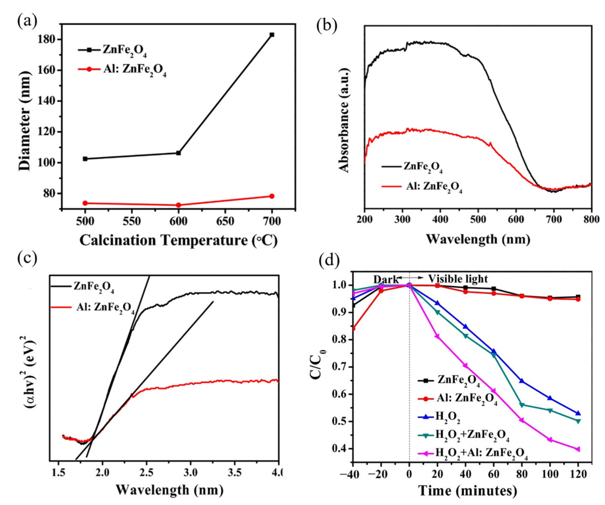
  95. Musa, M.A.; Xu, D.; Sun, F.; Shao, H.; Dong, X.T.; Azis, R.S.; Ugya, A.Y.; Ari, H.A. Electrospun ZnFe2O4/Al: ZnFe2O4 nanofibers for degradation of RhB via visible light photocatalysis and photo-Fenton processes. J. Mater. Sci. Mater. Electron. 2022, 33, 2375–2385. [Google Scholar]
  96. Reyes-Rodríguez, P.Y.; Ávila-Orta, C.A.; Andrade-Guel, M.; Cortés-Hernández, D.A.; Herrera-Guerrero, A.; Cabello-Alvarado, C.; Sánchez-Fuentes, J.; Ramos-Martínez, V.H.; Valdez-Garza, J.A.; Hurtado-López, G.F. Synthesis and characterization of magnetic nanoparticles Zn1-xMgxFe2O4 with partial substitution of Mg2+ (x= 0.0, 0.25, 0.5, 0.75 and 1.0) for adsorption of uremic toxins. Ceram. Int. 2020, 46, 27913–27921. [Google Scholar]
  97. Peymani-Motlagh, S.M.; Moeinian, N.; Rostami, M.; Fasihi-Ramandi, M.; Sobhani-Nasab, A.; Rahimi-Nasrabadi, M.; Eghbali-Arani, M.; Ganjali, M.R.; Jesionowski, T.; Ehrlich, H.; et al. Effect of Gd3+-, Pr3+- or Sm3+-substituted cobalte-zinc ferrite on photodegradation of methyl orange and cytotoxicity tests. J. Rare Earth 2019, 37, 1288–1295. [Google Scholar]
  98. Borhan, A.I.; Slatineanu, T.; Iordan, A.R.; Palamaru, M.N. Influence of chromium ion substitution on the structure and properties of zinc ferrite synthesized by the sol-gel auto-combustion method. Polyhedron 2013, 56, 82–89. [Google Scholar]
  99. Li, J.; Li, X.; Chen, X.; Yin, Z.; Li, Y.; Jiang, X. In situ construction of yolk-shell zinc ferrite with carbon and nitrogen co-doping for highly efficient solar light harvesting and improved catalytic performance. J. Colloid Interface Sci. 2019, 554, 91–102. [Google Scholar]
  100. Sonu; Dutta, V.; Sharma, S.; Raizada, P.; Hosseini-Bandegharaei, A.; Gupta, V.K.; Singh, P. Review on augmentation in photocatalytic activity of CoFe2O4 via heterojunction formation for photocatalysis of organic pollutants in water. J. Saudi Chem. Soc. 2019, 23, 1119–1136. [Google Scholar]
  101. Das, K.K.; Patnaik, S.; Mansingh, S.; Behera, A.; Mohanty, A.; Acharya, C.; Parida, K.M. Enhanced photocatalytic activities of polypyrrole sensitized zinc ferrite/graphitic carbon nitride n-n heterojunction towards ciprofloxacin degradation, hydrogen evolution and antibacterial studies. J. Colloid Interface Sci. 2020, 561, 551–567. [Google Scholar] [PubMed]
  102. Zhang, R.; Han, Q.; Li, Y.; Zhang, T.Q.; Liu, Y.; Zeng, K.L.; Zhao, C. Solvothermal synthesis of a peony flower-like dual Z-scheme PANI/BiOBr/ZnFe2O4 photocatalyst with excellent photocatalytic redox activity for organic pollutant under visible-light. Sep. Purif. Technol. 2020, 234, 116098. [Google Scholar]
  103. Wang, J.; Zhang, Q.; Deng, F.; Luo, X.B.; Dionysiou, D.D. Rapid toxicity elimination of organic pollutants by the photocatalysis of environment-friendly and magnetically recoverable step-scheme SnFe2O4/ZnFe2O4 nano-heterojunctions. Chem. Eng. J. 2020, 379, 122264. [Google Scholar]
  104. AL-Shwaiman, H.A.; Akshhayya, C.; Syed, A.; Bahkali, A.H.; Elgorban, A.M.; Das, A.; Varma, R.S.; Khan, S.S. Fabrication of intimately coupled CeO2/ZnFe2O4 nano-heterojunction for visible-light photocatalysis and bactericidal application. Mater. Chem. Phys. 2022, 279, 125759. [Google Scholar]
  105. Akshhayya, C.; Okla, M.K.; Thomas, A.M.; AL-ghamdi, A.A.; Abdel-Maksoud, M.A.; Almunqedhi, B.; AbdElgawad, H.; Raju, L.L.; Khan, S.S. Insights into photocatalytic mechanism for the rational design of p-n heterojunction by decorating mesoporous SnS2 over ZnFe2O4 nanocomposite for accelerated visible light photocatalysis. Mater. Chem. Phys. 2022, 277, 125464. [Google Scholar]
  106. Singh, C.; Goyal, A.; Malik, R.; Bansal, S.; Singhal, S. Envisioning the attachment of CdS nanoparticles on the surface of MFe2O4 (M = Zn, Co and Ni) nanocubes: Analysis of structural, optical, magnetic and photocatalytic properties. J. Alloys Compd. 2017, 695, 351–363. [Google Scholar]
  107. Piaskowski, K.; Zarzycki, P.K. Carbon-Based Nanomaterials as Promising Material for Wastewater Treatment Processes. Int. J. Environ. Res. Public Health 2020, 17, 5862. [Google Scholar]
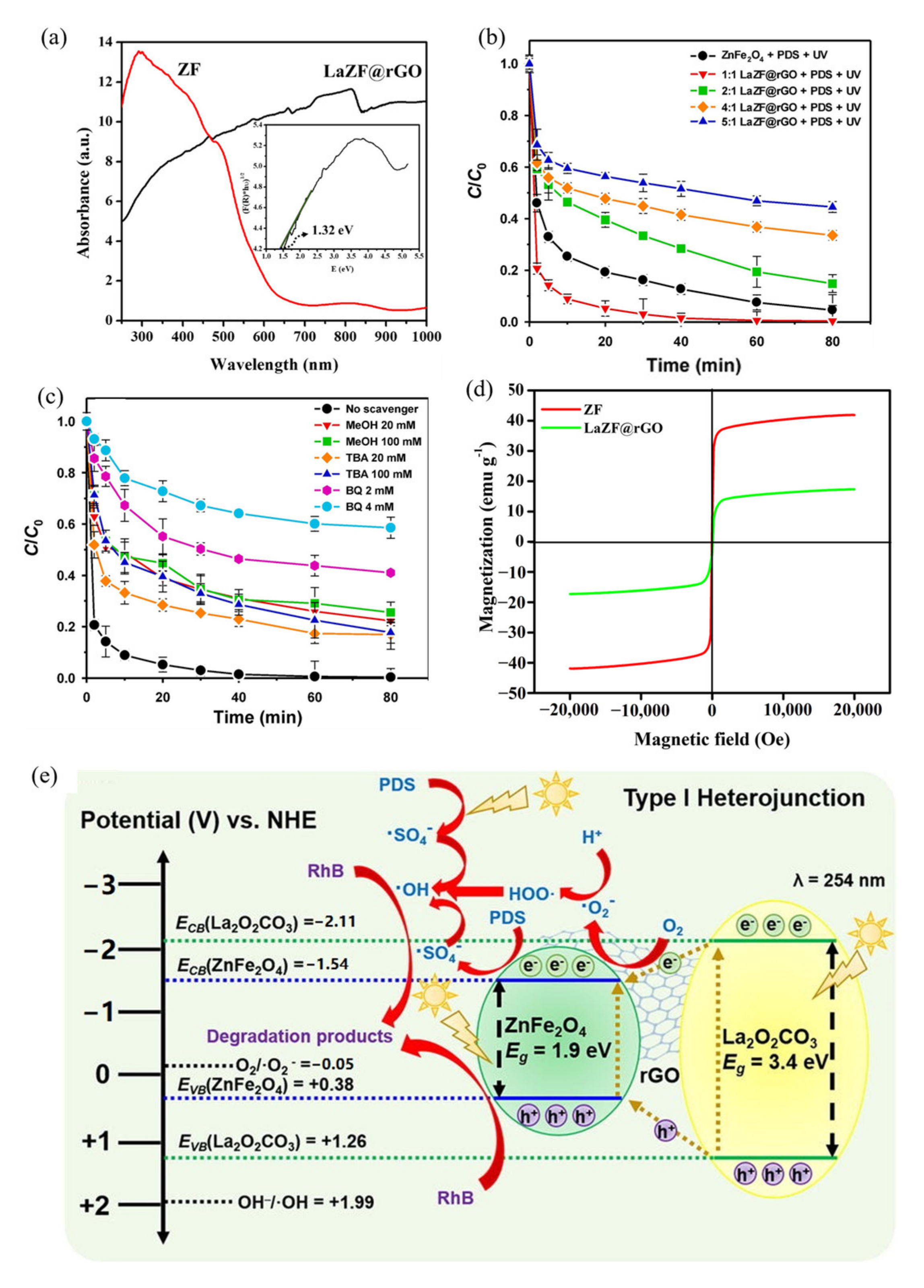
  108. Jun, B.M.; Elanchezhiyan, S.S.; Yoon, Y.; Wang, D.J.; Kim, S.; Prabhu, S.M.; Park, C.M. Accelerated photocatalytic degradation of organic pollutants over carbonate-rich lanthanum-substituted zinc spinel ferrite assembled reduced graphene oxide by ultraviolet (UV)-activated persulfate. Chem. Eng. J. 2020, 393, 124733. [Google Scholar]
  109. Baynosa, M.L.; Mady, A.H.; Nguyen, V.Q.; Kumar, D.R.; Sayed, M.S.; Tuma, D.; Shim, J.J. Eco-friendly synthesis of recyclable mesoporous zinc ferrite@reduced graphene oxide nanocomposite for efficient photocatalytic dye degradation under solar radiation. J. Colloid Interface Sci. 2020, 561, 459–469. [Google Scholar]
  110. Wang, W.R.; Guo, S.S.; Zhang, D.X.; Yang, Z. One-pot hydrothermal synthesis of reduced graphene oxide/zinc ferrite nanohybrids and its catalytic activity on the thermal decomposition of ammonium perchlorate. J. Saudi Chem. Soc. 2019, 23, 133–140. [Google Scholar]
  111. Gao, Y.; Wang, W.L.; Xu, M.; Hao, Y.; Zou, D.L. The magnetically recoverable C-ZnFe2O4 as a microwave absorption catalyst for the degradation of norfloxacin by persulfate: Mechanism, degradation pathway, toxicity, and stability. J. Environ. Chem. Eng. 2022, 10, 107434. [Google Scholar] [CrossRef]
  112. Ghazkoob, N.; Shoushtari, M.Z.; Kazeminezhad, I.; Baghal, S.M.L. Investigation of structural, magnetic, optical and photocatalytic properties of zinc ferrite nanowires/bismuth vanadate composite. J. Alloys Compd. 2022, 900, 163467. [Google Scholar] [CrossRef]
  113. Liu, H.Y.; Han, C.H.; Shao, C.L.; Yang, S.; Li, X.W.; Li, B.; Li, X.H.; Ma, J.G.; Liu, Y.C. ZnO/ZnFe2O4 Janus Hollow Nanofibers with Magnetic Separability for Photocatalytic Degradation of Water-Soluble Organic Dyes. ACS Appl. Nano Mater. 2019, 2, 4879–4890. [Google Scholar] [CrossRef]
  114. Chen, Y.J.; Jiang, H.Y.; Li, L.G.; Wang, Q.; Du, L.Z.; Liu, X.; Tian, G.H. Hierarchical NiS decorated CuO@ZnFe2O4 nanoarrays as advanced photocathodes for hydrogen evolution reaction. Int. J. Hydrogen Energy 2020, 45, 6174–6183. [Google Scholar] [CrossRef]
  115. Cao, Y.; Lei, X.Y.; Chen, Q.L.; Kang, C.; Li, W.X.; Liu, B.J. Enhanced photocatalytic degradation of tetracycline hydrochloride by novel porous hollow cube ZnFe2O4. J. Photoch. Photobio. A 2018, 364, 794–800. [Google Scholar] [CrossRef]
  116. Zhang, L.; Wang, Y.; Ni, Q.Q. Carbon nanotube template-assisted synthesis of zinc ferrite nanochains. Mater. Chem. Phys. 2010, 124, 1029–1033. [Google Scholar] [CrossRef]
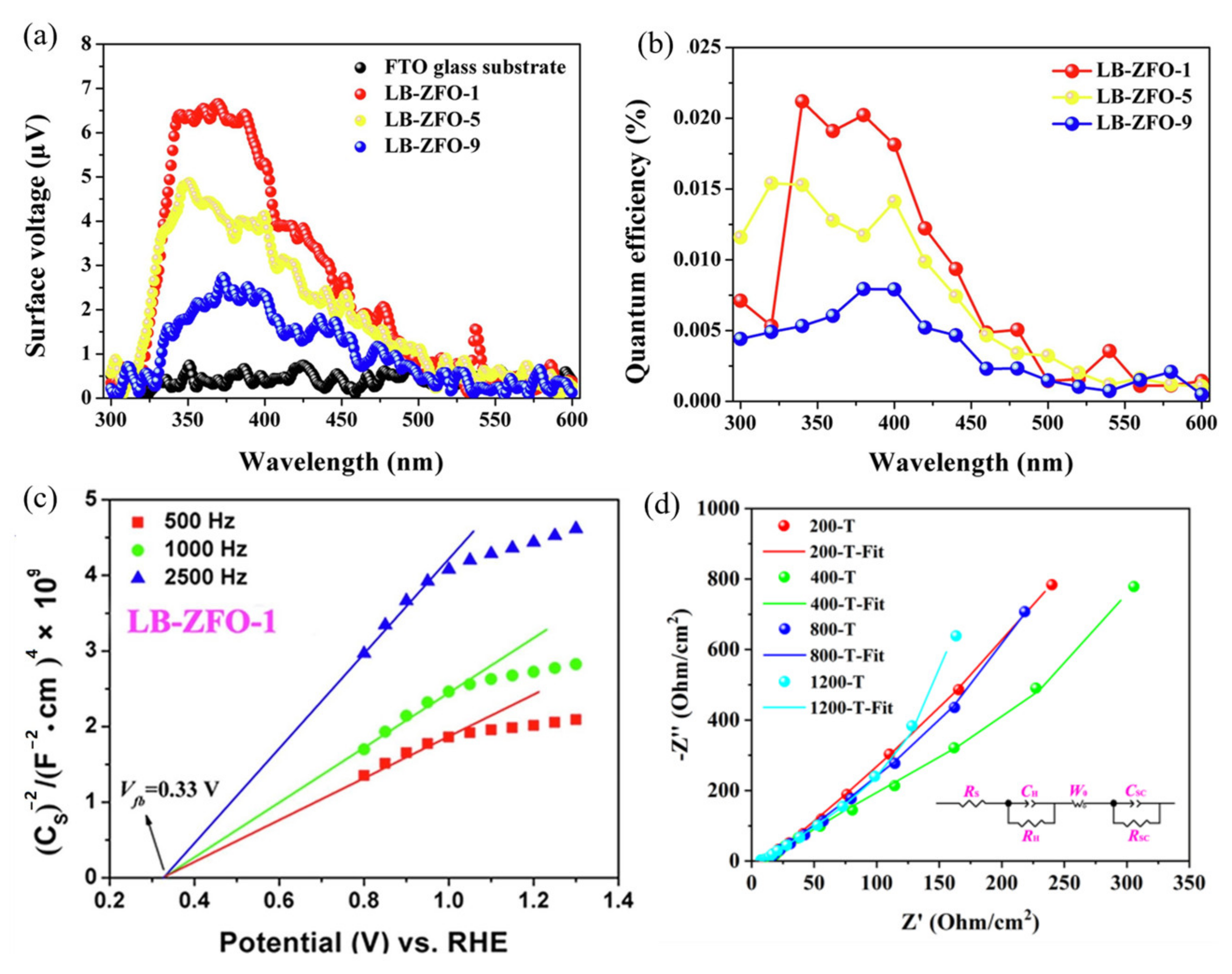
  117. Zhu, Y.Y.; Deng, W.; Chen, L.; Courtois, J.; Tian, Q.; Zhang, X.W.; Almasy, L.; Yan, M.H.; Xiong, K. Langmuir-Blodgett-assembled monolayer zinc ferrite nanoparticle film with unique photogenerated charge carrier separation efficiency and charge transfer behavior. Appl. Surf. Sci. 2020, 534, 147646. [Google Scholar] [CrossRef]
  118. Chen, Y.F.; Spoddig, D.; Ziese, M. Epitaxial thin film ZnFe2O4: A semi-transparent magnetic semiconductor with high Curie temperature. J. Phys. D Appl. Phys. 2008, 41, 205004. [Google Scholar] [CrossRef]
  119. Luo, X.; Duan, Z.F.; Zhu, Y.; Ni, J.Y.; Li, Y.L.; Zhang, J.Y.; Chen, Y.R.; Wang, X.H.; Zhao, G.Y. Enhanced visible-light catalytic activity of micro-patterned ZnFe2O4/Fe3+-TiO2 heterojunction composite thin films prepared by photolithography-assisted chemical solution deposition. Mater. Res. Bull. 2022, 155, 111951. [Google Scholar] [CrossRef]
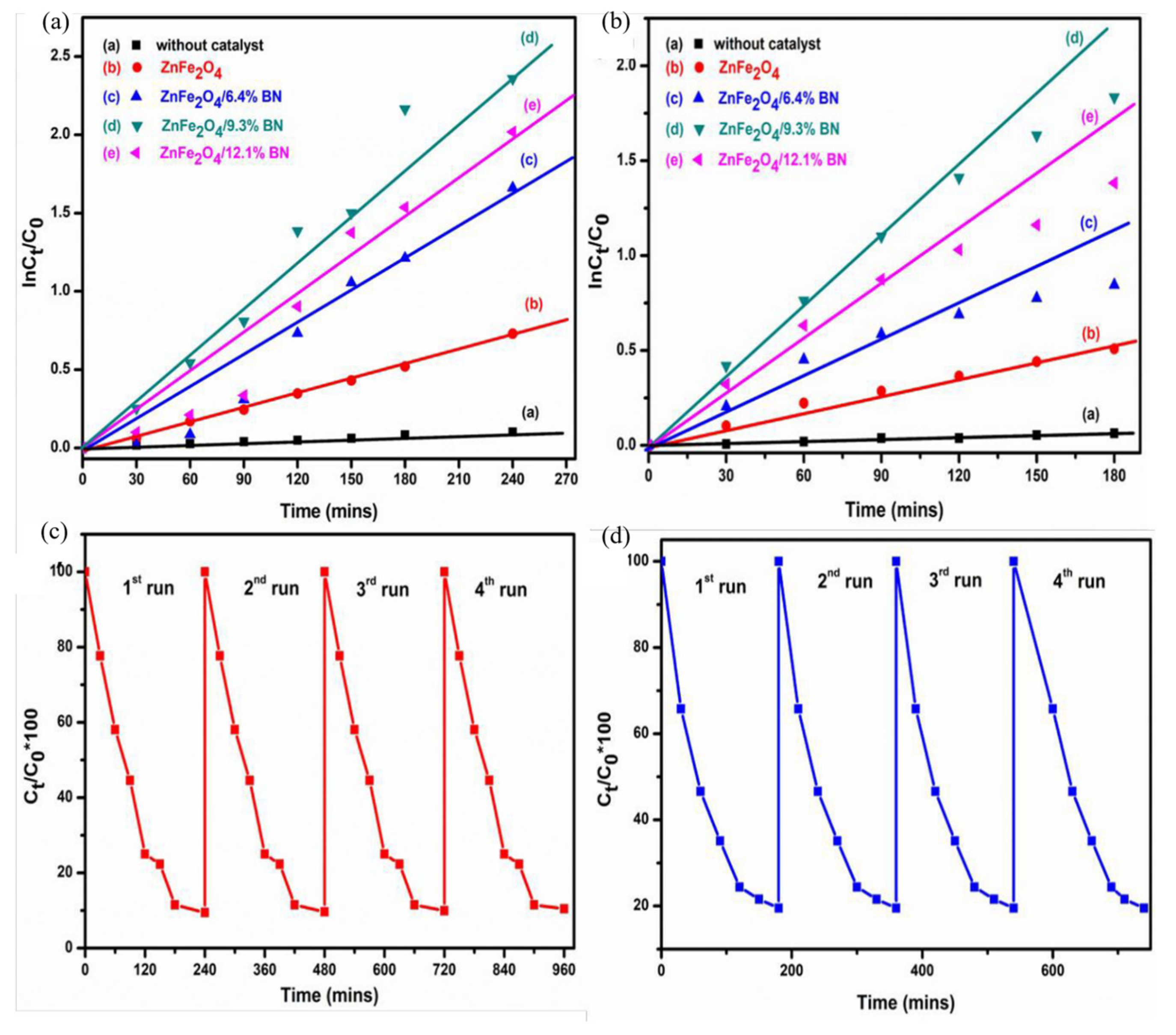
  120. Erusappan, E.; Thiripuranthagan, S.; Durai, M.; Kumaravel, S.; Vembuli, T. Photocatalytic performance of visible active boron nitride supported ZnFe2O4 (ZnFe2O4/BN) nanocomposites for the removal of aqueous organic pollutants. N. J. Chem. 2020, 44, 7758–7770. [Google Scholar] [CrossRef]
  121. Wang, G.J.; Li, Z.C.; Li, M.Y.; Feng, Y.M.; Li, W.; Lv, S.S.; Liao, J.C. Synthesizing vertical porous ZnO nanowires arrays on Si/ITO substrate for enhanced photocatalysis. Ceram. Int. 2018, 44, 1291–1295. [Google Scholar] [CrossRef]
  122. Zhu, C.Z.; He, Q.Y.; Wang, W.K.; Du, F.; Yang, F.; Chen, C.X.; Wang, C.H.; Wang, S.B.; Duan, X.G. S-scheme photocatalysis induced by ZnIn2S4 nanoribbons-anchored hierarchical CeO2 hollow spheres for boosted hydrogen evolution. J. Colloid Interface Sci. 2022, 620, 253–262. [Google Scholar] [CrossRef] [PubMed]
  123. Geng, Q.H.; Wang, H.C.; Wu, Y.; Lv, L.P.; Chen, S.Q.; Sun, W.W.; Wang, Y. Covalent-Induced Heterostructure of Covalent-Organic Frameworks and MXene as Advanced Electrodes with Motivated Pseudocapacitance Performance. Chemelectrochem 2022, 9, e202200340. [Google Scholar] [CrossRef]
  124. Geng, Q.H.; Wang, H.C.; Wang, J.L.; Hong, J.; Sun, W.W.; Wu, Y.; Wang, Y. Boosting the Capacity of Aqueous Li-Ion Capacitors via Pinpoint Surgery in Nanocoral-Like Covalent Organic Frameworks. Small Methods 2022, 6, 2200314. [Google Scholar] [CrossRef]
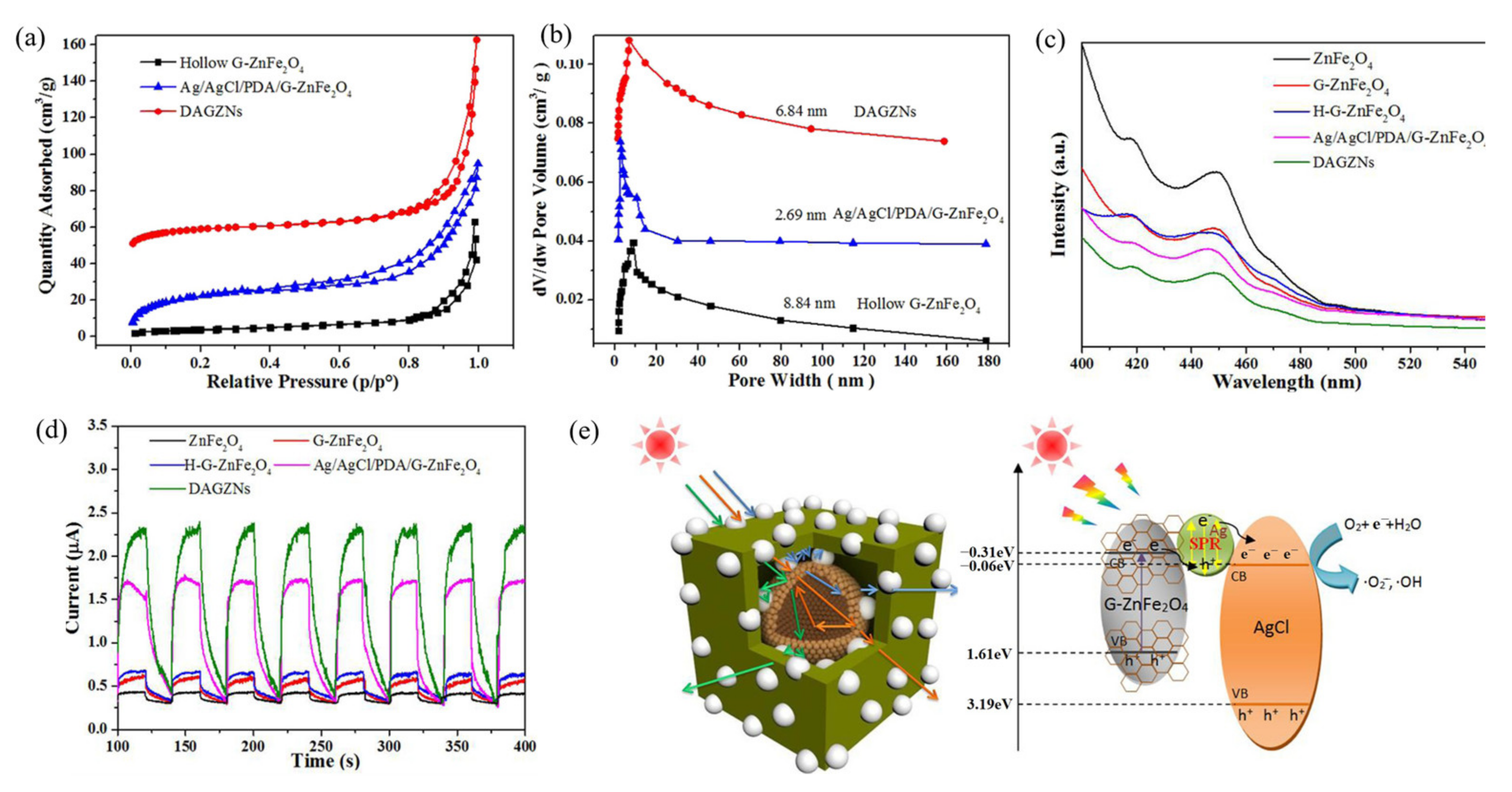
  125. Zhang, C.; Gu, Y.A.; Teng, G.X.; Wang, L.P.; Jin, X.D.; Qiang, Z.W.; Ma, W.G. Fabrication of a Double-Shell Ag/AgCl/G-ZnFe2O4 Nanocube with Enhanced Light Absorption and Superior Photocatalytic Antibacterial Activity. ACS Appl. Mater. Interfaces 2020, 12, 29883–29898. [Google Scholar] [CrossRef]
  126. Dihom, H.R.; Al-Shaibani, M.M.; Mohamed, R.M.S.R.; Al-Gheethi, A.A.; Sharma, A.; Khamidun, M.H.B. Photocatalytic degradation of disperse azo dyes in textile wastewater using green zinc oxide nanoparticles synthesized in plant extract: A critical review. J. Water Process Eng. 2022, 47, 102705. [Google Scholar] [CrossRef]
  127. Chahar, D.; Taneja, S.; Bisht, S.; Kesarwani, S.; Thakur, P.; Thakur, A.; Sharma, P.B. Photocatalytic activity of cobalt substituted zinc ferrite for the degradation of methylene blue dye under visible light irradiation. J. Alloys Compd. 2021, 851, 156878. [Google Scholar] [CrossRef]
  128. Shakil, M.; Inayat, U.; Khalid, N.R.; Tanveer, M.; Gillani, S.S.A.; Tariq, N.H.; Shah, A.; Mahmood, A.; Dahshan, A. Enhanced structural, optical, and photocatalytic activities of Cd-Co doped Zn ferrites for degrading methyl orange dye under irradiation by visible light. J. Phys. Chem. Solids 2022, 161, 110419. [Google Scholar] [CrossRef]
  129. Sun, X.F.; Wang, C.W.; Li, Y.B.; Wang, W.G.; Wei, J. Treatment of phenolic wastewater by combined UF and NF/RO processes. Desalination 2015, 355, 68–74. [Google Scholar] [CrossRef]
  130. Villegas, L.G.C.; Mashhadi, N.; Chen, M.; Mukherjee, D.; Taylor, K.E.; Biswas, N. A Short Review of Techniques for Phenol Removal from Wastewater. Curr. Pollut. Rep. 2016, 2, 157–167. [Google Scholar] [CrossRef]
  131. Mady, A.H.; Baynosa, M.L.; Tuma, D.; Shim, J.-J. Heterogeneous activation of peroxymonosulfate by a novel magnetic 3D γ-MnO2@ZnFe2O4/rGO nanohybrid as a robust catalyst for phenol degradation. Appl. Catal. B 2019, 244, 946–956. [Google Scholar] [CrossRef]
  132. Mathur, P.; Sanyal, D.; Callahan, D.L.; Conlan, X.A.; Pfeffer, F.M. Treatment technologies to mitigate the harmful effects of recalcitrant fluoroquinolone antibiotics on the environ-ment and human health. Environ. Pollut. 2021, 291, 118233. [Google Scholar] [CrossRef] [PubMed]
  133. Dhiman, P.; Rana, G.; Kumar, A.; Sharma, G.; Vo, D.V.N.; AlGarni, T.S.; Naushad, M.; ALOthman, Z.A. Nanostructured magnetic inverse spinel Ni-Zn ferrite as environmental friendly visible light driven photo-degradation of levofloxacin. Chem. Eng. Res. Des. 2021, 175, 85–101. [Google Scholar] [CrossRef]
  134. Vinosha, P.A.; Vinsla, J.V.A.; Madhavan, J.; Devanesan, S.; AlSalhi, M.S.; Nicoletti, M.; Xavier, B. Impact of dysprosium doped (Dy) zinc ferrite (ZnFe2O4) nanocrystals in photo-fenton exclusion of recalcitrant organic pollutant. Environ. Res. 2022, 203, 111913. [Google Scholar] [CrossRef]
  135. Ignat, M.; Rotaru, R.; Samoila, P.; Sacarescu, L.; Timpu, D.; Harabagiu, V. Relationship between the component synthesis order of zinc ferrite–titania nanocomposites and their performances as visible light-driven photocatalysts for relevant organic pollutant degradation. Comptes Rendus Chim. 2018, 21, 263–269. [Google Scholar] [CrossRef]
  136. Makofane, A.; Motaung, D.E.; Hintsho-Mbita, N.C. Photocatalytic degradation of methylene blue and sulfisoxazole from water using biosynthesized zinc ferrite nanoparticles. Ceram. Int. 2021, 47, 22615–22626. [Google Scholar] [CrossRef]
Figure 1. The crystal structure of spinel with different structures: (A) normal spinel; (B) inverse spinel; (C) complex spinel [31] (reprinted with permission from [31] Zhao et al., 2017).
Ijerph 19 10710 g001
Figure 2. The literature survey on ZnFe2O4 from 2012 to 2021: (a) fields of the relevant publications; (b) degree of attention to the different preparation methods used.
Ijerph 19 10710 g002
Figure 3. A schematic representation of the general steps used in the hydrothermal synthesis of ZnFe2O4.
Ijerph 19 10710 g003
Figure 4. The transmission electron microscope images of the ZnFe2O4 nanoparticles prepared at different hydrothermal temperatures: (a) 160; (b) 180; and (c) 200 °C [57] (reprinted with permission from [57] Zhu et al., 2021).
Ijerph 19 10710 g004
Figure 5. A schematic representation of the general steps used for the co-precipitation technique of ZnFe2O4.
Ijerph 19 10710 g005
Figure 6. A schematic representation of the general steps used for the sol-gel synthesis of ZnFe2O4.
Ijerph 19 10710 g006
Figure 7. (a) The TCP degradation using different g-C3N4/ZnFe2O4/Bi2S3 catalysts: PMS dosage; (b) determination of the optimum catalyst dosage; (c) determination of the optimum PMS dosage. Reaction conditions: [TCP] = 50 mg/L, Temperature [T] = 35 °C, visible light intensity: 80 W; (d) variation of residual TCP concentration for various initial concentration; (e) leached ion concentrations after consecutive cycles [81] (reprinted with permission from [81] Sarkar et al., 2022).
Ijerph 19 10710 g007
Figure 8. The ZnFe2O4 nanofibers and Al-doped ZnFe2O4 nanofibers: (a) plots of fiber diameters vs. calcination temperature; (b) UV-Vis DRS; (c) Tauc’s plots of calcined at 600 °C; (d) photocatalytic degradation rates of RhB by the pure and Al-doped ZnFe2O4 and their photo-Fenton process, with an H2O2 oxidant [95] (reprinted with permission from [95] Musa et al., 2022).
Ijerph 19 10710 g008
Figure 9. The band gap positioning of spinel ferrites upon interaction with an aqueous solution referenced with NHE (right pH ¼ 14 and left pH ¼ 0) relative to the standard potentials for the reduction and oxidation of water [100] (reprinted with permission from [100] Sonu et al., 2019).
Ijerph 19 10710 g009
Figure 10. (a) High resolution transmission electron microscope images of ZFCN@20PPY; (b) N2 adsorption-desorption isotherms obtained for PPY, CN, ZFCN, and ZFCN@20PPY; (c) pore size distribution; (d) photoluminescence (PL) spectroscopy; (e) a schematic representation of the reaction mechanism [101] (reprinted with permission from [101] Das et al., 2020).
Ijerph 19 10710 g010
Figure 11. PANI/BiOBr/ZnFe2O4: (a) scanning electron microscopy (SEM) image; (b) the UV-Vis absorption spectra changes of RhB solution during the photocatalytic process over 7% PANI; (c) the cyclic photodegradation curves; (d) dual Z-scheme electron transfer and pollutant degradation mechanism under visible-light exposure [102] (reprinted with permission from [102] Zhang et al., 2020).
Ijerph 19 10710 g011
Figure 12. The pure ZnFe2O4, pure SnFe2O4, and SnFe2O4/ZnFe2O4 nano-heterojunctions: (a) plots of (ahv)2 versus hv; (b) PL spectra; (c) photocurrent responses; (d) magnetization curve [103] (reprinted with permission from [103] Wang et al., 2020).
Ijerph 19 10710 g012
Figure 13. ZnFe2O4 and LaZF@rGO: (a) UV-Vis absorption spectra; (b) influence of La content in LaZF@rGO; (c) radical scavengers on RhB decolorization; (d) Magnetization curve; (e) a schematic representation of the RhB degradation mechanism [108] (reprinted with permission from [108] Jun et al., 2020).
Ijerph 19 10710 g013
Figure 14. The SEM images of ZnFe2O4 with different morphologies: (a) nanowire (field emission SEM) [112] (reprinted with permission from [112] Ghazkoob et al., 2022); (b) hollow nanofibers [113] (reprinted with permission from [113] Liu et al., 2019); (c) flower-shaped nanorods [91] (reprinted with permission from [91] Wang et al., 2019); (d) nanowire arrays [114] (reprinted with permission from [114] Chen et al., 2020); (e) yolk-shell [99] (reprinted with permission from [99] Li et al., 2019); (f) hollow cube skeleton [115] (reprinted with permission from [115] Cao et al., 2018).
Ijerph 19 10710 g014
Figure 15. (a) A schematic representation of the synthesis process of ZnFe2O4 nanochains [116] (reprinted with permission from [116] Zhang et al., 2010). (b) A schematic representation of the synthesis process of the ZnFe2O4 nanochains [113] (reprinted with permission from [113] Liu et al., 2019).
Ijerph 19 10710 g015
Figure 16. The LB-ZFO-1, LB-ZFO-5 and LB-ZFO-9 films: (a) surface photovoltage; (b) apparent quantum efficiency; (c) Mott–Schottky plots of LB-ZFO-1; (d) electrochemical impedance spectroscopy spectra [117] (reprinted with permission from [117] Zhu et al., 2020).
Ijerph 19 10710 g016
Figure 17. The kinetic plots in the photodegradation of (a) CR and (b) TC by the as-synthesized catalysts; photo-stability of the ZnFe2O4/9.3% BN composite catalyst in the degradation of (c) CR and (d) TC [120] (reprinted with permission from [120] Erusappan et al., 2020).
Ijerph 19 10710 g017
Figure 18. A schematic representation of the formation process of the CuO@ZnFe2O4 core/shell nanoarrays decorated with NiS quantum dots [114] (reprinted with permission from [114] Chen et al., 2020).
Ijerph 19 10710 g018
Figure 19. (a) The nitrogen adsorption–desorption isotherms of hollow G-ZnFe2O4, Ag/AgCl/PDA/G-ZnFe2O4, and DAGZNs; (b) Barrett–Joyner–Halenda (BJH) pore size distribution; (c) PL spectroscopy; (d) transient photocurrent; (e) schematic representation of the light scattering effect in DAGZN [125] (reprinted with permission from [125] Zhang et al., 2020).
Ijerph 19 10710 g019
Table 1. A comparison of the photodegradation rate of organic pollutants by ZnFe2O4 with different preparation methods.
ZnFe2O4 Dose (g/L)Synthesis MethodPollutants and Concentrations (mg/L)Time (min) and Degradation Efficiency (%)Refs.
0.2Biosynthesis method (Nyctanthes arbor-tristis flower extract)RhB, 530, 1002019 [68]
0.5Co-precipitationMethylene Blue (MB), 1050, 992018 [72]
——Biosynthesis method (Jatropha extract)Sudan, ————, 982020 [69]
——Biosynthesis method (Simbang Darah (Iresine herbstii) extract)Direct Red 81, 35120, 99.662021 [70]
0.5Biosynthesis method (Sugarcane juice)MB, 5 & RhB, 5120, 98 & 952018 [73]
0.6——Terramycin, 4060, 94.682022 [74]
2A single-mode microwave combustion processRhB, 1060, 52022 [75]
0.3Electrospinning and co-precipitationMB, 560, 29.7%2022 [76]
1HydrothermalMB, 2240, 202018 [77]
0.1Ultra-sonication assisted chemical precipitation methodMB, 25160, 66.4%2021 [78]
Table 2. The principles and the advantages and disadvantages of the different methods used to prepare the ZnFe2O4 photocatalysts.
MethodPreparation PrinciplesAdvantageShortcoming
Microwave-assisted methodUsing microwave heating, energy is directly transferred to the reactant particles through chemical interaction with electromagnetic radiation, providing a thermally uniform nucleation environment.The product has a very fine particle size, a high heating rate, high purity, low thermal inertia, and low cost.Complex equipment, different reactors are needed to apply high pressure, and the reactors take a long time to react.
Ball milling methodDry or wet milling of the metal salt mixture in a ball mill breaks up and combines the particles repeatedly.Low cost, simple operation, no use of toxic and expensive solutions, prevention of particle agglomeration, potential for large-scale production.Low product purity, risk of material contamination during milling, long production cycle, special equipment required.
Vapor deposition methodUsing thermal decomposition, precursor gases are converted to thin films, which are deposited on substrates.It has good conformal coverage on complex structures and can deposit various materials such as metals, oxides, semiconductors, etc.Crystallinity of the deposited layer is usually columnar and not bendable.
Sonochemical methodMechanical waves act on the fluid, producing a sharp motion that results in tiny bubbles that compress the solvent and sparse it, thereby speeding up chemical reactions.Low energy consumption; quick response; uniform size; simple and economical.The product has a low magnetic moment and poor crystallinity.
Pulsed laser methodDifferent substrates are bombarded with laser light, and the bombarded substances are deposited.High deposition rate, short test cycle, easy cleaning.Molten particles generated by the laser acting on the target can cause contamination of the material and uneven film thickness.
Ceramic methodAfter the two metal oxides (ZnO and Fe2O3) are mixed and ground into fine particles, they are pressed into particles using a press and sintered at a high temperature.——Some of the reactants and intermediate products remain. The purity is not high, the particle size is large, the reaction temperature is extremely high, the limitations are obvious, and the energy consumption is large.
Spray pyrolysis The raw materials are first dissolved and then atomized and volatilized to obtain the product.As raw materials are thermally decomposed during the preparation process, the reaction temperature is low, operation is quick and simple, and washing and grinding are unnecessary.Equipment is easily corroded by polluted reaction gases.
Table 3. The band gap change in the ZnFe2O4 photocatalysts after doping at different sites.
ResearcherA SiteB SiteModified CatalystEg/eVRefs.
Son et al.Ni--ZnFe2O41.81[89]
Zn0.75Ni0.25Fe2O41.73
Zn0.4Ni0.6Fe2O41.72
Somvanshi et al.Mg--ZnFe2O42.39[62]
Zn0.5Mg0.5Fe2O41.96
Fadl et al.Cr, Ni--Ni0.4Zn0.5Cr0.1Fe2O43.9[90]
Ni0.4Zn0.3Cr0.3Fe2O43.85
Ni0.4Cr0.6Fe2O43.78
Abdo et al.Co, CuYCo0.5Cu0.25Zn0.25Fe2.0O41.57[91]
Co0.5Cu0.25Zn0.25Y0.06Fe1.94O41.54
Co0.5Cu0.25Zn0.25Y0.1Fe1.9O41.52
Lemziouka et al.--LaZnFe1·99La0·01O41.945[92]
Tholkappiyan et al.--LaZnFe1·94La0·06O41.97[93]
Shoba et al.--ErZnFe2O41.85[94]
ZnFe1.8Er0.2O41.93
ZnFe1.6Er0.4O41.96
Table 4. The degradation performance of the ZnFe2O4 photocatalyst contaminants using different modification methods.
Sr. No.Catalyst and Dose (g/L)Modification MethodLight SourcePollutants and Concentrations (mg/L)Time (min) and Degradation Efficiency (%)StabilityRefs.
1Co0.5Zn0.5Fe2O4, 0.05Elemental doping (A site)300 W Tungsten lampMB, 1060 min, 77%——[127]
2Ni0.1Zn0.9Fe2O4, 0.4Elemental doping (A site)500 W Xenon lampLevofloxacin, 1090 min, 98.6%Five cycles 100%[133]
3Zn0.9Dy0.1Fe2O4, 1Elemental doping (A site)125 W Mercury lampMB, 2045 min, 97.3%Five cycles 100%[134]
4Co0.5Cu0.25Zn0.25Y0.1Fe1.9O4, 0.5Elemental doping (AB site)250 W Mercury lamp and 500 W Xenon lampMB, 2060 min, 95%Five cycles 99%[91]
5Zn0.5Cu0.5Fe1.95Ce0.05O4,1Elemental doping (AB site)250 W Mercury lamp and 500 W Tungsten lampBisphenol A, 10180 min, 91.3%Three cycles 97%[55]
6Cd0.75Zn0.25Co0.25Fe1.75O4, 0.2Elemental doping (AB site)400 W Mercury lampMO, 2120 min, 82%Six cycles 100%[128]
7ZFCN@20PPY, 0.2Compound modification
(conducting polymers)
Solar lightCiprofloxacin, 20120 min, 92%Four cycles 100%[101]
8PANI/BiOBr/ZnFe2O4, 1Compound modification
(conducting polymers)
Mercury lampRhB, 2060 min, 99%Three cycles 98%[102]
9ZnFe2O4/SnFe2O4, 0.3Compound modification
(metal oxide)
300 W Xenon lampPharmaceutical wastewater, ——720 min, 77.5%Four cycles 100%[103]
10ZnFe2O4/TiO2, 0.83Compound modification
(metal oxide)
150 W Mercury lampRhB, 5010 min, 97.87%——[135]
11ZnFe2O4/Fe2O3, 0.5Compound modification
(metal oxide)
300 W Xenon lampCiprofloxacin, 10180 min, 96.5%Five cycles 100%[56]
12CeO2/ZnFe2O4, 0.15Compound modification
(metal oxide)
500 W Tungsten lampMB, 2590 min, 90.48%Six cycles 92%[104]
13γ-MnO2@ZnFe2O4/rGO, 0.2Compound modification
(metal oxide and carbon material)
——Phenol, 2030 min, 100%Five cycles 100%[131]
14ZnFe2O4/SnS2, 0.03Compound modification
(metal sulfide)
500 W Tungsten lampMB, 2545 min, 91.78%Six cycles 100%[105]
15ZnFe2O4/CdS, 1Compound modification
(metal sulfide)
Visible lightRhB, 5120 min, 100%Three cycles 100%[106]
16LaZF@rGO, 0.25Compound modification
(carbon material)
11 W Tungsten lampRhB, 3060 min, 100%Four cycles 91%[108]
17ZnFe2O4@rGO, 0.5Compound modification
(carbon material)
150 W One-sun solar simulatorMB, 1060 min, 94%Three cycles 91%[109]
18C-ZnFe2O4, 1.5Compound modification
(carbon material)
——Noroxin, 540 min, 86.2%Four cycles 98%[111]
19ZnFe2O4, ——Morphology modification (nanosheets)——Sudan red, ——120 min, 67%——[69]
20ZnFe2O4, 0.25Morphology modification (nano stave)150 W UV Osram lampMB, 545 min, 99.8%Five cycles 100%[136]
21ZnFe2O4/BiVO4, 0.6Morphology modification (nanowires)100 W Xenon lampCR, 20105 min, 100%——[112]
22ZnO/Al2O3/ZnFe2O4, 1Morphology modification (nanofibers)150 W Xenon lampMB, 10120 min, 90%——[113]
23ZFO/FTO, ——Morphology modification (films)350 W Xenon lampMB, 10019 min, 100%Ten cycles 100%[119]
24ZnFe2O4, 0.5Morphology modification (hollow cube)300 W Xenon lampTetracycline hydrochloride, 4020 min, 80%——[115]
Publisher’s Note: MDPI stays neutral with regard to jurisdictional claims in published maps and institutional affiliations.

