Article

Bone Metastases of Endometrial Carcinoma Treated by Surgery: A Report on 13 Patients and a Review of the Medical Literature

1
Department of Obstetrics and Gynecology, Peking University People’s Hospital, Beijing 100044, China
2
Department of Orthopaedic Oncology, Peking University People’s Hospital, Beijing 100044, China
*
Author to whom correspondence should be addressed.
Int. J. Environ. Res. Public Health 2022, 19(11), 6823; https://doi.org/10.3390/ijerph19116823
Submission received: 17 March 2022 / Revised: 22 May 2022 / Accepted: 31 May 2022 / Published: 2 June 2022
(This article belongs to the Special Issue Gynecologic Oncology: Diagnosis, Treatment and Prevention)

Abstract

:
Background: The aim of this study was to describe the clinicopathological features of endometrial cancer (EC) patients with bone metastases treated with surgery and to systematically review the literature. Methods: We performed a retrospective study to include patients with bone metastases of EC at Peking University People’s Hospital from 2000 to 2019. Clinicopathological features and survival outcomes were collected. Results: Among the 1662 patients with EC, 14 (0.84%) were identified with bone metastases, and all were treated surgically. Thirteen cases were analyzed. Four had bone metastases when diagnosed, and the remaining nine cases had bone metastases when first relapsed, with a median time to recurrence of 13 months (range, 5–144). The median age of the 13 patients was 58 years old (range, 45–76). Twelve were endometrioid carcinoma. The majority of sites of bone metastases were the pelvis, followed by the spine. The median overall survival (OS) was 57 months. We further combined the 13 patients with another 24 cases identified from literature research. There was no significant difference in clinicopathological characteristics between the patients with bone metastases when diagnosed and when they first relapsed. The median OS was numerically longer for patients with bone metastases when diagnosed than when they first relapsed (57 vs. 36 months, p = 0.084). Conclusions: Patients with bone metastases of EC might benefit from comprehensive treatment based on surgery, as symptoms can be palliated and survival can probably be extended.

1. Introduction

Endometrial cancer (EC) is one of the most common gynecological cancers in the world, and its incidence has increased remarkably in recent decades, with approximately 63,230 new cases in 2018 [1].
In general, EC recurrence mostly occurs within the pelvis, with a recurrence rate following initial treatment of around 11–13% [2]. EC is more likely to metastasize to the lymph nodes, liver and lungs [3]. In contrast, bone involvement is quite rare, and its prevalence is reported between 0% and 15% [4,5]. The mechanism of metastasis of EC is thought to be related to hematologic dissemination by the retrograde flow of tumor emboli or Batson’s paravertebral valveless venous plexus [6]. However, the exact mechanism is not fully clear.
Bone metastases cause severe suffering to patients because of severe pain, physical disability and pathologic skeletal-related events. Thus, early diagnosis and appropriate therapy are needed to enhance the quality of life (QOL) of patients with osseous dissemination. However, because of its rarity, the factors influencing the survival and optimal therapeutic regime of primary bone metastases in patients with EC are not known yet. Some support noninvasive measures, while others suggest surgery can prolong the cancer-specific survival of patients due to the resistance to chemotherapy or radiotherapy. It was established that pain, pathological fracture and spinal cord compression are all indications for surgery [7]. All cases in our study were diagnosed according to symptoms, and all these patients had pain at the site of the bones involved. Thus, all of them were treated surgically.
The main purpose of this study is to describe the clinicopathological features and treatment results of the primary bone metastases of EC treated by surgery.

2. Materials and Methods

2.1. Patients

This was a retrospective study including EC patients with osseous disseminations. Patients diagnosed with EC were identified from January 2000 to December 2019 in Peking University People’s Hospital. Afterwards, we particularly identified those patients who were diagnosed as primary bone metastases, including those who had bone metastases either at presentation with EC simultaneously or as the first site of recurrence (with or without concomitant extra-osseous lesions). Those with osseous dissemination that was subsequent locations of EC relapse were excluded. Patients with uterine sarcomas or carcinosarcomas were also excluded from this study. This study was approved by the Institutional Review Board of Peking University People’s Hospital (2020PHB331-01). All patients provided written informed consent.

2.2. Follow Up

In the first year after surgery, the patients were examined every one to two months, then every three to six months for the next three years, and yearly afterwards, including routine and pelvic examinations, smear examinations of the upper part of the vagina, abdominopelvic ultrasonography and evaluations of serous CA125. In the first two years, a chest X-ray was carried out every six months, and then every year for the next three years. If necessary, a thorough evaluation was applied to make use of computed tomography (CT), pelvic magnetic resonance imaging (MRI) or positron emission tomography-computed tomography (PET-CT) scan of the chest, abdomen and pelvis.
The primary functional outcome was defined as an improvement in specific pains, and a visual analog pain scale (VAS) was used to evaluate the degree of pain both before and after surgery. In order to minimize the impact of cancer recurrence on the primary outcome of pain improvement, we focused the analysis on immediate pre-operative assessment and that occurring in the initial three months after surgical intervention.

2.3. Data Collection

Clinicopathological characteristics and survival outcomes of patients with primary bone metastases of EC were obtained from medical records. Patients were staged according to the 2009 International Federation of Gynecology and Oncology (FIGO) criteria. Description of tumor data was abstracted from primitive pathological reports.

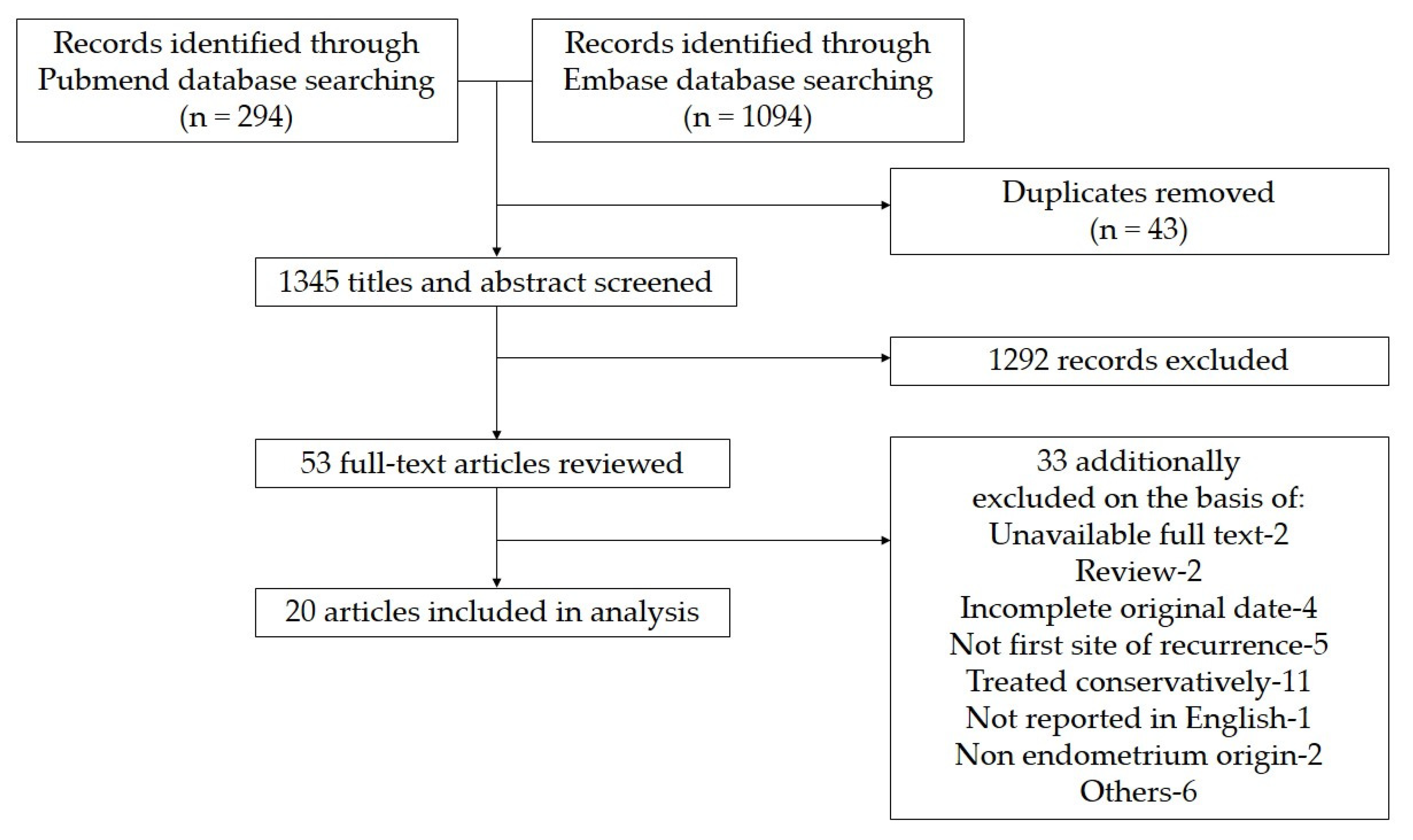
2.4. Literature Research

2.4.1. Search Strategy

A systematic review was carried out adhering to the Preferred Reporting Items for Systematic Reviews and Meta-Analysis (PRISMA) guidelines. We searched PubMed and Embase databases from database inception to the present. We used the following search query: “((endometrial cancer OR endometrial malignancy OR endometrial neoplasm OR endometrial carcinoma) AND (bone OR skeletal) AND metasta*)”. The search terms were designed to identify all publications found in this field and chose those composed of clinical case series or case reports that described patients with primary bone metastases of EC treated by surgical approach. Only papers reported in English were included. Electronic searches were supplemented by reviewing reference lists of all articles identified in the primary search to obtain more complete data.

2.4.2. Inclusion and Exclusion Criteria

Studies were included when the following criteria were met: cases on pathologically confirmed EC with primary bone metastasis; survival information was reported; cases treated with surgery; full study was available; studies obtained research ethics approval. Exclusion criteria were as follows: unavailable full text; review; republished literature; documents with incomplete original data and no corresponding data after contacting the author. Cases with osseous dissemination that was subsequent locations of EC relapse, or cases treated conservatively were also excluded.

2.4.3. Data Extraction and Quality Assessment

After the removal of duplicates, two investigators screened titles and abstracts independently for initial study selection. After abstract screening, the two investigators reviewed the entire publication to determine whether it met the inclusion or exclusion criteria. Any disagreement was resolved between both investigators to reach a consensus, and a review agreement was finally achieved. The following data were obtained from original articles: (1) general information including author and year of publication; (2) clinicopathological information including age, histology type, pathological grade, stage, symptoms, interval to bone metastasis, localization, extraosseous metastatic localization, therapy and available outcome data. We assessed the quality of the included studies by adapting items from the Joanna Briggs Institute Reviews Manual (http://www.joannabriggs.org/sumari.html accessed on 10 May 2022) by two investigators independently.

2.5. Statistical Analysis

Clinicopathological characteristics were analyzed using descriptive statistics, and SPSS (version 24.0) was used for data management and statistical analysis. The median survival was estimated by the Kaplan–Meier method. The overall survival (OS) after bone metastases was defined as the time from diagnosis of osseous dissemination to death or last follow-up. Statistically significant difference was defined as p < 0.05.

3. Results

3.1. Patient Characteristics

Among the 1662 patients with EC diagnosed in Peking University People’s Hospital from 2000 to 2019, 14 (0.84%) were identified with bone metastasis, and all of them were treated surgically. One patient was lost of follow-up after surgery. Therefore, the clinicopathological and survival data of the remaining 13 patients were finally reviewed. The characteristics of patients are described in detail in Table 1.
Among the 13 patients, 4 patients had bone metastases identified simultaneously with the diagnosis of EC. The remaining 9 patients had bone metastases at the primary recurrence of EC, with the median time from diagnosis to bone metastasis of 13 months (range, 5–144 months). The median age was 58 years old (range, 45–76 years old). Except for one case of clear cell carcinoma, the remaining 12 cases were endometrioid carcinoma.

3.2. Characteristics of Bone Metastasis

All cases were diagnosed according to symptoms, and all these patients had pain at the site of the bones involved. The total locations of bone metastases were 23 in these 13 patients, and the maximum amount of osseous dissemination found in a single patient was three. The majority of sites of bone metastasis were pelvic bones (17/23 sites), followed by the spine (5/23 sites). In five patients, the distribution of bone metastases involved the axial skeleton. Six patients had other metastases, including the lungs, the skin and the abdomen, besides bone metastasis. Five patients had a single bone lesion, and eight had multiple bone metastases. Only one case was found to have a single bone site and no extra-osseous dissemination. All patients were treated with surgical resection with or without adjuvant therapy as the salvage treatment after osseous dissemination. Four cases were treated with chemotherapy monotherapy in two patients, chemotherapy plus radiotherapy in one patient and chemotherapy plus radiotherapy and hormone therapy in one patient. Six patients died of the disease at the date of data analysis. The median OS after diagnosis of bone metastases was 57 months.
The mean VAS pain scale score was 5.6 (SD 1.5) preoperatively compared with 2.3 (SD 2.1) three months after the operation. There was a significant improvement in pain three months after surgical intervention (p < 0.001). Pain relief was observed in 12 patients (92.3%).

3.3. Literature Review

We further identified 20 studies [5,6,8,9,10,11,12,13,14,15,16,17,18,19,20,21,22,23,24,25] with a total of 24 cases of EC patients with bone metastases treated by surgery through literature research. The process of study recruitment is shown in Figure 1, and the detailed information on the included studies is displayed in Table 2. With the addition of our 13 cases, a sum of 37 cases was further analyzed. We then classified these cases into two groups: 24 cases with bone metastases when first relapsed and the other 13 cases with bone metastases when diagnosed as EC.
We then compared the clinicopathological features and the survival outcome between the two groups. The overall characteristics of the 37 patients are summarized in Table 3. In 24 cases, the median interval from the diagnosis of the EC to osseous dissemination was 18 months (range, 2 to 144 months). The predominant histopathological type was endometrioid carcinoma. Presenting symptoms were bone pain, swelling, throbbing, weakness, erythema, infection and lack of strength and sensation. Only 1 case was diagnosed as bone metastases without symptoms at the site involved. The most common affected location was the pelvic bones (23/59 sites (38.98%)). Bone recurrences were also seen in the vertebra, tarsus, talus, calcaneus, cranium, toe, sternum, femur, tibia, humerus, clavicle and ribs. At the diagnosis of bone metastasis, 11 (29.73%) patients had coexisting extra-osseous metastatic lesions, with the lungs (9/11) being the most frequent.
There was no significant difference in clinicopathological characteristics and survival outcomes between the two groups. The median OS after diagnosis of bone metastases for patients with bone metastases when first relapsed was 36 months, with the survival rate at 1 year, 2 years and 5 years being 79%, 59% and 31%, respectively, and the median OS for patients with bone metastases when diagnosed was 57 months, with the survival rate at 1 year, 2 years and 5 years being 92%, 92% and 46%, respectively. The median survival outcome of patients with bone metastasis, when diagnosed, was numerically longer than patients with bone metastasis when they first relapsed, while there was no significant difference (p = 0.084). In addition, the OS after bone metastasis was significantly longer in patients with endometrioid carcinoma than in those with type II carcinoma (57 vs. 4 months, p = 0.003). However, this may not be representative due to the small number of cases with type II carcinoma. Other clinicopathological features, including age, number of bone lesions, concomitant extra-osseous metastases, single bone involvement and no extra-osseous spread and metastases to the axial skeleton, were not associated with survival outcome.

4. Discussion

In the present study, we described the clinicopathological characteristics and survival outcomes of 13 EC patients with bone metastasis. We further combined the 13 patients with the 24 cases with bone metastasis identified from literature research. We found that the median OS after bone metastases was significantly associated with histology.
The occurrence of osseous dissemination secondary to EC is rarely seen, and the incidence is unknown. Although autopsy data shows an incidence up to 25%–27% [26], the clinical frequency is reported to be only 0.4%–1.8% [20,22,27]. Uccella et al. [22] also pointed out that the incidence of osseous dissemination in EC was less than 1%, according to the literature review. In contrast, in Takeshita’s cohort study [28], the incidence rate was up to 3.1%. The difference in sources of patients admitted to research centers might contribute to this distinction. In our series, the total incidence of bone metastases was 0.84% and 0.24% in patients with bone metastases at presentation with EC.
Although with no significant difference statistically, the survival outcome was better for the patients with osseous dissemination as a presenting feature of EC than as the first site of recurrence (57 vs. 36 months) in our study. The same result was reached in Vizzielli et al. [21] and Uccella et al.’s [22] research (28 vs. 21 months, and 20 vs. 10.5 months, respectively). In addition, the limited sample size of this study may not be sufficient enough to obtain significant results, and further research is needed to determine the clinical significance of patterns of osseous dissemination.
At the time of diagnosis, most patients with bone metastasis had pain [25]. In our study, all patients presented with symptoms of pain, and in our literature review, bone dissemination was discovered incidentally only in one case. The sacrum metastasis of the patient was found by a conventional follow-up computed tomography scan 37 months after the operation, and confirmation was made by computed tomography-guided biopsy. Given the low incidence of bone metastasis, there is no routine assessment of bone dissemination in the surveillance of EC. Despite this, there is a need to have a suspicion for metastasis in EC patients presenting bone tenderness and in patients with no history of cancer but with osteodynia responding poorly to conservative management, as EC may be an underlying cause [4]. The initial diagnosis may be difficult since the symptom of bone pain is more common in benign diseases such as trauma, soft tissue inflammation, arthritis and osteomyelitis [29]. While there is no standard approach to diagnose bone disease, plain radiograph, bone scan, MRI, positron emission tomography (PET), aspiration cytology and bone biopsy may help in the diagnosis. It is reported that technetium diphosphonate bone scans can be positive 18 months earlier before a lesion is detected on plain X-ray [30]. CT or MRI can be more helpful than bone scans since these methods may be useful to help diagnose other metastatic locations, which could change the therapeutic scheme [20]. PET/CT scans are used to determine malignancies of bone lesions, similar to the CT scans for other malignant tumors, including breast cancer [31]. Therefore, we propose that clinical and radiologic assessments should be carried out for patients with suspected lesions to rule out bone metastasis.
Most of the tumors that metastasize to the bones are endometrioid [6], instead of those histologic subtypes supposed to be more aggressive, such as papillary serous or clear cell carcinomas. In our study, the predominant histopathological type was endometrioid (86%, 32/37), 57% (21/37) of the tumors were moderately or poorly differentiated and 41% (15/37) were in FIGO stage IV. Some studies postulate that type II EC is associated with a worse prognosis [22,27] and is a predictor of hematogenous dissemination in EC [32]. Our study reached a similar conclusion that endometrioid carcinoma was associated with longer survival despite the small number of cases with type II carcinoma. It is suggested that EC with advanced stage and poorly differentiated grade is more likely to metastasize to bone [20]. Though rare, reports exist concerning bone recurrence in early-stage and well-differentiated EC [5]. In fact, different types of EC have specific histopathological and molecular characteristics. Traditional pathological analysis remains an important tool for risk stratification but brings the problem of undertreatment or overtreatment. The Cancer Genome Atlas (TCGA) Research Network identified four molecular subgroups of endometrial carcinoma: copy-number-low/p53-wild-type (p53wt), POLE-mutated/ultramutated (POLEmt), microsatellite-instability/hypermutated (MSI) and copy-number-high/p53-mutated (p53mt) in 2013 [33]. The high costs and complex technology required for sequencing techniques limited the application of the TCGA classification in clinical practice [34,35,36]. Therefore, the Proactive Molecular Risk Classifier for Endometrial Cancer (ProMisE) was developed to identify distinct molecular subgroups based on a combination of mutation analysis and immunohistochemistry [37]. Four molecular subtypes were identified, and this surrogate approach has the potential to improve the risk stratification in endometrial carcinoma. The subgroup of DNA polymerase epsilon (POLE), with mutations in the exonuclease domain of Polymerase-ε, shows an excellent prognosis, while the subgroup of p53 is abnormal (p53abn), with aberrant p53 immunohistochemical staining, demonstrates the worst prognosis. ECs with MMR-D, showing loss of one or more mismatch repair protein(s), or p53 wild-type (p53wt) have an intermediate prognosis [36]. In addition, traditional histological results of EC have significant value for the risk stratification independently of the molecular characterization, with non-endometrioid carcinoma having poor outcomes in each TCGA molecular subgroup. An integrated prognostic risk stratification combining traditional histological factors and molecular results was proposed in the European Society of Gynecological Oncology, European Society for Radiotherapy and Oncology and European Society of Pathology (ESGO/ESTRO/ESP) guidelines to assess the prognosis of EC [38,39]. Specifically, the prognosis of the MSI subgroup was the worst among non-endometrioid carcinomas, followed by non-endometrioid p53mt carcinomas. However, non-endometrioid carcinomas in the POLEmt subgroup showed a favorable prognosis instead. Considering only endometrioid histotypes, the survival outcome was different amongst the TCGA subgroup. ECs of the p53mt subgroup showed the worst prognosis, while the prognosis was the best in the POLEmt subgroup; endometrioid MSI carcinomas showed a significant overlap with the p53wt group [39]. These findings indicate that the histotype remains to have a strong impact on the prognosis in EC, and integration with molecular characterization for the risk stratification is worthy of support to optimize treatment [40,41]. In addition, there are other pathological features such as grade, myometrial invasion and lymphovascular space invasion (LVSI), which are proposed as independent prognostic factors [42,43,44,45,46]. Thus, a more tailored therapeutic regimen for EC patients can be achieved based on a combined risk assessment of these molecular and pathological prognostic factors. Stage I p53mt ECs are classified as intermediate risk or high risk according to the myometrial invasion status. For p53mt ECs without myometrial invasion, adjuvant treatment may be considered. However, for p53mt carcinomas with deep myometrial invasion, adjuvant treatment is strongly recommended. Instead, myometrial invasion has little effect on POLE-mt ECs prognostically [38]. Stage IA MMR-D ECs with negative or focal LVSI are classified as low risk following regular follow-up. On the other hand, stage IB MMR-D ECs will be upgraded from intermediate to high–intermediate with the presence of LVSI, and adjuvant treatment is recommended in these cases [47]. However, no effect of LVSI on the risk stratification were identified in POLE-mt and p53-mt ECs [38]. Moreover, further studies are needed to predict the risk of molecular factors to developing bone metastasis.
In some literature, the axial skeleton was the predominant metastatic site [20,22,27], while some researchers insisted that most bone metastases of EC affected the appendicular skeleton [21,48]. Similar to Gunsu Kimyon’s report [24], the involvement of the axial (46%) and appendicular (54%) skeleton was almost equally observed in our study. It showed that 70% of cases had single localization bone recurrence and most patients had isolated bone metastases without extra-osseous spread [6,23]. In the literature review, the most frequent sites of involvement were the vertebrate bones [48], with the pelvic bones being the second most common [21,22]. In our series, most patients had metastases in a single bone site (54%), and the pelvic bones (39%) were the most commonly affected bones. At the same time of diagnosis of osseous dissemination, 11 (30%) patients had coexisting extra-osseous metastatic lesions, with the lungs being the most frequent. In addition, Ucella et al. reported that patients with a single bone recurrence and the absence of extra-osseous spread have a better prognosis [22], and in some literature, the outcome appeared to be more favorable in those patients with isolated bone metastases [4,20]. However, we did not find a statistically significant relationship between these characteristics of bone metastases above and survival outcomes.
At present, the optimal therapeutic strategy has not been established yet for the patients with EC who developed bone metastases because of the few descriptions available in the literature due to their rarity and the various osseous sites being involved. Therapeutic schedules include surgical resection, directed radiotherapy, systemic chemotherapy and hormonal therapy if hormone receptor-positive. In accordance with literature from other solid tumors such as breast and prostate, another option as the treatment modality is a bisphosphonate (zoledronic acid) [49]. The selection of regimen differs according to previous therapy, the number and location of bones involved, concomitant extra-osseous dissemination and patient’s performance status [27].
The impact of surgery management on survival outcomes is not clear. Some researchers would argue that the tumor in the bone could just be a manifestation of a disseminated process and that it would be only a matter of time before other subclinical distant metastases became evident [50]. At the same time, the literature on the treatment of pulmonary metastases indicated that surgeons and oncologists treated some clinical criteria as a minimal standard of operability. These guidelines involve the feasibility of the complete removal of the primary lesion and all pulmonary metastatic tumors and the absence of verifiable extrathoracic metastases. In these conditions, resection of the lung metastases seems reasonable [51]. Imitating management, surgical resection of the bone metastases of EC may be justified. Thus, all patients were treated with wide resection and reconstruction to relieve symptoms palliatively, with or without adjuvant therapy in our study. The benefits of surgical treatment are well established, and the main objectives are the decompression of the spinal cord, better pain control, reconstruction and improvement in function and quality of life. Different reconstruction techniques are available, and the choice depends on the patient’s prognosis, size of the bone defect and the response of the tumor to adjuvant treatment.
When metastatic lesions involve the spine, the surgical resection and reconstruction of the vertebra should be undertaken following the division of Weinsteins. For patients undergoing vertebral body resection and subtotal resection, fixation through the anterior approach alone can support enough strength to reconstruct. For patients with total vertebrectomy, the stability of the spine is maintained through the anterior approach, combined with the posterior approach. If the tumor is confined to the vertebral body, accurate intraoperative localization and full exposure to the lesion is required. In the case of osteogenic metastases, a burr can be used to remove the lesions; for osteolytic metastases, curettes and suction devices are often used to remove the lesions. When removing the lesion, there should be no residual tumor until the posterior longitudinal ligament so as to relieve the compression on the dural sac. If there is nerve root compression, tumor resection and decompression around the nerve root should also be performed, and the resection of the tumor around the nerve root should also be performed to decompress the nerve root. According to the origin of the tumor and the degree of malignancy of the tumor, a bone graft or intramedullary needle bone cement is selected to fill the bone defect, and the corresponding anterior fixation is performed. If the appendix is also resected combined with the vertebral body, posterior Lugue rods or pedicle screws can also be used to enhance fixation in addition to anterior reconstruction. When vertebral body bone grafting or bone cement filling is used to reconstruct bone defects, care should be taken to avoid the compression of the posterior dural sac and the injury of important anterior vascular organs.
When metastatic lesions involve the pelvis, the surgery consists of the wide resection of en bloc removal of the bone lesion with an envelope of normal tissue and reconstruction with methy lacrylate alone or combined with Steinmann pins and a screw-rod system. The most common is the treatment of metastases around the acetabulum. Due to the short survival time and poor prognosis of patients, the treatment should be palliative rather than radical. Therefore, after the resection of metastatic acetabular tumors, simple and rapid methods are often used for pelvic and joint reconstruction. To achieve this goal, the best option is to use cement-based total hip replacement, which is often performed by curettage rather than mass excision. When total hip arthroplasty is used for acetabular metastases surgery, the acetabular part is usually fixed with bone cement. If the bone destruction is small and the displacement of the femoral head is not obvious, the application of ordinary bone cement acetabulum can achieve good results. The prosthesis of the femoral part can use conventional cemented total hip replacement stems.
In addition to EC, bone metastasis also occurs in other gynecological malignancies. Although the incidence of ovarian cancer is not as high as that of other cancers, such as EC, it is the most lethal of the female reproductive tract malignancies in the United States [52]. According to literature, the incidence of bone metastasis in ovarian cancer is less than 1%, with a median survival of fewer than eight months [53]. Moreover, the incidence is related to tumor stage, and bone metastasis is more common in advanced tumor [54]. However, the incidence is up to 10% based on the postmortem evaluation report [55]. In J Sehouli et al.’s study, the survival outcome was closely associated with the interval from the diagnosis of ovarian cancer to the occurrence of bone metastasis. When the interval was less than 12 months, the prognosis was poor (p < 0.01) [56]. The fact that patients would benefit from radiation or orthopedic surgery and the therapeutic effect of oral bisposphonates was affirmative. However, responsiveness to chemotherapy was not good. Bone metastasis occurred in 16% of patients with cervical cancer, with the lumbar and thoracic spine being the most frequently affected [57]. The prognosis of patients who received adjuvant chemotherapy or radiotherapy was better than that of the patients who received palliative care only. As for treatment, the combination of chemotherapy and radiotherapy was the most effective and radiotherapy can relieve pain effectively [58]. However, bone metastasis often occurs in the vicinity of previously irradiated areas; therefore, radiation therapy should be adhered to the indications strictly [59]. When gynecological malignancies metastasize to bone, the main purpose of treatment is to relieve pain and improve QOL. If indications have been met, palliative surgery can be selected, and adjuvant therapy can be undertaken postoperatively.
It is suggested that radiotherapy works on most occasions and may be curative [4,29]. Radiotherapy failed to improve survival due to the unresponsiveness of the aggressive tumors to the nonsurgical treatment [60]. However, the application of radiotherapy is still recommended to control local destruction and prevent fracture to the bone metastasis lesions [61]. In patients with single-site bone metastasis, local radiotherapy is curative indeed, while chemotherapy will be applied first in patients with multiple locations. Therefore, a combination of radiotherapy and chemotherapy is recommended, which may be useful in controlling tumor volume [62]. After that, zoledronic acid is chosen to relieve bony pain, although it does not inhibit tumor growth [63]. With regard to the treatment in our series, most of the cases operated on were treated with adjuvant therapy, while the other few did not.
In a review of the literature, a total of 37 cases who developed bone metastases of EC were treated by surgical approach with or without the adjuvant therapy, and the median survival was 57 months (range, 31–83 months). In contrast, according to previous literature reports, the median survival after the detection of osseous dissemination ranged from 10 months to 26 months [20,21,22]. Some experts reported that pain, pathological fracture and spinal cord compression were all indications for surgery [7]. When a cure is not achievable, palliation of symptoms should be a priority and pain control, a better performance grade and an improvement in QOL after operation of bone metastases were observed indeed [64]. In our study, the VAS was a significant index to assess the patient’s pain, and pain relief could be observed in 92.3% of patients according to VAS.
Moreover, the aggressive operation may prolong the median survival by keeping a good QOL. Similarly, the median survival of the 19 patients with primary bone metastases of EC treated at Mayo Clinic was 12 months, and only 1 of them received complete surgical excision and survived 41 months after the operation [22]. Currently, the issue of the best treatment plan remains controversial, but the main objective of treatment should be to palliate or even eliminate pain and extend survival. Taking the QOL into account, our case series may propose a probable therapeutic scheme of choice that the comprehensive treatment based on surgery be added to standard procedure for patients with bone dissemination of EC.
Our study was retrospective in nature, with the main limitations of the small study group and the heterogeneity of patients. Therefore, we acknowledge that it was not possible to draw any conclusions, but our results can be compared with various other published case reports in terms of clinicopathologic characteristics, treatment and survival outcome.

5. Conclusions

Patients with primary bone metastases of EC might benefit from comprehensive treatment based on surgery, as symptoms can be palliated and survival can probably be extended.

Author Contributions

J.W. (Jingyuan Wang): Conceptualization, Investigation, Writing—original draft, Data curation. Y.D.: Formal analysis. T.J.: Resources. W.G.: Project administration. Z.W.: Writing—Reviewing and Editing, Funding acquisition. J.W. (Jianliu Wang): Supervision, Funding acquisition. All authors have read and agreed to the published version of the manuscript.

Funding

This research was funded by the National Natural Science Foundation of China (81972426 to Z.W., 81202041 to Z.W., 81672571 to J.W. (Jianliu Wang), and 81874108 to J.W. (Jianliu Wang)), Special Projects for Strengthening Basic Research of Peking University (BMU2018JC005 to J.W. (Jianliu Wang)), National Key Technology Research and Development Program of China (2019YFC1005200 to J.W. (Jianliu Wang), 2019YFC1005201 to Z.W.).

Institutional Review Board Statement

The study was conducted in accordance with the Declaration of Helsinki and approved by the Institutional Review Board of Peking University People’s Hospital (2020PHB331-01).

Informed Consent Statement

Informed consent was obtained from all subjects involved in the study.

Data Availability Statement

Not applicable.

Conflicts of Interest

The authors declare no conflict of interest.

References

  1. Wang, J.; Zhang, L.; Jiang, W.; Zhang, R.; Zhang, B.; Silayiding, A.; Duan, X. MicroRNA-135a promotes proliferation, migration, invasion and induces chemoresistance of endometrial cancer cells. Eur. J. Obstet. Gynecol. Reprod. Biol. X 2020, 5, 100103. [Google Scholar] [CrossRef] [PubMed]
  2. Aalders, J.G.; Abeler, V.; Kolstad, P. Recurrent adenocarcinoma of the endometrium: A clinical and histopathological study of 379 patients. Gynecol. Oncol. 1984, 17, 85–103. [Google Scholar] [CrossRef]
  3. Fujimoto, T.; Nanjyo, H.; Fukuda, J.; Nakamura, A.; Mizunuma, H.; Yaegashi, N.; Sugiyama, T.; Kurachi, H.; Sato, A.; Tanaka, T. Endometrioid uterine cancer: Histopathological risk factors of local and distant recurrence. Gynecol. Oncol. 2009, 112, 342–347. [Google Scholar] [CrossRef] [PubMed]
  4. Kaya, A.; Olmezoglu, A.; Eren, C.S.; Bayol, U.; Altay, T.; Karapinar, L.; Ozturk, H.; Oztekin, D.; Guvenli, Y.; Karadogan, I. Solitary bone metastasis in the tibia as a presenting sign of endometrial adenocarcinoma: A case report and the review of the literature. Clin. Exp. Metastasis 2007, 24, 87–92. [Google Scholar] [CrossRef] [PubMed]
  5. Albareda, J.; Herrera, M.; Lopez Salva, A.; Garcia Donas, J.; Gonzalez, R. Sacral metastasis in a patient with endometrial cancer: Case report and review of the literature. Gynecol. Oncol. 2008, 111, 583–588. [Google Scholar] [CrossRef] [PubMed]
  6. Jiang, G.Q.; Gao, Y.N.; Gao, M.; Zheng, H.; Yan, X.; Wang, W.; An, N.; Cao, K. Clinicopathological features and treatment of extremity bone metastasis in patients with endometrial carcinoma: A case report and review. Chin. Med. J. 2011, 124, 622–626. [Google Scholar]
  7. Hoshi, M.; Takada, J.; Ieguchi, M.; Takahashi, S.; Nakamura, H. Prognostic factors for patients with solitary bone metastasis. Int. J. Clin. Oncol. 2013, 18, 164–169. [Google Scholar] [CrossRef]
  8. Ravault, P.P.; Lejeune, E.; Bouvier, M.; Vauzelle, J.L.; Ricard, R.; Bochu, M.; Jeanneret, J. Isolated metastasis of the tarsal scaphoid bone in the course of cancer of the uterine body. Rev. Rhum. Mal. Osteo-Articul. 1967, 34, 650–654. [Google Scholar]
  9. Petru, E.; Malleier, M.; Lax, S.; Lahousen, M.; Ehall, R.; Pickel, H.; Winter, R. Solitary metastasis in the tarsus preceding the diagnosis of primary endometrial cancer. A case report. Eur. J. Gynaecol. Oncol. 1995, 16, 387–391. [Google Scholar]
  10. Clarke, S.J.; Smith, T.P. Metastatic endometrial carcinoma of the foot. A case report. J. Am. Podiatr. Med. Assoc. 1996, 86, 331–333. [Google Scholar] [CrossRef]
  11. Mustafa, M.S.; Al-Nuaim, L.; Inayat-Ur-Rahman, N. Scalp and cranial bone metastasis of endometrial carcinoma: A case report and literature review. Gynecol. Oncol. 2001, 81, 105–109. [Google Scholar] [CrossRef] [PubMed]
  12. Neto, A.G.; Gupta, D.; Broaddus, R.; Malpica, A. Endometrial endometrioid adenocarcinoma in a premenopausal woman presenting with metastasis to bone: A case report and review of the literature. Int. J. Gynecol. Pathol. Off. J. Int. Soc. Gynecol. Pathol. 2002, 21, 281–284. [Google Scholar] [CrossRef] [PubMed]
  13. Arnold, J.; Charters, D.; Perrin, L. Prolonged survival time following initial presentation with bony metastasis in stage IVb endometrial carcinoma. Aust. N. Z. J. Obstet. Gynaecol. 2003, 43, 239–240. [Google Scholar] [CrossRef] [PubMed]
  14. Ali, Z.A.; Wimhurst, J.A.; Ali, A.A.; Tempest, M.E.; Edwards, D.J. Endometrial cancer metastasis presenting as a grossly swollen toe. Int. J. Gynecol. Cancer Off. J. Int. Gynecol. Cancer Soc. 2003, 13, 909–911. [Google Scholar] [CrossRef]
  15. Haraguchi, S.; Hioki, M.; Hisayoshi, T.; Yamashita, K.; Koizumi, K.; Shimizu, K. Resection of sternal metastasis from endometrial carcinoma followed by reconstruction with sandwiched marlex and stainless steel mesh: Report of a case. Surg. Today 2006, 36, 184–186. [Google Scholar] [CrossRef]
  16. Uharcek, P.; Mlyncek, M.; Ravinger, J. Endometrial adenocarcinoma presenting with an osseous metastasis. Gynecol. Obstet. Investig. 2006, 61, 200–202. [Google Scholar] [CrossRef] [PubMed]
  17. Qin, Y.; Peng, Z.; Gao, Y. Bilateral femur metastasis in endometrial adenocarcinoma. Saudi Med. J. 2008, 29, 766–769. [Google Scholar]
  18. Pakos, E.E.; Gartzonikas, D.N.; Tsekeris, P.G.; Xenakis, T.A. Solitary tibial osteolytic lesion. Case Rep. Med. 2009, 2009, 352085. [Google Scholar] [CrossRef] [Green Version]
  19. Chan, V.; Lau, J.; Rubens, F.D.; Dennie, C.; Ruel, M. Malignant invasion of sternotomy incision after cardiac operation. Ann. Thorac. Surg. 2010, 89, 1295–1296. [Google Scholar] [CrossRef]
  20. Kehoe, S.M.; Zivanovic, O.; Ferguson, S.E.; Barakat, R.R.; Soslow, R.A. Clinicopathologic features of bone metastases and outcomes in patients with primary endometrial cancer. Gynecol. Oncol. 2010, 117, 229–233. [Google Scholar] [CrossRef]
  21. Vizzielli, G.; Fanfani, F.; Costantini, B.; Gallotta, V.; Scambia, G.; Fagotti, A. External hemipelvectomy as treatment for solitary coxofemoral metastasis from endometrial carcinoma: Case report and review of the literature. J. Obstet. Gynaecol. Res. 2012, 38, 892–898. [Google Scholar] [CrossRef] [PubMed]
  22. Uccella, S.; Morris, J.M.; Bakkum-Gamez, J.N.; Keeney, G.L.; Podratz, K.C.; Mariani, A. Bone metastases in endometrial cancer: Report on 19 patients and review of the medical literature. Gynecol. Oncol. 2013, 130, 474–482. [Google Scholar] [CrossRef] [PubMed] [Green Version]
  23. Myriokefalitaki, E.; D’Costa, D.; Smith, M.; Ahmed, A.S. Primary bone metastasis as initial presentation of endometrial cancer (stage IVb). Arch. Gynecol. Obstet. 2013, 288, 739–746. [Google Scholar] [CrossRef]
  24. Kimyon, G.; Karalok, A.; Basaran, D.; Ureyen, I.; Celik, M.; Tasci, T.; Tulunay, G.; Turan, T. Bone recurrence rarely seen in endometrial cancer and review of the literature. J. Obstet. Gynaecol. Res. 2016, 42, 602–611. [Google Scholar] [CrossRef] [PubMed]
  25. Makris, G.M.; Mene, J.; Battista, M.J.; Chrelias, G.; Sergentanis, T.N.; Psyrri, A.; Chrelias, C. Endometrial carcinoma with tibial bone metastasis: A case report and literature review. J. Obstet. Gynaecol. J. Inst. Obstet. Gynaecol. 2018, 38, 1039–1047. [Google Scholar] [CrossRef]
  26. Abdul-Karim, F.W.; Kida, M.; Wentz, W.B.; Carter, J.R.; Sorensen, K.; Macfee, M.; Zika, J.; Makley, J.T. Bone metastasis from gynecologic carcinomas: A clinicopathologic study. Gynecol. Oncol. 1990, 39, 108–114. [Google Scholar] [CrossRef]
  27. Yoon, A.; Choi, C.H.; Kim, T.H.; Choi, J.K.; Park, J.Y.; Lee, Y.Y.; Kim, T.J.; Lee, J.W.; Bae, D.S.; Kim, B.G. Bone metastasis in primary endometrial carcinoma: Features, outcomes, and predictors. Int. J. Gynecol. Cancer Off. J. Int. Gynecol. Cancer Soc. 2014, 24, 107–112. [Google Scholar] [CrossRef]
  28. Takeshita, S.; Todo, Y.; Matsumiya, H.; Okamoto, K.; Yamashiro, K.; Kato, H. A prediction model of survival for patients with bone metastasis from uterine corpus cancer. Jpn. J. Clin. Oncol. 2016, 46, 973–978. [Google Scholar] [CrossRef]
  29. Cooper, J.K.; Wong, F.L.; Swenerton, K.D. Endometrial adenocarcinoma presenting as an isolated calcaneal metastasis. A rare entity with good prognosis. Cancer 1994, 73, 2779–2781. [Google Scholar] [CrossRef]
  30. Litton, G.J.; Ward, J.H.; Abbott, T.M.; Williams, H.J., Jr. Isolated calcaneal metastasis in a patient with endometrial adenocarcinoma. Cancer 1991, 67, 1979–1983. [Google Scholar] [CrossRef]
  31. Peterson, J.J. F-18 FDG-PET for detection of osseous metastatic disease and staging, restaging, and monitoring response to therapy of musculoskeletal tumors. Semin. Musculoskelet. Radiol. 2007, 11, 246–260. [Google Scholar] [CrossRef] [PubMed]
  32. Mariani, A.; Webb, M.J.; Keeney, G.L.; Calori, G.; Podratz, K.C. Hematogenous dissemination in corpus cancer. Gynecol. Oncol. 2001, 80, 233–238. [Google Scholar] [CrossRef] [PubMed]
  33. Kandoth, C.; Schultz, N.; Cherniack, A.D.; Akbani, R.; Liu, Y.; Shen, H.; Robertson, A.G.; Pashtan, I.; Shen, R.; Benz, C.C.; et al. Integrated genomic characterization of endometrial carcinoma. Nature 2013, 497, 67–73. [Google Scholar] [PubMed] [Green Version]
  34. Bosse, T.; Nout, R.A.; McAlpine, J.N.; McConechy, M.K.; Britton, H.; Hussein, Y.R.; Gonzalez, C.; Ganesan, R.; Steele, J.C.; Harrison, B.T.; et al. Molecular Classification of Grade 3 Endometrioid Endometrial Cancers Identifies Distinct Prognostic Subgroups. Am. J. Surg. Pathol. 2018, 42, 561–568. [Google Scholar] [CrossRef] [PubMed]
  35. Cosgrove, C.M.; Tritchler, D.L.; Cohn, D.E.; Mutch, D.G.; Rush, C.M.; Lankes, H.A.; Creasman, W.T.; Miller, D.S.; Ramirez, N.C.; Geller, M.A.; et al. An NRG Oncology/GOG study of molecular classification for risk prediction in endometrioid endometrial cancer. Gynecol. Oncol. 2018, 148, 174–180. [Google Scholar] [CrossRef] [Green Version]
  36. Kommoss, S.; McConechy, M.K.; Kommoss, F.; Leung, S.; Bunz, A.; Magrill, J.; Britton, H.; Kommoss, F.; Grevenkamp, F.; Karnezis, A.; et al. Final validation of the ProMisE molecular classifier for endometrial carcinoma in a large population-based case series. Ann. Oncol. Off. J. Eur. Soc. Med. Oncol. 2018, 29, 1180–1188. [Google Scholar] [CrossRef]
  37. Talhouk, A.; McConechy, M.K.; Leung, S.; Li-Chang, H.H.; Kwon, J.S.; Melnyk, N.; Yang, W.; Senz, J.; Boyd, N.; Karnezis, A.N.; et al. A clinically applicable molecular-based classification for endometrial cancers. Br. J. Cancer 2015, 113, 299–310. [Google Scholar] [CrossRef] [Green Version]
  38. Concin, N.; Matias-Guiu, X.; Vergote, I.; Cibula, D.; Mirza, M.R.; Marnitz, S.; Ledermann, J.; Bosse, T.; Chargari, C.; Fagotti, A.; et al. ESGO/ESTRO/ESP guidelines for the management of patients with endometrial carcinoma. Int. J. Gynecol. Cancer Off. J. Int. Gynecol. Cancer Soc. 2021, 31, 12–39. [Google Scholar] [CrossRef]
  39. Travaglino, A.; Raffone, A.; Stradella, C.; Esposito, R.; Moretta, P.; Gallo, C.; Orlandi, G.; Insabato, L.; Zullo, F. Impact of endometrial carcinoma histotype on the prognostic value of the TCGA molecular subgroups. Arch. Gynecol. Obstet. 2020, 301, 1355–1363. [Google Scholar] [CrossRef]
  40. Wortman, B.G.; Bosse, T.; Nout, R.A.; Lutgens, L.C.H.W.; van der Steen-Banasik, E.M.; Westerveld, H.; van den Berg, H.; Slot, A.; De Winter, K.A.J.; Verhoeven-Adema, K.W.; et al. Molecular-integrated risk profile to determine adjuvant radiotherapy in endometrial cancer: Evaluation of the pilot phase of the PORTEC-4a trial. Gynecol. Oncol. 2018, 151, 69–75. [Google Scholar] [CrossRef] [Green Version]
  41. Soumerai, T.E.; Donoghue, M.T.A.; Bandlamudi, C.; Srinivasan, P.; Chang, M.T.; Zamarin, D.; Cadoo, K.A.; Grisham, R.N.; O’Cearbhaill, R.E.; Tew, W.P.; et al. Clinical Utility of Prospective Molecular Characterization in Advanced Endometrial Cancer. Clin. Cancer Res. 2018, 24, 5939–5947. [Google Scholar] [CrossRef] [PubMed] [Green Version]
  42. Cho, K.R.; Cooper, K.; Croce, S.; Djordevic, B.; Herrington, S.; Howitt, B.; Hui, P.; Ip, P.; Koebel, M.; Lax, S.; et al. International Society of Gynecological Pathologists (ISGyP) Endometrial Cancer Project: Guidelines From the Special Techniques and Ancillary Studies Group. Int. J. Gynecol. Pathol. Off. J. Int. Soc. Gynecol. Pathol. 2019, 38 (Suppl. 1), S114–S122. [Google Scholar] [CrossRef] [PubMed]
  43. Stelloo, E.; Nout, R.A.; Osse, E.M.; Jürgenliemk-Schulz, I.J.; Jobsen, J.J.; Lutgens, L.C.; van der Steen-Banasik, E.M.; Nijman, H.W.; Putter, H.; Bosse, T.; et al. Improved Risk Assessment by Integrating Molecular and Clinicopathological Factors in Early-stage Endometrial Cancer-Combined Analysis of the PORTEC Cohorts. Clin. Cancer Res. 2016, 22, 4215–4224. [Google Scholar] [CrossRef] [Green Version]
  44. He, D.; Wang, H.; Dong, Y.; Zhang, Y.; Zhao, J.; Lv, C.; Zheng, X.; Li, D.; Li, T. POLE mutation combined with microcystic, elongated and fragmented (MELF) pattern invasion in endometrial carcinomas might be associated with poor survival in Chinese women. Gynecol. Oncol. 2020, 159, 36–42. [Google Scholar] [CrossRef] [PubMed]
  45. Loukovaara, M.; Pasanen, A.; Bützow, R. Mismatch repair protein and MLH1 methylation status as predictors of response to adjuvant therapy in endometrial cancer. Cancer Med. 2021, 10, 1034–1042. [Google Scholar] [CrossRef]
  46. Pasanen, A.; Loukovaara, M.; Bützow, R. Clinicopathological significance of deficient DNA mismatch repair and MLH1 promoter methylation in endometrioid endometrial carcinoma. Mod. Pathol. 2020, 33, 1443–1452. [Google Scholar] [CrossRef]
  47. Raffone, A.; Travaglino, A.; Raimondo, D.; Neola, D.; Maletta, M.; Santoro, A.; Insabato, L.; Casadio, P.; Fanfani, F.; Zannoni, G.F.; et al. Lymphovascular space invasion in endometrial carcinoma: A prognostic factor independent from molecular signature. Gynecol. Oncol. 2022, 165, 192–197. [Google Scholar] [CrossRef]
  48. Nguyen, M.L.; Lafargue, C.J.; Pua, T.L.; Tedjarati, S.S. Grade 1 endometrioid endometrial carcinoma presenting with pelvic bone metastasis: A case report and review of the literature. Case Rep. Obstet. Gynecol. 2013, 2013, 807205. [Google Scholar] [CrossRef] [Green Version]
  49. Rove, K.O.; Crawford, E.D. Evolution of treatment options for patients with CRPC and bone metastases: Bone-targeted agents that go beyond palliation of symptoms to improve overall survival. Oncology 2011, 25, 1362–1370, 1375–1381, 1387. [Google Scholar]
  50. Schöls, W.A.; Kock, H.C.; van Etten, F.H. Recurrent endometrial adenocarcinoma presenting as a solitary humeral metastasis. Gynecol. Oncol. 1995, 59, 148–150. [Google Scholar] [CrossRef]
  51. Fuller, A.F., Jr.; Scannell, J.G.; Wilkins, E.W., Jr. Pulmonary resection for metastases from gynecologic cancers: Massachusetts General Hospital experience, 1943–1982. Gynecol. Oncol. 1985, 22, 174–180. [Google Scholar] [CrossRef]
  52. Siegel, R.L.; Miller, K.D.; Jemal, A. Cancer statistics, 2020. CA A Cancer J. Clin. 2020, 70, 7–30. [Google Scholar] [CrossRef] [PubMed]
  53. Chen, Y.L.; Hsiao, S.M.; Lin, M.C.; Lin, H.H. Bone metastasis as the initial presentation in one case of ovarian cancer with two components of endometrioid adenocarcinoma and adenosarcoma. Taiwan. J. Obstet. Gynecol. 2009, 48, 298–301. [Google Scholar] [CrossRef] [Green Version]
  54. Zhang, M.; Sun, J. Bone metastasis from ovarian cancer. Clinical analysis of 26 cases. Saudi Med. J. 2013, 34, 1270–1273. [Google Scholar] [PubMed]
  55. Reed, E.; Zerbe, C.S.; Brawley, O.W.; Bicher, A.; Steinberg, S.M. Analysis of autopsy evaluations of ovarian cancer patients treated at the National Cancer Institute, 1972–1988. Am. J. Clin. Oncol. 2000, 23, 107–116. [Google Scholar] [CrossRef] [PubMed]
  56. Sehouli, J.; Olschewski, J.; Schotters, V.; Fotopoulou, C.; Pietzner, K. Prognostic role of early versus late onset of bone metastasis in patients with carcinoma of the ovary, peritoneum and fallopian tube. Ann. Oncol. Off. J. Eur. Soc. Med. Oncol. 2013, 24, 3024–3028. [Google Scholar] [CrossRef]
  57. Friedlander, M.; Grogan, M. Guidelines for the treatment of recurrent and metastatic cervical cancer. Oncologist 2002, 7, 342–347. [Google Scholar] [CrossRef] [Green Version]
  58. Matsuyama, T.; Tsukamoto, N.; Imachi, M.; Nakano, H. Bone metastasis from cervix cancer. Gynecol. Oncol. 1989, 32, 72–75. [Google Scholar] [CrossRef]
  59. Makino, H.; Nishio, S.; Tsubamoto, H.; Shimada, M.; Nishikawa, R.; Kai, K.; Ito, K.; Mizuno, T.; Ushijima, K.; Morishige, K. Treatment and prognosis of bone metastasis from cervical cancer (KCOG-G1202s). J. Obstet. Gynaecol. Res. 2016, 42, 701–706. [Google Scholar] [CrossRef]
  60. Balachandran, V.P.; Gonen, M.; Smith, J.J.; DeMatteo, R.P. Nomograms in oncology: More than meets the eye. Lancet Oncol. 2015, 16, e173–e180. [Google Scholar] [CrossRef] [Green Version]
  61. Vargas, R.; Rauh-Hain, J.A.; Clemmer, J.; Clark, R.M.; Goodman, A.; Growdon, W.B.; Schorge, J.O.; Del Carmen, M.G.; Horowitz, N.S.; Boruta, D.M., 2nd. Tumor size, depth of invasion, and histologic grade as prognostic factors of lymph node involvement in endometrial cancer: A SEER analysis. Gynecol. Oncol. 2014, 133, 216–220. [Google Scholar] [CrossRef] [PubMed]
  62. Dursun, P.; Gültekin, M.; Yüce, K.; Ayhan, A. Skeletal carcinomatosis in endometrial clear cell carcinoma at initial presentation: A case report. Int. J. Gynecol. Cancer Off. J. Int. Gynecol. Cancer Soc. 2006, 16, 891–895. [Google Scholar] [CrossRef] [PubMed]
  63. Shigemitsu, A.; Furukawa, N.; Koike, N.; Kobayashi, H. Endometrial cancer diagnosed by the presence of bone metastasis and treated with zoledronic Acid: A case report and review of the literature. Case Rep. Oncol. 2010, 3, 471–476. [Google Scholar] [CrossRef] [PubMed]
  64. Ji, T.; Eskander, R.; Wang, Y.; Sun, K.; Hoang, B.H.; Guo, W. Can surgical management of bone metastases improve quality of life among women with gynecologic cancer? World J. Surg. 2014, 12, 250. [Google Scholar] [CrossRef] [Green Version]
Figure 1. Flow diagram of the studies’ selection.
Ijerph 19 06823 g001
Table 1. Characteristics of 13 patients with primary bone metastases of EC treated by surgery at Peking University People’s Hospital.
Pt. No.Age (Years)Histology, Grade, StageSymptoms at PresentationCA-125 (u/mL)Interval to Bone Met (Months)No.
of Bone Met
SideLocalizationExtraosseous MetPalliative Surgical ResectionAdjuvant Therapy after SurgeryStatusSurvival after Bone Met (Months)VAS (Pre/Post-Operative)
145ADK,G2,IVpain84.6291MedianT12SkinReconstruction/Dead17/3
254ADK,NA,NAPainNA381MedianL2NoReconstruction/Dead204/1
358Clear cell,G3,IIPainNA482LAcetabulum, L1NoReconstruction/Dead48/3
476ADK,NA,IIIPain242.6101MedianL3LungReconstructionRTDead46/4
555ADK,NA,NAPain56.12131RTibiaLungReconstruction/Live476/2
665ADK,NA,NAPain22.061442BilateralIliumNoReconstruction/Dead164/1
774ADK,G2,IPain7.79133RSacrum, ilium, L5NoReconstruction/Live534/0
854ADK,G2,IVPain14.31At dx2LPubis, ischiumNoReconstructionRT + CHT + HTDead576/8
956ADK,G3,IVPain64.74At dx2RPubis, ischiumNoReconstructionRT + CHTLive145/1
1059ADK,G3,IVPain39.88At dx2LPubis, ischiumNoReconstructionCHTLive123/1
1163ADK,G1,IPainNA53RAcetabulum, pubis, ischiumLungReconstruction/Live357/3
1245ADK,G3,IVPain374.50At dx1RPubisAbdomenReconstructionCHTLive187/2
1372ADK,NA,NApain204.50362RAcetabulum, pubisLungReconstruction/Live46/1
Abbreviations: ADK, adenocarcinoma; CHT, chemotherapy; dx, diagnosis; HT, hormone therapy; L, left; met, metastases; NA, not available; Pt., pat1ient; R, right RT, radiotherapy; VAS, visual analog pain scale.
Table 2. Characteristics of 24 patients with primary bone metastases of EC treated by surgery reported in the literature.
AuthorPt. No.Age (Years)Histology, Grade, StageSymptoms at PresentationInterval to Bone Met (Months)LocalizationExtraosseous MetTherapyStatusSurvival after Bone Met (Months)
Ravault et al. [8]161NA,NA,NAPain36R tarsusNoSurgery, RTLive7
Petru et al. [9]261ADK,G1,IVPain, swellingAt dxL tarsusNoSurgery, CHT, HTLive10
Clarke and Smith [10]355ADK,NA,NAPain,
swelling
18R talus, calcaneusLungSurgery, RTDead36
Mustafa et al. [11]445ADK,G2,IInfection36CraniumLung, pelvic sidewallSurgery, HTDead6
Neto at al. [12]539ADK,G2,IVPain, tumbleAt dxR ischiumNoSurgery, RTLive36
Arnold et al. [13]663ADK,G1,IVPain, leg weaknessAt dxT12NoSurgery, RT, HTLive60
Ali et al. [14]777ADK,G3,IThrobbing, swelling24L 4th toe, distal phalanxLungSurgery, HTLive16
Haraguchi et al. [15]887NA,NA,NAPain108SternumNoSurgeryLive60
Uharcek et al. [16]967ADK,G1,IVPain, erythema, swellingAt dxR footNoSurgery, CHT, HTLive20
Albareda et al. [5]1062ADK,G1,INone37SacrumNoSurgery, HTLive26
Qin et al. [17]1148ADK,G3,IIPain22R and L femurNoSurgery, CHT, HT, RTLive42
Pakos et al. [18]1262ADK,G3,IIPain7R tibiaNoSurgeryLive27
Chan et al. [19]1362NA,NA,NAPain3SternumNASurgeryDead18
Kehoe et al. [20]1458ADK,G3,IPain10L4, L5NoSurgery, RT, CHTLive199
Kehoe et al. [20]1560Clear cell,G3,NAPain12Humerus, clavicleNoSurgery, RT, CHTDead13
Kehoe et al. [20]1655ADK,G3,IIIPain9RibNoSurgery, RTDead26
Kehoe et al. [20]1755ADK,G3,IVPainAt dxIschium, acetabulum, femurNoSurgery, RTDead10
Jiang et al. [6]1851ADK,G2,IVPain, swellingAt dxL tibia, calcaneus, tarsusLungSurgery, CHT, HTLive56
Vizzielli et al. [21]1962ADK,G1,IVPainAt dxThigh, acetabulum, ischiopubic boneLungSurgery, CHTLive30
Uccella et al. [22]2065ADK,G2,IVPain19R sternumNoSurgery, HTDead60
Uccella et al. [22]2165ADK,G2,NALack of strength and sensation18T5NoSurgery, RT, HTDead9
Myriokefalitaki et al. [23]2257ADK,G2,IVPainAt dxR femurNoSurgery, RTLive53
Kimyon et al. [24]2362ADK,G2,IPain2Tibia, femurNoSurgery, RT, CHTLive22
Makris et al. [25]2468ADK,G1,IVPainAt dxR tibiaNoSurgery, RT, CHTLive6
Abbreviations: ADK, adenocarcinoma; CHT, chemotherapy; dx, diagnosis; HT, hormone therapy; L, left; met, metastases; NA, not available; Pt., patient; R, right; and RT, radiotherapy.
Table 3. Overall characteristics of 37 patients with primary bone metastases of EC treated by surgery.
CharacteristicsBone Metastases at Primary Recurrence of EC (n = 24)Bone Metastases at the Diagnosis of EC (n = 13)p-Value
Age, median (range), years62 (45, 87)57 (39, 68)0.119
History0.513
Endometrioid1913
Nonendometrioid20
NA30
Grade 0.396
G125
G264
G374
NA90
No. of bone lesions0.985
Single137
Multiple116
Concomitant extraosseous metastases0.708
Yes83
No1510
Solitary bone metastasis without extraosseous involvement0.501
Yes86
No157
Metastases to the axial skeleton0.082
Yes143
No1010
Overall survival, median, months36.057.00.084
Abbreviations: NA, not available.
Publisher’s Note: MDPI stays neutral with regard to jurisdictional claims in published maps and institutional affiliations.

