Article

Virulence and Drug-Resistance of Staphylococcus aureus Strains Isolated from Venous Ulcers in Polish Patients

by
Mateusz Gajda
1,2,*,
Emilia Załugowicz
3,
Monika Pomorska-Wesołowska
4,
Tomasz Bochenek
5,
Barbara Gryglewska
6,
Dorota Romaniszyn
1,
Agnieszka Chmielarczyk
1 and
Jadwiga Wójkowska-Mach
1
1
Department of Microbiology, Faculty of Medicine, Jagiellonian University Medical College, Jagiellonian University, 31-121 Krakow, Poland
2
Doctoral School of Medical Sciences and Health Sciences, Jagiellonian University Medical College, Jagiellonian University, 31-530 Krakow, Poland
3
Faculty of Biology, Jagiellonian University, 30-387 Krakow, Poland
4
Department of Microbiology, Analytical and Microbiological Laboratory of Ruda Śląska KORLAB NZOZ, 41-703 Ruda Śląska, Poland
5
Department of Nutrition and Drug Research, Faculty of Health Sciences, Jagiellonian University Medical College, Jagiellonian University, 31-066 Krakow, Poland
6
Department of Internal Medicine and Gerontology, Faculty of Medicine, Jagiellonian University Medical College, Jagiellonian University, 31-531 Krakow, Poland
*
Author to whom correspondence should be addressed.
Int. J. Environ. Res. Public Health 2021, 18(9), 4662; https://doi.org/10.3390/ijerph18094662
Submission received: 24 February 2021 / Revised: 15 April 2021 / Accepted: 21 April 2021 / Published: 27 April 2021

Abstract

:
Infected chronic venous ulcers (VUs) represent a major health problem. We analysed the aerobic microbiome in the VUs, the virulence, and drug-resistance of Staphylococcus aureus (SA) strains. Swabs from 143 outpatients and inpatients Polish subjects were collected. SA strains were tested for drug sensitivity using a phenotyping method and for methicillin-resistant SA (MRSA) and macrolide-lincosamide-streptogramin B (MLSB) resistance using PCR. We analysed virulence genes, the genetic similarity of strains, and performed Staphylococcal cassette chromosome mec typing and Staphylococcal protein A typing. SA was isolated as a single one in 34.9% of cases, 31.5% paired with another pathogen, and 33.6% S. aureus combined with at least two other strains. The majority of SA isolates (68.5%) possessed the virulence lukE gene. Drug resistance was significantly common in hospitalised than in ambulatory patients (OR 3.8; 95%CI 1.8–7.91). MLSB (altogether in 19.6% isolates) were observed mostly in non-hospitalised patients (OR 9.1; 95%CI 1.17–71.02), while MRSA was detected in 11.9% of strains equally. Hospitalisation and patient’s age group (aged > 78.0 or < 54.5 years) were significant predictors of the multi-drug resistant SA (MDR-SA). Over 30% of the infected VUs were associated with multi-species biofilms and presence of potentially highly pathogenic microorganisms. Elderly hospitalised patients with chronic venous ulcers are prone to be infected with a MDR-SA.

1. Introduction

Venous ulceration (VU) of the lower extremity is an open sore in the lower leg’s skin, resulting from chronic venous insufficiency and high blood pressure in the leg veins [1]. The persistent venous hypertension causes retention of a high-protein fluid within tissues, which triggers an inflammatory process and activates the leucocytes. The resulting destruction of skin and subcutaneous tissue is observed as an ulceration. Leg ulcers have been most clearly defined by the WHO [2,3,4]. The prevalence of active leg ulcers is between 1.5 and 3.0 per 1000 population, but it rises with age to reach about 20 in people aged more than 80, as estimated on the British population. [5] Similar estimates have been made for the American adult population [6,7]. In a Polish study, involving more than 40,000 patients of general practitioners, venous ulcers in general were reported in 1.52% of patients, and the active ones—in 0.55% [8]. The latest Polish angiological recommendations indicate the prevalence of venous ulcers at the level of about 3.0 per 1000 [9].
Chronic leg ulcers may also be caused by other pathologies, e.g., arterial insufficiency, mixed arteriovenous disease, diabetes and rheumatoid arthritis, or, less frequently, autoimmune diseases, cancer, or tropical diseases. However, 70–81% of them are connected with venous insufficiency [10,11]. Due to a relatively common prevalence, chronic and painful character, as well as a long-term and complex treatment, venous leg ulcers constitute not only a challenging therapeutic problem, but also a socio-economic burden. With the aging population and the rising proportion of patients with other co-morbidities (diabetes mellitus, atherosclerosis) in the population, venous leg ulcers and their complications are going to pose a significant problem for healthcare systems.
The venous insufficiency usually lasts for years, and therefore recurrences can be observed in spite of a successful treatment. The treatment is based mostly on compressiotherapy, vein surgery, and skin grafting, but also on the local and systemic antibiotic therapy [3]. A common etiological factor of chronic leg ulcer infections is Staphylococcus aureus. It belongs to the natural microflora of the human mucosa, but at the same time it has many virulence features, which may contribute to pathogenicity and local infections. The data shows that approximately 30% of the human population is colonized with S. aureus [12]. The authors indicate the presence of this pathogen in wounds from 20% to over 50% of cases [13,14]. It evades host immune defences and facilitates the translocation to the bloodstream. Staphylococcal virulence factors are crucial to the invasive character of the strains. Panton-Valentine leucocidin, E and D leucocidin, and α-haemolysin have the capacity to lyse host cells. Exfoliative toxins facilitate a bacterial skin invasion, whereas enterotoxins and TSST-1, as superantigens, activate the lymphocyte T-cells. Testing the virulence profile of bacteria seems to be a reliable method of predicting the behaviour of S. aureus in wounds [15,16]. S. aureus infections of VUs may also develop numerous mechanisms of resistance to antibacterial drugs. According to our recent study, the antimicrobial resistance (AMR) remains a serious problem for the public health in Poland, where the system of monitoring the AMR and appropriate strategies to address the problem remain underdeveloped, while the role of microbiological diagnostics and the efforts to prevent infections are underestimated by physicians.
Polish population is characterized by a high consumption of antibiotics, the highest one of the studied European countries. The total consumption of antibacterials for systemic use, as well as the relative consumption of beta-lactamase-sensitive penicillins, were constantly rising in 2007–2016 [17]. In 2013, the prevalence of Methicillin-Resistant Staphylococcus aureus (MRSA) in Poland amounted to 16%, whereas in many European Union (EU) countries it exceeded 25% [18]. The high consumption of trimethoprim and sulfamethoxazole in Poland, accompanied by high levels of antibiotic resistance with MRSA prevalence 17.3%, was described by Pomorska-Wesołowska et al. in 2017 [19].
Considering the increasing antimicrobial resistance and chronic wound prevalence, there is an urgent need to understand the epidemiology and risk factors of S. aureus infections. The aim of our study was, then, to analyse the aerobic microbiome and investigate the virulence and drug-resistance of the S. aureus strains being an aetiological factor of chronic wound infections of outpatient and inpatient subjects. We also aimed to determine the scale of the problem of venous ulcer infections with S. aureus strains, either the multidrug-resistant ones or those carrying genes of extraordinary virulence.

2. Materials and Methods

This was a laboratory-based study. The samples were collected between 1 January and 31 December 2013, in collaboration with the Department of Microbiology Jagiellonian University Collegium Medicum, and 2 collaborative microbiological laboratories from the Silesia region: KORLAB NZOZ in Ruda Śląska and St. Barbara Specialised Regional Hospital No. 5, Sosnowiec. All patients with chronic wounds referred to the clinic or wound care unit (e.g., surgery clinic) who agreed to participate in the study were included in the study. Patients with pressure ulcers and under 18 years old were excluded from the study. We have also collected the relevant patient information, i.e., age, sex, place of treatment (ambulatory care or hospitals & long-term care facilities, LTCF) and type of hospitalisation (surgical or non-surgical wards, with LTCF classified as non-surgical wards).
Independent samples were collected from the hospitalised (95 cases) and non-hospitalised (outpatient care, infection treatment in home care, 48 cases) patients after the attending physician had diagnosed an ulcer infection. Chronic wounds were defined as those with a duration of >6 weeks. The infections were classified as “skin infection: cellulitis/soft tissue/wound infection,” in accordance with European Centre for Disease Prevention and Control (ECDC) definitions for long-term care facilities [18].
Susceptibility testing, DNA isolation, polymerase chain reaction (PCR) screening for resistance genes and for virulence factor genes screening, pulsed-field gel electrophoresis (PFGE), Spa and SCCmec typing were carried out using previously published methods (supplementary material).

2.1. Bacterial Isolates

The bacterial strains were collected and identified at the collaborating laboratories. In each case, two swabs were collected: one was used for the direct microscopic examination and the second was put into Amies or Stuart transport medium. Both swabs were, then, taken to the microbiological laboratory. The growth of bacteria was evaluated semi-quantitatively. Isolates were identified using the automated identification system (VITEK 2 COMPACT, bioMerieux, Marcy l’Etoile, France).
Altogether, 143 independent (a single strain was derived only from the first sample collected in the case of the first episode of infection) S. aureus strains were isolated. The samples were stored in the Department of Microbiology at the Jagiellonian University Medical College at −70 °C.

2.2. DNA Isolation

DNA was extracted from the isolates using the Genomic Mini kit (A&A Biotechnology, Gdynia, Poland), according to the manufacturer’s instructions.

2.3. Polymerase Chain Reaction (PCR) Screening for Resistance Genes

MRSA phenotype was determined by the detection of the mecA gene in PCR amplification using previously published primers [20]. Genes involved in erythromycin resistance (ermA, ermB, ermC and msr) were detected by multiplex PCR and amplification of a 456 bp fragment of the mupA gene performed by single PCR [21,22]. Relevant positive S. aureus ATCC 33591, ATCC BAA-1708 were included. Water was used for the negative control.
The spa gene amplicons were analysed by electrophoresis on a 1.5% agarose gel. The sequencing of PCR products was subcontracted to an external laboratory (Genomed, Warsaw, Poland). The nucleotide sequences were analysed to assign the isolates to various types using the spa typing website Ridom SpaServer (http://www.spaserver.ridom.de, accessed on 21 February 2014), developed by Ridom GmbH (Münster, Germany).

2.4. Susceptibility Testing

All strains were tested using disk-diffusion antimicrobial susceptibility methods on Mueller-Hinton agar plates, according to the current guidelines of the European Committee on Antimicrobial Susceptibility Testing (Clinical breakpoints tables v. 8.1; http://www.eucast.org v.8.1, accessed on 13 June 2018) A strain was considered drug-resistant if it was non-susceptible to one or more agent in any antimicrobial categories.

2.5. SCCmec Typing

Staphylococcal cassette chromosome mec (SCCmec) typing was performed as described by Kondo et al. (10). The following S. aureus strains were used as controls: ATCC BAA1762 (SCCmec IV), ATCC BAA2094 (SCCmec V) and ATCC BAA1681 (SCCmecII).

2.6. Pulsed-Field Gel Electrophoresis (PFGE)

The analysis of a genetic similarity between the S. aureus isolates was performed using PFGE in accordance with a protocol published by McDougal et al. [23] Restriction enzyme digestion was performed with 25 U of SmaI enzyme in Tango buffer (ThermoScientific, Waltham, MA, USA). Electrophoresis was conducted in a CHEFIII PFGE unit applying the parameters as in Supplementary Materials File S1. Isolates with more than 95% of similarity were clustered together as identical.

2.7. Statistical Methods

We have constructed a binary logistic regression model of the drug-resistance of the isolated Staphylococcus aureus strains, with 1 meaning that a strain was drug-resistant, and 0—that it was drug-sensitive. The independent variables were the age group of a patient and the need (taking the value of 1) or no need (0) of hospitalisation. A one-way ANOVA, with post-hoc tests, the Bonferroni correction and a classification tree (with a 50/50 a priori probability assumed), has shown statistically significant differences between the age groups. The statistical analysis was performed by means of the IBM SPSS Version 24 (http://www-01.ibm.com/software/uk/analytics/spss/, RRID:SCR_002865, accessed on 26 March 2014). The Odds ratios (Ors) and the 95% confidence intervals (Cis) were calculated at: https://www.medcalc.org/calc/odds_ratio.php (accessed on 28 March 2014).

2.8. Ethics

This work has been approved by the Bioethics Committee of Jagiellonian University Medical College in Cracow, Poland (approval no KBET/227/B/2012). All data analysed in the course of this study had previously been anonymised.

3. Results

The median age of the studied population was 62 (interquartile range, IQR 56;77), 27.3% were aged over 75, 53.2% were women. In the microbiological diagnostics of the 143 patient samples, we identified 50 cases where Staphylococcus aureus was the single aetiological factor (34.9%), 45 cases (31.5%) in which it was accompanied by another factor, and 48 (33.6%) cases with three or more aetiological factors (Figure 1). Apart from S. aureus, we identified other pathogens as in Table 1.
In four samples, we identified the presence of 5 microbe species which we could not identify, and those were excluded from the analysis.
MRSA screening and eradication based on strict rules had not been routinely performed (before surgical procedures or at the admission to the hospital) in any of the analysed hospitals.
Among the virulence factors, the pvl gene was observed the least frequently, in only 1.4% strains. The majority of isolates (68.5%) possessed the lukE gene, with no significant difference in prevalence between sensitive and drug-resistant strains. No difference was also detected in the prevalence of the tsst-1 (11.9%) and etA/B (16.0%) genes (p-value < 0.05) (Table 2). In the group of patients over 80 years of age, the ratio of resistance to susceptible strains increases significantly compared to the 60–75 age group. There was no association between the occurrence of drug-resistance strains and the gender of the patients. The drug-resistance was significantly more common in the hospitalised patients, but the type of care they received (medical or surgical) did not matter (Table 2).
Strains were generally susceptible to trimethoprim/sulfamethoxazole (94.6%) and fully susceptible to vancomycin, tigecycline and linezolid. The MIC50 for vancomycin was equal 1.0 mg/L and MIC90 was 1.5 mg/L. The MIC50 for tigecycline was 0.125 mg/mL and the MIC90 was 0.19 mg/mL. The resistance to other antibiotics was moderate or low, and the highest resistance was found for tobramycin (27.3%) and tetracycline (24.5%) (Table 3). Hospitalisation augmented the multidrug-resistance prevalence in comparison to the ambulatory patients but without statistical significance. Specifically, the resistance to chloramphenicol was two times higher in the hospitalised patients: 13.7% vs. 6.5%. The resistance to quinupristin–dalfopristin among the studied isolates has been observed in the hospitalised patients only, and amounted to 5.3% (Table 3).
A constitutive macrolide-lincosamide-streptogramin B (MLSB) resistance mechanism was detected among 28 (19.6%) isolates, mostly among non-hospitalized patients: OR 9.1; 95%CI 1.17–71.02. MRSA was detected in 11.9% of isolates, in both ambulatory and hospital care (Table 2).
The significant features (p < 0.001) of the logistic regression model of the presence of a drug-resistant S. aureus was age group.
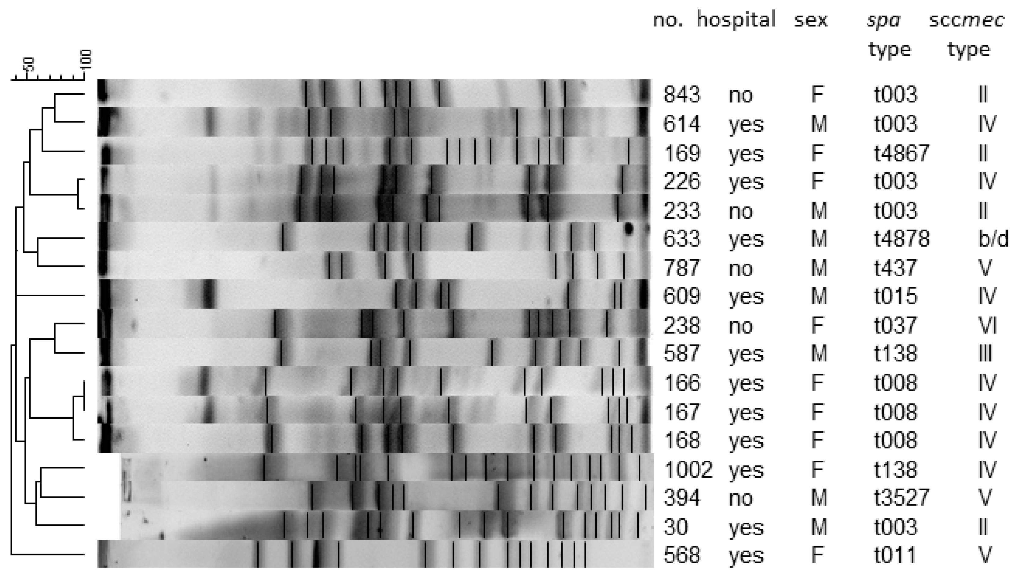
S. aureus isolates showed very different pulsotypes, no dominant clones were detected. Cluster analysis based on PFGE showed that pulsotypes were similar in less than 70%, suggesting a genotypically varied population (data not shown). Among MRSA strains, three isolates with identical pulsotypes were found. They came from different patients of the same LTCF unit and had the same spa-type (t008) and SCCmec type (IV) (Figure 2).
Overall, 10 different spa types (mostly t003, 29.4%) and five SCCmec (mostly SCCmec IV, 41.2%) were observed (Figure 2) in all MRSA strains.

4. Discussion

S. aureus is the most important aetiological agent in wound infections, including chronic wounds [24,25]. However, when a chronic wound is infected, bacteria often form polymicrobial biofilms, unlike in the case of acute wounds, such as surgical site infections. In two thirds of the studied population, no more than two microorganisms were isolated. Apart from S. aureus, the bacteria from Morganellaceae family were most frequently detected. In the remaining one third of cases, polymicrobial biofilms with a higher virulence compared to single-species biofilms were observed [26]. The common pathogens were Pseudomonas aeruginosa, Gram-positive cocci (GAS, GBS, EC, EF) and yeast-like fungi.
Microorganisms in chronic wounds prolong the healing [27]. Besides direct damage they cause to the tissue, bacteria attract leukocytes and activate inflammatory cytokines, proteases, and associated reactive oxygen species, thus both initiating and maintaining inflammatory cascades [28,29].
The strains we have analysed rarely contained genes responsible for specific types of virulence, which may point to their potentially low pathogenicity. The observed virulence was much lower than in previously studied Polish strains, isolated from invasive infections, such as surgical site infections. [18] The results do not show any correlation between the virulence and the presence of certain genes, in particular, between pvl genes and the spa type. The drug-resistance of the studied S. aureus strains does not raise any particular concerns either. Only one in five strains was multidrug-resistant, mostly to erythromycin and clindamycin (MLSB), as well as to tetracyclines and aminoglycosides. 1/3 of the strains were resistant to two or three antibiotic groups, but their resistance was weaker than in other patient groups in southern Poland, such as very-low-birth-weight infants or the geriatric patients of ambulatory care [18,30]. The high level of MLSB may result from a particularly very high consumption of macrolides, almost the highest in Poland compared to other EU at more than five DDD per 1000 inhabitants per day, while in the EU not more than three [17].
The author observed the high prevalence of S. aureus resistance to aminoglycosides and tetracyclines. These resistances are in part related to SCCmec modifications, but also other resistance mechanisms are known [31]. A lack of resistance to the group of antibiotics was observed significantly more often in the group of people not remaining for hospitalization. This suggests good practice towards rapid patient discharge due to the risk of infection or co-infection with potentially resistant pathogens [32]. This is important in preventing the spread of resistant infections, which was partly confirmed by our study, due to the coexistence of the same spa-type and SCCmec type in three patients. All staphylococcal strains were susceptible to vancomycin and linezolid. This is a positive result in the presented data due to the aforementioned abuse of antimicrobial agents in the Polish population, the more so as these drugs are more and more often the last alternative in the treatment of highly resistant strains. High resistance to fluoroquinolones in the obtained results (ciprofloxacin 17.5% and moxifloxacine 12.6%) may result from the high consumption of this group of antibiotics. In Europe, quinolones are the third most frequently used group of antibiotics after penicillins and beta-lactams [33]. The argument for the influence of exposure to antimicrobial agents is supported by an increase in the ratio of resistant to susceptible pathogens in the oldest group of patients in our study.
Effective surveillance of S. aureus can be achieved through a combination of the traditional or cultural standard methods and several molecular techniques, manly polymerase chain reactions (PCR)-based. Molecular techniques have been widely used in molecular epidemiology or outbreak investigation, and have the advantage that, they are rapid, less laborious, and more sensitive, specific and efficient compared to the conventional method. Typing of microorganisms covers the methods which enable to reproduce the transmission routes of pathogens as well as compare them with global spreading of specially virulent strains and the most important in everyday practice is use the multi locus seqence typing (MLST)—a reference method for establishing the basic genetic structure of S. aureus population, which is dominated by several large clone complexes and includes several hundred sequence types (ST)—or spa typing, based on the sequencing of short repetitive sequences of the polymorphic X region from the gene encoding protein A [34,35]. The ‘gold standard’ for the typing of Staphylococcus aureus is pulsed-field gel electrophoresis (PFGE) which allow to determine the spread of mi-croorganisms, which relies on separating the DNA fragments after restriction cutting, very often used for outbreak investigation in drug-resistant epidemic pathogens [36].
The genetic typing of MRSA and PFGE pointed to a high diversity among the strains, which means that colonisation was mostly not associated with the hospital environment. An MRSA clone was only found in three patients of one LTCF. The spa typing has confirmed the spa type t003 to be the most predominant among MRSA strains at hospitals in Southern Poland, as reported previously [37].
One of the problems we had to face when designing this study was how to collect the materials for analysis. The microbiological diagnostics of chronic wound infections largely depend on the way in which materials were collected. What is characteristic about chronic wounds is the presence of diverse bacteria on the surface, observed as a biofilm. The microbes in the biofilm are adapted to difficult conditions, and can survive different kinds of treatment, including the use of antibiotics [38,39]. A microbiological analysis should provide credible information on the aetiological factors of an infection, which are often located deep under the surface. That is why using the invasive methods, such as tissue aspirates or biopsies, rather than wound swabs is advised [40,41,42]. Invasive methods are optimal for a quantitative microbiology analysis and facilitate the identification of the aetiological factors of infections, no matter where they are located. However, they also have some downsides, since they are painful for patients and they disturb the wound healing. They may also cause the contamination of deeper located tissues or trigger a haemorrhage. Moreover, the specimen collection has to be performed by an experienced doctor, and an anaesthetic needs to be applied. That is why swabs are used so frequently, and why we decided to resort to them as well. They are non-invasive and can be collected by a nurse, not necessarily a doctor, in any conditions [43].
All in all, it seems that the best solution would be to use a method that is most appropriate given the specific conditions of each patient. The invasive (biopsies) and non-invasive (swabs) methods could complement one another in the clinical diagnostics. A wound swab may, in some situations, actually yield more meaningful results. Doctors should, then, always take advice from a microbiology consultant on the best method for specimen collection once they recognise the clinical signs of an infection [44].
One of the weaknesses of our study was the lack of confirmed presence of anaerobes and fastidious bacteria in the analysed materials [45,46]. We must also point out that the drug sensitivity was tested in vitro, with no regard to the specific conditions of a biofilm.

5. Conclusions

First of all, in our study, we pay attention to the identification of the same strains only among hospital patients. Therefore, special attention should be paid to the prevention of pathogen transmission between patients using the disinfection algorithms recommended by WHO.
The risk of multi-drug-resistant strains increases with age and is significantly the highest in the group of the oldest people. Mention was made of a possible coincidence of this phenomenon with increasing consumption of antibiotics. Therefore, special attention should be paid to rational antibiotic therapy, taking into account the guidelines for the use of antibiotic therapy in chronic wounds.
The optimal method of collecting material for research is a biopsy, but a properly performed swab can also provide clinically significant data.
Due to the constantly growing resistance of pathogens to the antimicrobial agents used, it can be concluded that this phenomenon will now be even more intense than in the results obtained.

Supplementary Materials

The following are available online at https://www.mdpi.com/article/10.3390/ijerph18094662/s1, File S1: precise Methodology description.

Author Contributions

Conceptualization, M.G. and J.W.-M.; data curation, E.Z., M.P.-W., D.R. and A.C.; formal analysis, E.Z., D.R. and A.C.; investigation, M.G., E.Z., M.P.-W., T.B., B.G. and J.W.-M.; methodology, M.G., D.R., A.C. and J.W.-M.; project administration, J.W.-M.; resources, M.P.-W.; supervision, J.W.-M.; writing—original draft, M.G. and B.G.; writing—review & editing, T.B. and J.W.-M. All authors have read and agreed to the published version of the manuscript.

Funding

The research project has been conducted under the supervision of Jadwiga Wójkowska-Mach. There has been no external funding for this manuscript.

Institutional Review Board Statement

This work has been approved by the Bioethics Committee of Jagiellonian University Medical College (approval no KBET/227/B/2012). All data analysed in the course of this study had previously been anonymised.

Informed Consent Statement

Not applicable.

Data Availability Statement

The datasets generated during and/or analysed during the current study are available from the corresponding author on reasonable request.

Acknowledgments

The research project has been conducted under the supervision of Jadwiga Wójkowska-Mach. The strains used as controls were kindly provided by Marek Gniadkowski, National Medicines Institute, Warsaw, Poland.

Conflicts of Interest

The authors declare no conflict of interest.

References

  1. Raffetto, J.D. The definition of the venous ulcer. J. Vasc. Surg. 2010, 52, 46S–49S. [Google Scholar] [CrossRef] [Green Version]
  2. Alavi, A.; Sibbald, R.G.; Phillips, T.J.; Miller, O.F.; Margolis, D.J.; Marston, W.; Woo, K.; Romanelli, M.; Kirsner, R.S. What’ s new: Management of venous leg ulcers Approach to venous leg ulcers. J. Am. Dermatol. 2019, 74, 627–640. [Google Scholar] [CrossRef]
  3. Nelson, E.A.; Adderley, U. Venous leg ulcers. BMJ Clin. Evid. 2016, 2016, 1902. [Google Scholar]
  4. Mohd-Zain, Z.; Mohd-Nawi, S.F.A.; Adnan, A.; Kumar, S. Frequency and molecular epidemiology of Panton-Valentine leukocidin gene in Staphylococcus aureus colonising HIV-infected patients. Malays. J. Pathol. 2017, 39, 115–122. [Google Scholar] [PubMed]
  5. Callam, M.J.; Harper, D.R.; Dale, J.J.; Ruckley, C.V. Chronic ulceration of the leg: Extent of the problem and provision of care. Br. Med. J. 1987, 290, 1855–1856. [Google Scholar] [CrossRef] [Green Version]
  6. Ruckley, C.V. Socioeconomic impact of chronic venous insufficiency and leg ulcers. Angiology 1997, 48, 67–69. [Google Scholar] [CrossRef]
  7. Xie, T.; Ye, J.; Rerkasem, K.; Mani, R. The venous ulcer continues to be a clinical challenge: An update. Burn. Trauma 2018, 6, 18. [Google Scholar] [CrossRef] [Green Version]
  8. Jawień, A.; Szewczyk, M.T.; Kaszuba, A.; Gaciong, Z.A.; Krasiński, Z.; Wroński, J.; Grzela, T.; Koblik, T. Wytyczne Grupy Ekspertów w sprawie gojenia owrzodzeń żylnych goleni. Leczenie Ran 2011, 8, 59–80. [Google Scholar]
  9. Tomkowski, W.; Kuca, P.; Urbanek, T.; Chmielewski, D.; Krasiński, Z.; Pruszczyk, P.; Windy, J.; Oszkinis, G.; Jawień, A.; Burakowski, J. Venous thromboembolism—recommendations on the prevention, diagnostic approach and management. The 2017 Polish Consensus Statement. Acta Angiol. 2017, 23, 35–71. [Google Scholar] [CrossRef]
  10. Bryant, R.A. Acute and Chronic Wounds, 2nd ed.; Mosby: St. Louis, MO, USA, 2000. [Google Scholar]
  11. Agale, S.V. Chronic Leg Ulcers: Epidemiology, Aetiopathogenesis, and Management. Ulcers 2013, 1–9. [Google Scholar] [CrossRef] [Green Version]
  12. Wertheim, H.F.; Melles, D.C.; Vos, M.C.; van Leeuwen, W.; van Belkum, A.; Verbrugh, H.A.; Nouwen, J.L. The role of nasal carriage in Staphylococcus aureus infections. Lancet Infect. Dis. 2005, 5, 751–762. [Google Scholar] [CrossRef]
  13. Rahim, K.; Saleha, S.; Zhu, X.; Huo, L.; Basit, A.; Franco, O.L. Bacterial Contribution in Chronicity of Wounds. Microb. Ecol. 2016, 73, 710–721. [Google Scholar] [CrossRef] [PubMed]
  14. Saseedharan, S.; Sahu, M.; Chaddha, R.; Pathrose, E.; Bal, A.; Bhalekar, P.; Sekar, P.; Krishnan, P. Epidemiology of diabetic foot infections in a reference tertiary hospital in India. Braz. J. Microbiol. 2018, 49, 401–406. [Google Scholar] [CrossRef] [PubMed]
  15. Dunyach-Remy, C.; Essebe, C.N.; Sotto, A.; Lavigne, J.P. Staphylococcus aureus toxins and diabetic foot ulcers: Role in pathogenesis and interest in diagnosis. Toxins 2016, 8, 209. [Google Scholar] [CrossRef] [Green Version]
  16. Bien, J.; Sokolova, O.; Bozko, P. Characterization of Virulence Factors of Staphylococcus aureus: Novel Function of Known Virulence Factors That are Implicated in Activation of Airway Epithelial Proinflammatory Response. J. Pathog. 2011, 2011, 1–13. [Google Scholar] [CrossRef] [Green Version]
  17. Wojkowska-Mach, J.; Godman, B.; Glassman, A.; Kurdi, A.; Pilc, A.; Rozanska, A.; Skoczyński, S.; Wałaszek, M.; Bochenek, T. Antibiotic consumption and antimicrobial resistance in Poland; Findings and implications 11 Medical and Health Sciences 1117 Public Health and Health Services. Antimicrob. Resist. Infect. Control 2018, 7. [Google Scholar] [CrossRef] [Green Version]
  18. ECDC. European Center for Disease Control and Prevention. Annual Surveillance Reports and Protocols-Antimicrobial Consumption. 2015. Available online: https://ecdc.europa.eu/en/antimicrobial-consumption/surveillance-anddisease-%0Adata/report-protocol (accessed on 16 August 2017).
  19. Pomorska-Wesołowska, M.; Rózańska, A.; Natkaniec, J.; Gryglewska, B.; Szczypta, A.; Dzikowska, M.; Chmielarczyk, A.; Wójkowska-Mach, J. Longevity and gender as the risk factors of methicillin-resistant Staphylococcus aureus infections in southern Poland. BMC Geriatr. 2017, 17, 1–7. [Google Scholar] [CrossRef] [PubMed] [Green Version]
  20. Pereira, E.M.; Schuenck, R.P.; Malvar, K.L.; Iorio, N.L.P.; Matos, P.D.M.; Olendzki, A.N.; Oelemann, W.M.R.; dos Santos, K.R.N. Staphylococcus aureus, Staphylococcus epidermidis and Staphylococcus haemolyticus: Methicillin-resistant isolates are detected directly in blood cultures by multiplex PCR. Microbiol. Res. 2010, 165, 243–249. [Google Scholar] [CrossRef]
  21. Sutcliffe, J.; Grebe, T.; Tait-Kamradt, A.; Wondrack, L. Detection of erythromycin-resistant determinants by PCR. Antimicrob. Agents Chemother. 1996, 40, 2562–2566. [Google Scholar] [CrossRef] [PubMed] [Green Version]
  22. Anthony, R.M.; Connor, A.M.; Power, E.G.M.; French, G.L. Use of the polymerase chain reaction for rapid detection of high-level mupirocin resistance in staphylococci. Eur. J. Clin. Microbiol. Infect. Dis. 1999, 18, 30–34. [Google Scholar] [CrossRef]
  23. Mcdougal, L.K.; Steward, C.D.; Steward, C.D.; Killgore, G.E.; Chaitram, J.M.; McAllister, S.K.; Tenover, F.C. Pulsed-Field Gel Electrophoresis Typing of Oxacillin-Resistant Staphylococcus aureus Isolates from the United States: Establishing a National Database. J. Clin. Microbiol. 2003, 41, 5113–5120. [Google Scholar] [CrossRef] [Green Version]
  24. Lipsky, B.A.; Hoey, C. Topical Antimicrobial Therapy for Treating Chronic Wounds. Clin. Infect. Dis. 2009, 49, 1541–1549. [Google Scholar] [CrossRef] [Green Version]
  25. Pugliese, D.J. Infection in Venous Leg Ulcers: Considerations for Optimal Management in the Elderly. Drugs Aging 2016, 33, 87–96. [Google Scholar] [CrossRef]
  26. Seth, A.K.; Geringer, M.R.; Hong, S.J.; Leung, K.P.; Mustoe, T.A. Comparative analysis of single-species and polybacterial wound biofilms using a quantitative, in vivo, rabbit ear model. PLoS ONE 2012, 7, 1–9. [Google Scholar] [CrossRef] [PubMed] [Green Version]
  27. Mudge, E.J. Recent accomplishments in wound healing. Int. Wound J. 2015, 12, 4–9. [Google Scholar] [CrossRef] [PubMed]
  28. Schreml, S.; Szeimies, R.M.; Prantl, L.; Karrer, S.; Landthaler, M.; Babilas, P. Oxygen in acute and chronic wound healing. Br. J. Dermatol. 2010, 163, 257–268. [Google Scholar] [CrossRef]
  29. James, G.A.; Swogger, E.; Wolcott, R.; Pulcini, E.D.; Secor, P.; Sestrich, J.; Costerton, J.W.; Stewart, P.S. Biofilms in chronic wounds. Wound Repair Regen. 2008, 16, 37–44. [Google Scholar] [CrossRef] [PubMed]
  30. Chmielarczyk, A.; Pilarczyk-Zurek, M.; Kamińska, W.; Pobiega, M.; Romaniszyn, D.; Ziółkowski, G.; Wojkowska-Mach, J.; Bulanda, M. Molecular Epidemiology and Drug Resistance of Acinetobacter baumannii Isolated from Hospitals in Southern Poland: ICU as a Risk Factor for XDR Strains. Microb. Drug Resist. 2016, 22, 328–335. [Google Scholar] [CrossRef]
  31. Foster, T.J. Antibiotic resistance in Staphylococcus aureus. Current status and future prospects. FEMS Microbiol. Rev. 2017, 41, 430–449. [Google Scholar] [CrossRef] [PubMed]
  32. Schreiber, P.W.; Sax, H.; Wolfensberger, A.; Clack, L.; Kuster, S.P.; Swissnoso. The preventable proportion of healthcare-associated infections 2005–2016: Systematic review and meta-analysis. Infect. Control Hosp. Epidemiol. 2018, 39, 1277–1295. [Google Scholar] [CrossRef] [PubMed] [Green Version]
  33. Olczak-Pieńkowska, A. Podsumowanie Aktualnych Danych nt. Konsumpcji Antybiotyków w Krajach unii Europejskiej–Dane Europejskiej Sieci Monitorowania Konsumpcji Antybiotyków ESAC-Net (listopad 2017) Zakład Epidemiologii i Mikrobiologii Klinicznej, Narodowy Instytut Leków, Warszawa. Available online: http://antybiotyki.edu.pl/wp-content/uploads/Biuletyn/biuletyn-npoa-2018_1.pdf (accessed on 15 June 2020).
  34. Mazi, W.; Sangal, V.; Sandstrom, G.; Saeed, A.; Yu, J. Evaluation of spa-typing of methicillin-resistant Staphylococcus aureus using high-resolution melting analysis. Int. J. Infect. Dis. 2015, 38, 125–128. [Google Scholar] [CrossRef] [PubMed] [Green Version]
  35. Sabat, A.J.; Budimir, A.; Nashev, D.; Sá-Leão, R.; van Dijl, J.M.; Laurent, F.; Grundmann, H.; Friedrich, A.W.; ESCMID Study Group of Epidemiological Mark-ers (ESGEM). Overview of molecular typing methods for outbreak detection and epi-demiological surveillance. Euro. Surveill. 2013, 18, 20380. [Google Scholar] [CrossRef] [PubMed] [Green Version]
  36. Go-pal Rao, G.; Batura, R.; Nicholl, R.; Coogan, F.; Patel, B.; Bassett, P.; Kearns, A.M. Outbreak re-port of investigation and control of an outbreak of Panton-Valentine Leukocidin-positive methicillin-sensitive Staphylococcus aureus (PVL-MSSA) infection in neo-nates and mothers. BMC Infect. Dis. 2019, 19, 178. [Google Scholar] [CrossRef]
  37. Chmielarczyk, A.; Pomorska-Wesołowska, M.; Szczypta, A.; Romaniszyn, D.; Pobiega, M.; Wójkowska-Mach, J. Molecular analysis of meticillin-resistant Staphylococcus aureus strains isolated from different types of infections from patients hospitalized in 12 regional, non-teaching hospitals in southern Poland. J. Hosp. Infect. 2017, 95, 259–267. [Google Scholar] [CrossRef] [PubMed]
  38. Chantelau, E.; Tanudjaja, T.; Altenhöfer, F.; Ersanli, Z.; Lacigova, S.; Metzger, C. Antibiotic treatment for uncomplicated neuropathic forefoot ulcers in diabetes: A controlled trial. Diabet. Med. 1996, 13, 156–159. [Google Scholar] [CrossRef]
  39. Öien, R.F.; Forssell, H.W. Ulcer healing time and antibiotic treatment before and after the introduction of the Registry of Ulcer Treatment: An improvement project in a national quality registry in Sweden. BMJ Open 2013, 3, 1–6. [Google Scholar] [CrossRef] [Green Version]
  40. Schultz, G.; Bjarnsholt, T.; James, G.A.; Leaper, D.J.; McBain, A.J.; Malone, M.; Stoodley, P.; Swanson, T.; Tachi, M.; Wolcott, R.D.; et al. Consensus guidelines for the identification and treatment of biofilms in chronic nonhealing wounds. Wound Repair Regen. 2017, 25, 744–757. [Google Scholar] [CrossRef]
  41. Gardner, S.E.; Frantz, R.A.; Saltzman, C.L.; Hillis, S.L.; Park, H.; Scherubel, M. Diagnostic validity of three swab techniques for identifying chronic wound infection. Wound Repair Regen. 2006, 14, 548–557. [Google Scholar] [CrossRef]
  42. Pellizzer, G.; Strazzabosco, M.; Presi, S.; Furlan, F.; Lora, L.; Benedetti, P.; Bonato, M.; Erle, G.; De Lalla, F. Deep tissue biopsy vs. superficial swab culture monitoring in the microbiological assessment of limb-threatening diabetic foot infection. Diabet. Med. 2001, 18, 822–827. [Google Scholar] [CrossRef]
  43. Miller, C.N.; Carville, K.; Newall, N.; Kapp, S.; Lewin, G.; Karimi, L.; Santamaria, N. Assessing bacterial burden in wounds: Comparing clinical observation and wound swabs. Int. Wound J. 2011, 8, 45–55. [Google Scholar] [CrossRef] [PubMed]
  44. Kelly, F. Reliability in Wound Swabbing. Br. J. Nurs. 2003, 12, 959–964. [Google Scholar] [CrossRef] [PubMed]
  45. Percival, S.L.; Malone, M.; Mayer, D.; Salisbury, A.-M.; Schultz, G. Role of anaerobes in polymicrobial communities and biofilms complicating diabetic foot ulcers. Int. Wound J. 2018, 15, 776–782. [Google Scholar] [CrossRef] [PubMed]
  46. Han, A.; Zenilman, J.M.; Melendez, J.H.; Shirtliff, M.E.; Agostinho, A.; James, G.; Stewart, P.S.; Mongodin, E.F.; Rao, D.; Rickard, A.H.; et al. The importance of a multifaceted approach to characterizing the microbial flora of chronic wounds. Wound Repair Regen. 2011, 19, 532–541. [Google Scholar] [CrossRef] [PubMed]
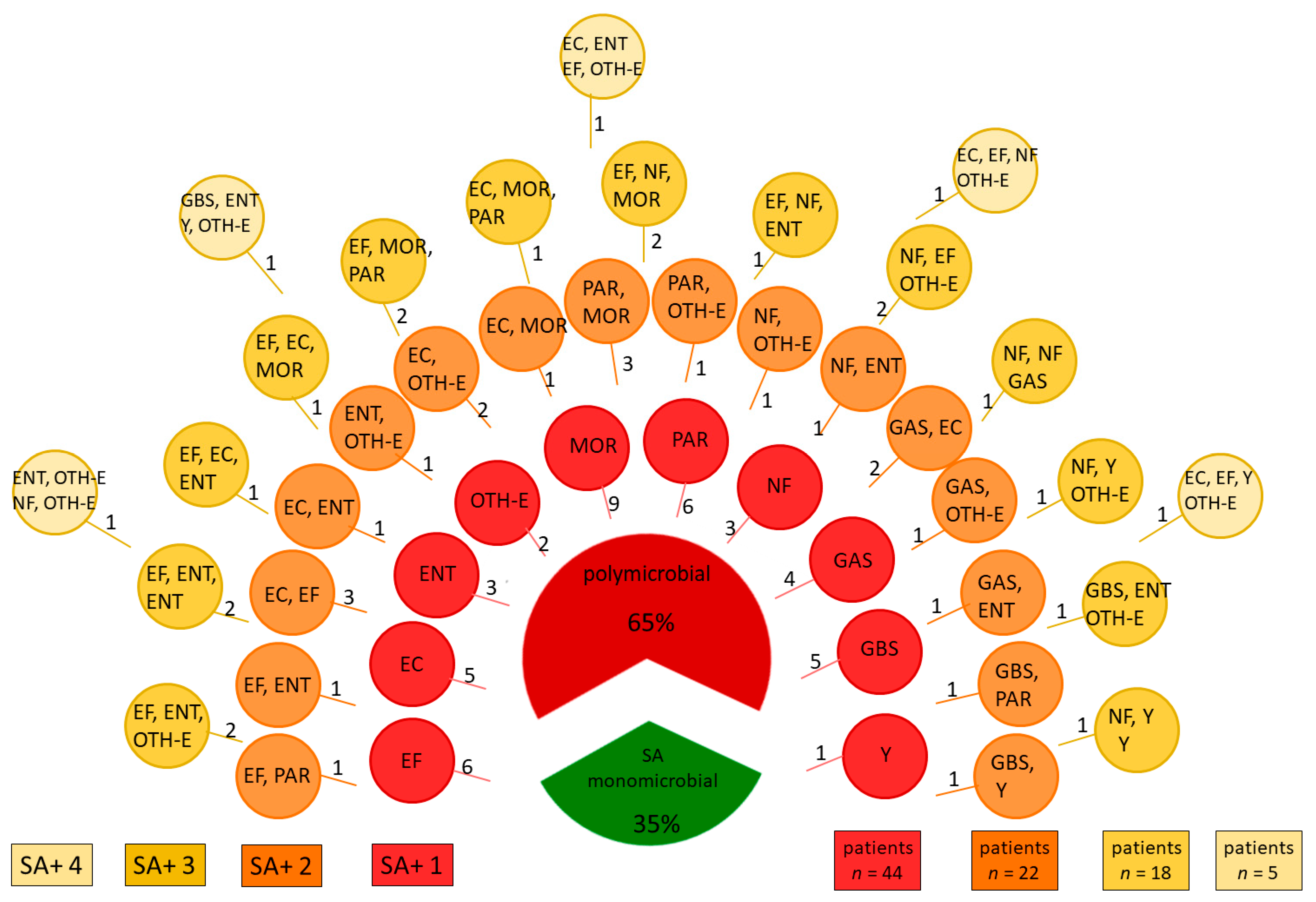
Figure 1. Microbiota isolated from infected venous ulcers from 143 patients, numbers indicate numbers of patients. Legend: EC Escherichia coli; EF Enterococcus faecalis; ENT Enterobacter spp.; GAS Streptococcus group A; GBS Streptococcus group B; MOR bacteria from Morganellaceae family; NF other non fermenting bacteria; OTH-E other bacteria from Enterobacteriaceae family; PAR Pseudomonas aeruginosa; SA Staphylococcus aureus; Y yeast. n—number of patients in the group with one, two, three or four pathogens in addition to SA in the polymicrobial group, respectively.
Ijerph 18 04662 g001
Figure 2. Analysis of the genetic similarity between S. aureus isolates by the PFGE (Pulsed-Field Gel Electrophoresis) method based on spa and sccmec genes.
Ijerph 18 04662 g002
Table 1. Other isolated pathogens.
Identified Pathogenn
Enterococcus faecalis (EF)30
Streptococcus agalactiae (GBS)10
Streptococcus pyogenes (GAS)9
Escherichia coli (EC)19
Enterobacter spp (ENT)19
OTH-E: Other Bacteria from Enterobacteriaceae Family
Klebsiella spp.12
Serratia spp.3
Citrobacter spp.6
Morganellaceae Family (MOR)
Proteus spp.17
Morganella morgani3
Providentia regretti1
Pseudomonas aeruginosa (PAR)15
Other Non-Fermenting Bacteria (NF)
Acinetobacter spp.13
Stenotrophomonas maltophilia4
Candida spp. (Y)7
Table 2. Characteristics of the study group according to drug sensitivity of Staphylococcus aureus.
Characteristics of the Study GroupTotal n = 143Sensitivity to AntibioticsOR (95% CI)
Yes n = 70 No n = 73
Age (Years) by Categories n [%]
≤59 years45 (31.5)23 (33.3)22 (30.1)0.8 (0.36–1.73)
60–75 years58 (40.6)33 (47.8)25 (34.2)ref.
76–80 years16 (11.2)6 (8.7)10 (13.7)0.5 (0.15–1.41)
≥81 years24 (16.8)7 (10.1)16 (21.9)0.3 (0.12–0.93)
Gender n [%]
Female 76 (53.2)34 (48.6)42 (57.5)0.7 (0.36–1.35)
Male (ref.) 67 (46.9)36 (51.4)31 (42.5)
Ambulatory Care n [%]
yes48 (32.2) 29 (41.4)19 (26.0)2.0 (0.99–4.08)
no95 (66.4)41 (58.6)54 (74.0)
Hospital Stay n [%]
surgical wards61 (42.7)26 (37.1)35 (47.8)0.9 (0.40–2.19)
non-surgical wards or LTCF34 (23.8)15 (21.4)19 (13.3)
The Presence of Resistance Genes n [;%], YES
mecA17 (11.9)n/a17 (11.9)n/a
ermA8 (5.6)n/a8 (5.6)n/a
ermB2 (1.4)n/a2 (1.4)n/a
msr1 (0.7)n/a1 (0.7)n/a
The Presence of Various Virulence Factors Genes n [;%], YES
lukE98 (68.5)45 (64.3)53 (72.6)0.7 (0.33–1.38)
tsst-117 (11.9)9 (12.9)8 (11.0)1.2 (0.43–3.30)
pvl2 (1.4)1 (1.4)1 (1.4)1.0 (0.06–17.01)
etA/B23 (16.1)10 (14.3)13 (17.8)0.8 (0.31–1.89)
Legend: etA/B exfoliative toxin A and/or B; LTCF long term care facilities; lukE LukDE leukocidin; n/a not applicable; OR—Odds ratio; (95%CI) 95% Confidence Interval; pvl Panton-Valentine leukocidin; ref.—reference; tsst-1 toxic shock syndrome toxin-1.
Table 3. Drug-resistance of Staphylococcus aureus isolated from hospitalised and non-hospitalised patients.
Antimicrobial CategoryAntimicrobial Agent Total n = 143Hospital StayOR (95%CI)
Yes, n = 95No, n = 48
Aminoglycosides Gentamicin 24 (16.8)16 (16.8)8 (16.7)0.9 (0.38–2.45)
Amikacin 29 (20.2)19 (20.0)10 (20.8)2.1 (0.71–5.89)
Tobramycin 39 (27.3)27 (28.4)12 (25.0)1.1 (0.51–2.49)
Anti-MRSA cephalosporinsCeftaroline0 (0)0 (0.0)0 (0.0)n/a
FluoroquinolonesCiprofloxacin25 (17.5)20 (21.1)5 (10.4)2.2 (0.76–6.26)
Moxifloxacin18 (12.6)14 (14.7)4 (8.3)0.5 (0.23–1.33)
Folate pathway inhibitors Trimethoprim/Sulfamethoxazole 8 (5.6)6 (6/3)2 (4.2)1.5 (0.29–7.65)
Lincosamides Clindamycin 29 (20.2)18 (18.9)11 (22.9)0.7 (0.32–1.74)
Macrolides Erythromycin 31 (21.6)17 (17.9)14 (29.2)0.6 (0.27–1.43)
GlycopeptidesVancomycin 0 (0)0 (0.0)0 (0.0)n/a
GlycylcyclinesTigecycline 0 (0)0 (0.0)0 (0.0)n/a
Oxazolidinones Linezolid 0 (0)0 (0.0)0 (0.0)n/a
PhenicolsChloramphenicol 16 (11.2)13 (13.7)3 (6.2)0.3 (0.13–0.69)
StreptograminsQuinupristin-dalfopristin 0 (0)0 (0.0)0 (0.0)n/a
Tetracyclines Tetracycline35 (24.5)27 (28.4)8 (16.7)1.9 (0.78–4.56)
Doxycycline 19 (13.3)13 (13.7)6 (12.5)1.1 (0.37–2.99)
Others (O)Mupirocin 8 (5.6)5 (5.3)3 (6.2)0.8 (0.18–3.49)
Multidrug Resistance n (;%)
MRSA, yes 17 (11.9)12 (12.6)5 (10.4)1.2 (0.39–3.59)
MLSB, yes 28 (19.6)16 (16.8)12 (25.0)9.1 (1.17–71.02)
Non-Susceptible to Antimicrobial Categories n (;%)
0 categories (fully susceptible)70 (49)41 (43.2)29 (60.4)0.4 (0.22–0.92)
1 category26 (18.2)23 (24.2)3 (6.2)4.8 (1.38–17.06)
Aminoglycosides9 (6.3)7 (7.4)2 (4.2)
Tetracyclines9 (6.3)9 (9.5)0 (0.0)
Phenicols4 (2.8)4 (4.2)0 (0.0)
Fluoroquinolones2 (1.4)2 (2.1)0 (0.0)
Lincosamides1 (0.7)1 (1.1)0 (0.0)
Macrolides1 (0.7)0 (0.0)1 (2.1)
2 categories19 (13.3)11 (11.6)8 (16.7)0.6 (0.23–1.67)
Macrolides + Lincosamides5 (3.5)2 (2.1)3 (6.3)
Tetracyclines + Phenicols3 (2.1)2 (2.1)1 (2.1)
Aminoglycosides + Fluoroquinolones3 (2.1)3 (3.2)0 (0.0)
Tetracyclines + Fluoroquinolones2 (1.4)2 (2.1)0 (0.0)
Aminoglycosides + Tetracycline2 (1.4)1 (1.1)1 (2.1)
Aminoglycosides A+ Phenicols2 (1.4)1 (1.1)1 (2.1)
Tetracyclines + Macrolides1 (0.7)0 (0.0)1 (2.1)
Aminoglycosides + Macrolides1 (0.7)0 (0.0)1 (2.1)
3 categories or more 28 (19.6)19 (20.0)8 (16.7)1.2 (0.48–2.96)
OR (95%CI) 95% confidence intervals of odds ratio; MRSA—methicillin resistant Staphylococcus aureus; MLSB—Macrolide-lincosamide-streptogramin B resistance phenotype.
Publisher’s Note: MDPI stays neutral with regard to jurisdictional claims in published maps and institutional affiliations.