Article

Disturbed Lipid Metabolism in Diabetic Patients with Manifest Coronary Artery Disease Is Associated with Enhanced Inflammation

1
Department of Cardiovascular Surgery, University Medical Center of the Johannes Gutenberg, University Mainz, Langenbeckstraße 1, 55131 Mainz, Germany
2
Department for Cardiology I, University Medical Center of the Johannes Gutenberg, University Mainz, Langenbeckstraße 1, 55131 Mainz, Germany
*
Author to whom correspondence should be addressed.
K. Buschmann and Y. Gramlich contributed equally to this study and are joint first authors.
A. Daiber and G.D. Duerr equally contributed as last authors.
Int. J. Environ. Res. Public Health 2021, 18(20), 10892; https://doi.org/10.3390/ijerph182010892
Submission received: 30 August 2021 / Revised: 26 September 2021 / Accepted: 12 October 2021 / Published: 17 October 2021

Abstract

:
Background: Diabetic vasculopathy plays an important role in the pathophysiology of coronary artery disease (CAD) with oxidative stress as a strong mediator. This study aims to elucidate the underlying pathomechanisms of diabetic cardiac vasculopathy leading to coronary disease with an emphasis on the role of oxidative stress. Therefore, novel insights into antioxidant pathways might contribute to new strategies in the treatment and prevention of diabetic CAD. Methods: In 20 patients with insulin-dependent or non-insulin dependent diabetes mellitus (IDDM/NIDDM) and 39 non-diabetic (CTR) patients, myocardial markers of oxidative stress, vasoactive proteins, endothelial nitric oxide synthase (eNOS), activated phosphorylated eNOS (p-eNOS), and antioxidant enzymes, e.g., tetrahydrobiopterin generating dihydrofolate reductase (DHFR), heme oxygenase (HO-1), as well as serum markers of inflammation, e.g., E-selectin, interleukin-6 (IL-6), and lipid metabolism, e.g., high- and low-density lipoptrotein (HDL- and LDL-cholesterol) were determined in specimens of right atrial tissue and in blood samples from type 2 diabetic and non-diabetic patients undergoing coronary artery bypass graft (CABG) surgery. Results: IDDM/NIDDM increased markers of inflammation (e.g., E-selectin, p = 0.005 and IL-6, p = 0.051), decreased the phosphorylated myocardial p-eNOS (p = 0.032), upregulated the myocardial stress response protein HO-1 (p = 0.018), and enhanced the serum LDL-/HDL-cholesterol ratio (p = 0.019). However, the oxidative stress markers in the myocardium and the expression of vasoactive proteins (eNOS, DHFR) showed only marginal adverse changes in patients with IDDM/NIDDM. Conclusion: Dyslipidemia and myocardial inflammation seem to be the major determinants of diabetic CAD complications. Dysregulation in pro-oxidative enzymes might be attributable to the severity of CAD and oxidative stress levels in all included patients undergoing CABG.

1. Introduction

Diabetic cardiomyopathy is characterized by microstructural changes: myocardial oxidative stress leads to molecular and cellular inflammation, myofibroblast induction, as well as cardiomyocyte loss with subsequent replacement fibrosis [1,2,3]. Those changes are usually associated with diastolic dysfunction followed by symptomatic progressive systolic dysfunction and cardiac insufficiency [2].
Recently, animal studies have shed new light on this process, suggesting a role for an exaggerated oxidative cardiac damage and inflammation induced by diabetes mellitus in the progress of diabetic cardiomyopathy [2,4,5,6,7]. In addition, an increased inflammatory response was observed in diabetic patients [8,9] that was associated with vascular oxidative stress [10] and endothelial dysfunction [11,12]. In the long-term, this may play a major role in the formation of atherosclerotic lesions [13] and enhance the risk for cardiovascular disorders [14,15]. In addition, most cytokines are associated with higher cardiovascular risk and represent independent cardiovascular risk factors [16,17], which is a concept that was more recently explored for pharmacological targeting, e.g., within the “CANTOS trial” using “Canakinumab”, a monocloncal antibody against interleukin-1β in patients with previous myocardial infarction and high C-reactive protein (CRP) levels [18] and many other published or ongoing studies (reviewed in [19]). Of note, “Canakinumab” therapy also improved the cardiovascular prognosis of diabetic patients [20].
Measuring the level of oxidative stress in myocardium is a demanding process and requires the identification of suitable and reliable markers. Reduction in the cellular generation of tetrahydrobiopterin (BH4), a cofactor of the endothelial nitric oxide synthase (eNOS), is well known to be associated with oxidative stress, impaired endothelial function, and higher cardiovascular risk [21,22]. Furthermore, the heme oxygenase 1 (HO-1) is an important cardiac antioxidant protein, which is responsible for the production of biliverdin, bilirubin, and carbon monoxide [23,24,25]. These species have anti-atherosclerotic, anti-aggregatory, and vasodilatory properties, also acting as a potent antioxidant by the long-term induction of ferritin, which is responsible for iron storage and contributes to the prevention of Fenton-type oxidative damage. Its inducible isoform HO-1 is considered to be an important part of the cardiac antioxidant system, playing a role in stress response pathways [26,27]. Hyperlipidemia is a common phenomenon associated with type 2 diabetes mellitus caused by the insulin-dependency of lipoprotein lipase. Increased levels of low-density lipoprotein (LDL) lead to cholesterol deposition in arteries, while high levels of high-density lipoprotein (HDL) prevent cholesterol deposition [28,29].
Thus, the current study aims to validate the findings of animal studies that suggest that diabetes mellitus is associated with dyslipidemia, increased inflammation, and oxidative cardiac damage, e.g., diabetic cardiomyopathy, being strongly associated with the development of CAD [3,30]. The translational approach from animal studies to human myocardium represents a major requirement to understand the mechanisms of human disease, as our human atrial tissue samples are barely accessible and rare: In this work, we used human samples obtained from the right atrium from diabetic (IDDM/NIDDM) and non-diabetic (CTR) patients undergoing CABG due to CAD. We used cardiac markers to quantify the oxidative stress and antioxidant/vascular-regulatory proteins as well as serum markers of inflammation in the above-described patients with associated CAD.

2. Materials and Methods

2.1. Ethical Aspect

This study was conducted with permission of the ethical board of Rhineland-Palatinate, Germany (Register Nr. 837.104.08 (6100)) and after obtaining written consents of all participants. The work has been carried out in accordance with The Code of Ethics of the World Medical Association (Declaration of Helsinki). Samples of this study were already used for association studies of body mass index (BMI) with markers of oxidative stress and vascular function [31].

2.2. Patient Selection and Tissue Harvesting

The following inclusion criteria were defined: elective isolated CABG patients, sinus rhythmus; age under 85 years; absence of relevant valvular disease; absence of pulmonary hypertension; sufficient renal function; no known neoplasms or chronic systematic inflammatory diseases; and absence of severe comorbidity. The patients’ baseline characteristics are summarized in Table 1. For more details, see the published protocol where patients were grouped by different degree of adiposity and obesity (by BMI) and markers of oxidative stress as well as vascular function were associated with the BMI [31].

2.3. Blood Samples and Tissue Harvesting

Blood serum samples and myocardial tissues were obtained from 59 CABG patients as described [31]. Twenty patients had diabetes mellitus with oral medication or insulin-dependent type 2 diabetes mellitus (IDDM/NIDDM). The other 39 patients with regular blood glucose levels formed the control group (CTR). After thoracotomy, a full heparin dose was applied (400 IE/kg body weight) before cannulation of the aorta ascendens prior to the right atrium for cardiopulmonary bypass. Approximately 1 cm3 of the right atrial appendages were collected. Right atrial tissue is usually removed and discarded during cannulation of the right atrium in order to install cardiopulmonary bypass, as previously described [32]. Then, these tissue samples were immediately stored in ice-cold NaCl 0.9% solution and subjected to analysis.

2.4. Isolation of Cardiac Mitochondria and Mitochondrial Aldehyde Dehydrogenase 2 Activity

A detailed description of the mitochondria isolation method was previously published [31]. Briefly, the human cardiac tissues were homogenized in HEPES buffer and then centrifuged (1500× g for 10 min, followed by 2000× g for 5 min). Then, the supernatant was re-centrifuged (20,000× g for 20 min), and the resulting precipitate was resuspended in 1 mL Tris buffer. The amount of protein was quantified by the Lowry method. The activity of aldehyde dehydrogenase 2 (ALDH-2) in isolated heart mitochondria (final protein content 1 mg/mL) was determined by measuring the conversion of 6-methoxy-2-naphthylaldehyde (Monal 62) to the fluorescent naphthoic acid product by an HPLC-based assay as described [6,33].

2.5. Western Blot Analysis

Protein expression and modification was assessed by standard SDS-PAGE, Western blot analysis using established protocols [31,34]. Cardiac protein samples were analyzed by Western blot analysis for endothelial NO-synthase (eNOS, mouse monoclonal, 1:1000, BD Biosciences, Heidelberg, Germany), phospho-Ser1177-eNOS (rabbit polyclonal, 1:1000, Cell Signaling, Danvers, MA, USA), dihydrofolate reductase (DHFR, mouse monoclonal, 1 μg/mL, Abnova Corp., Heidelberg, Germany), monoclonal mouse heme oxygenase-1 (HO-1) (4 μg/mL, Stressgen, San Diego, CA, USA), and polyclonal rabbit β-actin (both 1:2500, Sigma-Aldrich, St. Louis, MO, USA) for normalization of loading and transfer. Detection and quantification were performed by enhanced chemiluminescence (ECL) with peroxidase conjugated anti-mouse/rabbit (GAM-POX/GAR-POX, 1:10,000, Vector Lab., Burlingame, CA, USA) secondary antibodies. Densitometric quantification of antibody-specific bands was performed with a ChemiLux Imager (CsX-1400M, Intas, Göttingen, Germany) and Gel-Pro Analyzer software (Media Cybernetics, Bethesda, MD, USA).

2.6. ELISA

Quantikine ELISA Immunoassays were performed according to the manufacturers’ instructions and as published [31,35]. The assays were obtained from R&D Systems using human serum samples: human interleukin-6 (IL-6) (Catalog Number D6050), human soluble vascular cell adhesion molecule-1 (sVCAM-1)/CD106 (CatalogNumber DVC00), human sE-Selectin/CD62E (DSLE00), and human CD40L/TNFSF5 (CatalogNumber DCDL40).

2.7. CRP, Blood Lipids, and Lipoproteins

Human C-reactive protein (CRP) was analyzed in the Department of Clinical Chemistry, University Hospital Mainz, Germany, using the daily routine facilities for in-patient care. Serum cholesterol, triglyceride, HDL, and LDL levels were analyzed in the Department of Clinical Chemistry, University Hospital Mainz, Germany, using the daily routine facilities for in-patient care. In addition, lipoproteins were also quantified by Field-Flow Fractionation (FFF), as described [6]. Lipoproteins in serum were also determined by HF5 (Superon GmbH, Dernbach, Germany). Briefly, Field-Flow Fractionation (FFF) is a well-known family of separation methods that vary in the physical nature of the force field applied to generate separation [36]. Asymmetric Flow Field-Flow Fractionation (AF4) is the most popular type of FFF. It employs a flat or cylindrical separation channel equipped with an ultrafiltration membrane and covers a wide separation range (1 nm to 1 μm). In HF5, the solvent is pumped through a porous fiber allowing a part of the flow to penetrate the wall, thus creating a cross flow that is perpendicular to the main solvent flow, which has a parabolic profile and is directed to the fiber outlet. The combination of the two forces applied eventually results in the separation of the sample compound according to their respective diffusion coefficient (i.e., their hydrodynamic radius or molar mass, respectively). Similar to AF4, HF5 has a wide range of applications. It allows the separation of molecules in solution and particles in the same separation run. The separation takes place without the use of a stationary phase as in column chromatography. Consequently, there is less danger of sample adsorption or physical plugging of the separation channel. Another advantage of this technique is the low sample dilution due to the small channel volumes (<100 μL) and low detector flow rates. The literature shows promising results for protein, nanoparticle, and even whole cell fractionation.

2.8. HPLC Assay for Dihydroethidium Oxidation Products

Superoxide was measured by a modified HPLC-based method to quantify ethidium and 2-hydroxyethidium levels, as previously described [37]. Briefly, myocardial mitochondria (0.2 mg/mL) were incubated with 50 μM dihydroethidium (DHE) for 30 min at 37 °C in PBS buffer and stored at −80 °C. Upon thawing, DHE oxidation products were extracted by the addition of 50% acetonitrile and 50% PBS, incubated (10 min), centrifuged (20 min at 20,000× g), and filtered (30 kDa Millipore Filter, 45 min at 16,000× g). A 50 μL sample of this supernatant was subjected to HPLC analysis and measured, based on a previously described method [38,39].

2.9. Statistical Analysis

Data are presented as mean ± SD. After evaluating the normal distribution of the data (using the Kolmogorov–Smirnov test), we applied an unpaired and two-tailed t-test or, where appropriate, a Mann–Whitney U-test for comparative analysis of reactive oxygen species (ROS) detection, protein activity, and expression and serum parameters (Prism for Windows version 8). p values < 0.05 were considered statistically significant.

3. Results

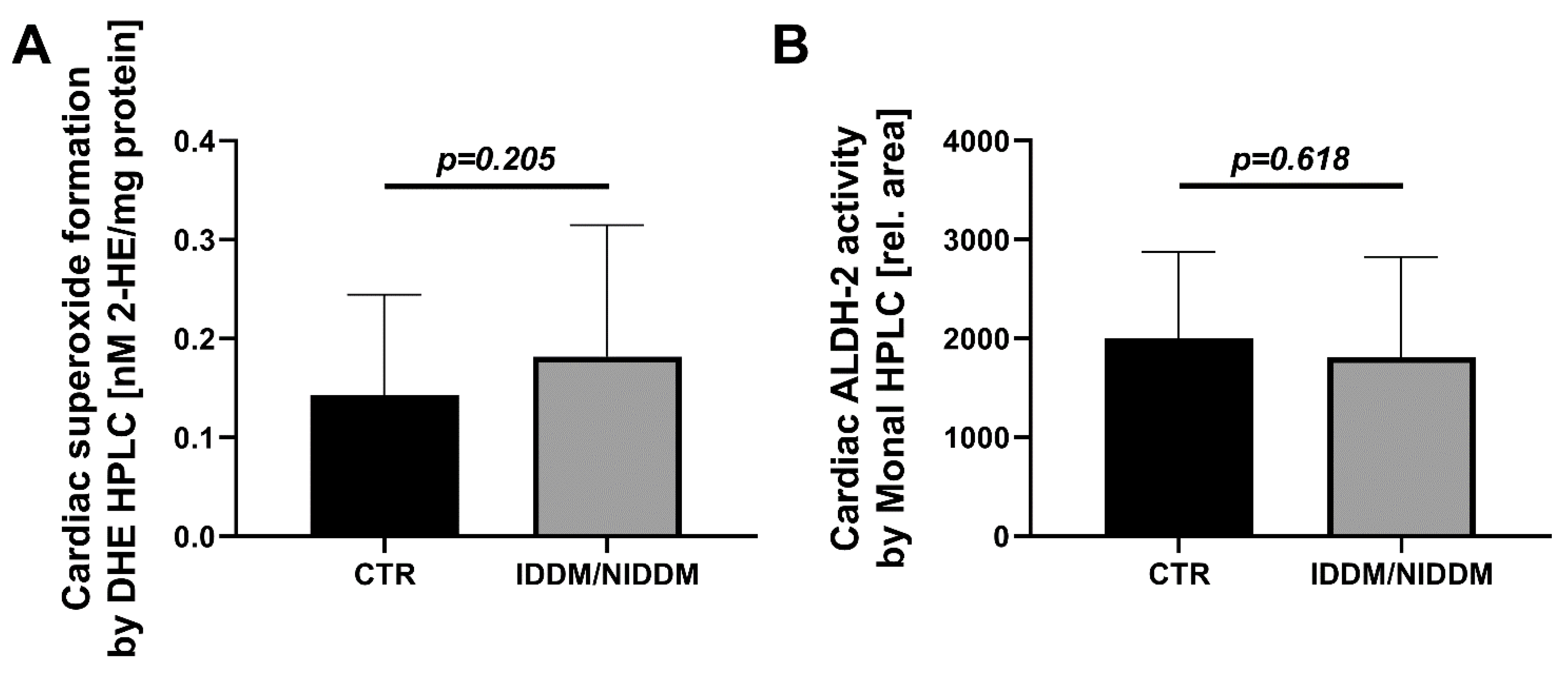
3.1. Myocardial Markers of Oxidative Stress

The primary markers of oxidative stress, direct measurement of mitochondrial superoxide formation and oxidative inactivation of ALDH-2, showed no significant changes of mitochondrial superoxide formation (DHE HPLC, p = 0.205) and cardiac ALDH-2 activity (Monal HPLC, p = 0.618) in the IDDM/NIDDM group (Figure 1).

3.2. Myocardial Vasoactive Regulatory Proteins and Antioxidant Enzymes

Cardiac eNOS protein showed a non-significant marginal increase in the diabetic subjects (p = 0.183) (Figure 2A). On the other hand, the phosphorylation of eNOS at serine 1177 (p = 0.032), an activation mark, was significantly decreased in the IDDM/NIDDM group (Figure 2B). Cardiac DHFR expression showed a non-significant marginal decrease in the diabetic subjects (p = 0.334) (Figure 2C). The antioxidant stress-response protein HO-1 was significantly upregulated in the IDDM/NIDDM group (p = 0.018) (Figure 2D).

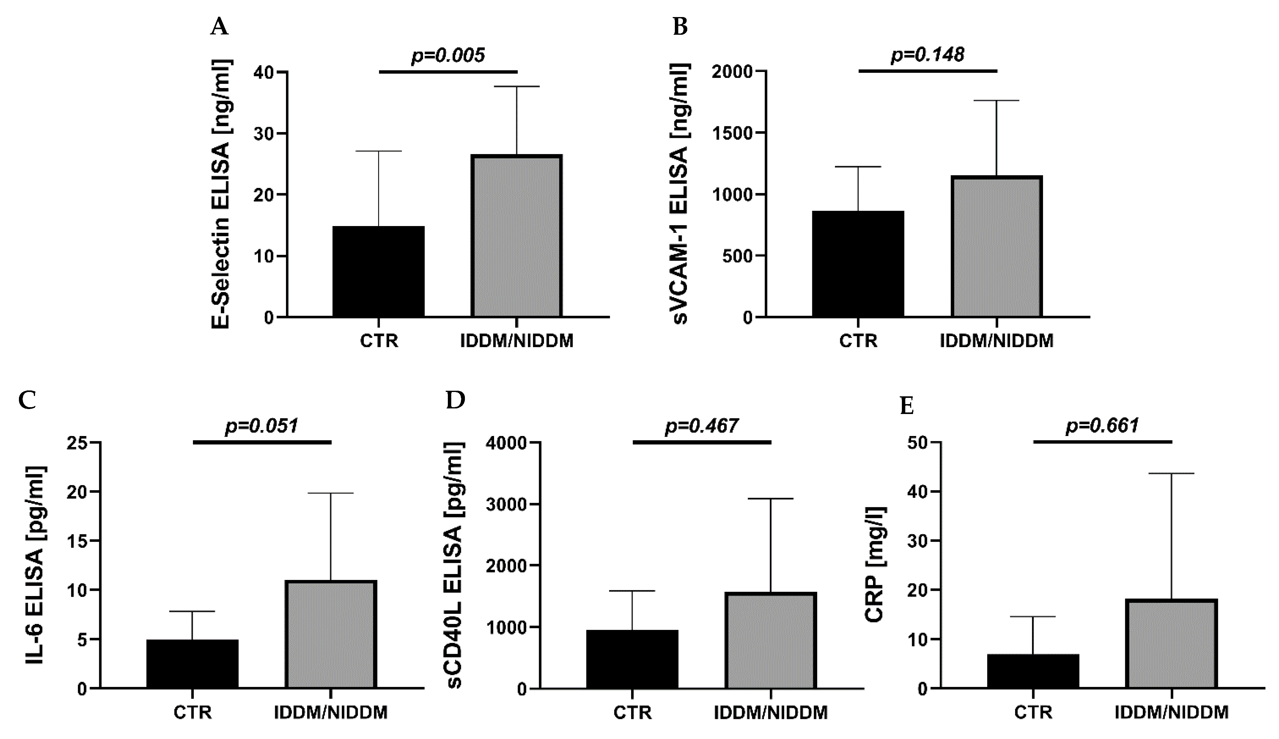
3.3. Serum Markers of Inflammation

While some inflammatory mediators were unchanged (sVCAM-1, p = 0.148 and sCD40L, p = 0.467 and CRP, p = 0.661), other serum markers of inflammation were increased in IDM/NIDDM (E-selectin, p = 0.005 and IL-6, p = 0.051) (Figure 3), pointing to a rather inflammatory and thrombotic phenotype in response to hyperglycemic conditions.

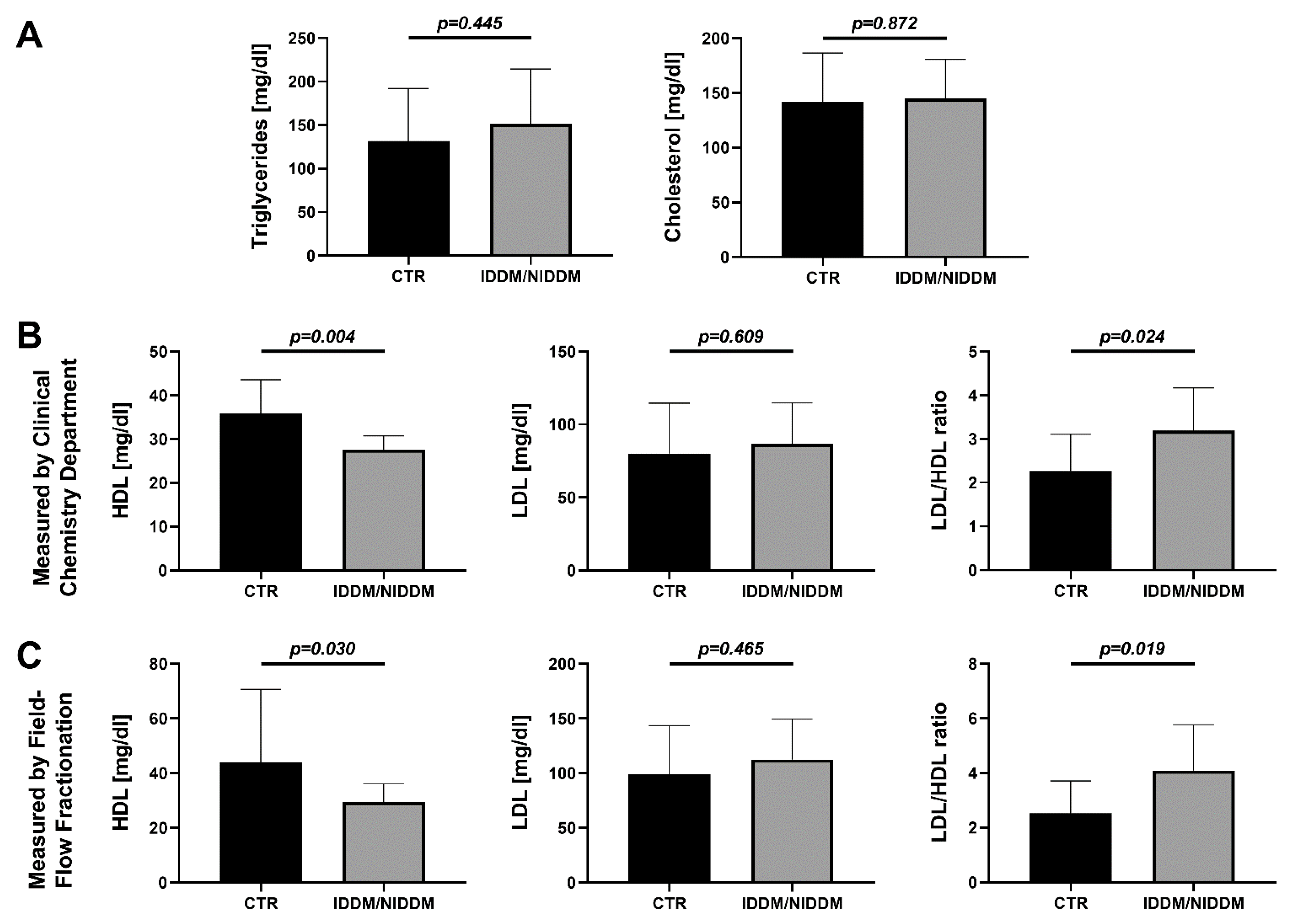
3.4. Serum Markers of Lipid Metabolism

Diabetic subjects showed no obvious increase in triglycerides (p = 0.445), total cholesterol (p = 0.872) (Figure 4A), or LDL-cholesterol (p = 0.609 in routine laboratory and p = 0.465 in Field-Flow Fractionation), but there was a significant lowered HDL-cholesterol (p = 0.03; Field-Flow Fractionation) and an imbalance between LDL and HDL, which was characterized by a higher LDL/HDL ratio (p = 0.024 and p = 0.019, respectively) in the IDM/NIDDM group (Figure 4B,C). The latter was mainly due to decreased HDL levels in the diabetic subjects, which was confirmed by two independent quantification methods for serum LDL and HDL (Clinical Chemistry routine measurement and Field-Flow Fractionation).

4. Discussion

The present study provides important insights into the contributing role of treated type 2 diabetes mellitus to the development of coronary artery disease in humans. Diabetic dyslipidemia is well known [40,41], and statin therapy should lower hyperlipidemia and prevent from the risk of CAD [40]. Here, we could show that dysregulated lipid metabolism and inflammation reactions are central pathomechanisms in diabetic vasculopathy. Our results provide evidence that the LDL-/HDL-cholesterol ratio and the E-selectin level in serum as a marker for vascular wall inflammation were raised in IDM/NIDDM patients compared to CTR with CAD. A higher LDL-/HDL-cholesterol ratio is a prognostic marker of higher atherosclerotic risk [42], and E-selectin is generally elevated in patients with CAD [43] and according to our present results further aggravated in patients with diabetes mellitus. As a general oxidative stress response in diabetes mellitus, the antioxidant HO-1 was upregulated, and there was less activated eNOS, resulting in endothelial dysfunction in accordance to animal experimental data reported previously [6].
In addition, we could show an increase in HO-1 protein levels in atrial tissue of diabetes mellitus. Gall et al. argued that hyperglycemia lowers HO-1 activity and increases superoxide production in the vasculature [44]. Interestingly, Oelze et al. have reported an upregulation of HO-1 in diabetic rats without therapy, assuming a general stress response, while rats under high-dose treatment with sodium-glucose cotransporter 2 inhibitor (SGLT2i) had a decline of HO-1 [6]. In this light, our data suggest either current antidiabetic therapy in diabetes mellitus protecting against HO-1 protein decrease or HO-1 providing a protective response, as plasma HO-1 levels are higher in patients with carotid plaques compared to healthy subjects, which probably indicates a possible protective response against carotid atherosclerosis, here against diabetic vasculopathy [44]. It remains unclear whether the HO-1 stress response is caused or affected by less activated eNOS in diabetes mellitus. The here-reported minor trend of eNOS protein upregulation in atrial tissue of diabetic patients is supported by similar observations in type 2 diabetic db/db mice [45,46]. Uncoupled eNOS leads to a lack of NO with disturbed endothelial function [47] and results in superoxide excess [48]. Probably HO-1 and eNOS protein upregulation compensates for the superoxide excess in IDDM/NIDDM, as we could not show an increased superoxide formation compared to CTR. However, we did clearly show less activated eNOS by decreased Ser1177 phosphorylation, as also shown in diabetic db/db mice [45,49] and an upregulation of HO-1 as an oxidative stress response. The predictive value of the regulatory enzymatic systems centered around eNOS was well documented in the past. Previously, it was shown that substituting BH4 improved the endothelial function in cell culture and animals [50] as well as patients with atherosclerosis [51]. Within the organelle, BH4 is produced by GTP cyclohydrolase I (GCH-1) and recycled from its oxidized form dihydrobiopterin (BH2) by DHFR [21,22]. Accordingly, BH4 levels or the expression levels of its main enzymatic sources, GCH-1 and DHFR, may be suitable to indirectly measure oxidative stress, although they are not changed significantly in the present study. Here, we observed a slight downregulation of DHFR protein in diabetic patients as also shown in diabetic db/db mice [52,53]. Altogether, our data support an uncoupled state of eNOS enzyme with futile compensatory upregulation of eNOS and DHFR proteins, which is compatible with the known impairment of endothelial function in diabetic subjects.
Dyslipidemia and cardiovascular inflammation are most likely the major pathomechanisms in diabetes mellitus with adverse impact on cardiovascular prognosis. Accordingly, it is not surprising that statin therapy improved the clinical outcome of patients with acute coronary syndromes, as this drug not only prevented dyslipidemia (decreased LDL levels) but also reduced inflammation (lower CRP levels), as shown by the PROVE IT-TIMI 22 study [54]. Statins have potent anti-inflammatory and antioxidant pleiotropic effects (e.g., via induction of NRF2/HO-1 pathway [55,56]), also in patients with diabetes [57,58,59,60]. Statins therapy also suppresses the activation of NADPH oxidase [61] and uncoupling of eNOS as well as subsequent endothelial dysfunction in animal models [62], all of which contribute to the beneficial antioxidant profile of statins [63]. In addition, statins mobilize endothelial progenitor cells, which could contribute to efficient repair of the endothelium, better recovery of coronary vessels from atherosclerotic insults, and thereby improved prognosis of patients with coronary artery disease [64].

5. Limitations of the Study

In the present study, we are talking about well-treated diabetes mellitus. We suggest the presence of a successful treatment without hyperglycemia in the IDDM/NIDDM group, although the proof by HbA1c < 6.5% was missed. Nevertheless, we can proclaim that patients with CAD and IDDM/NIDDM undergoing CABG should regularly present to a cardiac specialist, and medication for cardiovascular risk factors should be effective. The availability of left ventricular (LV) myocardium is rare and ethical considerations prohibit its use, as LV biopsies can be associated with severe bleeding and scar formation in the “working myocardium” and are thus considered harmful for patients. For this reason, our choice of myocardial specimen from the right atrial appendages has been reported before [32]. Due to the multiple measurements requiring large volumes, plasma samples were pooled for the ELISA assays in Figure 3 and for the determination of lipid parameters in Figure 4, explaining the reduced number of independent measurements for these parameters. The same applies for ALDH-2 activity measurement that also required the pooling of some of the heart samples. Finally, for protein expression, one to four data points are missing in each group due to insufficient blot quality (e.g., air bubbles caused incomplete transfer of the protein bands from the gel to the membrane during the Western blot procedure or a gel pocket was left empty before SDS-PAGE).

6. Conclusions

Inflammation and lipoprotein dysregulation remain the main triggers of diabetic coronary occlusive disease despite antidiabetic pharmacotherapy. Adjuvant pharmacological substances such as statins acting as anti-inflammatory agents and lipid blockers might diminish the detrimental effect of diabetes mellitus on the coronary inflammation and atherosclerosis. However, further studies are required to evaluate their true therapeutic value.

Author Contributions

Conceptualization, M.O., A.D., G.D.D.; methodology, A.D., M.O., K.B., Y.G.; software, M.O., U.H.; validation, Y.G., H.T., G.D.D., T.M.; formal analysis, M.O., T.M., R.C., Y.G.; investigation, K.B., Y.G., U.H.; resources, K.B., Y.G., R.C., H.T.; data curation, K.B., Y.G., R.C., M.O.; writing—original draft preparation, K.B., A.D.; writing—review and editing, G.D.D., H.T., T.M.; visualization, K.B., Y.G.; supervision, A.D., G.D.D.; project administration, K.B., A.D. All authors have read and agreed to the published version of the manuscript.

Funding

This research received no external funding.

Institutional Review Board Statement

The study was conducted according to the guidelines of the Declaration of Helsinki, and approved by the Ethical Committee of Rhineland-Palatinate, Germany (protocol code 837.104.08 (6100)).

Informed Consent Statement

Informed consent was obtained from all subjects involved in the study.

Data Availability Statement

The data used to support the findings of this study are available from the corresponding author upon request.

Acknowledgments

T.M. is PI of the DZHK (German Center for Cardiovascular Research), Partner Site Rhine-Main, Mainz, Germany. We thank Ulrich Rösch (Superon GmbH, Dernbach, Germany) for measurement of HDL and LDL by Field-Flow Fractionation method.

Conflicts of Interest

The authors declare no conflict of interest.

References

  1. Cai, L.; Kang, Y.J. Cell Death and Diabetic Cardiomyopathy. Cardiovasc. Toxicol. 2003, 3, 219–228. [Google Scholar] [CrossRef]
  2. Watanabe, K.; Thandavarayan, R.A.; Harima, M.; Sari, F.R.; Gurusamy, N.; Veeraveedu, P.T.; Mito, S.; Arozal, W.; Sukumaran, V.; Laksmanan, A.P.; et al. Role of Differential Signaling Pathways and Oxidative Stress in Diabetic Cardiomyopathy. Curr. Cardiol. Rev. 2010, 6, 280–290. [Google Scholar] [CrossRef]
  3. Duerr, G.D.; Heinemann, J.C.; Arnoldi, V.; Feisst, A.; Kley, J.; Ghanem, A.; Welz, A.; Dewald, O. Cardiomyocyte specific peroxisome proliferator-activated receptor-α overexpression leads to irreversible damage in ischemic murine heart. Life Sci. 2014, 102, 88–97. [Google Scholar] [CrossRef]
  4. Parrinello, C.M.; Lutsey, P.L.; Ballantyne, C.M.; Folsom, A.R.; Pankow, J.; Selvin, E. Six-year change in high-sensitivity C-reactive protein and risk of diabetes, cardiovascular disease, and mortality. Am. Heart J. 2015, 170, 380–389.e4. [Google Scholar] [CrossRef] [Green Version]
  5. Ofstad, A.P.; Gullestad, L.; Orvik, E.; Aakhus, S.; Endresen, K.; Ueland, T.; Aukrust, P.; Fagerland, M.W.; I Birkeland, K.; Johansen, O.E. Interleukin-6 and activin A are independently associated with cardiovascular events and mortality in type 2 diabetes: The prospective Asker and Bærum Cardiovascular Diabetes (ABCD) cohort study. Cardiovasc. Diabetol. 2013, 12, 126. [Google Scholar] [CrossRef] [Green Version]
  6. Oelze, M.; Kroller-Schon, S.; Welschof, P.; Jansen, T.; Hausding, M.; Mikhed, Y.; Stamm, P.; Mader, M.; Zinssius, E.; Agdauletova, S.; et al. The sodium-glucose co-transporter 2 inhibitor empagliflozin improves diabetes-induced vascular dysfunction in the streptozotocin diabetes rat model by inter-fering with oxidative stress and glucotoxicity. PLoS ONE 2014, 9, e112394. [Google Scholar] [CrossRef] [PubMed]
  7. A Badreldin, A.M.; Muehle, A.; Misic, J.; Tvildiani, T.; Duerr, G.D.; Paulini-Heine, B.; Peivandi, A.A. Objective method to evaluate the competency of residents in cardiac surgery. Eur. J. Cardio Thoracic Surg. 2021, 59, 1059–1068. [Google Scholar] [CrossRef]
  8. Daiber, A.; Steven, S.; Vujacic-Mirski, K.; Kalinovic, S.; Oelze, M.; Di Lisa, F.; Münzel, T. Regulation of Vascular Function and Inflammation via Cross Talk of Reactive Oxygen and Nitrogen Species from Mitochondria or NADPH Oxidase—Implications for Diabetes Progression. Int. J. Mol. Sci. 2020, 21, 3405. [Google Scholar] [CrossRef] [PubMed]
  9. Steven, S.; Frenis, K.; Oelze, M.; Vujacic-Mirski, K.; Bayo Jimenez, M.T.; Kalinovic, S.; Kroller-Schon, S.; Munzel, T.; Daiber, A. SGLT2 inhibitors, diabetes and oxidative stress. In Diabetes—Oxidative Stress and Dietary Antioxidants, 2nd ed.; Preedy, V., Ed.; Academic Press: Cambridge, MA, USA, 2020; pp. 1–12. [Google Scholar]
  10. Heitzer, T.; Finckh, B.; Albers, S.; Krohn, K.; Kohlschütter, A.; Meinertz, T. Beneficial effects of alpha-lipoic acid and ascorbic acid on endothelium-dependent, nitric oxide-mediated vasodilation in diabetic patients: Relation to parameters of oxidative stress. Free. Radic. Biol. Med. 2001, 31, 53–61. [Google Scholar] [CrossRef]
  11. Daiber, A.; Steven, S.; Weber, A.; Shuvaev, V.V.; Muzykantov, V.R.; Laher, I.; Li, H.; Lamas, S.; Münzel, T. Targeting vascular (endothelial) dysfunction. Br. J. Pharmacol. 2017, 174, 1591–1619. [Google Scholar] [CrossRef] [PubMed]
  12. Heitzer, T.; Krohn, K.; Albers, S.; Meinertz, T. Tetrahydrobiopterin improves endothelium-dependent vasodilation by in-creasing nitric oxide activity in patients with Type II diabetes mellitus. Diabetologia 2000, 43, 1435–1438. [Google Scholar] [CrossRef] [Green Version]
  13. Basta, G.; Schmidt, A.M.; De Caterina, R. Advanced glycation end products and vascular inflammation: Implications for accelerated atherosclerosis in diabetes. Cardiovasc. Res. 2004, 63, 582–592. [Google Scholar] [CrossRef]
  14. Ramanathan, K.; Abel, J.G.; Park, J.E.; Fung, A.; Mathew, V.; Taylor, C.M.; Mancini, G.J.; Gao, M.; Ding, L.; Verma, S.; et al. Surgical Versus Percutaneous Coronary Revascularization in Patients with Diabetes and Acute Coronary Syndromes. J. Am. Coll. Cardiol. 2017, 70, 2995–3006. [Google Scholar] [CrossRef]
  15. Stone, P.H.; E Muller, J.; Hartwell, T.; York, B.; Rutherford, J.D.; Parker, C.B.; Turi, Z.G.; Strauss, H.; Willerson, J.T.; Robertson, T.; et al. The effect of diabetes mellitus on prognosis and serial left ventricular function after acute myocardial infarction: Contribution of both coronary disease and diastolic left ventricular dysfunction to the adverse prognosis. J. Am. Coll. Cardiol. 1989, 14, 49–57. [Google Scholar] [CrossRef] [Green Version]
  16. Kaptoge, S.; Seshasai, S.R.K.; Gao, P.; Freitag, D.F.; Butterworth, A.S.; Borglykke, A.; Di Angelantonio, E.; Gudnason, V.; Rumley, A.; Lowe, G.D.O.; et al. Inflammatory cytokines and risk of coronary heart disease: New prospective study and updated meta-analysis. Eur. Heart J. 2013, 35, 578–589. [Google Scholar] [CrossRef] [PubMed] [Green Version]
  17. Karbach, S.; Wenzel, P.; Waisman, A.; Munzel, T.; Daiber, A. eNOS Uncoupling in Cardiovascular Diseases—The Role of Oxidative Stress and Inflammation. Curr. Pharm. Des. 2014, 20, 3579–3594. [Google Scholar] [CrossRef]
  18. Ridker, P.M.; Everett, B.M.; Thuren, T.; MacFadyen, J.G.; Chang, W.H.; Ballantyne, C.; Fonseca, F.; Nicolau, J.; Koenig, W.; Anker, S.D.; et al. Antiinflammatory Therapy with Canakinumab for Atherosclerotic Disease. N. Engl. J. Med. 2017, 377, 1119–1131. [Google Scholar] [CrossRef] [PubMed]
  19. Daiber, A.; Steven, S.; Euler, G.; Schulz, R. Vascular and cardiac oxidative stress and inflammation as targets for cardiopro-tection. Curr. Pharm. Des. 2021. [Google Scholar] [CrossRef] [PubMed]
  20. Ridker, P.M.; Howard, C.P.; Walter, V.; Everett, B.; Libby, P.; Hensen, J.; Thuren, T.; Group, C.P.I. Effects of interleukin-1beta inhibition with canakinumab on hemoglobin A1c, lipids, C-reactive protein, interleukin-6, and fibrinogen: A phase IIb randomized, placebo-controlled trial. Circulation 2012, 126, 2739–2748. [Google Scholar] [CrossRef] [PubMed] [Green Version]
  21. Schulz, E.; Jansen, T.; Wenzel, P.; Daiber, A.; Münzel, T. Nitric Oxide, Tetrahydrobiopterin, Oxidative Stress, and Endothelial Dysfunction in Hypertension. Antioxid. Redox Signal. 2008, 10, 1115–1126. [Google Scholar] [CrossRef] [PubMed]
  22. Bendall, J.K.; Douglas, G.; McNeill, E.; Channon, K.; Crabtree, M.J. Tetrahydrobiopterin in Cardiovascular Health and Disease. Antioxid. Redox Signal. 2014, 20, 3040–3077. [Google Scholar] [CrossRef] [PubMed] [Green Version]
  23. Abraham, N.G.; Kappas, A. Pharmacological and Clinical Aspects of Heme Oxygenase. Pharmacol. Rev. 2008, 60, 79–127. [Google Scholar] [CrossRef] [PubMed] [Green Version]
  24. Dulak, J.; Deshane, J.; Jozkowicz, A.; Agarwal, A. Heme oxygenase-1 and carbon monoxide in vascular pathobiology: Focus on angiogenesis. Circulation 2008, 117, 231–241. [Google Scholar] [CrossRef] [PubMed]
  25. Abraham, N.G.; Junge, J.M.; Drummond, G.S. Translational Significance of Heme Oxygenase in Obesity and Metabolic Syndrome. Trends Pharmacol. Sci. 2016, 37, 17–36. [Google Scholar] [CrossRef] [PubMed] [Green Version]
  26. Gozzelino, R.; Jeney, V.; Soares, M. Mechanisms of Cell Protection by Heme Oxygenase. Annu. Rev. Pharmacol. Toxicol. 2010, 50, 323–354. [Google Scholar] [CrossRef] [PubMed] [Green Version]
  27. Barbagallo, I.; Galvano, F.; Frigiola, A.; Cappello, F.; Riccioni, G.; Murabito, P.; D’Orazio, N.; Torella, M.; Gazzolo, D.; Volti, G.L. Potential Therapeutic Effects of Natural Heme Oxygenase-1 Inducers in Cardiovascular Diseases. Antioxid. Redox Signal. 2013, 18, 507–521. [Google Scholar] [CrossRef] [PubMed]
  28. Ference, B.; Kastelein, J.J.; Ray, K.K.; Ginsberg, H.N.; Chapman, M.J.; Packard, C.J.; Laufs, U.; Oliver-Williams, C.; Wood, A.; Butterworth, A.; et al. Association of Triglyceride-Lowering LPL Variants and LDL-C-Lowering LDLR Variants with Risk of Coronary Heart Disease. JAMA 2019, 321, 364–373. [Google Scholar] [CrossRef] [Green Version]
  29. Xu, Y.; He, Z.; King, G.L. Introduction of hyperglycemia and dyslipidemia in the pathogenesis of diabetic vascular com-plications. Curr. Diabetes Rep. 2005, 5, 1–7. [Google Scholar] [CrossRef]
  30. Kannel, W.B.; Hjortland, M.; Castelli, W.P. Role of diabetes in congestive heart failure: The Framingham study. Am. J. Cardiol. 1974, 34, 29–34. [Google Scholar] [CrossRef]
  31. Gramlich, Y.; Daiber, A.; Buschmann, K.; Oelze, M.; Vahl, C.-F.; Münzel, T.; Hink, U. Oxidative Stress in Cardiac Tissue of Patients Undergoing Coronary Artery Bypass Graft Surgery: The Effects of Overweight and Obesity. Oxidative Med. Cell. Longev. 2018, 2018, 6598326. [Google Scholar] [CrossRef] [Green Version]
  32. Denk, K.; Albers, J.; Kayhan, N.; Ister, D.; Bonz, A.; Werner, C.; Münzel, T.; Vahl, C.-F. Evidence for a negative inotropic effect of obesity in human myocardium? Eur. J. Cardio-Thoracic Surg. 2009, 36, 300–305. [Google Scholar] [CrossRef] [PubMed] [Green Version]
  33. Steven, S.; Oelze, M.; Hanf, A.; Kröller-Schön, S.; Kashani, F.; Roohani, S.; Welschof, P.; Kopp, M.; Gödtel-Armbrust, U.; Xia, N.; et al. The SGLT2 inhibitor empagliflozin improves the primary diabetic complications in ZDF rats. Redox Biol. 2017, 13, 370–385. [Google Scholar] [CrossRef] [PubMed]
  34. Kröller-Schön, S.; Daiber, A.; Steven, S.; Oelze, M.; Frenis, K.; Kalinovic, S.; Heimann, A.; Schmidt, F.P.; Pinto, A.; Kvandova, M.; et al. Crucial role for Nox2 and sleep deprivation in aircraft noise-induced vascular and cerebral oxidative stress, inflammation, and gene regulation. Eur. Heart J. 2018, 39, 3528–3539. [Google Scholar] [CrossRef] [PubMed]
  35. Steven, S.; Dib, M.; Hausding, M.; Kashani, F.; Oelze, M.; Kröller-Schön, S.; Hanf, A.; Daub, S.; Roohani, S.; Gramlich, Y.; et al. CD40L controls obesity-associated vascular inflammation, oxidative stress, and endothelial dysfunction in high fat diet-treated and db/db mice. Cardiovasc. Res. 2017, 114, 312–323. [Google Scholar] [CrossRef] [Green Version]
  36. Giddings, J.C. Field-flow fractionation: Analysis of macromolecular, colloidal, and particulate materials. Science 1993, 260, 1456–1465. [Google Scholar] [CrossRef]
  37. Zhao, H.; Joseph, J.; Fales, H.M.; Sokoloski, E.A.; Levine, R.L.; Vasquez-Vivar, J.; Kalyanaraman, B. Detection and charac-terization of the product of hydroethidine and intracellular superoxide by HPLC and limitations of fluorescence. Proc. Natl. Acad. Sci. USA 2005, 102, 5727–5732. [Google Scholar] [CrossRef] [Green Version]
  38. Wenzel, P.; Mollnau, H.; Oelze, M.; Schulz, E.; Wickramanayake, J.M.; Muller, J.; Schuhmacher, S.; Hortmann, M.; Baldus, S.; Gori, T.; et al. First evidence for a crosstalk between mitochondrial and NADPH oxi-dase-derived reactive oxygen species in nitroglycerin-triggered vascular dysfunction. Antioxid. Redox Signal. 2008, 10, 1435–1447. [Google Scholar] [CrossRef]
  39. Kroller-Schon, S.; Steven, S.; Kossmann, S.; Scholz, A.; Daub, S.; Oelze, M.; Xia, N.; Hausding, M.; Mikhed, Y.; Zinssius, E.; et al. Molecular mecha-nisms of the crosstalk between mitochondria and NADPH oxidase through reactive oxygen species-studies in white blood cells and in animal models. Antioxid. Redox Signal. 2014, 20, 247–266. [Google Scholar] [CrossRef] [Green Version]
  40. Verhulst, M.J.L.; Loos, B.G.; Gerdes, V.E.A.; Teeuw, W.J. Evaluating All Potential Oral Complications of Diabetes Mellitus. Front. Endocrinol. 2019, 10, 56. [Google Scholar] [CrossRef]
  41. Ma, Y.; Wang, Y.; Huang, Q.; Ren, Q.; Chen, S.; Zhang, A.; Zhao, L.; Zhen, Q.; Peng, Y. Impaired beta cell function in Chinese newly diagnosed type 2 diabetes mellitus with hyperlipidemia. J. Diabetes Res. 2014, 2014, 493039. [Google Scholar] [CrossRef] [Green Version]
  42. Hao, W.; Friedman, A. The LDL-HDL Profile Determines the Risk of Atherosclerosis: A Mathematical Model. PLoS ONE 2014, 9, e90497. [Google Scholar] [CrossRef] [Green Version]
  43. Martinovic, I.; Abegunewardene, N.; Seul, M.; Vosseler, M.; Horstick, G.; Buerke, M.; Darius, H.; Lindemann, S. Elevated Monocyte Chemoattractant Protein-1 Serum Levels in Patients at Risk for Coronary Artery Disease. Circ. J. 2005, 69, 1484–1489. [Google Scholar] [CrossRef] [Green Version]
  44. Gall, T.; Balla, G.; Balla, J. Heme, Heme Oxygenase, and Endoplasmic Reticulum Stress-A New Insight into the Pathophysiology of Vascular Diseases. Int. J. Mol. Sci. 2019, 20, 3675. [Google Scholar] [CrossRef] [Green Version]
  45. Lee, J.; Lee, S.; Zhang, H.; Hill, M.A.; Zhang, C.; Park, Y. Interaction of IL-6 and TNF-alpha contributes to endothelial dys-function in type 2 diabetic mouse hearts. PLoS ONE 2017, 12, e0187189. [Google Scholar]
  46. Einbinder, Y.; Ohana, M.; Benchetrit, S.; Zehavi, T.; Nacasch, N.; Bernheim, J.; Zitman-Gal, T. Glucagon-like peptide-1 and vitamin D: Anti-inflammatory response in diabetic kidney disease in db/db mice and in cultured endothelial cells. Diabetes/Metabolism Res. Rev. 2016, 32, 805–815. [Google Scholar] [CrossRef] [Green Version]
  47. Shah, M.S.; Brownlee, M. Molecular and Cellular Mechanisms of Cardiovascular Disorders in Diabetes. Circ. Res. 2016, 118, 1808–1829. [Google Scholar] [CrossRef] [PubMed] [Green Version]
  48. Tabit, C.E.; Chung, W.B.; Hamburg, N.; Vita, J.A. Endothelial dysfunction in diabetes mellitus: Molecular mechanisms and clinical implications. Rev. Endocr. Metab. Disord. 2010, 11, 61–74. [Google Scholar] [CrossRef] [PubMed] [Green Version]
  49. Tian, X.Y.; Wong, W.T.; Wang, N.; Lu, Y.; Cheang, W.S.; Liu, J.; Liu, L.; Liu, Y.; Lee, S.S.; Chen, Z.Y.; et al. PPARdelta activation protects endothelial function in diabetic mice. Diabetes 2012, 61, 3285–3293. [Google Scholar] [CrossRef] [PubMed] [Green Version]
  50. Daiber, A.; Xia, N.; Steven, S.; Oelze, M.; Hanf, A.; Kröller-Schön, S.; Münzel, T.; Li, H. New Therapeutic Implications of Endothelial Nitric Oxide Synthase (eNOS) Function/Dysfunction in Cardiovascular Disease. Int. J. Mol. Sci. 2019, 20, 187. [Google Scholar] [CrossRef] [Green Version]
  51. Tiefenbacher, C.P.; Bleeke, T.; Vahl, C.; Amann, K.; Vogt, A.; Kübler, W. Endothelial Dysfunction of Coronary Resistance Arteries Is Improved by Tetrahydrobiopterin in Atherosclerosis. Circulation 2000, 102, 2172–2179. [Google Scholar] [CrossRef] [Green Version]
  52. Yamamoto, E.; Nakamura, T.; Kataoka, K.; Tokutomi, Y.; Dong, Y.-F.; Fukuda, M.; Nako, H.; Yasuda, O.; Ogawa, H.; Kim-Mitsuyama, S. Nifedipine prevents vascular endothelial dysfunction in a mouse model of obesity and type 2 diabetes, by improving eNOS dysfunction and dephosphorylation. Biochem. Biophys. Res. Commun. 2010, 403, 258–263. [Google Scholar] [CrossRef]
  53. Wang, C.; Chao, Y.; Xu, W.; Liang, M.; Deng, S.; Zhang, D.; Huang, K. CTRP13 Preserves Endothelial Function by Targeting GTP Cyclohydrolase 1 in Diabetes. Diabetes 2019, 69, 99–111. [Google Scholar] [CrossRef] [PubMed]
  54. Ridker, P.M.; Cannon, C.P.; Morrow, D.; Rifai, N.; Rose, L.M.; McCabe, C.H.; Pfeffer, M.A.; Braunwald, E. C-Reactive Protein Levels and Outcomes after Statin Therapy. N. Engl. J. Med. 2005, 352, 20–28. [Google Scholar] [CrossRef] [PubMed]
  55. Habeos, I.G.; Ziros, P.G.; Chartoumpekis, D.; Psyrogiannis, A.; Kyriazopoulou, V.; Papavassiliou, A.G. Simvastatin activates Keap1/Nrf2 signaling in rat liver. J. Mol. Med. 2008, 86, 1279–1285. [Google Scholar] [CrossRef] [PubMed]
  56. Ali, F.; Zakkar, M.; Karu, K.; Lidington, E.A.; Hamdulay, S.S.; Boyle, J.J.; Zloh, M.; Bauer, A.; Haskard, D.O.; Evans, P.C.; et al. Induction of the cytoprotective enzyme heme oxygenase-1 by statins is enhanced in vascular endothelium ex-posed to laminar shear stress and impaired by disturbed flow. J. Biol. Chem. 2009, 284, 18882–18892. [Google Scholar] [CrossRef] [PubMed] [Green Version]
  57. Steven, S.; Münzel, T.; Daiber, A. Exploiting the Pleiotropic Antioxidant Effects of Established Drugs in Cardiovascular Disease. Int. J. Mol. Sci. 2015, 16, 18185–18223. [Google Scholar] [CrossRef] [Green Version]
  58. Takemoto, M.; Liao, J.K. Pleiotropic Effects of 3-Hydroxy-3-Methylglutaryl Coenzyme A Reductase Inhibitors. Arterioscler. Thromb. Vasc. Biol. 2001, 21, 1712–1719. [Google Scholar] [CrossRef] [PubMed] [Green Version]
  59. Ray, K.K.; Cannon, C.P. Pathological Changes in Acute Coronary Syndromes: The Role of Statin Therapy in the Modulation of Inflammation, Endothelial Function and Coagulation. J. Thromb. Thrombolysis 2004, 18, 89–101. [Google Scholar] [CrossRef]
  60. Patel, T.N.; Shishehbor, M.H.; Bhatt, D.L. A review of high-dose statin therapy: Targeting cholesterol and inflammation in atherosclerosis. Eur. Heart J. 2007, 28, 664–672. [Google Scholar] [CrossRef]
  61. Adam, O.; Laufs, U. Rac1-mediated effects of HMG-CoA reductase inhibitors (statins) in cardiovascular disease. Antioxid. Redox Signal. 2014, 20, 1238–1250. [Google Scholar] [CrossRef]
  62. Wenzel, P.; Daiber, A.; Oelze, M.; Brandt, M.; Closs, E.; Xu, J.; Thum, T.; Bauersachs, J.; Ertl, G.; Zou, M.H.; et al. Mechanisms underlying recoupling of eNOS by HMG-CoA reductase inhibition in a rat model of streptozoto-cin-induced diabetes mellitus. Atherosclerosis 2008, 198, 65–76. [Google Scholar] [CrossRef] [PubMed] [Green Version]
  63. Margaritis, M.; Channon, K.M.; Antoniades, C. Statins as regulators of redox state in the vascular endothelium: Beyond lipid lowering. Antioxid. Redox Signal. 2014, 20, 1198–1215. [Google Scholar] [CrossRef] [PubMed] [Green Version]
  64. Hibbert, B.; Simard, T.; Ramirez, F.D.; Pourdjabbar, A.; Raizman, J.E.; Maze, R.; Wilson, K.R.; Hawken, S.; O’Brien, E.R. The effect of statins on circulating endothelial progenitor cells in humans: A systematic review. J. Cardiovasc. Pharmacol. 2013, 62, 491–496. [Google Scholar] [CrossRef]
Figure 1. Markers of oxidative stress in myocardium. (A) Marginal higher concentration of mitochondrial superoxide formation (DHE HPLC) in the diabetic (IDDM/NIDDM) group versus control (CTR). (B) No difference in ALDH-2 activity after oxidative inactivation in the IDDM/NIDDM versus CTR group. 2-HE, 2-hydroxyethidium; ALDH-2, mitochondrial aldehyde dehydrogenase. Data are the mean ± SD of n = 38 (CTR) and 20 (IDDM/NIDDM) (A) and n = 16 (CTR) and 11 (IDDM/NIDDM) (B) patients. Mann–Whitney test in (A) and unpaired t-test in (B). CTR, control; IDDM, insulin-dependent diabetes mellitus type 2; NIDDM, non-insulin dependent diabetes mellitus type 2.
Ijerph 18 10892 g001
Figure 2. Expression of vasoactive regulatory protein and antioxidant enzyme/response in myocardium. (A) Absent upregulation of dysfunctional enzyme (eNOS protein) in the diabetic (IDDM/NIDDM) versus the control (CTR) group. (B) Significant decrease in activated eNOS (Ser1177 phosphorylation) in the IDDM/NIDDM versus CTR group. (C) Slightly lower DHFR expression in the IDDM/NIDDM versus CTR group. (D) Significant upregulation of the antioxidant stress-response enzyme HO-1 in the IDDM/NIDDM versus CTR group. Data are mean ± SD of n = 37–39 (CTR) and 17 (IDDM/NIDDM) (A,B) and n = 38 (CTR) and 16 (IDDM/NIDDM) (C) and n = 37 (CTR) and 16 (IDDM/NIDDM) (D) patients. Unpaired t-test in (AC) and Mann–Whitney test in (D). CTR, control; IDDM, insulin-dependent diabetes mellitus type 2; NIDDM, non-insulin dependent diabetes mellitus type 2.
Ijerph 18 10892 g002
Figure 3. Serum markers of inflammation. (A) Significant raise of E-selectin in the diabetic (IDDM/NIDDM) versus the control (CTR) group. (B) Enhanced concentration of VCAM-1 in the IDDM/NIDDM versus CTR group (n.s). (C) Increased concentration of interleukin-6 (IL-6) in the IDM/NIDDM versus CTR group (n.s.). (D) Increase in concentration of CD40L and (E) C-reactive protein (CRP) in the DM versus CTR group (n.s.). Data are mean ± SD of n = 15 (CTR) and 11 (IDDM/NIDDM) patients (AD) or n = 11–12 (E). Mann–Whitney test in (A,D,E) and unpaired t test in (B,C). CTR, control; IDDM, insulin-dependent diabetes mellitus type 2; NIDDM, non-insulin-dependent diabetes mellitus type 2.
Ijerph 18 10892 g003
Figure 4. Serum markers of lipometabolism. (A) No obvious increase in triglycerides or total cholesterol in the IDDM/NIDDM versus CTR group. (B) (measurement by Clinical Chemistry Department) and (C) (measurement by Field-Flow Fractionation). Significant lower concentration of HDL-cholesterol together with a slightly higher concentration of LDL-cholesterol resulting in a significant raise of LDL/HDL ratio in the IDDM/NIDDM versus CTR group. Data are mean ± SD of n = 12 (CTR) and 11 (IDDM/NIDDM) (A,B) and n = 12 (CTR) and 10 (IDDM/NIDDM) (C) patients. Unpaired t-test in (AC) [LDL and LDL/HDL ratio] and Mann–Whitney test in (C) [HDL]. CTR, control; IDDM, insulin-dependent diabetes mellitus type 2; NIDDM, non-insulin-dependent diabetes mellitus type 2.
Ijerph 18 10892 g004
Table 1. Patients’ baseline characteristics. Summary of preoperative patients’ characteristics. Values are expressed as mean ± SD or as percentage (in brackets). Significant changes are displayed in italics. Abbreviations: ACE, angiotensin-converting enzyme; AT1, angiotensin-1; BMI, body mass index. Chi2 test was performed for categorial variables, and an unpaired, two-tailed t-test was applied for comparison of metric variables. Significant changes are displayed in italics.
CTRIDDM/NIDDMp-Value
Age (years)65.15 ± 9.0866.90 ± 9.830.5782
Female (%)61.54%61.11%0.9754
Height (cm)1.694 ± 0.0811.681 ± 0.1040.5856
Body weight (kg)80.62 ± 13.579.56 ± 12.60.7794
BMI (ratio)27.99 ± 3.6228.43 ± 5.600.7234
Waist circumference (cm)102.4 ± 8.69102.6 ± 9.530.9257
Hip circumference (cm)100.5 ± 6.49104.9 ± 8.590.1357
Waist/Hip ratio1.018 ± 0.0770.986 ± 0.0960.3452
Oral anti-diabetics (%)0.00%88.89%<0.0001
Insulin dependence (%)0.00%16.67%0.0088
ACE inhibitors (%)35.90%50.00%0.3131
AT1 antagonists (%)12.82%22.22%0.3656
Beta-blocker (%)58.97%55.56%0.8080
Calcium-channel blocker/%)20.51%16.67%0.7323
Spironolactone (%)2.56%0.00%0.4931
Statins (%)64.10%50.00%0.3131
Publisher’s Note: MDPI stays neutral with regard to jurisdictional claims in published maps and institutional affiliations.

