
Association of Decreased Physical Activity with Rheumatoid Mid-Hindfoot Deformity/Destruction

Abstract
:1. Introduction
2. Materials and Methods
2.1. Study Design and Patient Population
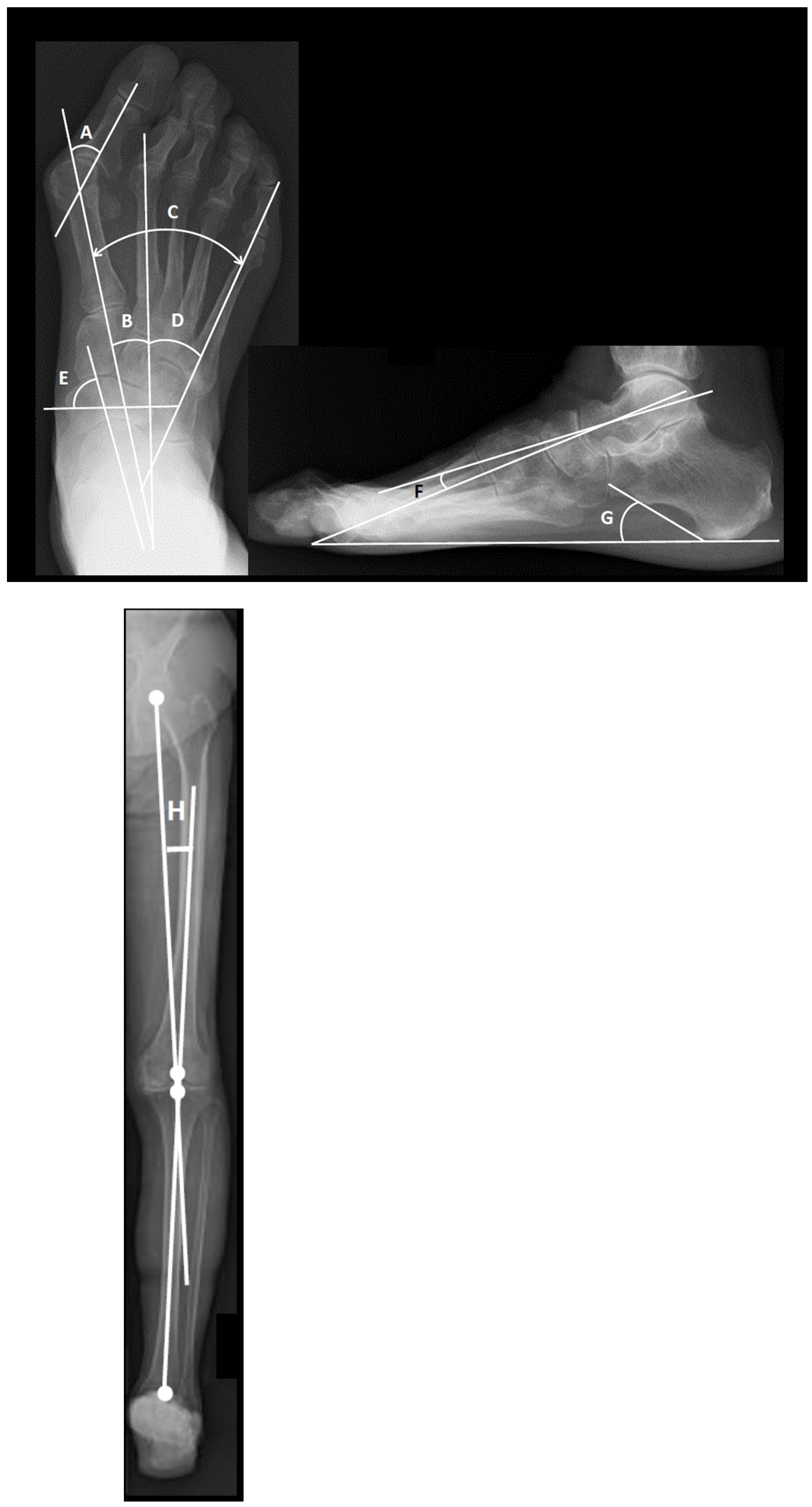
2.2. Radiographic Assessment
2.3. Clinical Assessment
2.4. Statistical Analysis
3. Results
3.1. Correlations between Knee and Entire Foot Deformities, and Clinical Parameters
3.2. Correlations between Destruction of Each Joint and Clinical Parameters
3.3. Correlations among Clinical Parameters
3.4. Correlations among Joints with Destruction
4. Discussion
5. Conclusions
Author Contributions
Funding
Institutional Review Board Statement
Informed Consent Statement
Data Availability Statement
Acknowledgments
Conflicts of Interest
References
- Wechalekar, M.D.; Lester, S.; Proudman, S.M.; Cleland, L.G.; Whittle, S.; Rischmueller, M.; Hill, C.L. Active foot synovitis in patients with rheumatoid arthritis: Applying clinical criteria for disease activity and remission may result in underestimation of foot joint involvement. Arthritis Rheum. 2012, 64, 1316–1322. [Google Scholar] [CrossRef] [PubMed]
- Simonsen, M.B.; Hørslev-Petersen, K.; Cöster, M.C.; Jensen, C.; Bremander, A. Foot and Ankle Problems in Patients with Rheumatoid Arthritis in 2019: Still an Important Issue. ACR Open Rheumatol. 2021, 3, 396–402. [Google Scholar] [CrossRef] [PubMed]
- López, L.D.; Pérez, R.M.; Ruano, R.A.; Losa, I.M.E.; Becerro, D.B.V.R.; Romero, M.C.; Calvo, L.C.; Navarro, F.E. Impact of quality of life related to foot problems: A case–control study. Sci. Rep. 2021, 11, 14515. [Google Scholar] [CrossRef]
- López, L.D.; Becerro, D.B.V.R.; Losa, I.M.E.; Palomo, L.P.; Rodríguez, S.D.; Brandariz, P.J.M.; Calvo, L.C. Evaluation of foot health related quality of life in individuals with foot problems by gender: A cross-sectional comparative analysis study. BMJ Open 2018, 8, e023980. [Google Scholar] [CrossRef] [PubMed] [Green Version]
- Tovaruela, C.N.; Vallejo, R.B.D.B.; Losa, I.M.E.; López, P.P.; Munuera, M.P.V.; Pérez, G.S.; López, L.D. Accurately Determining Proper Shoe Size in Patients With Rheumatoid Arthritis. Rehabil. Nurs. 2018, 43, 285–289. [Google Scholar] [CrossRef]
- Louwerens, J.W.K.; Schrier, J.C.M. Rheumatoid forefoot deformity: Pathophysiology, evaluation and operative treatment options. Int. Orthop. 2013, 37, 1719–1729. [Google Scholar] [CrossRef] [Green Version]
- Matricali, A.G.; Boonen, A.; Verduyckt, J.; Taelman, V.; Verschueren, P.; Sileghem, A.; Corluy, L.; Westhovens, R. The presence of forefoot problems and the role of surgery in patients with rheumatoid arthritis. Ann. Rheum. Dis. 2006, 65, 1254–1255. [Google Scholar] [CrossRef] [Green Version]
- Michelson, J.; Easley, M.; Wigley, F.M.; Hellmann, D. Foot and Ankle Problems in Rheumatoid Arthritis. Foot Ankle Int. 1994, 15, 608–613. [Google Scholar] [CrossRef]
- Van Der Leeden, M.; Steultjens, M.P.M.; Ursum, J.; Dahmen, R.; Roorda, L.D.; Van Schaardenburg, D.; Dekker, J. Prevalence and course of forefoot impairments and walking disability in the first eight years of rheumatoid arthritis. Arthritis Rheum. 2008, 59, 1596–1602. [Google Scholar] [CrossRef]
- Matsumoto, T.; Nakamura, I.; Miura, A.; Momoyama, G.; Ito, K. Radiologic Patterning of Joint Damage to the Foot in Rheumatoid Arthritis. Arthritis Rheum. 2014, 66, 499–507. [Google Scholar] [CrossRef]
- Hirao, M.; Ebina, K.; Tsuboi, H.; Noguchi, T.; Hashimoto, J.; Yoshikawa, H. Appearance of hindfoot valgus deformity and recurrence of hallux valgus in the very early period after hallux valgus surgery in a poorly controlled rheumatoid arthritis case: A case report. Mod. Rheumatol. 2016, 29, 367–369. [Google Scholar] [CrossRef]
- Hirao, M.; Hirai, Y.; Ebina, K.; Shi, K.; Noguchi, T.; Tsuboi, H.; Hashimoto, J.; Yoshikawa, H. Hallux valgus deformity after total ankle arthroplasty for rheumatoid arthritis: A case report. Mod. Rheumatol. 2018, 28, 890–892. [Google Scholar] [CrossRef]
- Stockley, I.; Betts, R.; Rowley, D.; Getty, C.; Duckworth, T. The importance of the valgus hindfoot in forefoot surgery in rheumatoid arthritis. J. Bone Jt. Surg. Br. Vol. 1990, 72-B, 705–708. [Google Scholar] [CrossRef] [PubMed] [Green Version]
- Yamada, S.; Hirao, M.; Tsuboi, H.; Akita, S.; Matsushita, M.; Ohshima, S.; Saeki, Y.; Hashimoto, J. Involvement of valgus hindfoot deformity in hallux valgus deformity in rheumatoid arthritis. Mod. Rheumatol. 2013, 24, 851–854. [Google Scholar] [CrossRef] [PubMed]
- Hirao, M.; Tsuboi, H.; Akita, S.; Matsushita, M.; Ohshima, S.; Saeki, Y.; Hashimoto, J. Effect of correction of hindfoot valgus deformity on ankle joint pain relief in rheumatoid arthritis cases: A report of two cases. Sage Open Med. Case Rep. 2014, 2. [Google Scholar] [CrossRef] [PubMed] [Green Version]
- Baan, H.; Dubbeldam, R.; Nene, A.V.; van de Laar, M.A. Gait Analysis of the Lower Limb in Patients with Rheumatoid Arthritis: A Systematic Review. Semin. Arthritis Rheum. 2012, 41, 768–788.e8. [Google Scholar] [CrossRef] [PubMed]
- Brenton-Rule, A.; Dalbeth, N.; Menz, H.B.; Bassett, S.; Rome, K. Foot and ankle characteristics associated with falls in adults with established rheumatoid arthritis: A cross-sectional study. BMC Musculoskelet. Disord. 2016, 17, 1–8. [Google Scholar] [CrossRef] [Green Version]
- Hirao, M.; Ebina, K.; Tsuboi, H.; Nampei, A.; Kushioka, J.; Noguchi, T.; Tsuji, S.; Owaki, H.; Hashimoto, J.; Yoshikawa, H. Outcomes of modified metatarsal shortening offset osteotomy for forefoot deformity in patients with rheumatoid arthritis: Short to mid-term follow-up. Mod. Rheumatol. 2017, 27, 981–989. [Google Scholar] [CrossRef] [PubMed]
- Hardy, R.H.; Clapham, J.C. Observations on hallux valgus. J. Bone Jt. Surg. Br. Vol. 1951, 33-B, 376–391. [Google Scholar] [CrossRef]
- Ueki, Y.; Sakuma, E.; Wada, I. Pathology and management of flexible flat foot in children. J. Orthop. Sci. 2019, 24, 9–13. [Google Scholar] [CrossRef]
- Cobey, J.C. Posterior roentgenogram of the foot. Clin. Orthop. Relat. Res. 1976, 118, 202–207. [Google Scholar]
- Xie, K.; Jiang, X.; Han, X.; Ai, S.; Qu, X.; Yan, M. Association between Knee Malalignment and Ankle Degeneration in Patients With End-Stage Knee Osteoarthritis. J. Arthroplast. 2018, 33, 3694–3698.e1. [Google Scholar] [CrossRef]
- Kunugiza, Y.; Tomita, T.; Hirao, M.; Hamada, M.; Hosono, N. Amelioration in Ankle Pain and Improvement in Function after Total Knee Arthroplasty for Ipsilateral Knee and Ankle Osteoarthritis: A Report of Two Cases. Arthroplast. Today 2020, 6, 925–930. [Google Scholar] [CrossRef] [PubMed]
- Larsen, A.; Dale, K.; Eek, M. Radiographic Evaluation of Rheumatoid Arthritis and Related Conditions by Standard Reference Films. Acta Radiol. Diagn. 1977, 18, 481–491. [Google Scholar] [CrossRef] [PubMed]
- Inoue, E.; Yamanaka, H.; Hara, M.; Tomatsu, T.; Kamatani, N. Comparison of Disease Activity Score (DAS)28- erythrocyte sedimentation rate and DAS28- C-reactive protein threshold values. Ann. Rheum. Dis. 2007, 66, 407–409. [Google Scholar] [CrossRef]
- Podsiadlo, D.; Richardson, S. The Timed “Up & Go”: A Test of Basic Functional Mobility for Frail Elderly Persons. J. Am. Geriatr. Soc. 1991, 39, 142–148. [Google Scholar] [CrossRef] [PubMed]
- Pincus, T.; Summey, J.A.; Soraci, S.A.; Wallston, K.A.; Hummon, N.P. Assessment of patient satisfaction in activities of daily living using a modified stanford health assessment questionnaire. Arthritis Rheum. 1983, 26, 1346–1353. [Google Scholar] [CrossRef]
- Niki, H.; Tatsunami, S.; Haraguchi, N.; Aoki, T.; Okuda, R.; Suda, Y.; Takao, M.; Tanaka, Y. Validity and reliability of a self-administered foot evaluation questionnaire (SAFE-Q). J. Orthop. Sci. 2013, 18, 298–320. [Google Scholar] [CrossRef] [PubMed] [Green Version]
- Kim, E.-K.; Kim, J.S. The effects of short foot exercises and arch support insoles on improvement in the medial longitudinal arch and dynamic balance of flexible flatfoot patients. J. Phys. Sci. 2016, 28, 3136–3139. [Google Scholar] [CrossRef] [Green Version]

| N = 59 | |
|---|---|
| Age (y) | 67.1 ± 12.0 |
| Male–female ratio (n) | 0:59 |
| Disease duration (y) | 21.3 ± 13.1 |
| Weight (kg) | 49.2 ± 8.5 |
| BMI (kg/m2) | 21.5 ± 3.44 |
| Steinbrocker stage (I, II, III, IV) (n) | 0, 7, 12, 40 |
| Steinbrocker class (I, II, III, IV) (n) | 0, 36, 23, 0 |
| DAS28-CRP score | 2.77 ± 0.85 |
| Prednisolone usage (%) | 50.8 |
| Prednisolone dosage (mg/day) | 1.9 ± 2.4 (0–10) |
| Methotrexate usage (%) | 67.8 |
| Biologics usage (%) | 23.7 |
| Biologics used (n) | TCZ: 5, IFX: 3, ETN: 3, ABT: 3 |
| SAFE-Q (Physical functioning/Social functioning) | 62.55 ± 19.11/63.95 ± 25.40 |
| mHAQ score (points) | 0.71 ± 0.7 |
| TUG average time (seconds) | 14.9 ± 12.1 |
| HKA (hip–knee–ankle) angle (°) | 0.4 ± 5.3 (−15–8) |
| Tibiocalcaneal angle (°) | 6.3 ± 5.6 (−10–28) |
| Pronated foot index (°) | 73.4 ± 12.4 (40–101) |
| Talo-1st metatarsal angle (°) | 14.0 ± 11.5 (−17–51) |
| Calcaneal pitch angle (°) | 15.4 ± 6.4 (−2–31) |
| Intermetatarsal angle between 1st and 2nd metatarsal bones (°) | 11.9 ± 5.0 (2–30) |
| Intermetatarsal angle between 1st and 5th metatarsal bones (°) | 32.2 ± 6.8 (13–47) |
| Intermetatarsal angle between 2nd and 5th metatarsal bones (°) | 20.3 ± 5.7 (7–33) |
| Hallux valgus angle (°) | 30.7 ± 18.2 (−4–66) |
| Hip Larsen (0, I, II, III, IV, V) (n) | 75, 15, 8, 1, 2, 0 |
| Knee Larsen (0, I, II, III, IV, V) (n) | 73, 11, 3, 5, 6, 3 |
| Ankle Larsen (0, I, II, III, IV, V) (n) | 26, 37, 5, 12, 16, 5 |
| Talo-navicular Larsen (0, I, II, III, IV, V) (n) | 20, 31, 15, 11, 20, 4 |
| Subtalar Larsen (0, I, II, III, IV, V) (n) | 44, 31, 11, 9, 3, 3 |
| (A) | |||||||
|---|---|---|---|---|---|---|---|
| HKA Angle | TCA | PFI | Talo-1st Metatarsal Angle | Calcaneal Pitch Angle | HVA | M1-M5A | |
| TUG time | r = −0.527 (p < 0.001) | r = −0.082 (p = 0.532) | r = −0.024 (p = 0.857) | r = 0.64 (p < 0.001) | r = −0.433 (p < 0.001) | r = −0.054 (p = 0.681) | r = −0.345 (p = 0.007) |
| Knee Larsen | Subtalar Larsen | pVAS | mHAQ | SAFE-Q Physical functioning | SAFE-Q Social functioning | ||
| TUG time | r = 0.286 (p = 0.025) | r = 0.329 (p = 0.01) | r = 0.479 (p = 0.003) | r = 0.586 (p < 0.001) | r = 0.061 (p = 0.821) | r = 0.194 (p = 0.472) | |
| (B) | |||||||
| HKA Angle | Talo-1st Metatarsal Angle | Calcaneal Pitch Angle | Knee Larsen | Talo-Navicular Larsen | Subtalar Larsen | ||
| mHAQ | r = −0.256 (p = 0.013) | r = 0.232 (p = 0.025) | r = −0.233 (p = 0.024) | r = 0.249 (p = 0.016) | r = 0.234 (p = 0.023) | r = 0.338 (p = 0.001) | |
| pVAS | dVAS | CRP | DAS28-CRP | TUG | |||
| mHAQ | r = 0.447 (p < 0.001) | r = 0.326 (p = 0.001) | r = 0.366 (p < 0.001) | r = 0.558 (p < 0.001) | r = 0.581 (p < 0.001) | ||
| (C) | |||||||
| Disease Duration | HKA Angle | Talo-1st Metatarsal Angle | HVA | M1-M5A | |||
| SAFE-Q (Physical functioning) | r = −0.444 (p = 0.034) | r = 0.41 (p = 0.009) | r = −0.076 (p = 0.645) | r = −0.042 (p = 0.799) | r = −0.203 (p = 0.214) | ||
| Hip Larsen | Subtalar Larsen | mHAQ | TUG | ||||
| SAFE-Q (Physical functioning) | r = −0.37 (p = 0.02) | r = 0.296 (p = 0.068) | r = −0.11 (p = 0.636) | r = 0.061 (p = 0.821) | |||
| (D) | |||||||
| Disease Duration | HKA Angle | Talo-1st Metatarsal Angle | HVA | ||||
| SAFE-Q (Social functioning) | r = −0.317 (p = 0.14) | r = 0.266 (p = 0.102) | r = −0.101 (p = 0.54) | r = −0.239 (p = 0.143) | |||
| M1-M5A | Subtalar Larsen | mHAQ | TUG | ||||
| SAFE-Q (Social functioning) | r = −0.4 (p = 0.012) | r = 0.332 (p = 0.039) | r = −0.195 (p = 0.396) | r = 0.194 (p = 0.472) | |||
| (A) | |||
|---|---|---|---|
| β | 95% CI | p value | |
| Age | 0.29 | 0.087–0.326 | 0.001 |
| HKA angle | −0.277 | −0.77–−0.194 | 0.002 |
| M2-M5A | −0.256 | −0.806–0.108 | 0.011 |
| Ankle Larsen | 0.214 | 0.535–3.286 | 0.007 |
| Talo-1st metatarsal angle | 0.452 | 0.183–0.604 | <0.001 |
| Calcaneal pitch angle | −0.326 | 0.187–0.92 | 0.004 |
| mHAQ | 0.281 | 1.88–7.401 | 0.001 |
| (B) | |||
| β | 95% CI | p value | |
| Calcaneal pitch angle | −0.29 | −0.051–−0.009 | 0.006 |
| pVAS | 0.288 | 0.019–0.125 | 0.009 |
| dVAS | 0.328 | 0.053–0.226 | 0.002 |
| TUG | 0.297 | 0.005–0.031 | 0.007 |
| (C) | |||
| β | 95% CI | p value | |
| HKA angle | 0.185 | 0.095–1.799 | 0.03 |
| Hip Larsen | 0.189 | 0.657–9.521 | 0.026 |
| SAFE-Q (Social functioning) | 0.755 | 0.454–0.715 | <0.001 |
| (D) | |||
| β | 95% CI | p value | |
| M1-M2A | −0.347 | −2.665–0.988 | <0.001 |
| Subtalar Larsen | 0.17 | 0.18–8.938 | 0.042 |
| SAFE-Q (Physical functioning) | 0.756 | 0.767–1.178 | <0.001 |
| (A) | |||
|---|---|---|---|
| β | 95% CI | p value | |
| Talo-navicular Larsen | 0.423 | 0.114–0.684 | 0.007 |
| (B) | |||
| β | 95% CI | p value | |
| Ankle Larsen | 0.611 | 0.352–1.297 | 0.002 |
| (C) | |||
| β | 95% CI | p value | |
| Ankle Larsen | 0.437 | 0.033–0.711 | 0.033 |
Publisher’s Note: MDPI stays neutral with regard to jurisdictional claims in published maps and institutional affiliations. |
© 2021 by the authors. Licensee MDPI, Basel, Switzerland. This article is an open access article distributed under the terms and conditions of the Creative Commons Attribution (CC BY) license (https://creativecommons.org/licenses/by/4.0/).
Share and Cite
Noguchi, T.; Hirao, M.; Tsuji, S.; Ebina, K.; Tsuboi, H.; Etani, Y.; Akita, S.; Hashimoto, J. Association of Decreased Physical Activity with Rheumatoid Mid-Hindfoot Deformity/Destruction. Int. J. Environ. Res. Public Health 2021, 18, 10037. https://doi.org/10.3390/ijerph181910037
Noguchi T, Hirao M, Tsuji S, Ebina K, Tsuboi H, Etani Y, Akita S, Hashimoto J. Association of Decreased Physical Activity with Rheumatoid Mid-Hindfoot Deformity/Destruction. International Journal of Environmental Research and Public Health. 2021; 18(19):10037. https://doi.org/10.3390/ijerph181910037
Chicago/Turabian StyleNoguchi, Takaaki, Makoto Hirao, Shigeyoshi Tsuji, Kosuke Ebina, Hideki Tsuboi, Yuki Etani, Shosuke Akita, and Jun Hashimoto. 2021. "Association of Decreased Physical Activity with Rheumatoid Mid-Hindfoot Deformity/Destruction" International Journal of Environmental Research and Public Health 18, no. 19: 10037. https://doi.org/10.3390/ijerph181910037
APA StyleNoguchi, T., Hirao, M., Tsuji, S., Ebina, K., Tsuboi, H., Etani, Y., Akita, S., & Hashimoto, J. (2021). Association of Decreased Physical Activity with Rheumatoid Mid-Hindfoot Deformity/Destruction. International Journal of Environmental Research and Public Health, 18(19), 10037. https://doi.org/10.3390/ijerph181910037