Article

Association between Statins and Retinal Vascular Occlusion: A Population-Based Cohort Study

1
Department of Ophthalmology, Tri-Service General Hospital, National Defense Medical Center, Taipei City 114202, Taiwan
2
Department of Internal Medicine, Tri-Service General Hospital, National Defense Medical Center, Taipei City 114202, Taiwan
3
School of Public Health, National Defense Medical Center, Taipei City 114201, Taiwan
4
Department of Medical Research, Tri-Service General Hospital, National Defense Medical Center, Taipei City 114202, Taiwan
5
Taiwanese Injury Prevention and Safety Promotion Association, Taipei City 114201, Taiwan
6
Department of Public Health, College of Medicine, Fu-Jen Catholic University, New Taipei City 242062, Taiwan
7
Big Data Research Center, College of Medicine, Fu-Jen Catholic University, New Taipei City 242062, Taiwan
8
Graduate Institute of Life Sciences, National Defense Medical Center, Taipei City 114201, Taiwan
*
Authors to whom correspondence should be addressed.
Int. J. Environ. Res. Public Health 2021, 18(18), 9864; https://doi.org/10.3390/ijerph18189864
Submission received: 29 July 2021 / Revised: 13 September 2021 / Accepted: 14 September 2021 / Published: 18 September 2021

Abstract

:
Retinal vascular occlusion (RVO), including retinal arterial occlusion and retinal vein occlusion, is a common retinal vascular disease that causes visual disturbance. The exact pathogenesis of RVO remains unclear. In all types of RVO patients, hyperlipidemia is more than twofold more common than in controls. Statins have been used to control blood cholesterol levels and have been found to reduce the risk of cardiovascular morbidity and mortality. Moreover, the immunomodulatory functions of statins may play a role in treating inflammatory diseases. This study aimed to evaluate whether patients taking statins have a lower risk of developing RVO compared to patients not taking statins. Adult patients with statins usage on the index date identified from the Taiwan National Health Insurance Research Database (NHIRD) between 2000 and 2013 were included. A threefold matched group was selected using age, sex, and year of index date for comparison. During the mean follow-up period of 12.87 ± 1.88 years, the cumulative incidence of RVO was significantly lower in the statin-user group (29.96 per 105 person-years [PYs]) than in the non-statin-user group (39.35 per 105 PYs). The results showed a lower cumulative incidence rate of RVO in patients prescribed statins than in those not prescribed statins (log-rank test, p = 0.020). The adjusting hazard ratio (HR) was significantly greater for RVO in the statin-user group (adjusted HR, 0.704; 95% CI, 0.591–0.873). Statin users had a decreased risk for all types of RVO development, including central retinal artery occlusion, arterial branch occlusion, central retinal vein occlusion, and branch retinal vein occlusion. In conclusion, patients undergoing statin treatment have a lower risk of developing RVO compared to patients not taking statins.

1. Introduction

Retinal vascular occlusion (RVO), including retinal arterial occlusion and retinal vein occlusion, is the second most common retinal vascular disease. Retinal artery occlusion is associated with blockage of the retinal artery, often by an embolus, thrombus, or vasculitis-associated event. Retinal vein occlusion occurs when a thrombus occludes the retinal vein or secondary to atherosclerosis of the retinal artery, compressing and occluding the retinal vein [1]. Most of the cases of RVO develop at an elderly age [2]. Risk factors for RVO are diabetes mellitus (DM), hypertension (HTN), dyslipidemia [3], high body mass index, and smoking [4]. Moreover, atherosclerosis contributes to the pathophysiology of RVO [5]. Hyperlipidemia is defined as an imbalance of cholesterol levels, including low-density lipoprotein cholesterol and high-density lipoprotein cholesterol, in the blood [6]. In all types of RVO patients, hyperlipidemia is more than twofold more common than in controls [7]. Elevated serum lipid levels affect plasma viscosity and platelet function, predisposing to thrombosis and blood stasis [8]. Although the exact pathogenesis of RVO remains elusive, it is likely caused by thrombosis, which can be described by Virchow’s triad: hypercoagulability, hemodynamic changes, and endothelial injury/dysfunction [9].
RVO is the second most common form of retinal vascular disease that causes vision loss [10]. The main cause of visual impairment in RVO is macular edema, while neovascularization of the optic disc and retina are the most serious complications. Additionally, these complications lead to vitreous hemorrhage, retinal detachment, and neovascular glaucoma. Due to its multifactorial nature, RVO management remains challenging [5].
Statins, 3-hydroxy-3-methylglutaryl coenzyme A (HMG-CoA) reductase inhibitors, have been used to control blood cholesterol levels and have been found to lower cardiovascular morbidity and mortality [11,12]. The mechanism of action of statins in vascular disease is established [13,14]. More evidence of statins activities is accumulating regarding alternative immunomodulatory functions which may enhance their cardiovascular effect. Additionally, statins play a role in treating inflammatory diseases [15]. There is evidence supporting the anti-inflammatory role of statins in the pathophysiology of several eye diseases. The effects of statins have been studied in cataract [16], diabetic retinopathy [17], glaucoma [18], and age-related macular degeneration (AMD) [19].
Growth factors and inflammatory cytokines are increased in the onset of retinal ischemia [20]. This may lead to chronic inflammation [21]. In addition to their anti-inflammatory action, statins may have a protective effect on the microvascular endothelium in retinal ischemia models, thereby preventing pathological neovascularization [22]. Low doses of statins promoted vascular repair mechanisms by upregulating nitric oxide (NO) and vascular endothelial growth factor (VEGF) levels [23].
However, there is little clinical evidence evaluating the role of statins in the prevention or management of RVO. In a retrospective case–control study, no clear preventative or therapeutic benefit of statins for RVO could be found [24].
This study aimed to determine whether statins can prevent or protect against RVO, providing useful information to all physicians.

2. Materials and Methods

2.1. Data Sources

A nationwide population-based study was conducted using the Longitudinal Health Insurance Database (LHID), a subset of the National Health Insurance Research Database (NHIRD) managed by the Taiwanese National Health Research Institutions, in Taiwan from 2000 to 2013. The National Health Insurance program of Taiwan covers healthcare services for more than 99% of Taiwan’s 23 million residents [25]. Previous studies have demonstrated that this database is suitable for use in pharmacoepidemiologic research [26]. This study was approved by the Institutional Review Board of the Tri-Service General Hospital, Taipei, Taiwan (TSGH IRB No. B-110-28). To ensure confidentiality, the identifying data of all patients in the database were encrypted prior to their release for research purposes. Because the data from the NHI were deidentified, signed informed consent of the included patients was waived.

2.2. Inclusion and Exclusion Criteria

A retrospective cohort study of patients in the LHID was performed with a follow-up period from 1 January 2001 to 31 December 2013. We extracted data from the LHID for patients with complete sex information over 20 years of age. The statin group was defined as patients who underwent statin therapy for at least 3 months between cohort entry and index date before enrollment. A group of control patients who were threefold matched with the statin group was selected as controls, based on age (each 5-year span), sex, and index date year. Patients with or without statin use that were diagnosed with RVO prior to the index year were excluded from the analysis.

2.3. Study Variables

The following baseline comorbidities were identified: hypertension, diabetes mellitus, hyperlipidemia, ischemic heart disease, cerebrovascular disease, renal disease, malignancies, metabolic syndrome, hypercoagulable state, and ischemic stroke. Patients with ophthalmic diseases reported to have an impact on RVO development were enrolled, including patients with cataract [27], glaucoma [28], diabetic retinopathy [29], and age-related macular degeneration (AMD) (details of the ICD-9 codes are listed in Table S1). In addition, the urbanization level of the residency was accessible. Other considered covariates in our study were economic status, insurance cost, and the revised Charlson Comorbidity Index (CCI_R) [30], which is generally regarded as a score for the severity of systemic diseases.

2.4. Study Outcome

The two groups were followed until the end of 2013 to determine whether they subsequently developed RVO, including central retinal artery occlusion, arterial branch occlusion, central retinal vein occlusion, and branch retinal vein occlusion (ICD-9-CM codes, 362.31–362.32, 362.35–362.36). Subgroup analysis was conducted to ascertain the association between statin use and the incidence of different subtypes of RVO.

2.5. Statistical Analysis

Group differences between the statin-user group and the non-statin-user control group were analyzed using two-sample t-tests (for continuous variables) and chi-squared tests (for categorical variables). A survival analysis using the Kaplan–Meier estimator with a log-rank test was applied to describe and compare the cumulative hazard curves of RVO development. The Cox proportional hazard model was used to estimate the hazard ratio (HR) for the occurrence of RVO according to each variable in the univariate and multivariate analyses. SPSS 22nd edition (IBM Corp., Armonk, NY, USA) was used for all statistical analyses.

3. Results

3.1. Demographic Characteristics of the Study Sample

A total of 32,025 patients with statin use and 96,075 matched controls without statin use were enrolled in the study. Figure 1 shows the overall workflow indicating how cases and controls were drawn from the population databases, with the exclusion criteria used for both groups.
Table 1 shows the demographics of the two groups. The mean age in both groups was 52.90 ± 15.84 years. The statin-user group had a higher CCI, higher percentage of DM, higher percentage of HTN, higher percentage of hyperlipidemia, higher ischemic heart disease (IHD) percentage, higher cardiovascular disease (CVD) percentage, higher renal disease, higher malignancy, higher percentage of metabolic syndrome (MetS), higher percentage of hypercoagulable state, higher ischemic stroke rate, higher cataract rate, higher glaucoma rate, higher diabetic retinopathy rate, and higher AMD rate. During the mean follow-up period of 12.87 ± 1.88 years (details of years of follow-up in the two groups are listed in Table S2), the cumulative incidence of RVO was significantly lower in the statin-user group (29.96 per 105 Person-years [PYs]) than in the non-statin-user group (39.35 per 105 PYs).

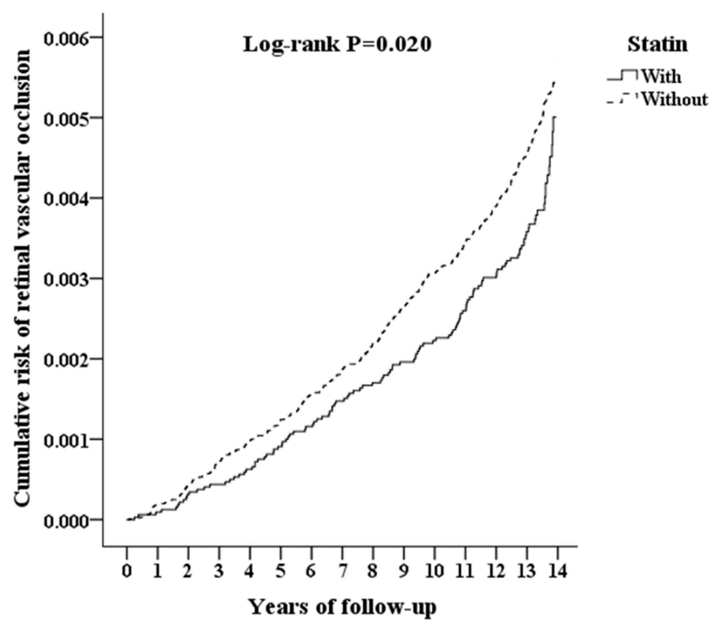
3.2. Cumulative Hazard Curves by the Kaplan–Meier Method

The incidence of RVO during the follow-up period was illustrated using a Kaplan–Meier plot. The results showed a lower cumulative incidence rate of RVO in patients who were prescribed statins compared with those who were not prescribed statins (log-rank test, p = 0.020, Figure 2).

3.3. Univariate and Multivariate Analyses by the Cox Regression Model

The unadjusted HR for RVO was 0.792 times lower in the statin-user group than in the control group (95% confidence interval [CI], 0.650–0.965). After adjusting for covariates, a significantly greater hazard for RVO in the statin-user group remained (adjusted HR, 0.704; 95% CI, 0.591–0.873). Age was a significant risk factor for RVO development in both univariate and multivariate analyses. Patients with DM and HTN were likely to develop RVO (adjusted HR, 1.714; 95% CI, 1.512–1.888, and adjusted HR, 1.976; 95% CI, 1.501–2.630, respectively). In the multivariate analysis, patients with IHD, CVD, renal disease, MetS, and ischemic stroke had a significantly higher risk of developing RVO. Considering ophthalmic diseases, the occurrence of cataract, glaucoma, and diabetic retinopathy appeared to be risk factors with statistical significance for developing RVO in the multivariate analysis. CCI was a significant risk factor for RVO development in both univariate and multivariate analyses (crude HR, 1.234; 95% CI, 1.125–1.290; and adjusted HR, 1.200; 95% CI, 1.093–1.248) (Table 2).

3.4. Subgroup Analysis

We performed subgroup analyses to investigate the use of statins and the incidence of different types of RVO and found that statin users had a decreased risk for all types of RVO development, including central retinal artery occlusion (adjusted HR, 0.679; 95% CI, 0.564–0.841), arterial branch occlusion (adjusted HR, 0.620; 95% CI, 0.518–0.779), central retinal vein occlusion (adjusted HR, 0.758; 95% CI, 0.640–0.952), and branch retinal vein occlusion (adjusted HR, 0.743; 95% CI, 0.629–0.939) (Table 3).

4. Discussion

Our study included 32,025 Chinese patients using statins and 96,075 matched controls, with a 14-year follow-up period. We found that the cumulative incidence of all types of RVO (including central retinal artery occlusion, arterial branch occlusion, central retinal vein occlusion, and branch retinal vein occlusion) was significantly lower in the statin-user group than in the non-statin-user group (Table 3). To our knowledge, this is the first large population study focusing on the role of statins in preventing RVO. During the follow-up period, the cumulative incidence of RVO (including central retinal artery occlusion, arterial branch occlusion, central retinal vein occlusion, and branch retinal vein occlusion) was significantly lower in the statin-user group than in the non-statin-user group. In addition, the incidence of RVO during the follow-up period in the Kaplan–Meier plot showed a lower cumulative incidence rate of RVO in patients who were prescribed statins than in patients who were not prescribed statins (log-rank test, p value = 0.020, Figure 2). The adjusted HR for RVO was 0.704 times lower in the statin-user group than in the control group. Therefore, we found that patients receiving statin treatment have a lower risk of developing RVO compared to patients not taking statins.
The exact pathogenesis of RVO remains unclear. Inflammation has been hypothesized to play an important role in the etiology of RVO [31]. Atherosclerosis is a chronic, low-grade inflammatory condition and has been found to be related to RVO. Macrophages and T lymphocytes contribute to the initial pathology and to progress to thrombus formation later [32]. The induction of hypercoagulability is another mechanism proposed to lead to RVO. Several inflammatory chemokines/cytokines, such as tumor necrosis factor alpha, interleukin-1 beta, and interleukin-6, are prothrombogenic, being activators of the extrinsic coagulation pathway [33,34]. Moreover, local inflammation in the eye is associated with the pathogenesis of RVO. In in vivo assessment of the vitreous fluid in patients with RVO, elevated levels of proinflammatory mediators and lower levels of anti-inflammatory cytokines were found [35,36]. Patients with RVO had elevated levels of interleukin-6, interleukin-8, and monocyte chemoattractant protein-1 in a study on inflammatory immune mediators in vitreoretinal diseases [37]. Vascular endothelial growth factor was significantly elevated in patients with retinal vein occlusion [38]. All these chemokines/cytokines are considered highly proinflammatory [37,39,40]. The abovementioned evidence suggests that inflammation plays an important role in RVO.
Statins, HMG-CoA reductase inhibitors, inhibit the rate-limiting enzyme in cholesterol biosynthesis, i.e., HMG-CoA reductase [41]. Statins have been used to control blood cholesterol levels. Further, statins lower the risk of cardiovascular morbidity and mortality [11,12]. Moreover, several studies on statins have focused on the immunomodulatory effects of statins [15,23,42]. Statins exert their immunomodulatory effect not only by interfering with T cell proliferation and inhibiting the expression of costimulatory molecules on B lymphocytes [43], but also by acting as immunomodulators through the inhibition of cellular interactions and signaling molecules such as tumor necrosis factor alpha [44]. Statins can modulate the immune response by shifting the chemokine/cytokine balance from proinflammatory to anti-inflammatory [45,46].
Evidence has supported the role of inflammation in the pathophysiology of several eye diseases. The effects of statins have been researched in cataract [16], glaucoma [18], diabetic retinopathy [17], and AMD [19], with mixed results. In AMD patients, there is inadequate evidence supporting the prevention by statins of AMD or delayed disease progression [47]. Statins retarded the progression of retinopathy and improved diabetic macular edema in diabetic patients significantly [48,49]. In patients with uveitis, statins have shown a protective effect by possibly reducing uveitis development according to a retrospective population-based study [50].
However, there is little clinical evidence evaluating the role of statins in the prevention or management of RVO. A retrospective, case–control study conducted by Matei et al. analyzed 43 eyes and 129 control eyes after 43 months of follow-up in the United States. They found no clear preventative or therapeutic benefit of statins for retinal vein occlusion in high-risk patients (defined as those with hypertension and primary open-angle glaucoma) [24]. In their study, the focus was only on retinal vein occlusion with a 4-year follow-up period.
A major strength of our study is the large population (a total of 32,025 patients with statin use and 96,075 matched controls without statin use) that we examined, with a long-term follow-up (mean follow-up of 12.87 ± 1.88 years). This study used a sample from a nationwide Taiwanese population. This design reduced the selection bias, make the results applicable to the general population of Taiwan, and accurately evaluated the role of statins in RVO. To the best of our knowledge, this is the first large-size study focusing on the role of statins in the prevention of RVO.
However, the present study has a few limitations. The NHIRD database does not provide laboratory or clinical information, such as color fundus examination and fluorescein angiography for the classification of disease severity. Furthermore, statin dosage was not recorded in the NHIRD database, and it was not possible to evaluate the benefit of statins. We also did not measure the levels of cholesterol in this study. Therefore, it is uncertain whether statin usage or cholesterol is associated with RVO. Moreover, hyperlipidemia being a chronic disease, statin is prescribed to patients in Taiwan for at least 3 months with follow-up. Therefore, we defined the statin group as patients who underwent statin therapy for at least 3 months. Finally, the results are limited by the uniform ethnic background.

5. Conclusions

In conclusion, patients undergoing statin treatment may have a lower risk of developing RVO compared to patients not taking statins. More studies are needed to evaluate the potential benefits of statins in RVO prevention.

Supplementary Materials

The following are available online at https://www.mdpi.com/article/10.3390/ijerph18189864/s1, Table S1: Abbreviation, ICD-9-CM, and definition; Table S2: Years of follow-up.

Author Contributions

Conceptualization, K.-H.C. and W.-C.C.; methodology, C.-H.C., C.-A.S. and W.-C.C.; software, C.-H.C., C.-A.S. and W.-C.C.; validation, C.-C.C., K.-H.C. and W.-C.C.; formal analysis, C.-H.C., C.-A.S. and W.-C.C.; investigation, C.-C.C.; resources, W.-C.C.; data curation, C.-H.C., C.-A.S. and W.-C.C.; writing, original draft preparation, C.-C.C.; writing, review and editing, C.-C.C., K.-H.C., C.-H.C., C.-A.S. and W.-C.C.; visualization, C.-C.C. and P.-H.C.; supervision, K.-H.C., C.-H.C., C.-A.S. and W.-C.C.; project administration, K.-H.C., C.-H.C., C.-A.S. and W.-C.C.; and funding acquisition, K.-H.C. and W.-C.C. All authors have read and agreed to the published version of the manuscript.

Funding

This study was supported by the Tri-Service General Hospital Research Foundation (TSGH-B-110-012 and TSGH-D-110-115) and Ministry of National Defense Medical Affairs Bureau (MND-MAB-100-102), and the sponsor had no role in the study design, data collection and analysis, decision to publish, or preparation of the manuscript.

Institutional Review Board Statement

The study was conducted according to the guidelines of the Declaration of Helsinki and approved by the Institutional Review Board of TSGH IRB No. B-110-28.

Informed Consent Statement

Patient consent was waived because the National Health Insurance (NHI) dataset consists of de-identified secondary data used for research purposes, and the IRB provided a formal written waiver of the need for consent.

Data Availability Statement

Data are contained within the article and Supplementary Materials.

Acknowledgments

We thank Hung-Jie Jhou, M.D., for manuscript editing. We also appreciate the Health and Welfare Data Science Center, Ministry of Health and Welfare (HWDC, MOHW), Taiwan, for providing the National Health Insurance Research Database (NHIRD).

Conflicts of Interest

The authors declare no conflict of interest.

References

  1. Scott, I.U.; Campochiaro, P.A.; Newman, N.J.; Biousse, V. Retinal vascular occlusions. Lancet 2020, 396, 1927–1940. [Google Scholar] [CrossRef]
  2. Hayreh, S.S.; Zimmerman, M.B.; Podhajsky, P. Incidence of various types of retinal vein occlusion and their recurrence and demographic characteristics. Am. J. Ophthalmol. 1994, 117, 429–441. [Google Scholar] [CrossRef]
  3. Wong, T.Y.; Larsen, E.K.; Klein, R.; Mitchell, P.; Couper, D.J.; Klein, B.E.; Hubbard, L.D.; Siscovick, D.S.; Sharrett, A.R. Cardiovascular risk factors for retinal vein occlusion and arteriolar emboli: The Atherosclerosis Risk in Communities & Cardiovascular Health studies. Ophthalmology 2005, 112, 540–547. [Google Scholar] [CrossRef] [PubMed]
  4. Risk factors for central retinal vein occlusion. The Eye Disease Case-Control Study Group. Arch. Ophthalmol. 1996, 114, 545–554. [CrossRef]
  5. Rehak, M.; Wiedemann, P. Retinal vein thrombosis: Pathogenesis and management. J. Thromb. Haemost. 2010, 8, 1886–1894. [Google Scholar] [CrossRef] [PubMed]
  6. Karr, S. Epidemiology and management of hyperlipidemia. Am. J. Manag. Care 2017, 23, S139–S148. [Google Scholar]
  7. O’Mahoney, P.R.; Wong, D.T.; Ray, J.G. Retinal vein occlusion and traditional risk factors for atherosclerosis. Arch. Ophthalmol. 2008, 126, 692–699. [Google Scholar] [CrossRef] [Green Version]
  8. Dodson, P.M.; Galton, D.J.; Hamilton, A.M.; Blach, R.K. Retinal vein occlusion and the prevalence of lipoprotein abnormalities. Br. J. Ophthalmol. 1982, 66, 161–164. [Google Scholar] [CrossRef] [Green Version]
  9. Yau, J.W.; Lee, P.; Wong, T.Y.; Best, J.; Jenkins, A. Retinal vein occlusion: An approach to diagnosis, systemic risk factors and management. Intern. Med. J. 2008, 38, 904–910. [Google Scholar] [CrossRef]
  10. Prisco, D.; Marcucci, R. Retinal vein thrombosis: Risk factors, pathogenesis and therapeutic approach. Pathophysiol. Haemost. Thromb. 2002, 32, 308–311. [Google Scholar] [CrossRef]
  11. Chou, R.; Dana, T.; Blazina, I.; Daeges, M.; Jeanne, T.L. Statins for Prevention of Cardiovascular Disease in Adults: Evidence Report and Systematic Review for the US Preventive Services Task Force. JAMA 2016, 316, 2008–2024. [Google Scholar] [CrossRef] [PubMed]
  12. Singh, V.; Deedwania, P. Reducing morbidity and mortality in high risk patients with statins. Vasc. Health Risk Manag. 2009, 5, 495–507. [Google Scholar] [CrossRef] [Green Version]
  13. Koh, K.K. Effects of statins on vascular wall: Vasomotor function, inflammation, and plaque stability. Cardiovasc. Res. 2000, 47, 648–657. [Google Scholar] [CrossRef]
  14. Zhou, Q.; Liao, J.K. Statins and cardiovascular diseases: From cholesterol lowering to pleiotropy. Curr. Pharm. Des. 2009, 15, 467–478. [Google Scholar] [CrossRef] [Green Version]
  15. Jain, M.K.; Ridker, P.M. Anti-inflammatory effects of statins: Clinical evidence and basic mechanisms. Nat. Rev. Drug Discov. 2005, 4, 977–987. [Google Scholar] [CrossRef] [PubMed]
  16. Erie, J.C.; Pueringer, M.R.; Brue, S.M.; Chamberlain, A.M.; Hodge, D.O. Statin Use and Incident Cataract Surgery: A Case-Control Study. Ophthalmic Epidemiol. 2016, 23, 40–45. [Google Scholar] [CrossRef] [PubMed] [Green Version]
  17. Zhang, W.; Yan, H. Simvastatin increases circulating endothelial progenitor cells and reduces the formation and progression of diabetic retinopathy in rats. Exp. Eye Res. 2012, 105, 1–8. [Google Scholar] [CrossRef] [PubMed]
  18. Talwar, N.; Musch, D.C.; Stein, J.D. Association of Daily Dosage and Type of Statin Agent With Risk of Open-Angle Glaucoma. JAMA Ophthalmol. 2017, 135, 263–267. [Google Scholar] [CrossRef]
  19. Vavvas, D.G.; Daniels, A.B.; Kapsala, Z.G.; Goldfarb, J.W.; Ganotakis, E.; Loewenstein, J.I.; Young, L.H.; Gragoudas, E.S.; Eliott, D.; Kim, I.K.; et al. Regression of Some High-risk Features of Age-related Macular Degeneration (AMD) in Patients Receiving Intensive Statin Treatment. EBioMedicine 2016, 5, 198–203. [Google Scholar] [CrossRef] [Green Version]
  20. Lee, W.J.; Kang, M.H.; Seong, M.; Cho, H.Y. Comparison of aqueous concentrations of angiogenic and inflammatory cytokines in diabetic macular oedema and macular oedema due to branch retinal vein occlusion. Br. J. Ophthalmol. 2012, 96, 1426–1430. [Google Scholar] [CrossRef] [PubMed]
  21. Noma, H.; Funatsu, H.; Mimura, T.; Harino, S.; Hori, S. Aqueous humor levels of vasoactive molecules correlate with vitreous levels and macular edema in central retinal vein occlusion. Eur. J. Ophthalmol. 2010, 20, 402–409. [Google Scholar] [CrossRef]
  22. Medina, R.J.; O’Neill, C.L.; Devine, A.B.; Gardiner, T.A.; Stitt, A.W. The pleiotropic effects of simvastatin on retinal microvascular endothelium has important implications for ischaemic retinopathies. PLoS ONE 2008, 3, e2584. [Google Scholar] [CrossRef] [PubMed] [Green Version]
  23. Al-Janabi, A.; Lightman, S.; Tomkins-Netzer, O. Statins in retinal disease. Eye 2018, 32, 981–991. [Google Scholar] [CrossRef] [PubMed]
  24. Matei, V.M.; Xia, J.Y.; Nguyen, C. Poor outcomes despite aspirin or statin use in high-risk patients with retinal vein occlusion. Graefes Arch. Clin. Exp. Ophthalmol. 2017, 255, 761–766. [Google Scholar] [CrossRef]
  25. Hsieh, C.Y.; Su, C.C.; Shao, S.C.; Sung, S.F.; Lin, S.J.; Kao Yang, Y.H.; Lai, E.C. Taiwan’s National Health Insurance Research Database: Past and future. Clin. Epidemiol. 2019, 11, 349–358. [Google Scholar] [CrossRef] [Green Version]
  26. Cheng, C.L.; Kao, Y.H.; Lin, S.J.; Lee, C.H.; Lai, M.L. Validation of the National Health Insurance Research Database with ischemic stroke cases in Taiwan. Pharmacoepidemiol. Drug Saf. 2011, 20, 236–242. [Google Scholar] [CrossRef]
  27. Fischer, C.; Bruggemann, A.; Hager, A.; Callizo Planas, J.; Roider, J.; Hoerauf, H. Vascular Occlusions following Ocular Surgical Procedures: A Clinical Observation of Vascular Complications after Ocular Surgery. J. Ophthalmol. 2017, 2017, 9120892. [Google Scholar] [CrossRef] [PubMed] [Green Version]
  28. Yin, X.; Li, J.; Zhang, B.; Lu, P. Association of glaucoma with risk of retinal vein occlusion: A meta-analysis. Acta Ophthalmol. 2019, 97, 652–659. [Google Scholar] [CrossRef] [PubMed]
  29. Chen, S.N.; Chao, C.C.; Hwang, J.F.; Yang, C.M. Clinical manifestations of central retinal artery occlusion in eyes of proliferative diabetic retinopathy with previous vitrectomy and panretinal photocoagulation. Retina 2014, 34, 1861–1866. [Google Scholar] [CrossRef]
  30. Simard, M.; Sirois, C.; Candas, B. Validation of the Combined Comorbidity Index of Charlson and Elixhauser to Predict 30-Day Mortality Across ICD-9 and ICD-10. Med. Care 2018, 56, 441–447. [Google Scholar] [CrossRef]
  31. Deobhakta, A.; Chang, L.K. Inflammation in retinal vein occlusion. Int. J. Inflam. 2013, 2013, 438412. [Google Scholar] [CrossRef] [Green Version]
  32. Stary, H.C.; Chandler, A.B.; Glagov, S.; Guyton, J.R.; Insull, W., Jr.; Rosenfeld, M.E.; Schaffer, S.A.; Schwartz, C.J.; Wagner, W.D.; Wissler, R.W. A definition of initial, fatty streak, and intermediate lesions of atherosclerosis. A report from the Committee on Vascular Lesions of the Council on Arteriosclerosis, American Heart Association. Circulation 1994, 89, 2462–2478. [Google Scholar] [CrossRef] [Green Version]
  33. Neumann, F.J.; Ott, I.; Marx, N.; Luther, T.; Kenngott, S.; Gawaz, M.; Kotzsch, M.; Schomig, A. Effect of human recombinant interleukin-6 and interleukin-8 on monocyte procoagulant activity. Arter. Thromb. Vasc. Biol. 1997, 17, 3399–3405. [Google Scholar] [CrossRef] [PubMed]
  34. Ulfhammer, E.; Larsson, P.; Karlsson, L.; Hrafnkelsdottir, T.; Bokarewa, M.; Tarkowski, A.; Jern, S. TNF-alpha mediated suppression of tissue type plasminogen activator expression in vascular endothelial cells is NF-kappaB- and p38 MAPK-dependent. J. Thromb. Haemost. 2006, 4, 1781–1789. [Google Scholar] [CrossRef] [PubMed]
  35. Noma, H.; Funatsu, H.; Mimura, T.; Harino, S.; Eguchi, S.; Hori, S. Pigment epithelium-derived factor and vascular endothelial growth factor in branch retinal vein occlusion with macular edema. Graefes Arch. Clin. Exp. Ophthalmol. 2010, 248, 1559–1565. [Google Scholar] [CrossRef] [PubMed]
  36. Noma, H.; Funatsu, H.; Mimura, T.; Eguchi, S.; Shimada, K. Inflammatory factors in major and macular branch retinal vein occlusion. Ophthalmologica 2012, 227, 146–152. [Google Scholar] [CrossRef]
  37. Yoshimura, T.; Sonoda, K.H.; Sugahara, M.; Mochizuki, Y.; Enaida, H.; Oshima, Y.; Ueno, A.; Hata, Y.; Yoshida, H.; Ishibashi, T. Comprehensive analysis of inflammatory immune mediators in vitreoretinal diseases. PLoS ONE 2009, 4, e8158. [Google Scholar] [CrossRef]
  38. Ehlken, C.; Rennel, E.S.; Michels, D.; Grundel, B.; Pielen, A.; Junker, B.; Stahl, A.; Hansen, L.L.; Feltgen, N.; Agostini, H.T.; et al. Levels of VEGF but not VEGF(165b) are increased in the vitreous of patients with retinal vein occlusion. Am. J. Ophthalmol. 2011, 152, 298–303.e1. [Google Scholar] [CrossRef]
  39. Kany, S.; Vollrath, J.T.; Relja, B. Cytokines in Inflammatory Disease. Int. J. Mol. Sci. 2019, 20, 6008. [Google Scholar] [CrossRef] [Green Version]
  40. Maloney, J.P.; Gao, L. Proinflammatory Cytokines Increase Vascular Endothelial Growth Factor Expression in Alveolar Epithelial Cells. Mediat. Inflamm. 2015, 2015, 387842. [Google Scholar] [CrossRef] [Green Version]
  41. Hall, A. Rho GTPases and the actin cytoskeleton. Science 1998, 279, 509–514. [Google Scholar] [CrossRef] [PubMed] [Green Version]
  42. Palinski, W.; Tsimikas, S. Immunomodulatory effects of statins: Mechanisms and potential impact on arteriosclerosis. J. Am. Soc. Nephrol. 2002, 13, 1673–1681. [Google Scholar] [CrossRef] [PubMed] [Green Version]
  43. Youssef, S.; Stuve, O.; Patarroyo, J.C.; Ruiz, P.J.; Radosevich, J.L.; Hur, E.M.; Bravo, M.; Mitchell, D.J.; Sobel, R.A.; Steinman, L.; et al. The HMG-CoA reductase inhibitor, atorvastatin, promotes a Th2 bias and reverses paralysis in central nervous system autoimmune disease. Nature 2002, 420, 78–84. [Google Scholar] [CrossRef]
  44. Sadeghi, M.M.; Tiglio, A.; Sadigh, K.; O’Donnell, L.; Collinge, M.; Pardi, R.; Bender, J.R. Inhibition of interferon-gamma-mediated microvascular endothelial cell major histocompatibility complex class II gene activation by HMG-CoA reductase inhibitors. Transplantation 2001, 71, 1262–1268. [Google Scholar] [CrossRef]
  45. Bu, D.X.; Griffin, G.; Lichtman, A.H. Mechanisms for the anti-inflammatory effects of statins. Curr. Opin. Lipidol. 2011, 22, 165–170. [Google Scholar] [CrossRef]
  46. Kwak, B.; Mulhaupt, F.; Myit, S.; Mach, F. Statins as a newly recognized type of immunomodulator. Nat. Med. 2000, 6, 1399–1402. [Google Scholar] [CrossRef]
  47. Gehlbach, P.; Li, T.; Hatef, E. Statins for age-related macular degeneration. Cochrane Database Syst. Rev. 2016. [Google Scholar] [CrossRef] [Green Version]
  48. Gupta, A.; Gupta, V.; Thapar, S.; Bhansali, A. Lipid-lowering drug atorvastatin as an adjunct in the management of diabetic macular edema. Am. J. Ophthalmol. 2004, 137, 675–682. [Google Scholar] [CrossRef] [PubMed]
  49. Sen, K.; Misra, A.; Kumar, A.; Pandey, R.M. Simvastatin retards progression of retinopathy in diabetic patients with hypercholesterolemia. Diabetes Res. Clin. Pract. 2002, 56, 1–11. [Google Scholar] [CrossRef]
  50. Borkar, D.S.; Tham, V.M.; Shen, E.; Parker, J.V.; Uchida, A.; Vinoya, A.C.; Acharya, N.R. Association between statin use and uveitis: Results from the Pacific Ocular Inflammation study. Am. J. Ophthalmol. 2015, 159, 707–713. [Google Scholar] [CrossRef] [PubMed] [Green Version]
Figure 1. Flowchart of the study sample selection.
Ijerph 18 09864 g001
Figure 2. Kaplan–Meier curve for cumulative risk of retinal vascular occlusion among patients aged 18 and over stratified by statin use with a log-rank test.
Ijerph 18 09864 g002
Table 1. Characteristics of the two groups at baseline.
StatinWithWithoutp
Variablesn%n%
Total32,025 25.00 96,075 75.00
Gender 0.999
Male15,096 47.14 45,288 47.14
Female16,929 52.86 50,787 52.86
Age (years)54.57 ± 13.5152.34 ± 16.510.088
DM <0.001 *
Without23,513 73.42 87,195 90.76
With8512 26.58 8880 9.24
HTN <0.001 *
Without23,600 73.69 85,693 89.19
With8425 26.31 10,382 10.81
Hyperlipidemia <0.001 *
Without7027 21.94 95,078 98.96
With24,998 78.06 997 1.04
IHD <0.001 *
Without29,513 92.16 91,352 95.08
With2512 7.84 4723 4.92
CVD <0.001 *
Without28,939 90.36 90,548 94.25
With3086 9.64 5527 5.75
Renal disease <0.001 *
Without29,180 91.12 94,770 98.64
With2845 8.88 1305 1.36
Tumor <0.001 *
Without31,01996.86 94,07797.92
With10063.1419982.08
MetS <0.001 *
Without29,671 92.65 93,333 97.15
With2354 7.35 2742 2.85
Hypercoagulable state 0.004 *
Without31,639 98.79 95,104 98.99
With386 1.21 971 1.01
Ischemic stroke <0.001 *
Without29,040 90.68 90,623 94.33
With2985 9.32 5452 5.67
Cataract <0.001 *
Without30,710 95.89 92,749 96.54
With1315 4.11 3326 3.46
Glaucoma <0.001 *
Without29,240 91.30 89,064 92.70
With2785 8.70 7011 7.30
Diabetic retinopathy <0.001 *
Without31,243 97.56 95,084 98.97
With782 2.44 991 1.03
AMD <0.001 *
Without31,379 97.98 94,931 98.81
With646 2.02 1144 1.19
CCI0.05 ± 0.180.03 ± 0.15<0.001 *
* indicates a significant difference between the two groups with/without statin usage, p < 0.05. a presented as mean ± SD. CCI: Charlson comorbidity index, DM: diabetes mellitus, HTN: hypertension, IHD: ischemic heart disease, CVD: cardiovascular disease, MetS: metabolic syndrome, AMD: age-related macular degeneration.
Table 2. Risk factors for retinal vascular occlusion by using Cox regression.
VariablesCrude HR95% CI95% CIpAdjusted HR95% CI95% CIp
Statin
WithoutReference Reference
With0.792 0.650 0.965 0.020 * 0.704 0.591 0.873 <0.001 *
Gender
Male0.953 0.812 1.118 0.552 0.970 0.822 1.111 0.703
FemaleReference Reference
Age (yrs)1.037 1.031 1.042 <0.001 *1.026 1.019 1.031 <0.001 *
DM
WithoutReference Reference
With1.798 1.546 1.986 <0.001 *1.714 1.512 1.888 <0.001 *
HTN
WithoutReference Reference
With2.068 1.573 2.676 <0.001 *1.976 1.501 2.630 <0.001 *
Hyperlipidemia
WithoutReference Reference
With1.234 1.004 1.308 0.046 *0.819 0.478 1.335 0.586
IHD
WithoutReference Reference
With1.462 1.297 1.682 <0.001 *1.369 1.194 1.617 <0.001 *
CVD
WithoutReference Reference
With1.350 1.104 1.553 <0.001 *1.271 1.043 1.495 0.007 *
Renal disease
WithoutReference Reference
With1.668 1.321 2.095 <0.001 *1.609 1.225 1.962 <0.001 *
Tumor
WithoutReference Reference
With1.032 0.862 1.345 0.184 1.031 0.860 1.352 0.176
MetS
WithoutReference Reference
With2.065 1.482 2.798 <0.001 *1.531 1.045 2.088 0.003 *
Hypercoagulable state
WithoutReference Reference
With1.526 0.862 2.060 0.289 1.306 0.671 1.884 0.462
Ischemic stroke
WithoutReference Reference
With1.398 1.167 1.592 <0.001 *1.288 1.050 1.516 0.001 *
Cataract
WithoutReference Reference
With1.411 1.086 1.702 0.004 *1.352 1.015 1.670 0.032 *
Glaucoma
WithoutReference Reference
With1.525 1.126 1.978 <0.001 *1.488 1.034 1.826 0.018 *
Diabetic retinopathy
WithoutReference Reference
With1.279 1.089 1.465 <0.001 *1.186 1.003 1.395 0.046 *
AMD
WithoutReference Reference
With1.382 0.979 1.891 0.072 1.297 0.875 1.742 0.088
CCI_R1.234 1.125 1.290 <0.001 *1.200 1.093 1.248 <0.001 *
* indicates a significant difference between the two groups with/without statin usage, p < 0.05. a presented as mean ± SD. CCI: Charlson comorbidity index, DM: diabetes mellitus, HTN: hypertension, IHD: ischemic heart disease, CVD: cardiovascular disease, MetS: metabolic syndrome, AMD: age-related macular degeneration.
Table 3. Risk for retinal vascular occlusion subgroups by Cox regression analysis.
StatinWithWithout (Reference)With vs. Without (Reference)
Retinal Vascular Occlusion SubgroupsEventsPYsRate (per 105 PYs)EventsPYsRate (per 105 PYs)Adjusted HR95% CI95% CIp
Overall124 413,862.59 29.96 486 1,235,055.74 39.35 0.704 0.591 0.873 <0.001 *
Central Retinal Artery Occlusion31 413,862.59 7.49 126 1,235,055.74 10.20 0.679 0.564 0.841 <0.001 *
Arterial Branch Occlusion27 413,862.59 6.52 119 1,235,055.74 9.64 0.620 0.518 0.779 <0.001 *
Central Retinal Vein Occlusion34 413,862.59 8.22 124 1,235,055.74 10.04 0.758 0.640 0.952 0.008 *
Branch Retinal Vein Occlusion32 413,862.59 7.73 117 1,235,055.74 9.47 0.743 0.629 0.939 <0.001 *
* indicates a significant difference between the two groups with/without statin usage, p < 0.05. a presented as mean ± SD. PYs = Person-years; Adjusted HR = Adjusted Hazard ratio: Adjusted for the variables listed in Table 2; CI = confidence interval.
Publisher’s Note: MDPI stays neutral with regard to jurisdictional claims in published maps and institutional affiliations.

