
Double-Negative T-Cell Reaction in a Case of Listeria Meningitis

 ,
,  , ,
, ,  and
and 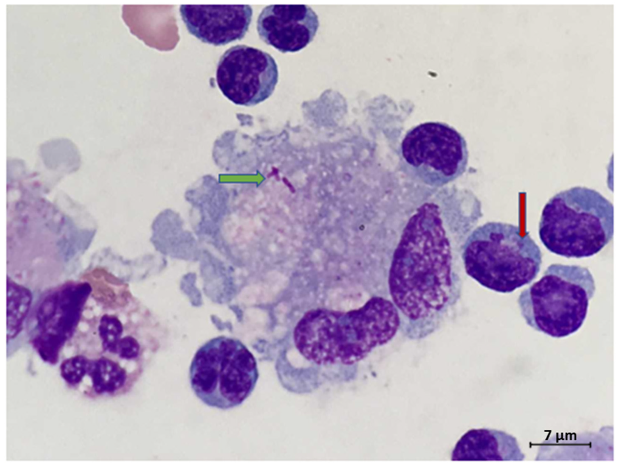
Abstract
1. Introduction
2. Case Report
3. Discussion
4. Conclusions
Author Contributions
Funding
Institutional Review Board Statement
Informed Consent Statement
Data Availability Statement
Conflicts of Interest
References
- Munk, M.E.; Elser, C.; Kaufmann, S.H. Human gamma/delta T-cell response to Listeria monocytogenes protein components in vitro. Immunology 1996, 87, 230–235. [Google Scholar] [CrossRef] [PubMed]
- Zhu, Y.; Wang, H.; Xu, Y.; Hu, Y.; Chen, H.; Cui, L.; Zhang, J.; He, W. Human γδ T cells augment antigen presentation in Listeria Monocytogenes infection. Mol. Med. 2016, 22, 737–746. [Google Scholar] [CrossRef] [PubMed]
- Wei, P.; Bao, R.; Fan, Y. Brainstem Encephalitis Caused by Listeria monocytogenes. Pathogens 2020, 9, 715. [Google Scholar] [CrossRef] [PubMed]
- Nakamura, F.; Nasu, R. Listeria monocytogenes septicemia and meningoencephalitis associated with relapsed and refractory follicular lymphoma. J. Infect. Chemother. 2020, 26, 619–621. [Google Scholar] [CrossRef] [PubMed]
- Carrique-Mas, J.J.; Hökeberg, I.; Andersson, Y.; Arneborn, M.; Tham, W.; Danielsson-Tham, M.L.; Osterman, B.; Leffler, M.; Steen, M.; Eriksson, E.; et al. Febrile gastroenteritis after eating on-farm manufactured fresh cheese—An outbreak of listeriosis? Epidemiol Infect. 2003, 130, 79–86. [Google Scholar] [CrossRef] [PubMed]
- Koopmans, M.M.; Bijlsma, M.W.; Brouwer, M.C.; van de Beek, D.; van der Ende, A. Listeria monocytogenes meningitis in the Netherlands, 1985-2014: A nationwide surveillance study. J. Infect. 2017, 75, 12–19. [Google Scholar] [CrossRef] [PubMed]
- Pagliano, P.; Ascione, T.; Boccia, G.; De Caro, F.; Esposito, S. Listeria monocytogenes meningitis in the elderly: Epidemiological, clinical and therapeutic findings. Infez Med. 2016, 24, 105–111. [Google Scholar] [PubMed]
- Gelfand, M.S. Clinical Manifestations and Diagnosis of Listeria monocytogenes Infection. UpToDate (en Línea) (Consultado el 30/03/2016). 2007. Available online: www.uptodate.com/contents/clinical-Manifestations-and-diagnosis-of-listeria-monocytogenes-infection (accessed on 12 April 2021).
- Ooi, S.T.; Lorber, B. Gastroenteritis due to Listeria monocytogenes. Clin. Infect. Dis. 2005, 40, 1327–1332. [Google Scholar] [CrossRef] [PubMed]
- Matereke, L.T.; Okoh, A.I. Listeria monocytogenes Virulence, Antimicrobial Resistance and Environmental Persistence: A Review. Pathogens 2020, 9, 528. [Google Scholar] [CrossRef]
- Matsuzaki, G.; Hiromatsu, K.; Yoshikai, Y.; Muramori, K.; Nomoto, K. Characterization of T-cell receptor gamma delta T cells appearing at the early phase of murine Listeria monocytogenes infection. Immunology 1993, 78, 22–27. [Google Scholar] [PubMed]
- Skeen, M.J.; Rix, E.P.; Freeman, M.M.; Ziegler, H.K. Exaggerated proinflammatory and Th1 responses in the absence of gamma/delta T cells after infection with Listeria monocytogenes. Infect. Immun. 2001, 69, 7213–7223. [Google Scholar] [CrossRef]
- Brouwer, M.C.; van de Beek, D.; Heckenberg, S.G.; Spanjaard, L.; de Gans, J. Community-acquired Listeria monocytogenes meningitis in adults. Clin. Infect. Dis. 2006, 43, 1233–1238. [Google Scholar] [CrossRef] [PubMed]
- Bazooyar, B. Rhombencephalitis by Listeria monocytogens in Two Diabetic Patients. Arch. Iran. Med. 2015, 18, 613–615. [Google Scholar] [PubMed]
- Uldry, P.A.; Kuntzer, T.; Bogousslavsky, J.; Regli, F.; Miklossy, J.; Bille, J.; Francioli, P.; Janzer, R. Early symptoms and outcome of Listeria monocytogenes rhombencephalitis: 14 adult cases. J. Neurol. 1993, 240, 235–242. [Google Scholar] [CrossRef] [PubMed]
- Nieman, R.E.; Lorber, B. Listeriosis in adults: A changing pattern. Report of eight cases and review of the literature, 1968–1978. Rev. Infect. Dis. 1980, 2, 207–227. [Google Scholar] [CrossRef] [PubMed]
- Jubelt, B.; Mihai, C.; Li, T.M.; Veerapaneni, P. Rhombencephalitis / brainstem encephalitis. Curr. Neurol. Neurosci. Rep. 2011, 11, 543–552. [Google Scholar] [CrossRef] [PubMed]
- Belles, C.; Kuhl, A.K.; Donoghue, A.J.; Sano, Y.; O’Brien, R.L.; Born, W.; Bottomly, K.; Carding, S.R. Bias in the gamma delta T cell response to Listeria monocytogenes. V delta 6.3+ cells are a major component of the gamma delta T cell response to Listeria monocytogenes. J. Immunol. 1996, 156, 4280–4289. [Google Scholar] [PubMed]
- Vantourout, P.; Hayday, A. Six-of-the-best: Unique contributions of γδ T cells to immunology. Nat. Rev. Immunol. 2013, 13, 88–100. [Google Scholar] [CrossRef] [PubMed]
- Guo, Y.; Ziegler, H.K.; Safley, S.A.; Niesel, D.W.; Vaidya, S.; Klimpel, G.R. Human T-cell recognition of Listeria monocytogenes: Recognition of listeriolysin O by TcR alpha beta + and TcR gamma delta + T cells. Infect. Immun. 1995, 63, 2288–2294. [Google Scholar] [CrossRef] [PubMed]


Publisher’s Note: MDPI stays neutral with regard to jurisdictional claims in published maps and institutional affiliations. |
© 2021 by the authors. Licensee MDPI, Basel, Switzerland. This article is an open access article distributed under the terms and conditions of the Creative Commons Attribution (CC BY) license (https://creativecommons.org/licenses/by/4.0/).
Share and Cite
Ullah, A.; Patterson, G.T.; Mattox, S.N.; Cotter, T.; Patel, N.G.; Savage, N.M. Double-Negative T-Cell Reaction in a Case of Listeria Meningitis. Int. J. Environ. Res. Public Health 2021, 18, 6486. https://doi.org/10.3390/ijerph18126486
Ullah A, Patterson GT, Mattox SN, Cotter T, Patel NG, Savage NM. Double-Negative T-Cell Reaction in a Case of Listeria Meningitis. International Journal of Environmental Research and Public Health. 2021; 18(12):6486. https://doi.org/10.3390/ijerph18126486
Chicago/Turabian StyleUllah, Asad, G. Taylor Patterson, Samantha N. Mattox, Thomas Cotter, Nikhil G. Patel, and Natasha M. Savage. 2021. "Double-Negative T-Cell Reaction in a Case of Listeria Meningitis" International Journal of Environmental Research and Public Health 18, no. 12: 6486. https://doi.org/10.3390/ijerph18126486
APA StyleUllah, A., Patterson, G. T., Mattox, S. N., Cotter, T., Patel, N. G., & Savage, N. M. (2021). Double-Negative T-Cell Reaction in a Case of Listeria Meningitis. International Journal of Environmental Research and Public Health, 18(12), 6486. https://doi.org/10.3390/ijerph18126486