A Narrative Review of the Histological and Histomorphometrical Evaluation of the Peri-Implant Bone in Loaded and Unloaded Dental Implants. A 30-Year Experience (1988–2018)

 ,
, 


Abstract
1. Introduction
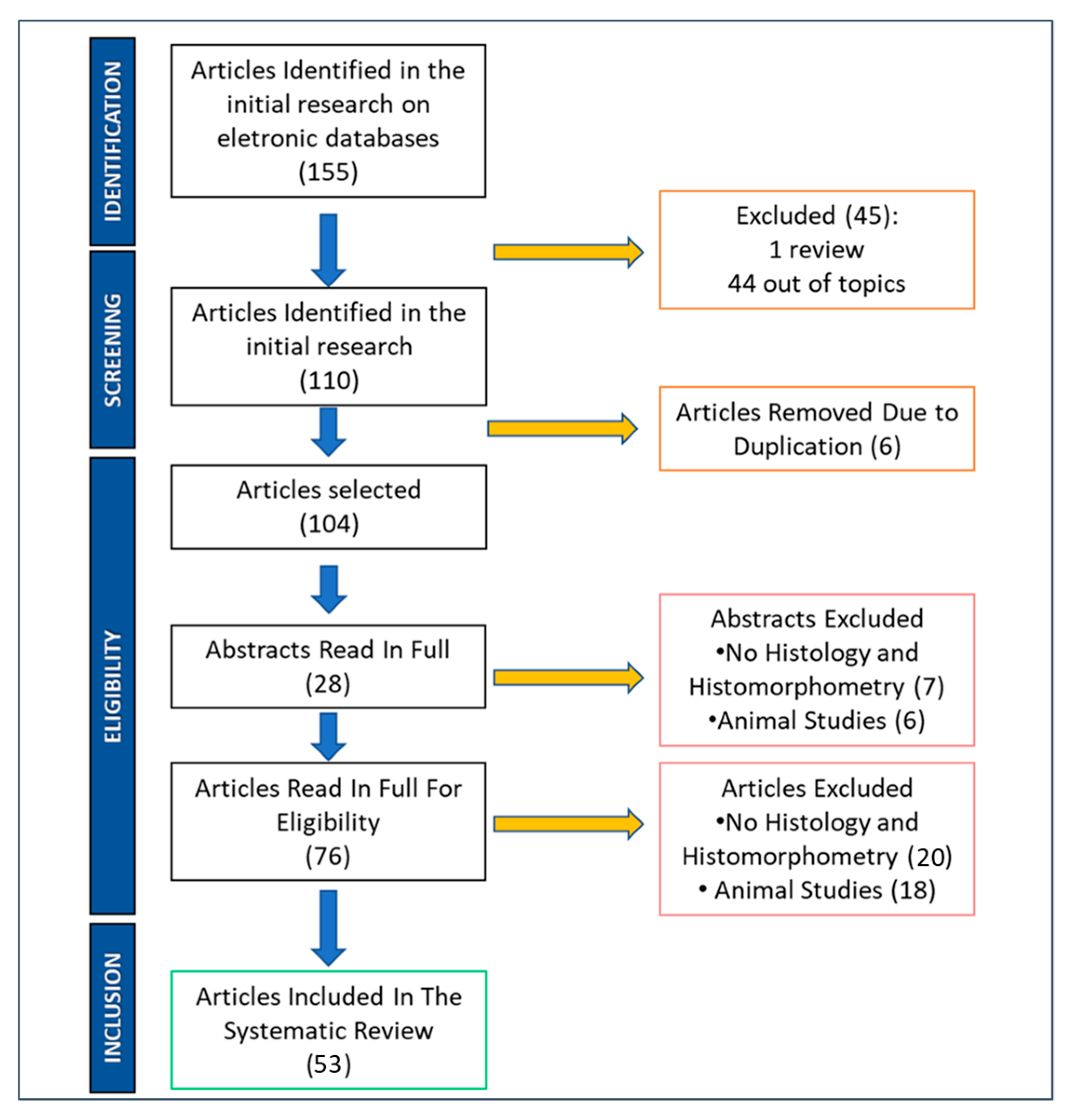
2. Materials and Methods
2.1. Search Strategy
2.2. Inclusion Criteria
2.3. Selection of the Studies
2.4. Data Extraction
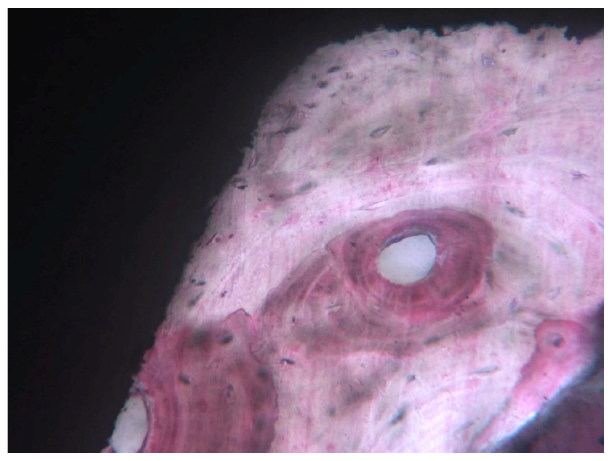
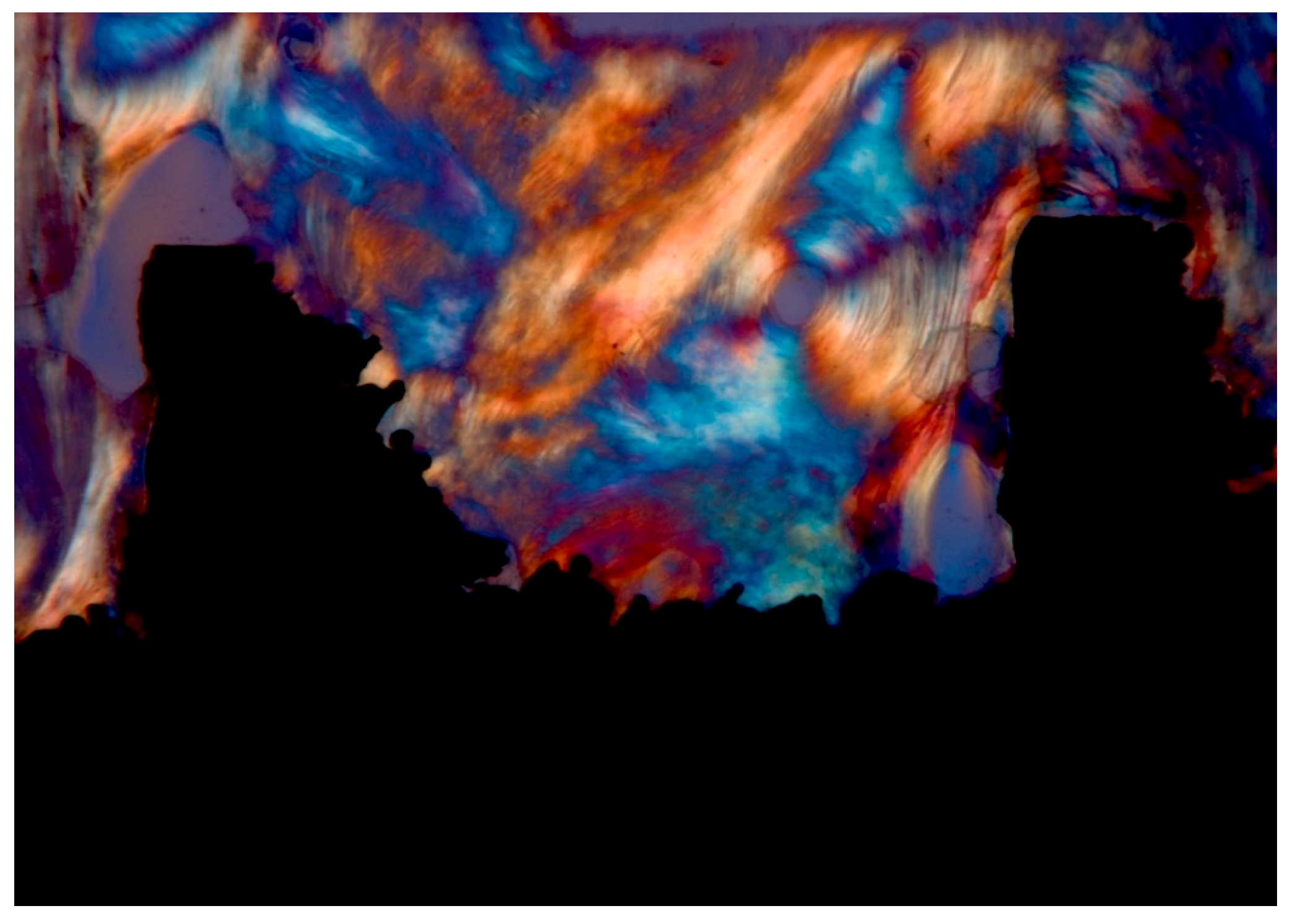
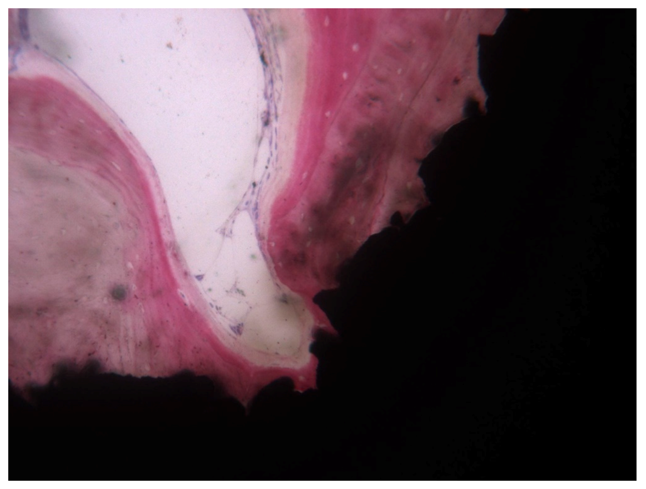
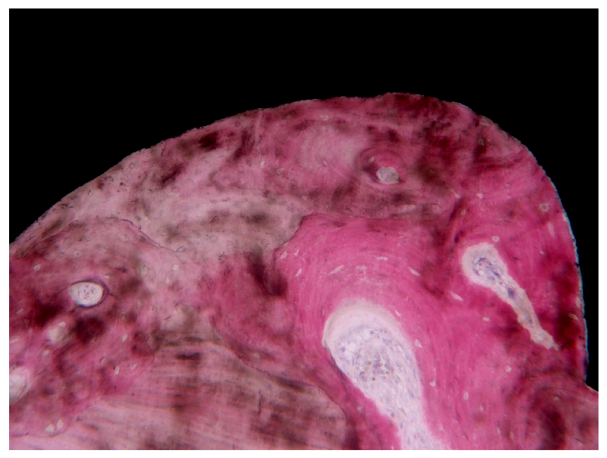
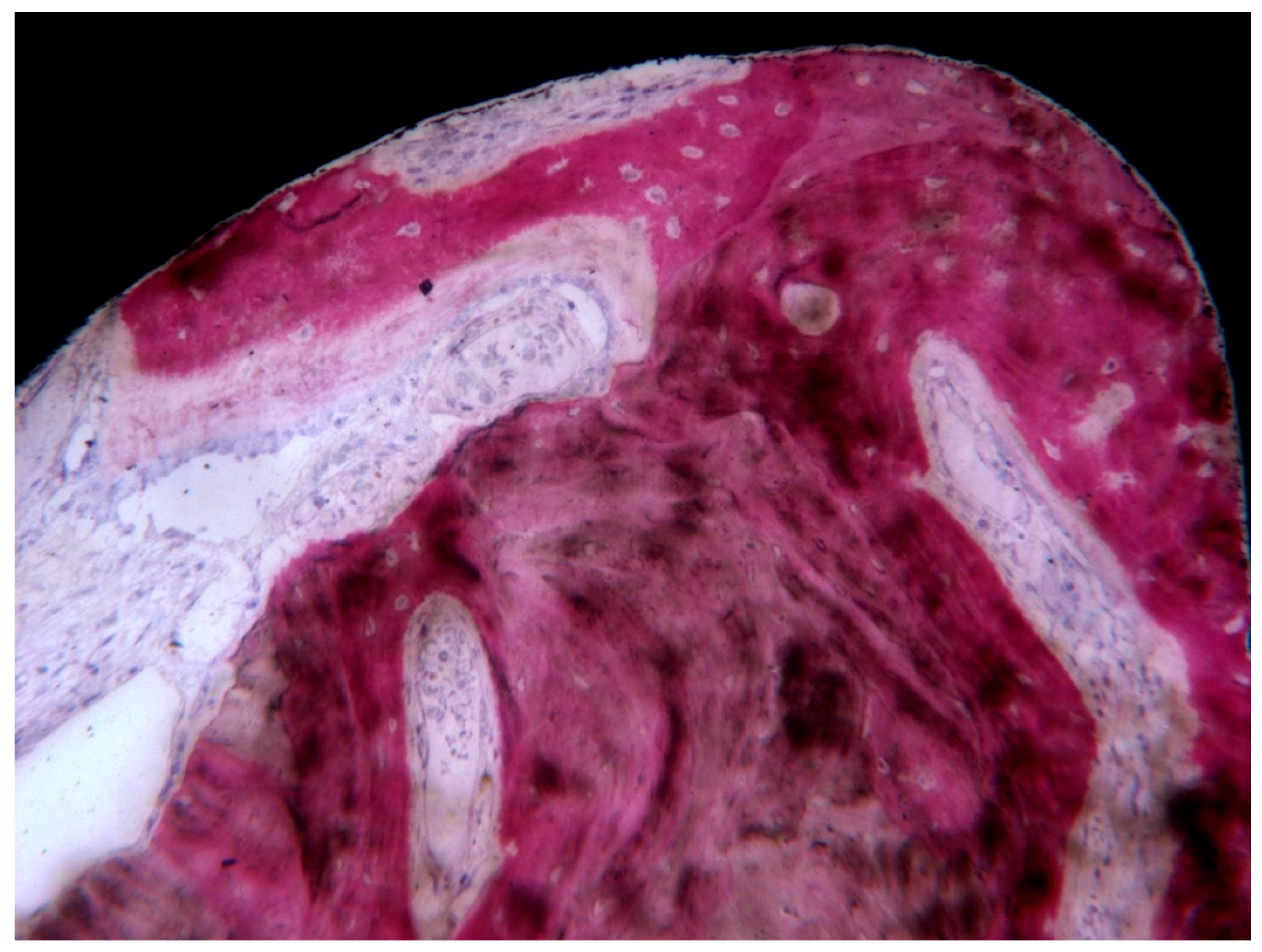
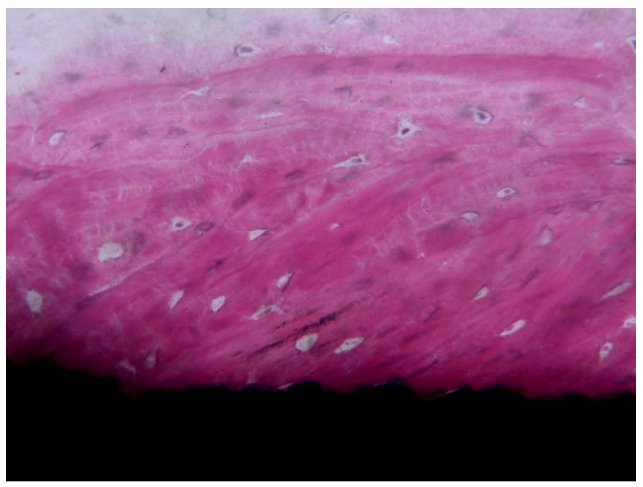
3. Results
4. Discussion
5. Conclusions
Author Contributions
Funding
Conflicts of Interest
References
- Shah, F.A.; Nilson, B.; Brånemark, R.; Thomsen, P.; Palmquist, A. The bone-implant interface-nanoscale analysis of clinically retrieved dental implants. Nanomedicine 2014, 10, 1729–1737. [Google Scholar] [CrossRef]
- Schüpbach, P.; Glauser, R.; Rocci, A.; Martignoni, M.; Sennerby, L.; Lundgren, A.; Gottlow, J. The human bone-oxidized titanium implant interface: A light microscopic, scanning electron microscopic, back-scatter scanning electron microscopic, and energy-dispersive x-ray study of clinically retrieved dental implants. Clin. Implant. Dent. Relat. Res. 2005, 7 (Suppl. 1), S36–S43. [Google Scholar]
- Coelho, P.G.; Marin, C.; Granato, R.; Suzuki, M. Histomorphologic analysis of 30 plateau root form implants retrieved after 8 to 13 years in function. A human retrieval study. J. Biomed. Mater. Res. Part B Appl. Biomater. 2009, 91, 975–979. [Google Scholar] [CrossRef] [PubMed]
- Tarnow, D.P.; Chu, S.J. Human histologic verification of osseointegration of an immediate implant placed into a fresh extraction socket with excessive gap distance without primary flap closure, graft, or membrane: A case report. Int. J. Periodontics Restor. Dent. 2011, 31, 515–521. [Google Scholar] [PubMed]
- Nevins, M.; Nevins, M.L.; Schupbach, P.; Fiorellini, J.; Lin, Z.; Kim, D.M. The impact of bone compression on bone-to-implant contact of an osseointegrated implant: A canine study. Int. J. Periodontics Restor. Dent. 2012, 32, 637–645. [Google Scholar] [PubMed]
- Kim, J.-C.; Lee, J.; Kim, S.; Koo, K.-T.; Kim, H.-Y.; Yeo, I.-S.L. Influence of implant-abutment connection structure on peri-implant bone level in a second molar: A 1-year randomized controlled trial. J. Adv. Prosthodont. 2019, 11, 147–154. [Google Scholar] [CrossRef]
- Sağirkaya, E.; Kucukekenci, A.S.; Karasoy, D.; Akça, K.; Eckert, S.E.; Çehreli, M.C. Comparative assessments, meta-analysis, and recommended guidelines for reporting studies on histomorphometric bone-implant contact in humans. Int. J. Oral. Maxillofac. Implants 2013, 28, 1243–1253. [Google Scholar]
- Berketa, J.W.; James, H.; Langlois, N.E.I.; Richards, L.C. A study of osseointegrated dental implants following cremation. Aust. Dent. J. 2014, 59, 149–155. [Google Scholar] [CrossRef]
- Shemtov-Yona, K.; Rittel, D. On the mechanical integrity of retrieved dental implants. J. Mech. Behav. Biomed. Mater. 2015, 49, 290–299. [Google Scholar] [CrossRef]
- Albrektsson, T.; Eriksson, A.R.; Friberg, B.; Lekholm, U.; Lindahl, L.; Nevins, M.; Oikarinen, V.; Roos, J.; Sennerby, L.; Astrand, P. Histologic investigations on 33 retrieved Nobelpharma implants. Clin. Mater. 1993, 12, 1–9. [Google Scholar] [CrossRef]
- Coelho, P.G.; Bonfante, E.A.; Marin, C.; Granato, R.; Giro, G.; Suzuki, M. A human retrieval study of plasma-sprayed hydroxyapatite-coated plateau root form implants after 2 months to 13 years in function. J. Long. Term. Eff. Med. Implants 2010, 20, 335–342. [Google Scholar] [CrossRef] [PubMed]
- Abbott, J.R.; Marino, V.; Bartold, P.M. Human cadaveric histomorphological and metallurgical analysis of dental implants following 12.5 years of service. Clin. Oral. Implants Res. 2014, 25, 266–271. [Google Scholar] [CrossRef] [PubMed]
- Romanos, G.E.; Toh, C.G.; Siar, C.H.; Wicht, H.; Yacoob, H.; Nentwig, G.-H. Bone-implant interface around titanium implants under different loading conditions: A histomorphometrical analysis in the Macaca fascicularis monkey. J. Periodontol. 2003, 74, 1483–1490. [Google Scholar] [CrossRef] [PubMed]
- Podaropoulos, L.; Veis, A.A.; Trisi, P.; Papadimitriou, S.; Alexandridis, C.; Kalyvas, D. Bone reactions around dental implants subjected to progressive static load: An experimental study in dogs. Clin. Oral. Implants Res. 2016, 27, 910–917. [Google Scholar] [CrossRef]
- Joos, U.; Büchter, A.; Wiesmann, H.-P.; Meyer, U. Strain driven fast osseointegration of implants. Head. Face Med. 2005, 1, 6. [Google Scholar] [CrossRef]
- Delgado-Ruiz, R.A.; Abboud, M.; Romanos, G.; Aguilar-Salvatierra, A.; Gomez-Moreno, G.; Calvo-Guirado, J.L. Peri-implant bone organization surrounding zirconia-microgrooved surfaces circularly polarized light and confocal laser scanning microscopy study. Clin. Oral. Implants Res. 2015, 26, 1328–1337. [Google Scholar] [CrossRef]
- Neugebauer, J.; Traini, T.; Thams, U.; Piattelli, A.; Zöller, J.E. Peri-implant bone organization under immediate loading state. Circularly polarized light analyses: A minipig study. J. Periodontol. 2006, 77, 152–160. [Google Scholar] [CrossRef]
- Baldassarri, M.; Bonfante, E.; Suzuki, M.; Marin, C.; Granato, R.; Tovar, N.; Coelho, P.G. Mechanical properties of human bone surrounding plateau root form implants retrieved after 0.3-24 years of function. J. Biomed. Mater. Res. Part B Appl. Biomater. 2012, 100, 2015–2021. [Google Scholar] [CrossRef]
- Palmquist, A.; Grandfield, K.; Norlindh, B.; Mattsson, T.; Brånemark, R.; Thomsen, P. Bone-titanium oxide interface in humans revealed by transmission electron microscopy and electron tomography. J. R. Soc. Interface 2012, 9, 396–400. [Google Scholar] [CrossRef]
- Nevins, M.; Camelo, M.; Koo, S.; Lazzara, R.J.; Kim, D.M. Human histologic assessment of a platform-switched osseointegrated dental implant. Int. J. Periodontics Restor. Dent. 2014, 34 (Suppl. 3), s71–s73. [Google Scholar]
- Gandolfi, M.G.; Zamparini, F.; Iezzi, G.; Degidi, M.; Botticelli, D.; Piattelli, A.; Prati, C. Microchemical and Micromorphologic ESEM-EDX Analysis of Bone Mineralization at the Thread Interface in Human Dental Implants Retrieved for Mechanical Complications After 2 Months to 17 Years. Int. J. Periodontics Restor. Dent. 2018, 38, 431–441. [Google Scholar] [CrossRef] [PubMed]
- Botticelli, D.; Perrotti, V.; Piattelli, A.; Iezzi, G. Four Stable and Functioning Dental Implants Retrieved for Fracture After 14 and 17 Years from the Same Patient: A Histologic and Histomorphometric Report. Int. J. Periodontics Restor. Dent. 2019, 39, 83–88. [Google Scholar] [CrossRef] [PubMed]
- Yonezawa, D.; Piattelli, A.; Favero, R.; Ferri, M.; Iezzi, G.; Botticelli, D. Bone Healing at Functionally Loaded and Unloaded Screw-Shaped Implants Supporting Single Crowns: A Histomorphometric Study in Humans. Int. J. Oral Maxillofac Implants 2018, 33, 181–187. [Google Scholar] [CrossRef] [PubMed]
- Mangano, F.G.; Pires, J.T.; Shibli, J.A.; Mijiritsky, E.; Iezzi, G.; Piattelli, A.; Mangano, C. Early Bone Response to Dual Acid-Etched and Machined Dental Implants Placed in the Posterior Maxilla: A Histologic and Histomorphometric Human Study. Implant Dent. 2017, 26, 24–29. [Google Scholar] [CrossRef] [PubMed]
- Mangano, C.; Shibli, J.A.; Pires, J.T.; Luongo, G.; Piattelli, A.; Iezzi, G. Early Bone Formation around Immediately Loaded Transitional Implants Inserted in the Human Posterior Maxilla: The Effects of Fixture Design and Surface. Biomed. Res. Int. 2017, 2017, 4152506. [Google Scholar] [CrossRef] [PubMed]
- Mangano, F.; Mangano, C.; Piattelli, A.; Iezzi, G. Histological Evidence of the Osseointegration of Fractured Direct Metal Laser Sintering Implants Retrieved after 5 Years of Function. Biomed. Res. Int. 2017, 2017, 9732136. [Google Scholar] [CrossRef]
- Mangano, F.G.; Iezzi, G.; Shibli, J.A.; Pires, J.T.; Luongo, G.; Piattelli, A.; Mangano, C. Early bone formation around immediately loaded implants with nanostructured calcium-incorporated and machined surface: A randomized, controlled histologic and histomorphometric study in the human posterior maxilla. Clin. Oral. Investig. 2017, 21, 2603–2611. [Google Scholar] [CrossRef]
- Iezzi, G.; Piattelli, A.; Mangano, C.; Degidi, M.; Testori, T.; Vantaggiato, G.; Fiera, E.; Frosecchi, M.; Floris, P.; Perroni, R.; et al. Periimplant Bone Response in Human-Retrieved, Clinically Stable, Successful, and Functioning Dental Implants After a Long-Term Loading Period: A Report of 17 Cases From 4 to 20 Years. Implant Dent. 2016, 25, 380–386. [Google Scholar] [CrossRef]
- Mangano, C.; Piattelli, A.; Mortellaro, C.; Mangano, F.; Perrotti, V.; Iezzi, G. Evaluation of Peri-Implant Bone Response in Implants Retrieved for Fracture After More Than 20 Years of Loading: A Case Series. J. Oral. Implantol. 2015, 41, 414–418. [Google Scholar] [CrossRef]
- Traini, T.; Mangano, C.; Perrotti, V.; Caputi, S.; Coelho, P.; Piattelli, A.; Iezzi, G. Human bone reactions around implants with adverse interfacial bone strain over 20 years. J. Biomed. Mater. Res. Part B Appl. Biomater. 2014, 102, 1342–1352. [Google Scholar] [CrossRef]
- Piattelli, A.; Artese, L.; Penitente, E.; Iaculli, F.; Degidi, M.; Mangano, C.; Shibli, J.A.; Coelho, P.G.; Perrotti, V.; Iezzi, G. Osteocyte density in the peri-implant bone of implants retrieved after different time periods (4 weeks to 27 years). J. Biomed. Mater. Res. Part B Appl. Biomater. 2014, 102, 239–243. [Google Scholar] [CrossRef] [PubMed]
- Iezzi, G.; Piattelli, A.; Mangano, C.; Shibli, J.A.; Vantaggiato, G.; Frosecchi, M.; Di Chiara, C.; Perrotti, V. Peri-implant bone tissues around retrieved human implants after time periods longer than 5 years: A retrospective histologic and histomorphometric evaluation of 8 cases. Odontology 2014, 102, 116–121. [Google Scholar] [CrossRef] [PubMed]
- Mangano, C.; Piattelli, A.; Mangano, F.; Rustichelli, F.; Shibli, J.A.; Iezzi, G.; Giuliani, A. Histological and synchrotron radiation-based computed microtomography study of 2 human-retrieved direct laser metal formed titanium implants. Implant Dent. 2013, 22, 175–181. [Google Scholar] [CrossRef] [PubMed]
- Mangano, C.; Perrotti, V.; Raspanti, M.; Mangano, F.; Luongo, G.; Piattelli, A.; Iezzi, G. Human dental implants with a sandblasted, acid-etched surface retrieved after 5 and 10 years: A light and scanning electron microscopy evaluation of two cases. Int. J. Oral. Maxillofac. Implants 2013, 28, 917–920. [Google Scholar] [CrossRef] [PubMed][Green Version]
- Iezzi, G.; Degidi, M.; Shibli, J.A.; Vantaggiato, G.; Piattelli, A.; Perrotti, V. Bone response to dental implants after a 3- to 10-year loading period: A histologic and histomorphometric report of four cases. Int. J. Periodontics Restor. Dent. 2013, 33, 755–761. [Google Scholar] [CrossRef]
- Iezzi, G.; Degidi, M.; Piattelli, A.; Shibli, J.A.; Perrotti, V. A histological and histomorphometrical evaluation of retrieved human implants with a wettable, highly hydrophilic, hierarchically microstructured surface: A retrospective analysis of 14 implants. Implant Dent. 2013, 22, 138–142. [Google Scholar] [CrossRef]
- Degidi, M.; Daprile, G.; Nardi, D.; Piattelli, A. Buccal bone plate in immediately placed and restored implant with Bio-Oss(®) collagen graft: A 1-year follow-up study. Clin. Oral. Implants Res. 2013, 24, 1201–1205. [Google Scholar] [CrossRef]
- Shibli, J.A.; Mangano, C.; Mangano, F.; Rodrigues, J.A.; Cassoni, A.; Bechara, K.; Ferreia, J.D.B.; Dottore, A.M.; Iezzi, G.; Piattelli, A. Bone-to-implant contact around immediately loaded direct laser metal-forming transitional implants in human posterior maxilla. J. Periodontol. 2013, 84, 732–737. [Google Scholar] [CrossRef]
- Mangano, C.; Mangano, F.G.; Shibli, J.A.; Ricci, M.; Perrotti, V.; d’Avila, S.; Piattelli, A. Immediate loading of mandibular overdentures supported by unsplinted direct laser metal-forming implants: Results from a 1-year prospective study. J. Periodontol. 2012, 83, 70–78. [Google Scholar] [CrossRef]
- Iezzi, G.; Vantaggiato, G.; Shibli, J.A.; Fiera, E.; Falco, A.; Piattelli, A.; Perrotti, V. Machined and sandblasted human dental implants retrieved after 5 years: A histologic and histomorphometric analysis of three cases. Quintessence Int. 2012, 43, 287–292. [Google Scholar]
- Degidi, M.; Perrotti, V.; Piattelli, A.; Iezzi, G. Mineralized bone-implant contact and implant stability quotient in 16 human implants retrieved after early healing periods: A histologic and histomorphometric evaluation. Int. J. Oral. Maxillofac. Implants 2010, 25, 45–48. [Google Scholar] [PubMed]
- Shibli, J.A.; Mangano, C.; D’avila, S.; Piattelli, A.; Pecora, G.E.; Mangano, F.; Onuma, T.; Cardoso, L.A.; Ferrari, D.S.; Aguiar, K.C.; et al. Influence of direct laser fabrication implant topography on type IV bone: A histomorphometric study in humans. J. Biomed. Mater. Res. A 2010, 93, 607–614. [Google Scholar] [CrossRef] [PubMed]
- Mangano, C.; Piattelli, A.; d’Avila, S.; Iezzi, G.; Mangano, F.; Onuma, T.; Shibli, J.A. Early human bone response to laser metal sintering surface topography: A histologic report. J. Oral. Implantol. 2010, 36, 91–96. [Google Scholar] [CrossRef] [PubMed]
- Shibli, J.A.; Grassi, S.; Piattelli, A.; Pecora, G.E.; Ferrari, D.S.; Onuma, T.; d’Avila, S.; Coelho, P.G.; Barros, R.; Iezzi, G. Histomorphometric evaluation of bioceramic molecular impregnated and dual acid-etched implant surfaces in the human posterior maxilla. Clin. Implant Dent. Relat. Res. 2010, 12, 281–288. [Google Scholar] [CrossRef] [PubMed]
- Degidi, M.; Piattelli, A.; Shibli, J.A.; Perrotti, V.; Iezzi, G. Early bone formation around immediately restored implants with and without occlusal contact: A human histologic and histomorphometric evaluation. Case report. Int. J. Oral. Maxillofac. Implants 2009, 24, 734–739. [Google Scholar]
- Degidi, M.; Piattelli, A.; Shibli, J.A.; Perrotti, V.; Iezzi, G. Bone formation around immediately loaded and submerged dental implants with a modified sandblasted and acid-etched surface after 4 and 8 weeks: A human histologic and histomorphometric analysis. Int. J. Oral. Maxillofac. Implants 2009, 24, 896–901. [Google Scholar]
- Barros, R.R.M.; Degidi, M.; Novaes, A.B.; Piattelli, A.; Shibli, J.A.; Iezzi, G. Osteocyte density in the peri-implant bone of immediately loaded and submerged dental implants. J. Periodontol. 2009, 80, 499–504. [Google Scholar] [CrossRef]
- Iezzi, G.; Pecora, G.; Scarano, A.; Perrotti, V.; Piattelli, A. Immediately loaded screw implant retrieved after a 12-year loading period: A histologic and histomorphometric case report. J. Osseointegr. 2009, 1, 54–59. [Google Scholar]
- Vantaggiato, G.; Iezzi, G.; Fiera, E.; Perrotti, V.; Piattelli, A. Histologic and histomorphometric report of three immediately loaded screw implants retrieved from man after a three-year loading period. Implant Dent. 2008, 17, 192–199. [Google Scholar] [CrossRef]
- Guida, L.; Iezzi, G.; Annunziata, M.; Salierno, A.; Iuorio, G.; Costigliola, G.; Piattelli, A. Immediate placement and loading of dental implants: A human histologic case report. J. Periodontol. 2008, 79, 575–581. [Google Scholar] [CrossRef]
- Degidi, M.; Iezzi, G.; Scarano, A.; Piattelli, A. Immediately loaded titanium implant with a tissue-stabilizing/maintaining design (’beyond platform switch’) retrieved from man after 4 weeks: A histological and histomorphometrical evaluation. A case report. Clin. Oral. Implants Res. 2008, 19, 276–282. [Google Scholar] [CrossRef] [PubMed]
- Traini, T.; Degidi, M.; Iezzi, G.; Artese, L.; Piattelli, A. Comparative evaluation of the peri-implant bone tissue mineral density around unloaded titanium dental implants. J. Dent. 2007, 35, 84–92. [Google Scholar] [CrossRef] [PubMed]
- Iezzi, G.; Pecora, G.; Scarano, A.; Perrotti, V.; Piattelli, A. Histologic evaluation of 3 retrieved immediately loaded implants after a 4-month period. Implant Dent. 2006, 15, 305–312. [Google Scholar] [CrossRef] [PubMed]
- Di Stefano, D.; Iezzi, G.; Scarano, A.; Perrotti, V.; Piattelli, A. Immediately loaded blade implant retrieved from a after a 20-year loading period: A histologic and histomorphometric case report. J. Oral. Implantol. 2006, 32, 171–176. [Google Scholar] [CrossRef]
- Traini, T.; De Paoli, S.; Caputi, S.; Iezzi, G.; Piattelli, A. Collagen fiber orientation near a fractured dental implant after a 5-year loading period: Case report. Implant Dent. 2006, 15, 70–76. [Google Scholar] [CrossRef]
- Traini, T.; Pecora, G.; Iezzi, G.; Piattelli, A. Preferred collagen fiber orientation human peri-implant bone after a short- and long-term loading period: A case report. J. Oral. Implantol. 2006, 32, 177–181. [Google Scholar] [CrossRef]
- Romanos, G.E.; Testori, T.; Degidi, M.; Piattelli, A. Histologic and histomorphometric findings from retrieved, immediately occlusally loaded implants in humans. J. Periodontol. 2005, 76, 1823–1832. [Google Scholar] [CrossRef]
- Degidi, M.; Scarano, A.; Iezzi, G.; Piattelli, A. Histologic analysis of an immediately loaded implant retrieved after 2 months. J. Oral. Implantol. 2005, 31, 247–254. [Google Scholar] [CrossRef]
- Iezzi, G.; Degidi, M.; Scarano, A.; Perrotti, V.; Piattelli, A. Bone response to submerged, unloaded implants inserted in poor bone sites: A histological and histomorphometrical study of 8 titanium implants retrieved from man. J. Oral. Implantol. 2005, 31, 225–233. [Google Scholar] [CrossRef]
- Degidi, M.; Scarano, A.; Iezzi, G.; Piattelli, A. Histologic and histomorphometric analysis of an immediately loaded implant retrieved from man after 14 months of loading. J. Long. Term Eff. Med. Implants 2005, 15, 489–498. [Google Scholar] [CrossRef]
- Traini, T.; Degidi, M.; Strocchi, R.; Caputi, S.; Piattelli, A. Collagen fiber orientation near dental implants in human bone: Do their organization reflect differences in loading? J. Biomed. Mater. Res. Part B Appl. Biomater. 2005, 74, 538–546. [Google Scholar] [CrossRef] [PubMed]
- Traini, T.; Degidi, M.; Caputi, S.; Strocchi, R.; Di Iorio, D.; Piattelli, A. Collagen fiber orientation in human peri-implant bone around immediately loaded and unloaded titanium dental implants. J. Periodontol. 2005, 76, 83–89. [Google Scholar] [CrossRef] [PubMed]
- Degidi, M.; Scarano, A.; Piattelli, M.; Perrotti, V.; Piattelli, A. Bone remodeling in immediately loaded and unloaded titanium dental implants: A histologic and histomorphometric study in humans. J. Oral. Implantol. 2005, 31, 18–24. [Google Scholar] [CrossRef] [PubMed]
- Degidi, M.; Petrone, G.; Lezzi, G.; Piattelli, A. Histologic evaluation of 2 human immediately loaded and 1 titanium implants inserted in the posterior mandible and submerged retrieved after 6 months. J. Oral. Implantol. 2003, 29, 223–229. [Google Scholar] [CrossRef]
- Degidi, M.; Scarano, A.; Petrone, G.; Piattelli, A. Histologic analysis of clinically retrieved immediately loaded titanium implants: A report of 11 cases. Clin. Implant Dent. Relat. Res. 2003, 5, 89–93. [Google Scholar] [CrossRef]
- Degidi, M.; Scarano, A.; Iezzi, G.; Piattelli, A. Periimplant bone in immediately loaded titanium implants: Histologic and histomorphometric evaluation in human. A report of two cases. Clin. Implant Dent. Relat. Res. 2003, 5, 170–175. [Google Scholar] [CrossRef]
- Degidi, M.; Petrone, G.; Iezzi, G.; Piattelli, A. Histologic evaluation of a human immediately loaded titanium implant with a porous anodized surface. Clin. Implant Dent. Relat. Res. 2002, 4, 110–114. [Google Scholar] [CrossRef]
- Testori, T.; Szmukler-Moncler, S.; Francetti, L.; Del Fabbro, M.; Scarano, A.; Piattelli, A.; Weinstein, R.L. Immediate loading of Osseotite implants: A case report and histologic analysis after 4 months of occlusal loading. Int. J. Periodontics Restor. Dent. 2001, 21, 451–459. [Google Scholar]
- Piattelli, A.; Scarano, A.; Piattelli, M.; Bertolai, R.; Panzoni, E. Histologic aspects of the bone and soft tissues surrounding three titanium non-submerged plasma-sprayed implants retrieved at autopsy: A case report. J. Periodontol. 1997, 68, 694–700. [Google Scholar] [CrossRef]
- Piattelli, A.; Degidi, M.; Marchetti, C.; Scarano, A. Histologic analysis of the interface of a titanium implant retrieved from a nonvascularized mandibular block graft after a 10-month loading period. Int. J. Oral. Maxillofac. Implants 1997, 12, 840–843. [Google Scholar]
- Piattelli, A.; Paolantonio, M.; Corigliano, M.; Scarano, A. Immediate loading of titanium plasma-sprayed screw-shaped implants in man: A clinical and histological report of two cases. J. Periodontol. 1997, 68, 591–597. [Google Scholar] [CrossRef] [PubMed]
- Piattelli, A.; Corigliano, M.; Scarano, A. Microscopical observations of the osseous responses in early loaded human titanium implants: A report of two cases. Biomaterials 1996, 17, 1333–1337. [Google Scholar] [CrossRef]
- Piattelli, A.; Trisi, P.; Romasco, N.; Emanuelli, M. Histologic analysis of a screw implant retrieved from man: Influence of early loading and primary stability. J. Oral. Implantol. 1993, 19, 303–306. [Google Scholar] [PubMed]
- Trisi, P.; Quaranta, M.; Emanuelli, M.; Piattelli, A. A light microscopy, scanning electron microscopy, and laser scanning microscopy analysis of retrieved blade implants after 7 to 20 years of clinical function. A report of 3 cases. J. Periodontol. 1993, 64, 374–378. [Google Scholar] [CrossRef] [PubMed]


| Research Studies | Results | Platform | Loading | N | Test | Control | Loading Time |
|---|---|---|---|---|---|---|---|
| Gandolfi et al. Int J Periodontics Restorative Dent 2018 [21] | Higher degree of mineralization of 2-month immediately loaded implant then 2-month submerged one | Screwed | Functionally loaded implants | 9 implants | Functionally Loaded | Unloaded Implants | 2 months to 17 years |
| Botticelli et al. Int J Periodontics Restorative Dent. 2019 [22] | The BIC levels of the evaluated fixtures were 83%, 66%, 74%, and 65%. | Screwed | Functionally loaded implants | 4 implants | Functionally Loaded | ________ | 14 and 17 years |
| Yonezawa et al. Int J Oral Maxillofac Implants 2018 [23] | BIC%: 86.8% ± 6.5% loaded. 84.6% ± 3.7% unloaded New Bone (NB): 85.5% ± 6.7% loaded; 83.4% ± 3.9% unloaded. Bone density (BD) 76.8% ± 8.3% loaded, 74.1% ± 10.5% unloaded | Screwed | Functionally loaded implants | 10 implants | Functionally Loaded | Unloaded Implants | 4 months |
| Mangano et al. Implant Dent 2017 [24] | Machined fixture: the mean BIC, bone density in the threaded area (BDTA), bone density (BD) were 21.76, 28.58, and 21.54. Dual acid-etched (DAE) fixtures were 37.49, 30.59, and 31.60. | Screwed | Functionally loaded implants | 14 implants | Dual Acid-Etched (DAE) | Machined (MA) surface | 2 months |
| Mangano C et al. Biomed res. 2017 [25] | The BIC quantity was 66.1% (±4.5%). | Screwed | Functionally loaded implants | 1 patient | Metal Laser Sintered Implants | _______ | 5 years |
| Mangano F Biomed Res Int 2017 [26] | BIC% and BD%: 35.9 (±9.1) and 31.8 (±7.5). The control BIC% and BD% 29.9 (±7.6) and 32.5 (±3.9) | Screwed | Functionally loaded implants | 10 patients | Anyridge®, Megagen, | EZPlus®, (Megagen) | 8 weeks |
| Mangano et al. Clin Oral Invest 2017 [27] | A difference was reported between the two surfaces about BIC: no differences about bone density around fixtures. | Screwed | Functionally loaded implants | 15 patients/ 24 implants | Nanostructured Calcium-Incorporated | Machined | 8 weeks |
| Iezzi et al. Implant Dent. 2016 [28] | High BIC (more than 50%) was present | Screwed | Functionally loaded implants | 4 implants | (A) Osseotite (3i); (B) TIXOS (Leader Italia); (C) Screw; (D) Sandblasted | _______ | 4/20 years function |
| Mangano et al. J Oral Implantol. 2015 [29] | BIC percentage varied from 37.2% to 76% | Screwed | Functionally loaded implants | 4 implants | Titanium Retrieved Fixture | _______ | 20 years |
| Traini T J Biomed Mater Res B Appl Biomater. 2014 [30] | The bone-remodeling rate (BRR) was 51.9% (±10), and bone transverse collagen fibers orientation (CFO) was 13.0% (±9.7) | Screwed | Functionally loaded implants | 5 implants | Implant Fractured Retreved | _______ | 20 years |
| Piattelli A J Biomed Mater Res B Appl Biomater. 2014 [31] | Osteocyte density higher at 1–5 years and decreased at 14–27 years loading | Screwed | Functionally loaded implants | 18 implants | Titanium Implant | _______ | 4 weeks to 27 years loading |
| Iezzi et al. Odontology. 2014 [32] | The BIC of the three best threads for all implants varied from 94% to 100% | Cone morse | Functionally loaded implant | 8 implants | Retrieved Implants | _______ | 8 years loading |
| Mangano et al. Implant Dent. 2013 [33] | Superficial debris and particle inclusions around the tissues close to the bone | Screwed | Functionally loaded implants | 2 implants | Laser Metal Sintering | _______ | 8 weeks |
| Mangano et al. Int J Oral Maxillofac Implants. 2013 [34] | Compact, mature lamellar bone was present. | Screwed | Functionally loaded implants | 2 implants | Sandblasted, Acid-Etched | _______ | 5, 10 years |
| Iezzi et al. Int J Periodontics Restorative Dent. 2013 [35] | An increased BIC was reported around the dental microstructured fixtures | Cone morse | Functionally loaded implants | 4 implants | Titanium Fixture | _______ | >1 year |
| Iezzi et al. Implant Dent. 2013 [36] | No epithelial ingrowth was reported. A very high BIC quantity was reported | One piece | Functionally loaded implants | 14 implants | Titanium Fixture | _______ | 4–8 weeks |
| Degidi et al. Clin Implant Dent Relat Res. 2013 [37] | Variable torque work (VTW) shows a negative significant correlation with initial BIC in bone I and a positive significant correlation in bone II and III. | Screwed | Immediately loaded implant | 90 implants | Porous Anodized Surface. | _______ | Immediate |
| Shibli et al. J Periodontol. 2013 [38] | BIC levels were 45.20% ± 7.68% and 34.10% ± 7.85% for immediately loaded (IL) and unloaded (UI) implants | Screwed | Immediately loaded implants | 12 patients, 24 implants | Laser Implants Immediately Loaded | Direct laser Implants | 8 weeks |
| Mangano et al. J Periodontol. 2012 [39] | The first visible bone contact was 0.28 ± 0.30 mm (95% confidence interval, 0.24 to 0.32) | One-piece | Immediately loaded implant | 96 implants | Tixos Nano Ovd | _______ | 1 year |
| Iezzi et al. Quintessence Int. 2012 [40] | The BIC of machined fixture was 92.7%. The sandblasted fixture reported a BIC of 85.9% and 76.6%. | Screwed | Functionally loaded implants | 3 implants | Two-Stage Submerged Implant | _______ | 5 years |
| Degidi et al. Int J Oral Maxillofac Implants. 2010 [41] | A statistically nonsignificant correlation was present between resonance frequency analysis (RFA) values and BIC. | Screwed | Functionally loaded implants | 16 implants | Sandblasted And Acid-Etched | ______ | 4 or 8 weeks |
| Shibli et al. J Biomed Mater Res A. 2010 [42] | The BA% was higher for the direct laser fabrication (DFL) surface, although there was no difference with the SAE surface. | Cone morse | Functionally loaded implants | 30 patients | Direct Laser Fabrication Implant | ______ | 8 weeks |
| Mangano et al. J Oral Implantol. 2010 [43] | The mean of BIC percentage was 69.51%. | One piece | Unloaded implant | 1 implant | Laser Sintering Procedure | _______ | _________ |
| Shibli et al. Clin Implant Dent Relat Res. 2010 [44] | Increased BIC and osteocyte index (Oi) test group and no BA% differences | Cone morse | Functionally loaded implants | 20 implants | Dual Acid-Etched Surface And Bioceramic Molecular Impregnated | _______ | 2 months |
| Degidi et al. Int J Oral Maxillofac Implants. 2009 [45] | The BIC in implant A reported a value of 51.2% ± 4.5% whereas, in implant B, was 55.1% ± 2.3% | Screwed | Functionally loaded implants | 2 implants | Functionally Loading Implants | Unloaded Implants | 5 weeks |
| Degidi et al. Int J Oral Maxillofac Implants. 2009 [46] | The IL BIC was 76.2% and the submerged BIC 62.3% | Screwed | Immediately loaded implants | 4 patients | Submerged Dental Implants | _______ | 4 and 8 weeks |
| Barros et al. J Periodontol. 2009 [47] | A higher quantity of osteocytes was reported around IL implants | Screwed | Immediately loaded implant | 14 patients, 28 implants | Immediately Loaded | Submerged dental | 8 weeks |
| Iezzi et al. J Osseointegr 2009 [48] | The BIC value was 75% ± 4% | One piece | Immediately loaded implant | 1 implant | Immediately Loaded | _________ | 12 years |
| Vantaggiato Implant Dent. 2008 [49] | The BIC percentage was 56.3% ± 5% | Screwed | Immediately loaded implants | 3 implants | Immediately Loaded Screw Implants | ________ | 3 years |
| Guida et al. J Periodontol. 2008 [50] | The BICs of control and test were 58% ± 4.0% and 52% ± 3.2% | Screwed | Immediately loaded implant | 1 implant | Loaded Immediately | _________ | 6 months |
| Degidi et al. Clin Oral Implants Res. 2008 [51] | The BIC levels were 65.3% ± 4.8% | Cone Morse | Immediately loaded implants | 3 implants | Cone Connection | _________ | 4 weeks |
| Traini et al. J Dent. 2007 [52] | The low mineral density index (LMDI) was of 29.2 ± 3.1, while the high mineral density index (HMDI) was of 88.2 ± 3.6 | Screwed | Unloaded implants | 5 implants | Micro-Structured Surface Xive Implant | _________ | 6 months |
| Iezzi et al. Implant Dent. 2006 [53] | The bone showed lamellae that tended to run parallel to the implant surface | Screwed | Immediately loaded implants | 3 implants | Immediately Loaded Implants | _________ | 4 months |
| Di Stefano et al. J Oral Implantol 2006 [54] | The BIC was 51% ± 6% | Screwed | Immediately loaded implant | 1 implant | Immediately Loaded Blade Implant | _________ | 20 years |
| Traini Implant Dent. 2006 [55] | The BIC percentage was 81.6% ± 1.5% | Screwed | Functionally loaded implant | 1 implant | 1 Fractured Screw-Shaped Implant | _________ | 5 years |
| Traini et al. J Oral Implantol. 2006 [56] | The BIC of implant A was 67.9% ± 9.5% whereas, for implant B, was 74.6% ± 11.2% | Screwed | Functionally loaded implants | 2 implants | 1 Short-Term Implant (Implant A) | 1 long-term implant (implant B) | 4 months, 12 years |
| Romanos J Periodontol. 2005 [57] | A high BIC level of 66.8% (±8.9%) was reported | Screwed | Immediately loaded implants | 29 implants | Immediately Loaded | _________ | 2 and 10 months. |
| Degidi et al. J Oral Implantol. 2005 [58] | The BIC level was 71% ± 3.2% | Screwed | Immediately loaded implant | case report | Immediately Loaded Implant | _________ | 2 months |
| Iezzi J Oral Implantol. 2005 [59] | The BIC level varied from 30% to 96% | Screwed | Unloaded | 8 implants | Submerged, Unloaded Implants | _________ | 12 months |
| Degidi et al. J Long Term Eff Med Implants. 2005 [60] | BIC percentage was 72.6% (±2.7%) | Screwed | Immediately loaded implant | case report | Immediately Loaded Implant | _________ | 14 months |
| Traini et al. J Biomed Mater Res B Appl Biomater. 2005 [61] | Peri-implant bone area (PB) 32.96% (3.208 ± 0.435 mm) with transverse collagen fibers 19.70% (1.957 ± 0.253 mm) by longitudinally collagen fibers | Screwed | Immediately loaded implants | 10 patients, 20 implants | Implants Retrieved | _________ | 6 months |
| Traini J Periodontol. 2005 [62] | Transverse collagen fiber 45,481 ± 3037 pixel2 and longitudinal collagen fibers 13,676 ± 2232 | Screwed | Immediately loaded implants | 10 implants | Immediately Loaded Implant | unloaded implant | 6 months |
| Degidi et al. J Oral Implantol. 2005 [63] | Lamellar bone, osteoblasts, and bone tetracycline labeling higher loaded implants | Screwed | Immediately loaded implants | 12 patients | Immediately Loaded | Unloaded implant | 6 months |
| Degidi et al. J Oral Implantol. 2003 [64] | The BIC percentage was about 65% to 70% | Screwed | Immediately loaded implants | 3 implants | Immediately Loaded | submerged | 6 months |
| Degidi et al. Clin Implant Dent Relat Res. 2003 [65] | The BIC level was 60 to 65% for all fixtures | Screwed | Functionally loaded implants | 6 patients, 11 implants | Immediately Loaded Titanium Implants | _________ | 10-month |
| Degidi et al. Clin Implant Dent Relat Res. 2003 [66] | BIC level was 80.6% ± 4.7%. | Screwed | Immediately loaded implant | 2 implants | Immediately Loaded Titanium Implants | _________ | 6 months |
| Degidi Clin Implant Dent Relat Res. 2002 [67] | The BIC percentage was about 60% | Screwed | Immediately loaded implant | case report | Porous Anodized surface | _________ | 6 months |
| Testori et al. Int J Periodontics Restorative Dent. 2001 [68] | BIC ranging from 78% to 85% | Screwed | Immediately loaded implants | 12 implants | Immediate Loading | Submerged | 6 months |
| Piattelli J Periodontol. 1997 [69] | The BIC percentage of about 60% | Screwed | Functionally loaded implant | 1 patient | Titanium Non-Submergedplasma-Sprayed Implants | _________ | 10-month |
| Piattelli Int J Oral Maxillofac Implants. 1997 [70] | Mature bone at the level of the surface of the implant | Screwed | Functionally loaded implant | 1 patient | Retrieved Dental Implants | _________ | 10 years loading |
| Piattelli J Periodontol. 1997 [71] | The BIC percentage ranged 60 to 70%. | Screwed | Immediately loaded implants | 2 patients | Titanium Plasma-Sprayed (TPS) | Machined | 8 and 9 months |
| Piattelli Biomaterials. 1996 [72] | The fixtures were surrounded by compact and mature lamellar bone | Screwed | Early loaded implants | 2 patients | Titanium Implants | _______ | 18 and 42 months |
| Piattelli J Oral Implantol. 1993 [73] | The BIC of 86.69% (SD = 5.43) | Screwed | Early loaded implant | 1 patient | Early Loaded | _______ | 7 years |
| Trisi J Periodontol. 1993 [74] | Structures similar to bone reversal lines were observed at the edge of the bone side of the interface | Screwed | Functionally loaded implants | 1 patient 2 implants | Blade Implant | _______ | 7 to 20 years |
© 2020 by the authors. Licensee MDPI, Basel, Switzerland. This article is an open access article distributed under the terms and conditions of the Creative Commons Attribution (CC BY) license (http://creativecommons.org/licenses/by/4.0/).
Share and Cite
Tumedei, M.; Piattelli, A.; Degidi, M.; Mangano, C.; Iezzi, G. A Narrative Review of the Histological and Histomorphometrical Evaluation of the Peri-Implant Bone in Loaded and Unloaded Dental Implants. A 30-Year Experience (1988–2018). Int. J. Environ. Res. Public Health 2020, 17, 2088. https://doi.org/10.3390/ijerph17062088
Tumedei M, Piattelli A, Degidi M, Mangano C, Iezzi G. A Narrative Review of the Histological and Histomorphometrical Evaluation of the Peri-Implant Bone in Loaded and Unloaded Dental Implants. A 30-Year Experience (1988–2018). International Journal of Environmental Research and Public Health. 2020; 17(6):2088. https://doi.org/10.3390/ijerph17062088
Chicago/Turabian StyleTumedei, Margherita, Adriano Piattelli, Marco Degidi, Carlo Mangano, and Giovanna Iezzi. 2020. "A Narrative Review of the Histological and Histomorphometrical Evaluation of the Peri-Implant Bone in Loaded and Unloaded Dental Implants. A 30-Year Experience (1988–2018)" International Journal of Environmental Research and Public Health 17, no. 6: 2088. https://doi.org/10.3390/ijerph17062088
APA StyleTumedei, M., Piattelli, A., Degidi, M., Mangano, C., & Iezzi, G. (2020). A Narrative Review of the Histological and Histomorphometrical Evaluation of the Peri-Implant Bone in Loaded and Unloaded Dental Implants. A 30-Year Experience (1988–2018). International Journal of Environmental Research and Public Health, 17(6), 2088. https://doi.org/10.3390/ijerph17062088