Trabecular Bone Assessment Using Magnetic-Resonance Imaging: A Pilot Study

 ,
, 

Abstract
1. Introduction
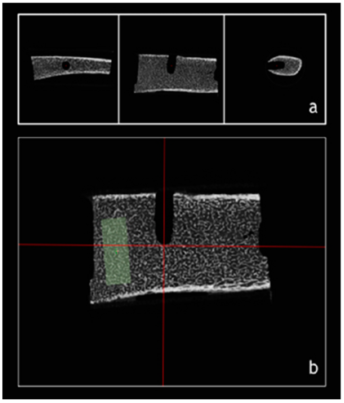
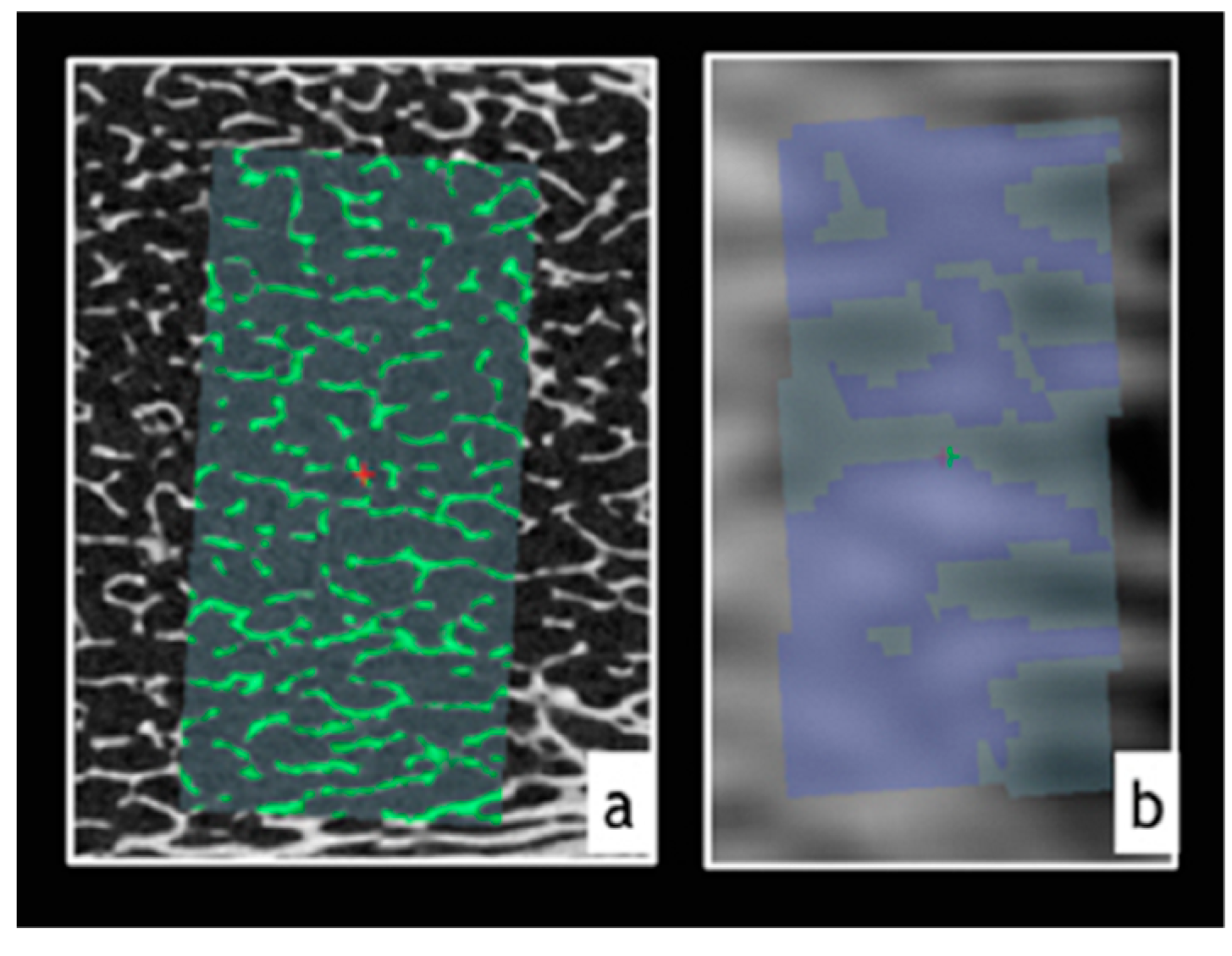
2. Materials and Methods
3. Results
4. Discussion
5. Conclusions
Author Contributions
Funding
Conflicts of Interest
References
- Lekholm, U.; Zarb, G.A. Tissue Integrated Protheses: Osseointegration in Clinical Dentistry; Quintessence: Batavia, NY, USA, 1985. [Google Scholar]
- Oh, J.S.; Kim, S.G. Clinical study of the relationship between implant stability measurements using Periotest and Osstell mentor and bone quality assessment. Oral Surg. Oral Med. Oral Pathol. Oral Radiol. 2012, 113, 35–40. [Google Scholar] [CrossRef] [PubMed]
- Ribeiro-Rotta, R.F.; de Oliveira, R.C.; Dias, D.R.; Lindh, C.; Leles, C.R. Bone tissue microarchitectural characteristics at dental implant sites part 2: Correlation with bone classification and primary stability. Clin. Oral Implant. Res. 2014, 25, 47–53. [Google Scholar] [CrossRef] [PubMed]
- Van Dessel, J.; Nicolielo, L.F.; Huang, Y.; Coudyzer, W.; Salmon, B.; Lambrichts, I.; Jacobs, R. Accuracy and reliability of different cone beam computed tomography (CBCT) devices for structural analysis of alveolar bone in comparison with multislice CT and micro-CT. Eur. J. Oral Implantol. 2017, 10, 95–105. [Google Scholar] [PubMed]
- Benic, G.I.; Mokti, M.; Chen, C.J.; Weber, H.P.; Hammerle, C.H.; Gallucci, G.O. Dimensions of buccal bone and mucosa at immediately placed implants after 7 years: A clinical and cone beam computed tomography study. Clin. Oral Implant. Res. 2012, 23, 560–566. [Google Scholar] [CrossRef]
- Behnia, H.; Motamedian, S.R.; Kiani, M.T.; Morad, G.; Khojasteh, A. Accuracy and reliability of cone beam computed tomographic measurements of the bone labial and palatal to the maxillary anterior teeth. Int. J. Oral Maxillofac. Implant. 2015, 30, 1249–1255. [Google Scholar] [CrossRef]
- Bohner, L.O.L.; Tortamano, P.; Marotti, J. Accuracy of linear measurements around dental implants by means of cone beam computed tomography with different exposure parameters. Dentomaxillofac. Radiol. 2017, 46, 20160377. [Google Scholar] [CrossRef]
- Hendee, W.R.; Edwards, F.M. ALARA and an integrated approach to radiation protection. Semin. Nucl. Med. 1986, 16, 142–150. [Google Scholar] [CrossRef]
- Duttenhoefer, F.; Mertens, M.E.; Vizkelety, J.; Gremse, F.; Stadelmann, V.A.; Sauerbier, S. Magnetic resonance imaging in zirconia-based dental implantology. Clin. Oral Implant. Res. 2015, 26, 1195–1202. [Google Scholar] [CrossRef]
- Fluegge, T.; Hovener, J.B.; Ludwig, U.; Eisenbeiss, A.K.; Spittau, B.; Hennig, J.; Schmelzeisen, R.; Nelson, K. Magnetic resonance imaging of intraoral hard and soft tissues using an intraoral coil and FLASH sequences. Eur. Radiol. 2016, 26, 4616–4623. [Google Scholar] [CrossRef]
- Hilgenfeld, T.; Prager, M.; Schwindling, F.S.; Heil, A.; Kuchenbecker, S.; Rammelsberg, P.; Bendszus, M.; Heiland, S. Artefacts of implant-supported single crowns - Impact of material composition on artefact volume on dental MRI. Eur. J. Oral Implantol. 2016, 9, 301–318. [Google Scholar]
- Wanner, L.; Ludwig, U.; Hovener, J.B.; Nelson, K.; Flugge, T. Magnetic resonance imaging-a diagnostic tool for postoperative evaluation of dental implants: A case report. Oral Surg. Oral Med. Oral Pathol. Oral Radiol. 2018, 125, e103–e107. [Google Scholar] [CrossRef] [PubMed]
- Demirturk Kocasarac, H.; Kursun-Cakmak, E.S.; Ustaoglu, G.; Bayrak, S.; Orhan, K.; Noujeim, M. Assessment of signal-to-noise ratio and contrast-to-noise ratio in 3 T magnetic resonance imaging in the presence of zirconium, titanium, and titanium-zirconium alloy implants. Oral Surg. Oral Med. Oral Pathol. Oral Radiol. 2020, 129, 80–86. [Google Scholar] [CrossRef] [PubMed]
- Boeddinghaus, R.; Whyte, A. Trends in maxillofacial imaging. Clin. Radiol. 2018, 73, 4–18. [Google Scholar] [CrossRef] [PubMed]
- Gray, C.F.; Redpath, T.W.; Smith, F.W.; Staff, R.T. Advanced imaging: Magnetic resonance imaging in implant dentistry. Clin. Oral Implant. Res. 2003, 14, 18–27. [Google Scholar] [CrossRef] [PubMed]
- Bryll, A.; Urbanik, A.; Chrzan, R.; Jurczak, A.; Kwapinska, H.; Sobiecka, B. MRI disturbances caused by dental materials. Neuroradiol. J. 2007, 20, 9–17. [Google Scholar] [CrossRef] [PubMed]
- Eley, K.A.; Watt-Smith, S.R.; Golding, S.J. “Black bone” MRI: A potential alternative to CT when imaging the head and neck: Report of eight clinical cases and review of the Oxford experience. Br. J. Radiol. 2012, 85, 1457–1464. [Google Scholar] [CrossRef] [PubMed]
- Klinke, T.; Daboul, A.; Maron, J.; Gredes, T.; Puls, R.; Jaghsi, A.; Biffar, R. Artifacts in magnetic resonance imaging and computed tomography caused by dental materials. PLoS ONE 2012, 7, e31766. [Google Scholar] [CrossRef]
- Blankenstein, F.; Truong, B.T.; Thomas, A.; Thieme, N.; Zachriat, C. Predictability of magnetic susceptibility artifacts from metallic orthodontic appliances in magnetic resonance imaging. J. Orofac. Orthop. 2015, 76, 14–29. [Google Scholar] [CrossRef]
- Blankenstein, F.H.; Asbach, P.; Beuer, F.; Glienke, J.; Mayer, S.; Zachriat, C. Magnetic permeability as a predictor of the artefact size caused by orthodontic appliances at 1.5 T magnetic resonance imaging. Clin. Oral Investig. 2017, 21, 281–289. [Google Scholar] [CrossRef]
- Pompa, V.; Galasso, S.; Cassetta, M.; Pompa, G.; De Angelis, F.; Di Carlo, S. A comparative study of Magnetic Resonance (MR) and Computed Tomography (CT) in the pre-implant evaluation. Ann. Stomatol. (Roma) 2010, 1, 33–38. [Google Scholar]
- Aguiar, M.F.; Marques, A.P.; Carvalho, A.C.; Cavalcanti, M.G. Accuracy of magnetic resonance imaging compared with computed tomography for implant planning. Clin. Oral Implant. Res. 2008, 19, 362–365. [Google Scholar] [CrossRef] [PubMed]
- Jungmann, P.M.; Agten, C.A.; Pfirrmann, C.W.; Sutter, R. Advances in MRI around metal. J. Magn. Reson. Imaging 2017, 46, 972–991. [Google Scholar] [CrossRef] [PubMed]
- Folkesson, J.; Krug, R.; Goldenstein, J.; Issever, A.S.; Fang, C.; Link, T.M.; Majumdar, S. Evaluation of correction methods for coil-induced intensity inhomogeneities and their influence on trabecular bone structure parameters from MR images. Med. Phys. 2009, 36, 1267–1274. [Google Scholar] [CrossRef] [PubMed]
- Bohner, L.; Meier, N.; Gremse, F.; Tortamano, P.; Kleinheinz, J.; Hanisch, M. Magnetic resonance imaging artifacts produced by dental implants with different geometries. Dentomaxillofac. Radiol. 2020, 49, 20200121. [Google Scholar] [CrossRef] [PubMed]
- Faul, F.; Erdfelder, E.; Lang, A.G.; Buchner, A. G*Power 3: A flexible statistical power analysis program for the social, behavioral, and biomedical sciences. Behav. Res. Methods 2007, 39, 175–191. [Google Scholar] [CrossRef] [PubMed]
- Gremse, F.; Stärk, M.; Ehling, J.; Menzel, J.R.; Lammers, T.; Kiessling, F. Imalytics Preclinical: Interactive Analysis of Biomedical Volume Data. Theranostics 2016, 6, 328–341. [Google Scholar] [CrossRef] [PubMed]
- Probst, F.A.; Schweiger, J.; Stumbaum, M.J.; Karampinos, D.; Burian, E.; Probst, M. Magnetic resonance imaging based computer-guided dental implant surgery—A clinical pilot study. Clin. Oral Implant. Res 2020, 22, 612–621. [Google Scholar] [CrossRef]
- Hilgenfeld, T.; Juerchott, A.; Jende, J.M.; Rammelsberg, P.; Heiland, S.; Bendszus, M.; Schwindling, F.S. Use of dental MRI for radiation-free guided dental implant planning: A prospective, in vivo study of accuracy and reliability. Eur. Radiol. 2020, 30, 6392–6401. [Google Scholar] [CrossRef]
- Sambataro, S.; Cervino, G.; Bocchieri, S.; La Bruna, R.; Cicciu, M. TMJ dysfunctions systemic implications and postural assessments. A review of recent literature. J. Funct. Morphol. Kinesiol. 2019, 4, 58. [Google Scholar] [CrossRef]
- Song, D.; Shujaat, S.; Zhao, R.; Huang, Y.; Shaheen, E.; Van Dessel, J.; Orhan, K.; Coropciuc, R.; Pauwels, R.; Vande Velde, G.; et al. In vivo quantification of mandibular bone remodeling and vascular changes in a Wistar rat model: A novel HR-MRI and micro-CT fusion technique. Imaging Sci. Dent. 2020, 50, 199–208. [Google Scholar] [CrossRef]


| Measurement | Abbreviation | Measurement Unit | Description |
|---|---|---|---|
| Trabecular bone volume | BV | mm3 | Trabecular bone volume at VOI, determined by gray values above threshold value |
| Total bone volume | TV | mm3 | Total bone volume (VOI) |
| Trabecular volume fraction | BvTv | % | Ratio between trabecular and total bone volume |
| Bone specific surface | BsBv | 1/cm | Bone surface to bone volume ratio |
| Trabecular thickness | TbTh | cm | Trabecular bone thickness determined by distance between bone surface above threshold value |
| Trabecular separation | TbSp | cm | Mean distance between trabeculae, determined by gray values under threshold value |
| Bone Measurements | Imaging Device | Mean ± SD |
|---|---|---|
| BV (mm3) | ||
| µCT | 0.51 ± 0.22 | |
| MRI | 0.82 ± 0.28 * | |
| BvTv (%) | ||
| µCT | 29.48 ± 7.95 | |
| MRI | 48.86 ± 11.66 * | |
| BsBv (1/cm) | ||
| µCT | 146.23 ± 42.95 | |
| MRI | 30.06 ± 8.50 * | |
| TbTh (cm) | ||
| µCT | 0.25 ± 0.07 | |
| MRI | 1.28 ± 0.89 * | |
| TbSp (cm) | ||
| µCT | 0.58 ± 0.07 | |
| MRI | 1.19 ± 0.21 * |
| Paired T-Test | Pearson Correlation | |||||
|---|---|---|---|---|---|---|
| Bone Measurements | t-Value | df | 95% CI | r | p Value | |
| Inf | Sup | |||||
| BV | −5.8 | 6 | −0.46 | −0.189 | 0.826 | 0.01 |
| BvTv | −4.49 | 6 | −29.92 | −8.81 | 0.274 | 0.27 |
| BsBv | 7.93 | 7 | 81.53 | 150.80 | 0.083 | 0.43 |
| TbTh | −3.17 | 6 | −1.82 | −0.23 | 0.522 | 0.11 |
| TbSp | −8.45 | 6 | −0.79 | −0.43 | 0.464 | 0.14 |
Publisher’s Note: MDPI stays neutral with regard to jurisdictional claims in published maps and institutional affiliations. |
© 2020 by the authors. Licensee MDPI, Basel, Switzerland. This article is an open access article distributed under the terms and conditions of the Creative Commons Attribution (CC BY) license (http://creativecommons.org/licenses/by/4.0/).
Share and Cite
Bohner, L.; Tortamano, P.; Meier, N.; Gremse, F.; Kleinheinz, J.; Hanisch, M. Trabecular Bone Assessment Using Magnetic-Resonance Imaging: A Pilot Study. Int. J. Environ. Res. Public Health 2020, 17, 9282. https://doi.org/10.3390/ijerph17249282
Bohner L, Tortamano P, Meier N, Gremse F, Kleinheinz J, Hanisch M. Trabecular Bone Assessment Using Magnetic-Resonance Imaging: A Pilot Study. International Journal of Environmental Research and Public Health. 2020; 17(24):9282. https://doi.org/10.3390/ijerph17249282
Chicago/Turabian StyleBohner, Lauren, Pedro Tortamano, Norbert Meier, Felix Gremse, Johannes Kleinheinz, and Marcel Hanisch. 2020. "Trabecular Bone Assessment Using Magnetic-Resonance Imaging: A Pilot Study" International Journal of Environmental Research and Public Health 17, no. 24: 9282. https://doi.org/10.3390/ijerph17249282
APA StyleBohner, L., Tortamano, P., Meier, N., Gremse, F., Kleinheinz, J., & Hanisch, M. (2020). Trabecular Bone Assessment Using Magnetic-Resonance Imaging: A Pilot Study. International Journal of Environmental Research and Public Health, 17(24), 9282. https://doi.org/10.3390/ijerph17249282