Article

First Report of Canine Infection by Leishmania (Viannia) guyanensis in the Brazilian Amazon

by
Francisco J. A. Santos
1,
Luciana C. S. Nascimento
1,2,
Wellington B. Silva
3,
Luciana P. Oliveira
1,
Walter S. Santos
1,
Délia C. F. Aguiar
4 and
Lourdes M. Garcez
1,2,*
1
Seção de Parasitologia, Instituto Evandro Chagas, Secretaria de Vigilância em Saúde, Ministério da Saúde, Ananindeua 67030-000, Pará, Brazil
2
Centro de Ciências Biológicas e da Saúde, Universidade do Estado do Pará, Belém 66095-662, Pará, Brazil
3
Centro Nacional de Primatas, Instituto Evandro Chagas, Ministério da Saúde, Secretaria de Vigilância em Saúde, Ananindeua 67030-000, Pará, Brazil
4
Instituto de Ciências Biológicas, Universidade Federal do Pará, Belém 66075-110, Pará, Brazil
*
Author to whom correspondence should be addressed.
Int. J. Environ. Res. Public Health 2020, 17(22), 8488; https://doi.org/10.3390/ijerph17228488
Submission received: 11 September 2020 / Revised: 20 October 2020 / Accepted: 22 October 2020 / Published: 16 November 2020
(This article belongs to the Section Infectious Disease Epidemiology)

Abstract

:
The American cutaneous (CL) and visceral leishmaniasis (VL) are zooanthroponoses transmitted by sand flies. Brazil records thousands of human leishmaniasis cases annually. Dogs are reservoirs of Leishmania infantum, which causes VL, but their role in the transmission cycle of CL is debatable. Wild mammals are considered reservoirs of the aetiological agents of CL (Leishmania spp.). Objective: To describe the aetiology of leishmaniasis in dogs in an endemic area for CL and VL in the Amazon, Brazil. Methods: Clinical evaluation and blood collection of 40 dogs from the villages Ubim (20) and Socorro (20), city of Tomé-Açu, state of Pará, were carried out. The DNA extracted from the blood was used for PCR with Leishmania-specific primers targeting the hsp70-234 gene sequence. Products were sequenced (ABI3500XL), and the sequences were aligned, edited (BioEdit), and analyzed (Blastn). Results: Of the 34 amplified samples, 21 were sequenced, namely Leishmania infantum (12), L. guyanensis (5), L. braziliensis (3), and Leishmania sp. (01). Conclusion: Given the diversity of circulating pathogens, elucidation of the role of the dog in the Leishmania spp. cycle in Amazonian villages is imperative to the surveillance of CL in the region. We present the first report in Brazil, confirmed by sequencing, of canine infection by L. guyanensis, a species highly resistant to treatment in humans, with the drug of first choice (Glucantime®).

1. Introduction

Leishmaniasis occurs in more than 90 countries. There are two main clinical forms of the disease, namely visceral leishmaniasis (VL), which compromises internal organs and produces high lethality in untreated cases, and cutaneous leishmaniasis (CL), characterized by skin lesions in most cases (>90%); they may be associated with mucosal involvement or late skin manifestations. The aetiological agents are obligate intracellular protozoa of vertebrates (Kinetoplastida: Leishmania spp.). Its incidence is predominant in the poorest regions of the planet [1,2].
The primitive hosts of Leishmania spp. are wild mammals of different orders which include Rodentia, Marsupialia, Edentata, Cingulata, Carnivora, Primates, and Chiroptera. Normally, there are no pathological effects of Leishmania spp. in wild animals [3]. The wild enzootic cycle of the parasites is maintained between these vertebrates and sand flies (Psychodidae: Phlebotominae). However, synanthropic or domestic wild animals can play an important role as a source of infection to Leishmania spp. in the peridomicile (reservoirs), which increases the risk of transmission to humans.
In the Americas, zooanthroponotic cycles of leishmaniasis transmission prevail, where a vector acquires the protozoan by feeding on the blood of animals and then transmits it to humans and domestic animals. In the Amazon region, in Brazil, foxes are incriminated as the main wild reservoirs of Leishmania infantum [4], the aetiological agent of VL. However, domestic dogs are the reservoirs with real epidemiological importance in the urban and peri-urban environment, as they are highly susceptible to infection by this species of protozoan and often have high parasitemia and parasitic load on skin tissue [5]. The presence of the vector Lutzomyia longipalpis, normally abundant in the home, favors the transmission of VL to people, which occurs mainly in this environment.
In the Amazon, transmission of CL occurs mainly in the forest, with transmission cycles involving sand flies and wild mammals. However, dogs can also become infected and develop skin lesions or an asymptomatic condition. Some of the species causing CL infecting dogs in the Americas are Leishmania (Viannia) guyanensis, Leishmania (Viannia) panamensis, Leishmania (Viannia) braziliensis, and Leishmania (Viannia) peruviana [6,7,8]. Both the species diversity of CL agents and the role of the domestic dog as a reservoir of these Leishmania species in the peridomicile remain to be elucidated [9]. In Suriname, it is suspected that the dog is an important link in the chain of transmission of L. (V.) guyanensis to humans [10].
Several molecular markers and molecular techniques are available for leishmaniasis diagnosis by detection, identification, discrimination, and quantification of Leishmania parasites. Some of the most used are PCR, RFLP-PCR, and sequencing of hsp70 and ITS1 markers [11]. The choice of the diagnostic test and the molecular target depends on the objective of the study. In endemic areas with few pathogenic Leishmania species circulating, where simple detection or identification of the predominant species is desired, several markers can be successfully used in PCR techniques. An example is a PCR combining the markers SSUr and G6PD to identify Leishmania genus, Leishmania (Viannia) subgenus, and L. (V.) braziliensis species, but lacking the power for discriminating other Leishmania species, which are not actually relevant in such an area [12].
Nevertheless, in the Amazon we strongly need to discriminate species since therapy can differ according to the species [13]. The high Leishmania intraspecific variability in this region [14] makes it difficult to discriminate species using a PCR (or RFLP-PCR) alone with hsp70-234, ITS-1, or other marker, since most of the results for clinical samples are inconclusive or expressed by similar RFLP profiles for different Leishmania species [15]. Thus, the sequencing of PCR products cannot be dispensed with to ensure the correct aetiological diagnosis of human or canine leishmaniasis [15].
In Brazil, knowledge about the epidemiological importance of dogs in the transmission chain of human CL is scarce, since the diagnosis of canine infection by Leishmania spp., recommended by the Ministry of Health in surveillance actions, does not distinguish between species.
In Tomé-Açu city, Northeast Pará State, the rural villages Socorro and Ubim are the only areas where both clinical forms of human disease are reported. In this Amazonian city, cases of CL associated with infection by Leishmania (Viannia) shawi, L. (V.) braziliensis, and L. (L.) infantum [14] have been described, but no molecular analysis was performed on dog samples living there.
Using DNA extracted from the blood of dogs, we sequenced the hsp70-234 gene region of Leishmania spp. in order to elucidate the aetiology of canine leishmaniasis in the two Amazonian villages of Socorro and Ubim.

2. Material and Methods

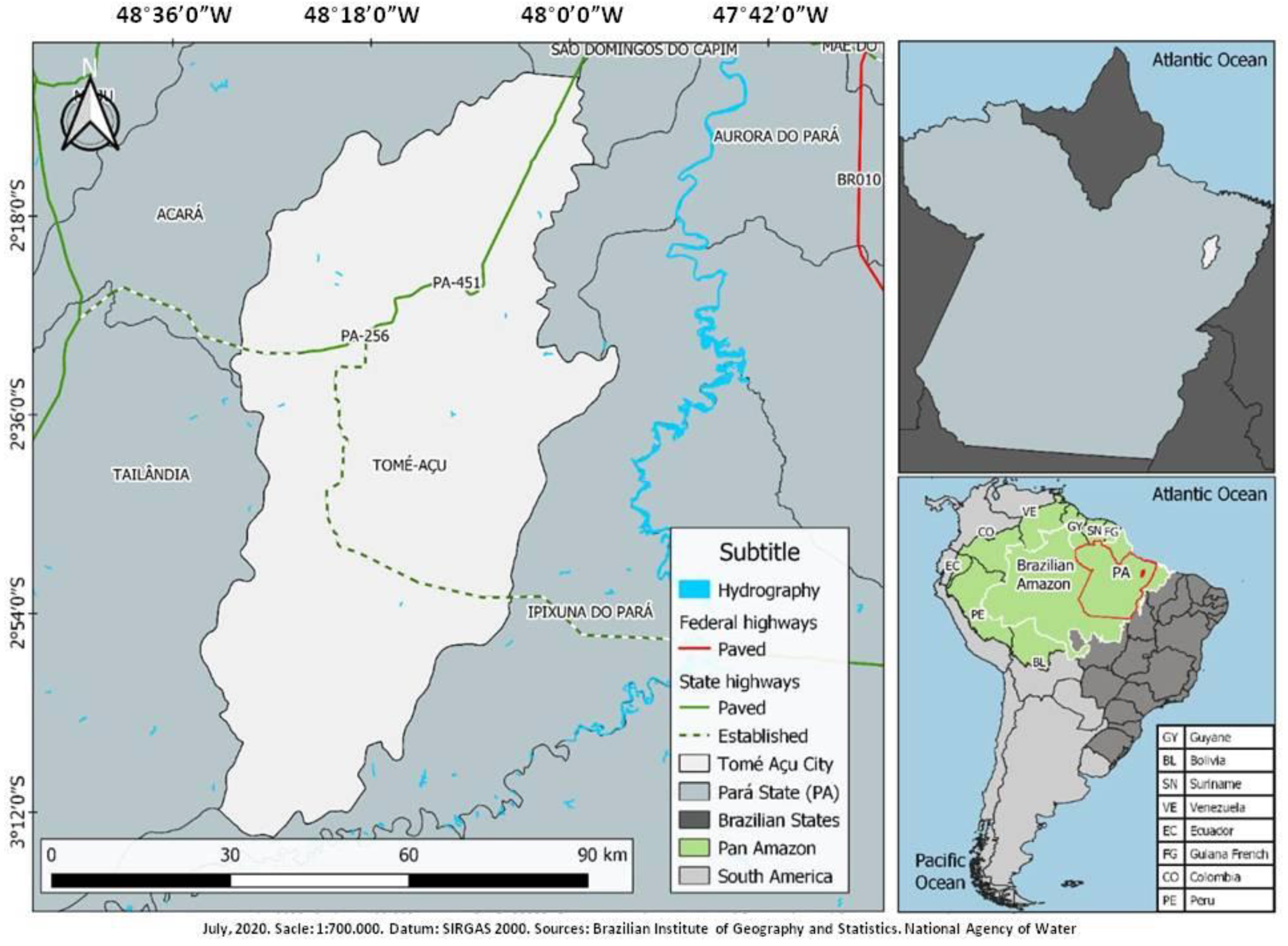
2.1. Characterization of the Tomé-Açu City and the Study Areas

Tomé-Açu is an agricultural pole in the northeast of the state of Pará. The Amazonian winter is characterized by heavy rains that occur from December to May, representing 80% of the rains, and the Amazonian summer is the season with the sparsest rainfall, which extends from June to November. The average annual temperature is 26 °C, and the average rainfall is 2300 mm/year. The population in 2019 was estimated at 63,447 inhabitants [16,17]. Figure 1 identifies the location of Tomé-Açu in the northeast of the state of Pará and its distance from the state capital, the city of Belém.
The study areas were the Amazonian villages of Socorro and Ubim, with populations of 1200 and 564 people, respectively, located in the city of Tomé-Açu, state of Pará (Figure 1). They are the only towns in the city where the two forms of the disease coexist. In the other endemic areas of the city of Tomé-Açu, only CL is present.

2.2. Dogs, Clinical Examination, and Blood Collection

A series of 40 mixed-breed dogs, resident in the rural villages Socorro (20) and Ubim (20), in May 2019 were analyzed. The animals were examined for the presence of six clinical signs of leishmaniasis, namely alopecia, skin ulcerations, dermatitis, onychogryphosis, conjunctivitis, and lymphadenopathy. Each signal was scored on a semi-quantitative scale from 0 (absent) to 3 (severe). The sum of the values corresponding to the six signs allowed classification of each dog into asymptomatic (0 to 2), oligosymptomatic (3 to 6), or polysymptomatic (7 to 18), as per previously described methods [18]. Then, a sample of peripheral blood (5 mL) from each animal was obtained and stored in a vacuum tube, containing the EDTA anticoagulant (−20 °C), until the moment of analysis. This study was ethically approved according to opinion No. 04/2019 (CEUA-IEC/SVS/MS).

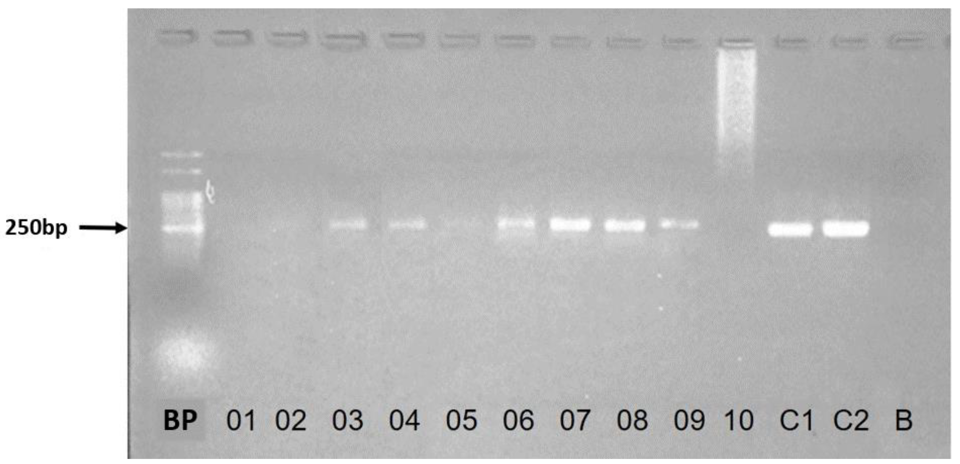
2.3. Molecular Methodology

DNA extractions from peripheral blood samples were performed with the Mini Spin 50 Extraction kit (Kasvi®, São José dos Pinhais, Brazil), according to the manufacturer’s recommendations, with a final volume of 50 µL.
The samples were analyzed using conventional PCR for the hsp70-234 target region. The mix, with a final volume equal to 50 µL, contained Taq DNA polymerase 0.03U/µL, 1.5 mM MgCl2, 0.25 mM of each dNTP, 1X buffer solution with KCl, 0.25% DMSO, 0.2 pmol of primers F (5′GGACGAGATCGAGCGCATGGT 3′) and R (5′TCCTTCGACGCCTCCTGGTTG 3′) to amplify the 234 bp [19,20] fragment, and 3.0 µL of DNA from the samples. The test conditions were initial denaturation at 94 °C/5 min, followed by 32 cycles of 94 °C/30 s for denaturation, 61 °C/1 min for annealing, and extension at 72 °C/1 min. The final extension occurred at 72 °C/10 min [19,20]. Two positive controls containing genomic DNA from L. infantum (MCER/BR/79/M6445), L. braziliensis (MHOM/BR/75/M2904), and a blank control without DNA were included. The amplified DNA samples were subjected to electrophoresis on 2% agarose gel in TAE (Tris acetate 0.004 M; EDTA 0.001 M, pH 8.0) containing GelRed ™ (Biotium®, San Francisco, CA, USA) at a concentration of 0.5 μg/mL and, subsequently, observed in a transilluminator for qualitative analysis of the results.

2.4. Sequencing

Sequence analysis of the hsp70-234 gene region was performed for 21 out of 40 samples. For sequencing, the PCR products were purified with the enzyme Illustra ExoProStar 1—Step (GE Healthcare®, Buckinghamshire, UK) and prepared for sequencing using the BigDye Terminator Kit v3.1 (Thermo Fisher Scientific®, Foster City, CA, USA), following the manufacturer’s specifications. The automatic DNA analyzer model ABI3500XL (Thermo Fisher Scientific®) was used to obtain the nucleotide sequences. Each amplicon was sequenced with forward and reverse primers, and the nucleotide sequences were edited and aligned with the aid of the BioEdit [21] program. The final sequences were compared with nucleotide sequences present in GenBank, by observing identity, coverage, e-value, and score, using the BLASTn search tool from NCBI (National Center for Biotechnology Information) to discriminate Leishmania species.

3. Results

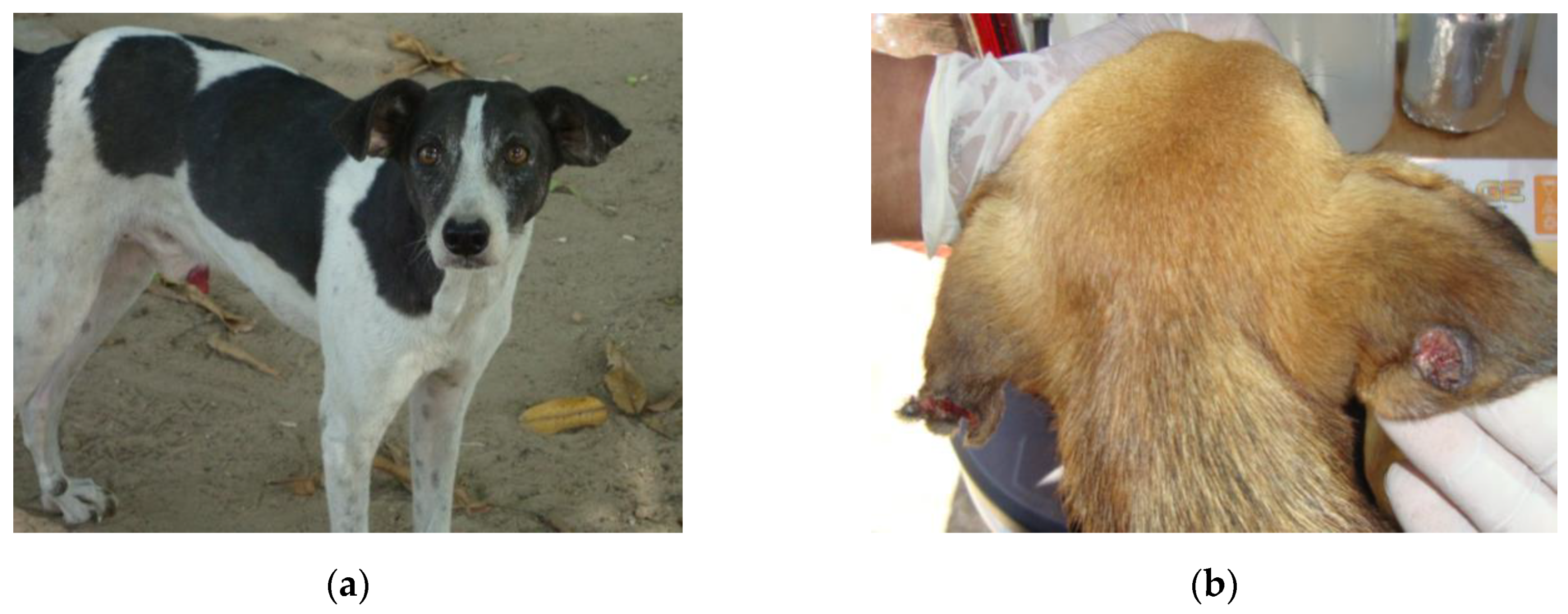
Clinical Status, Frequency, and Aetiology of Canine Infection

Most dogs did not show signs of canine leishmaniasis (36/40). Among the symptomatic (04/40), three were oligosymptomatic and one was polysymptomatic. Figure 2 depicts an asymptomatic animal (a) and another with skin lesions (b).
PCR-hsp70-234 for the detection of the Leishmania genus in DNA extracted from blood (Figure 3) revealed an identical frequency of positives (17/20; 85%) in Socorro and Ubim villages. Of the total number of PCR-hsp70-234 positives (34/40; 95%), the majority (30/34; 88%) were asymptomatic. The sequencing of the hsp70-234 gene region, performed in 21 of the 34 samples amplified by PCR-hsp70-234, allowed discrimination between three species, namely L. (L.) infantum and two other dermotropic species, L. (V.) braziliensis and L. (V.) guyanensis (Table 1). It was not possible to identify the Leishmania species in the sample of the polysymptomatic dog, but it was characterized as Leishmania (Leishmania) sp. (Table 1).

4. Discussion

Surveys carried out in zoonotic VL surveillance actions in Brazil allow the diagnosis of Leishmania infection in dogs, but without discriminating between species of the parasite. There are few surveys in support of surveillance that address this issue, despite the Ministry of Health of Brazil recommending the discrimination of Leishmania species circulating in areas where human VL and CL coexist [17]. In this study, we used molecular methods to elucidate the aetiology of canine Leishmania infection in Amazonian villages, endemic to CL and VL. The two villages, Socorro and Ubim, are located in the city of Tomé-Açu, northeast of the state of Pará, in the Brazilian Amazon.
Of the 40 animals included in the study, 36 had no symptoms, but 30 of those asymptomatic (83%) were positive by hsp70-234 PCR. Subclinical canine leishmaniasis is a common finding in areas with VL transmission in the Amazon [22,23]. Symptomatic dogs are more likely to infect sand fly vectors than asymptomatic ones, but the latter contribute to the maintenance of the Leishmania transmission cycle and can also develop high parasitic loads [23]. Therefore, once infected, symptomatic or not, they will be able to transmit the parasite to the sand fly vectors that feed on their blood [24]. These infected vectors may transmit the parasite to humans during a new blood meal.
The period between infection of the dog by a certain species of Leishmania and the first clinical manifestations is quite variable. Although it usually lasts for weeks or months, some animals can take several years to become symptomatic [25].
Analysis of DNA extracted from whole-blood samples from 21 of the 34 positive dogs in the two study areas (30 asymptomatic and 4 symptomatic) demonstrated that canine leishmaniasis was associated with at least three species of Leishmania, namely L. (L.) infantum, L. (V.) braziliensis, and L. (V.) guyanensis. For the only polysymptomatic dog, the analysis of the hsp70-234 gene region did not allow discrimination between the species of the parasite, but the subgenus Leishmania (Leishmania) sp. It could be a variant of L. (L.) infantum, which need to be studied, since there are reports of genetic diversity related to the geographical origin of L. infantum isolates in Brazil [26].
In the small series of 21 samples sequenced among the 34 positive, eight animals (38%) were infected with species of the subgenus Leishmania (Viannia). Although the record of L. (V.) guyanensis in dogs is known in other South American countries, no studies have been found to confirm canine infection by sequencing molecular markers for this species in Brazil. This finding reinforces the importance of using methodologies capable of discriminating between protozoan species for surveillance of LT in areas with great biodiversity such as the Amazon.
In Brazil, the infection of dogs in areas with leishmaniasis transmission is usually attributed to L. infantum. The dog, a domestic reservoir of this species, is the target of surveillance and preventive actions for human VL. However, canine infection by Leishmania species causing human CL has been increasingly reported in rural villages. Studies indicate that the presence of a dog in the home is a risk factor for the transmission of these pathogens to people [27,28]. In Suriname, the dog may be the domestic reservoir of species of the subgenus Leishmania (Viannia) [10]. In Colombia, the report of infection in one dog by L. (V.) guyanensis and infection in two others by L. (V.) braziliensis, among 21 positive and sequenced samples (7SL RNA gene), increased the suspicion about its role as reservoir of this pathogen [6]. In both countries, the natural reservoirs of L. (V.) guyanensis are unknown, but in the state of Pará, Brazil, the main reservoirs of L. (V.) guyanensis are Edentates (sloths and anteaters) and several small wild rodents [29].
For humans, infection by L. (V.) guyanensis, very prevalent in Northern Amazonia, represents a high risk of complications, given the parasite’s resistance to the drug of choice for treatment in Brazil (i.e., meglumine antimoniate (Glucantime®)) [13]. In Brazilian cities or states where L. (V.) guyanensis predominates, the drug for systemic treatment of the disease should be pentamidine isothyanate, as already occurs in the Amapá state [20].
L. (V.) braziliensis is considered as the main cause of canine cutaneous leishmaniasis and also human cutaneous leishmaniasis in Brazil, given its wide distribution and variety of vectors. In humans, it can cause complications, with extensive or disseminated lesions, which are difficult to resolve, including those in mucous membranes [13].
Leishmaniasis by L. (V.) braziliensis in the dog compromises mainly the skin and, less frequently, can progress to systemic conditions [30]. Some studies on the aetiology of canine infection have been conducted in extra-Amazonian states. In Bahia, a survey in the city of Ilhéus covering 560 dogs confirmed L. (V.) braziliensis in 54.72% of the animals. Most have little or no symptoms [30]. In Pernambuco, 48.7% of the dogs were infected with L. (V.) braziliensis [31]. Other studies with similar results suggest that the clinical course of infection caused by L. (V.) braziliensis is milder compared to that observed in canine infection by L. (L.) infantum [32,33].
Some circumstances may also favor the occurrence of outbreaks of canine leishmaniasis. In Colombia, an outbreak was detected by L. (V.) braziliensis and L. (V.) guyanensis in army animals that entered the forest during training [7].
In the Amazon, potential vectors of pathogens of the subgenus Leishmania (Viannia) are close to human dwellings, colonizing microenvironments in varying abundance around the home, as we have already observed in the study locations [34]. When dogs living in rural villages accompany their owners to the forest to hunt or to collect food, they are more easily exposed to bites from insects infected with Leishmania, which increases the chance of acquiring the infection. Over time, dogs infected with dermotropic species of the parasite may become infective to sand fly vectors. This scenario would increase the risk of transmission of the parasite to humans in the home.

5. Conclusions

Given the diversity of pathogens infecting dogs in the study areas, elucidation of the role of the animal in the transmission cycle of Leishmania species in Amazonian villages is imperative to the surveillance of CL in the region. Although L. (L.) infantum is the main agent of canine leishmaniasis in the Americas, L. (V.) guyanensis and L. (V.) braziliensis have been associated with 38% of the confirmed canine leishmaniasis cases in Socorro and Ubim villages, where CL and VL coexist. The absence of an association between clinical manifestations and laboratory diagnosis reinforces the indication of the use of molecular tests with discriminatory power for zoonotic leishmaniasis surveillance actions in these areas. This is the first report in Brazil of canine infection by L. (V.) guyanensis, a species of Amazonian distribution highly resistant to treatment in humans with the drug of first choice (Glucantime®).
Additionally, this is the first report of L. (V.) guyanensis infecting dogs in Brazil, confirmed by sequencing.

Author Contributions

Conceptualization, L.M.G.; Formal analysis, F.J.A.S., L.C.S.N., L.P.O., W.S.S. and L.M.G.; Methodology, F.J.A.S., L.C.S.N., W.B.S., L.P.O., D.C.F.A. and L.M.G.; Project administration, L.C.S.N. and L.M.G.; Supervision, L.M.G.; Writing—original draft, F.J.A.S. and L.M.G.; Writing—review & editing, F.J.A.S., L.C.S.N., W.B.S., L.P.O., Walter Souza Santos, D.C.F.A. and L.M.G. All authors have read and agreed to the published version of the manuscript.

Funding

This research was financed in part by the Coordenação de Aperfeiçoamento de Pessoal de Nível Superior—Brasil (CAPES), through the Programa de Pós-graduação em Epidemiologia e Vigilância em Saúde (PPGEVS/IEC/SVS/MS), Conselho Nacional de Desenvolvimento Científico e Tecnológico (CNPq), which awarded a scholarship to the first author and the Instituto Evandro Chagas/SVS/MS for paying the translation and publication fees.

Acknowledgments

To the Secretaria Municipal de Saúde de Tomé-Açu (State of Pará, Brazil) for logistical support during field activities.

Conflicts of Interest

The authors declare no conflict of interest.

References

  1. Alvar, J.; Vélez, I.D.; Bern, C.; Herrero, M.; Desjeux, P.; Cano, J.; Jannin, J.; Boer, M.D. Who Leishmaniasis Control the WHO Leishmaniasis Control Team Leishmaniasis Worldwide and Global Estimates of Its Incidence. PLoS ONE 2012, 7, e35671. [Google Scholar] [CrossRef] [PubMed]
  2. World Health Organization. Investing to Overcome the Global Impact of Neglected Tropical Diseases: Third WHO Report on Neglected Tropical Diseases 2015; World Health Organization: Geneva, Switzerland, 2015; pp. 118–126. [Google Scholar]
  3. Roque, A.L.R.; Jansen, A.M. Wild and synanthropic reservoirs of Leishmania species in the Americas. Int. J. Parasitol. Parasites Wildl. 2014, 3, 251–262. [Google Scholar] [CrossRef] [PubMed] [Green Version]
  4. Lainson, R.; Shaw, J.; Silveira, F.; Braga, R. American visceral leishmaniasis: On the origin of Leishmania (Leishmania) chagasi. Trans. R. Soc. Trop. Med. Hyg. 1987, 81, 517. [Google Scholar] [CrossRef]
  5. Ciaramella, P.; Corona, M. Canine leishmaniasis: Clinical and diagnostic aspects. VetLearn 2003, 25, 358–368. [Google Scholar]
  6. Santaella, J.; Ocampo, C.; Saravia, N.G.; Méndez, F.; Góngora, R.; Gomez, M.A.; Munstermann, L.E.; Quinnell, R.J. Leishmania (Viannia) Infection in the Domestic Dog in Chaparral, Colombia. Am. J. Trop. Med. Hyg. 2011, 84, 674–680. [Google Scholar] [CrossRef] [PubMed] [Green Version]
  7. Velez, I.D.; Carrillo, L.M.; López, L.; Rodríguez, E.; Robledo, S.M. An epidemic outbreak of canine cutaneous leishmaniasis in Colombia caused by Leishmania braziliensis and Leishmania panamanensis. Am. J. Trop. Med. Hyg. 2012, 86, 807–811. [Google Scholar] [CrossRef] [PubMed]
  8. Desjeux, P.; World Health Organization. Information on the Epidemiology and Control of the Leishmaniases by Country and Territory/by P. Desjeux; World Health Organization: Geneva, Switzerland, 1991; pp. 35–47. [Google Scholar]
  9. Dantas-Torres, F. The role of dogs as reservoirs of Leishmania parasites, with emphasis on Leishmania (Leishmania) infantum and Leishmania (Viannia) braziliensis. Veter. Parasitol. 2007, 149, 139–146. [Google Scholar] [CrossRef]
  10. Kent, A.; Ramkalup, P.; Mans, D.; Schallig, H. Is the Dog a Possible Reservoir for Cutaneous Leishmaniasis in Suriname? J. Trop. Med. 2013, 2013, 1–5. [Google Scholar] [CrossRef] [Green Version]
  11. Akhoundi, M.; Downing, T.; Votýpka, J.; Kuhls, K.; Lukeš, J.; Cannet, A.; Ravel, C.; Marty, P.; Delaunay, P.; Kasbari, M.; et al. Leishmania infections: Molecular targets and diagnosis. Mol. Asp. Med. 2017, 57, 1–29. [Google Scholar] [CrossRef]
  12. Garcez, L.M.; Soares, D.C.; Chagas, A.P.; Souza, G.C.R.; Miranda, J.F.C.; Fraiha, H.; Flöeter-Winter, L.M.; Nunes, H.M.; Zampieri, R.A.; Shaw, J.J. Etiology of cutaneous leishmaniasis and anthropophilic vectors in Juruti, Pará State, Brazil. Cad. Saúde Pública 2009, 25, 2291–2295. [Google Scholar] [CrossRef] [Green Version]
  13. BRASIL. Manual de Vigilância da Leishmaniose Tegumentar, 1st ed.; Ministério da Saúde: Brasilia, Brasil, 2017; p. 191.
  14. Garcez, L.M.; Hueb, M.; Cordies, N.; Sanchez, L.; Nascimento, L.; Santos, R. Cutaneous Leishmaniasis in Brazil: Undeniable Diversity of Species is Still Poorly Known. Available online: http://worldleish2017.org/documentos/Abstracts_Book_WL6_2017.pdf (accessed on 8 October 2019).
  15. Soares, D.C. Etiologia da Leishmaniose Tegumentar na Mesorregião do Baixo Amazonas do Estado Do Pará, Brasil. Ph.D. Thesis, Universidade Federal do Pará, Belém, Brazil, March 2014. [Google Scholar]
  16. Brazilian Institute of Geography and Statistics. Características Sobre o Município de Tomé-Açu. Disponível em. Available online: https://cidades.ibge.gov.br/brasil/pa/tome-acu (accessed on 15 June 2000).
  17. Pachêco, N.A.; Bastos, T.X.; Creão, L. Boletim Agrometeorológico de 2008 Para Tomé-Açu, PA. Embrapa Amazônia Oriental-Documentos (INFOTECA-E). 2009. Available online: https://www.infoteca.cnptia.embrapa.br/bitstream/doc/697089/1/Doc361.pdf (accessed on 15 June 2020).
  18. Quinnell, R.J.; Courtenay, O.; Garcez, L.M.; Kaye, P.M.; Shaw, M.A.; Dye, C.; Day, M.J. IgG subclass responses in a longitudinal study of canine visceral leishmaniasis. Veter. Immunol. Immunopathol. 2003, 91, 161–168. [Google Scholar] [CrossRef]
  19. Da Graça, G.C.; Volpini, A.C.; Romero, G.A.S.; Neto, M.P.D.O.; Hueb, M.; Porrozzi, R.; Boité, M.C.; Cupolillo, E. Development and validation of PCR-based assays for diagnosis of American cutaneous leishmaniasis and identificatio nof the parasite species. Memórias Instituto Oswaldo Cruz 2012, 107, 664–674. [Google Scholar] [CrossRef] [PubMed]
  20. De Almeida, A.N.F.; Nascimento, L.D.C.S.D.; Sousa, E.S.M.D.M.; De Oliveira, A.J.D.; De Sena, M.G.; De Resende, B.M.; Chaves, R.C.G.; Garcez, L.M. Vigilância da leishmaniose cutânea em amostras clínicas: Distribuição da Leishmania guyanensis no estado do Amapá, 2018. Epidemiol. Serv. Saude. 2020, 29, e2018504. [Google Scholar] [CrossRef] [PubMed] [Green Version]
  21. Hall, B.G. Phylogenetics Trees Made Easy. A How-To Manual, 3rd ed.; Sinauer Associates: Sunderland, MA, USA, 2007; p. 203. [Google Scholar]
  22. Quinnell, R.J.; Carson, C.; Reithinger, R.; Garcez, L.M.; Courtenay, O. Evaluation of rK39 Rapid Diagnostic Tests for Canine Visceral Leishmaniasis: Longitudinal Study and Meta-Analysis. PLoS Negl. Trop. Dis. 2013, 7, e1992. [Google Scholar] [CrossRef] [Green Version]
  23. Courtenay, O.; Carson, C.; Calvo-Bado, L.; Garcez, L.M.; Quinnell, R.J. Heterogeneities in Leishmania infantum Infection: Using Skin Parasite Burdens to Identify Highly Infectious Dogs. PLoS Negl. Trop. Dis. 2014, 8, e2583. [Google Scholar] [CrossRef]
  24. Quaresma, P.F.; Rêgo, F.D.; Botelho, H.A.; Da Silva, S.R.; Moura, A.J.; Neto, R.G.T.; Madeira, F.M.; Carvalho, M.B.; Paglia, A.P.; Melo, M.N.; et al. Wild, synanthropic and domestic hosts of Leishmania in an endemic area of cutaneous leishmaniasis in Minas Gerais State, Brazil. Trans. R. Soc. Trop. Med. Hyg. 2011, 105, 579–585. [Google Scholar] [CrossRef] [PubMed]
  25. Reiner, S.L.; Locksley, R.M. The regulation of immunity to Leishmania major. Annu. Rev. Immunol. 1995, 13, 151–177. [Google Scholar] [CrossRef]
  26. Segatto, M.; Ribeiro, L.S.; Costa, D.L.; Costa, C.H.N.; De Oliveira, M.R.; Carvalho, S.F.G.; Macedo, A.M.; Valadares, H.M.S.; Dietze, R.; De Brito, C.F.A.; et al. Genetic diversity of Leishmania infantum field populations from Brazil. Memórias Instituto Oswaldo Cruz 2012, 107, 39–47. [Google Scholar] [CrossRef] [Green Version]
  27. Loiseau, R.; Nabet, C.; Simon, S.; Ginouves, M.; Brousse, P.; Blanchet, D.; Demar, M.; Couppie, P.; Blaizot, R. American cutaneous leishmaniasis in French Guiana: An epidemiological update and study of environmental risk factors. Int. J. Dermatol. 2019, 58, 1323–1328. [Google Scholar] [CrossRef]
  28. Membrive, N.A.; Rodrigues, G.; Gualda, K.P.; Bernal, M.V.Z.; Oliveira, D.M.; Lonardoni, M.V.C.; Teodoro, U.; Teixeira, J.J.V.; Silveira, T.G.V. Environmental and Animal Characteristics as Factors Associated with American Cutaneous Leishmaniasis in Rural Locations with Presence of Dogs, Brazil. PLoS ONE 2012, 7, e47050. [Google Scholar] [CrossRef] [Green Version]
  29. Lainson, R.; Shaw, J.; Póvoa, M. The importance of edentates (sloths and anteaters) as primary reservoirs of Leishmania braziliensis guyanensis, causative agent of “pianbois” in north Brazil. Trans. R. Soc. Trop. Med. Hyg. 1981, 75, 611–612. [Google Scholar] [CrossRef]
  30. Carvalho, F.; Wenceslau, A.; Albuquerque, G.; Munhoz, A.D.; Gross, E.; Carneiro, P.; Oliveira, H.; Rocha, J.; Santos, I.; Rezende, R. Leishmania (Viannia) braziliensis in dogs in Brazil: Epidemiology, co-infection, and clinical aspects. Genet. Mol. Res. 2015, 14, 12062–12073. [Google Scholar] [CrossRef] [PubMed]
  31. Dantas-Torres, F.; De Paiva-Cavalcanti, M.; Figueredo, L.A.; Melo, M.F.; Da Silva, F.J.; Da Silva, A.L.; Almeida, E.L. Brandão-Filho, S.P. Cutaneous and visceral leishmaniosis in dogs from a rural community in northeastern Brazil. Veter. Parasitol. 2010, 170, 313–317. [Google Scholar] [CrossRef] [PubMed]
  32. Figueredo, L.A.; De Paiva-Cavalcanti, M.; Almeida, E.L.; Brandão-Filho, S.P.; Dantas-Torres, F. Clinical and hematological findings in Leishmania braziliensis-infected dogs from Pernambuco, Brazil. Rev. Bras. Parasitol. Veterinária 2012, 21, 418–420. [Google Scholar] [CrossRef] [Green Version]
  33. Solano-Gallego, L.; Koutinas, A.; Miró, G.; Cardoso, L.; Pennisi, M.; Ferrer, L.; Bourdeau, P.; Oliva, G.; Baneth, G. Directions for the diagnosis, clinical staging, treatment and prevention of canine leishmaniosis. Veter. Parasitol. 2009, 165, 1–18. [Google Scholar] [CrossRef]
  34. Santos, W.S.; Ortega, F.D.; Alves, V.R.; Garcez, L.M. Flebotomíneos (Psychodidae: Phlebotominae) de área endêmica para leishmaniose cutânea e visceral no nordeste do estado do Pará, Brasil. Rev. Pan-Amaz. Saúde 2019, 10, 1–8. [Google Scholar] [CrossRef] [Green Version]
Figure 1. Map of the spatial location of the Amazonian city of Tomé-Açu, state of Pará, Brazil.
Ijerph 17 08488 g001
Figure 2. Apparently healthy dog (a) and dog with skin lesions (b) suggestive of leishmaniasis. Animals from Vila Socorro and Ubim, places where human visceral and cutaneous leishmaniasis coexist, in the Amazon city of Tomé Açu, state of Pará, Brazil (May 2019).
Ijerph 17 08488 g002
Figure 3. Profile of hsp70-234 PCR bands visualized on 2% agarose gel. Samples 03, 04, 05, 06, 07, 08, and 09 showed a band pattern close to 250 bp. The standard size of amplicon for this gene region is 234 bp. BP: band pattern 50 bp; C1: positive control, standard strain of Leishmania (Leishmania) infantum (MCER/BR/79/M6445); C2: positive control, standard strain of Leishmania (Viannia) braziliensis (MHOM/BR/75/M2904); B: blank; 01, 02, 03, 04, 05, 06, 07, 08, 09, and 10: blood samples from dogs.
Ijerph 17 08488 g003
Table 1. Canine leishmaniasis: aetiological agents and respective clinical status of dogs in endemic areas for both cutaneous and visceral leishmaniasis. Dogs living in two small Amazonian villages, North Brazil (May 2019).
VillagesAgentsDogsTotal
AsymptomaticsPoly/Oligosymptomatics
SocorroL. (L.) infantum516
L. (V.) guyanensis202
L. (V.) braziliensis000
Leishmania (L.) sp.01 1
Subtotal729
UbimL. (L.) infantum516
L. (V.) guyanensis213
L. (V.) braziliensis303
Subtotal10212
Total17421
Single polysymptomatic dog.
Publisher’s Note: MDPI stays neutral with regard to jurisdictional claims in published maps and institutional affiliations.

