Article

Effects of Different Components of PM2.5 on the Expression Levels of NF-κB Family Gene mRNA and Inflammatory Molecules in Human Macrophage

1
Department of Occupational and Environmental Health, School of Public Health, Jilin University, Changchun 130000, China
2
Clinical Teaching and Research Laboratory, Medical School, Xilingol Vocational College, Inner Mongolia 026000, China
*
Author to whom correspondence should be addressed.
Int. J. Environ. Res. Public Health 2019, 16(8), 1408; https://doi.org/10.3390/ijerph16081408
Submission received: 5 April 2019 / Revised: 14 April 2019 / Accepted: 15 April 2019 / Published: 19 April 2019

Abstract

:
Background: Studies have found that exposure to fine particulate matter with sizes below 2.5 µm (PM2.5) might cause inflammation response via the NF-κB pathway. To date, only a few studies have focused on the toxicity of different components of PM2.5. We aimed to explore the effects of PM2.5 with different components on the expression levels of NF-κB family gene mRNA and inflammatory molecules in human macrophages. Methods: Human monocytic cell line THP-1-derived macrophages were exposed to water-soluble (W-PM2.5), fat-soluble (F-PM2.5), and insoluble (I-PM2.5) PM2.5. The cell survival rate was measured by 3-(4,5-dimethylthiazol-2-yl)-2,5-diphenyltetrazolium bromide (MTT) assay. The levels of inflammatory molecules were determined by enzyme-linked immunosorbent assay (ELISA), and the relative mRNA levels of the NF-κB family gene were determined by real time PCR. Results: PM2.5 could decrease the cell viability. After exposure to W-PM2.5, the levels of interleukins (IL)-1β and IL-12 p70 significantly increased. After exposure to F-PM2.5, the levels of IL-12 p70 significantly increased. The levels of IL-12 p70 and TNF-α after exposure to I-PM2.5 were significantly higher than that in W- and F-PM2.5 treatment groups. The levels of IL-8, C reactive protein (CRP), and cyclooxygenase (COX)-2 increased only after exposure to I-PM2.5. F-PM2.5 increased the mRNA levels of NF-κB genes, especially NF-κB1 and RelA. Conclusions: PM2.5 can decrease the cell survival rate and up-regulate the expression of NF-κB family gene mRNA and inflammatory molecules. The main toxic components of PM2.5 related to inflammatory response in macrophages were the I-PM2.5.

1. Introduction

Epidemiological and experimental studies have proven that exposure to atmospheric particulate matter (PM), especially exposure to fine particulate matter with sizes below 2.5 µm (PM2.5), is closely associated with population mortality [1,2,3]. PM is considered to have contributed to the global burden of respiratory system diseases and cardiovascular diseases [4]. This is because PM2.5 can be inhaled deeply into the lung alveoli, and some of them can even enter the bloodstream [5].
PM2.5 can induce many pro-inflammatory responses in vivo, including increasing the production of reactive oxygen species (ROS), altering the transcription of inflammation-related genes and the polarization of macrophages, and overproducing pro-inflammatory molecules [6,7,8]. PM2.5 can cause inflammatory responses and oxidative stress in human bronchial epithelial cells (16HBE) and human lung epithelial type 2 cells (A549) in vitro and in rat lungs in vivo [9,10,11]. Zhao et al. found that PM2.5 could induce increasing levels of interleukin (IL)-6, IL-1β and tumor necrosis factor-α (TNF-α) in macrophages [12]. PM2.5 also increases the levels of IL-1β, IL-6, IL-8, and cyclooxygenase (COX)-2 in a dose-dependent manner [13]. Exposure to PM2.5 can induce the release of proinflammatory molecules, such as IL-8 in bronchial epithelial cells [14]. In addition, the mRNA expression levels of IL-6, IL-1β, and IL-5 in liver of mice were also up-regulated after being exposed to PM2.5 [15]. The PM2.5-induced pro-inflammatory responses were considered the main reason for respiratory damage [16]. Exposure to PM2.5 can activate the nuclear factor-κB (NF-κB) pathway [17,18,19], and up-regulate the expression levels of NF-κB1 and RelA and inflammatory molecules such as IL-1β amd IL-6 in human bronchial epithelial cells [13,20].
NF-κB is known to be a critical signal pathway regulating immunity and inflammation [21]. The NF-κB family includes NF-κB1, NF-κB2, RelA, RelB, and c-Rel. On one hand, NF-κB can regulate gene transcriptions of pro-inflammatory molecules and chemokines, such as IL-1β, IL-6, and TNF-α, and inflammatory enzymes, such as COX-2 and inducible nitric oxide synthase (iNOS) [22]. On the other hand, inflammatory action can promote the activation of the NF-κB signal pathway. Certain NF-κB-dependent pro-inflammatory molecules, such as TNF-α, IL-1β and IL-6, can promote acute or chronic inflammation via the NF-κB signaling pathway [23]. When PM2.5 reaches the alveoli through the trachea, the alveolar macrophages can phagocytose the particles and secrete inflammatory molecules. The NF-κB signaling pathway is activated and participates in the inflammatory response.
The sources of PM2.5 are very wide-ranging, and the components are complex (sulfate, nitrate, organic chemicals, metals, crystal elements, etc.) [24]. Current studies mainly focus on the toxic effects of total PM2.5, and only a few studies have explored the effects of different components of PM2.5 [25,26]. The mechanism of inflammation caused by different components of PM2.5 remains to be studied.
Therefore, the present study explored the effects of different components of PM2.5 on the levels of NF-κB family gene mRNA expression and inflammatory molecules in human monocytic cell (THP-1)-derived macrophages. This study contributes to fully understanding the toxicity of PM2.5, and provides a basis for the prevention and treatment of respiratory diseases caused by different types of PM2.5.

2. Materials and Methods

2.1. PM2.5 Sampling and Constituent Preparation

2.1.1. PM2.5 Sampling

Ambient PM2.5 was collected from one of Changchun City’s PM2.5 monitoring points between March and June 2015, using an air sampler with a flow rate of 100 L/min. The microfiber filters were replaced every 24 h. The standardized pressure, humidity, and sample volume were recorded. After sampling, the microfiber filters were dried for 24 h at a stable temperature and weighed. The filters were then marked and stored in sealed bags at −20 °C.

2.1.2. PM2.5 Component Extraction and Solution Preparation

The microfiber filters containing PM2.5 were irradiated by ultraviolet radiation for 30 min and cut into pieces with a diameter of 1 cm. The pieces were immersed in deionized water, vibrated by ultrasonic concussion for 30 min, and filtered to form the filtrates which were lyophilized to powder. The PM2.5 collected from all microfiber filters in the three months was mixed for subsequent sample preparation. The blank microfiber filters were processed in the same way. The blank microfiber filters were diluted with sterile phosphate-buffered saline (PBS, GIBCO, Grand Island, NY, USA) as control. A certain amount of PM2.5 powder was diluted by sterile PBS to form a suspension with a concentration of 1000 μg/mL. The PM2.5 suspension was centrifuged at 13,000 rpm for 30 min, and the supernatant was collected as the water-soluble PM2.5 (W-PM2.5). After centrifugation, the precipitates were freeze-dried and weighed. One hundred percent (100%) dimethylsulfoxide (DMSO) was used to prepare the fat-soluble PM2.5 (F-PM2.5), with a concentration of 20 mg/mL diluted by the culture medium. Ultrasound was used for 60 min to fully elute organic components attached to PM2.5 particles. The precipitate was vacuumized and dried again, dissolved in sterilized PBS solution, and vibrated by ultrasound for 30 min to prepare the insoluble PM2.5 (I-PM2.5) with a concentration of 1000 μg/mL. Different components of PM2.5 were stored at 4 °C. The suspension was vibrated again before use and diluted to the desired concentration by RPMI 1640 supplemented with fetal bovine serum (FBS).

2.2. Cell Culture and Exposure to PM2.5

The human monocytic cell line THP-1 was purchased from the Shanghai Chinese Academy of Science cell bank. The THP-1 cells were subcultured using RPMI 1640 complete medium (GIBCO, Grand Island, NY, USA) containing 10% FBS (BI, Kibbutz Beit Haemek, Israel) at 37 °C and 5% CO2. The growth of cells was viewed under an inverted microscope. When the cells were in good condition and the concentration of the cells was adjusted to 1.0 × 10⁶/mL, phorbol myristate acetate (PMA, Sigma, St. Louis., MO, USA) was added to the cells to induce the cells for 72 h, and the final concentration was 100 ng/mL. The differentiation of cells was observed under a microscope. When the differentiation rate of cells was over 95%, the following experiment could be carried out. The differentiated cells were washed by sterilized PBS three times to remove PMA, and were randomly divided into control, water-soluble, fat-soluble, and insoluble. The cells of each condition were continuously treated with the corresponding PM2.5 components and concentrations (Table 1). In order to control the effect of DMSO on cell growth, the final concentration of DMSO was about 0.35% (<1%). The cell supernatant and cells were collected and marked at 12, 24, and 48 h after treatment, and were stored at −80 °C for the detection of inflammatory molecules and the extraction of mRNA.

2.3. MTT for the Cell Survival Rate

The cell survival rate was measured by 3-(4,5-dimethylthiazol-2-yl)-2,5-diphenyltetrazolium bromide (MTT) assay to evaluate the effects of W-PM2.5, F-PM2.5, and I-PM2.5. The cells were seeded in 96 well plates and then treated with different doses of various components of PM2.5 (Table 1) for 12, 24, and 48 h. After exposure, 20 μL of MTT (1 mg/mL in PBS) was added to each well and incubated continuously for 2 h at 37 °C. The cells were then treated with 100 μL of DMSO. The absorbance was measured at 570 nm using a microplate reader (Thermo MK3, Winosky, VT, USA). Data were expressed as a percentage of the value obtained for the solvent control (0.1% DMSO), which was set to 100%.

2.4. ELISA for Inflammatory Molecules and Cytokines in the Supernatant

After exposure to PM2.5 for 12 and 24 h, the supernatant was collected to detect the measurement of IL-1β, IL-6, IL-8, IL-12 p70, TNF-α, CRP, IL-2, and COX-2. The measurement of inflammatory molecules and cytokines in the supernatant was performed using ELISA kits (Shanghai Hengyuan Biological Technology Co., Ltd., Shanghai, China) according to the manufacturer’s instructions.

2.5. Real-Time Quantitative PCR for the Expression Levels of NF-κB Family Genes

Macrophages were treated with PM2.5 at different doses and types of PM2.5 for 24 h. After cell lysis, total RNA isolation was performed by Trizol (Ambion, Shanghai, China) extraction. The mRNA, used as the template, was reverse-transcribed into cDNA using primers (TaKaRa, Dalian, China) and reverse transcriptase to perform PCR amplification. Real-time PCR was conducted in the Step One Plus TM apparatus. The PCR parameters were set as Segment 1: 95 °C, 30 s, 1 cycle; Segment 2, 95 °C, 5 s, 60 °C, 30 s, 40 cycles; Segment 3, 95 °C, 15 s, 60 °C, 1 min, 95 °C, 15 s, 1 cycle. The relative mRNA levels were relatively quantified via 2−ΔΔCT. Table 2 is the sequence of primers that was used to amplify candidate genes and internal reference genes.

2.6. Statistical Analyses

All statistical analyses were performed with IBM SPSS software version 24.0 (IBM, Arnmonk, NY, USA). Data were expressed as mean ± SD for normal continuous variables. Two-way analysis of variance (ANOVA) was used to analyze the differences in mRNA expression levels and inflammatory cytokine levels among different PM2.5 components and concentration groups, and least significant difference or Tamhane test was used to compare the difference between two groups. The relationship between NF-κB mRNA levels and inflammatory-molecule levels was analyzed by linear correlation analysis.

3. Results

3.1. Differentiation of Monocytes

The morphology of the cells was observed under microscope. The undifferentiated THP-1 cells were spherical and suspended (Figure 1A). After treatment with 100 ng/mL PMA for 72 h, mature macrophages differentiated from THP-1 cells adhered to the bottom of the flask and presented an irregular shape with pseudopod (Figure 1B).

3.2. Effects of PM2.5 on Cell Survival Rate

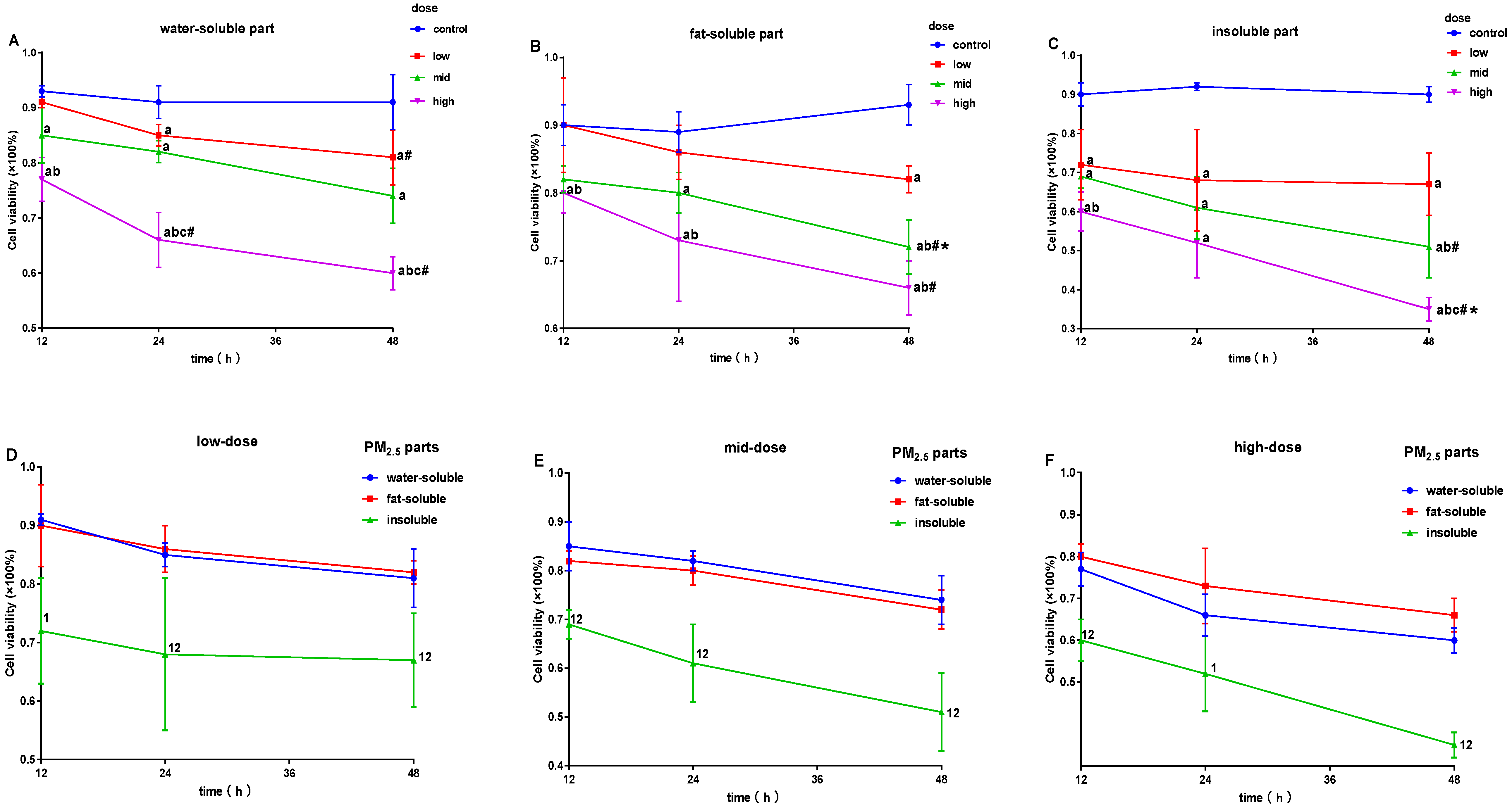
Figure 2 shows that the survival rates of cells significantly decreased after exposure to W-PM2.5, compared to the control, except for the low dose for 12 h (p < 0.05, Figure 2A). After exposure to F-PM2.5 for 24 and 48 h, cell survival rates were significantly lower than that of the control (p < 0.05, Figure 2B). After exposure to I-PM2.5, survival rates of cells decreased significantly (p < 0.05, Figure 2C). In addition, the cell survival rate decreased earlier and more dramatically after exposure to I-PM2.5 than W-PM2.5 or F-PM2.5 (p < 0.05, Figure 2D–F).

3.3. Effects of PM2.5 on the Levels of IL-1β, IL-6, IL-8, IL-12 p70, TNF-α, CRP, IL-2, and COX-2

Figure 3 shows the effects of exposure to PM2.5 for 12 h on the levels of inflammatory molecules. The levels of IL-6, IL-12 p70, and TNF-α increased significantly after exposure to a high dose of I-PM2.5 for 12 h (Figure 3B,D,E). After exposure to W- and F-PM2.5 for 12 h, levels of inflammatory molecules saw no significant changes.
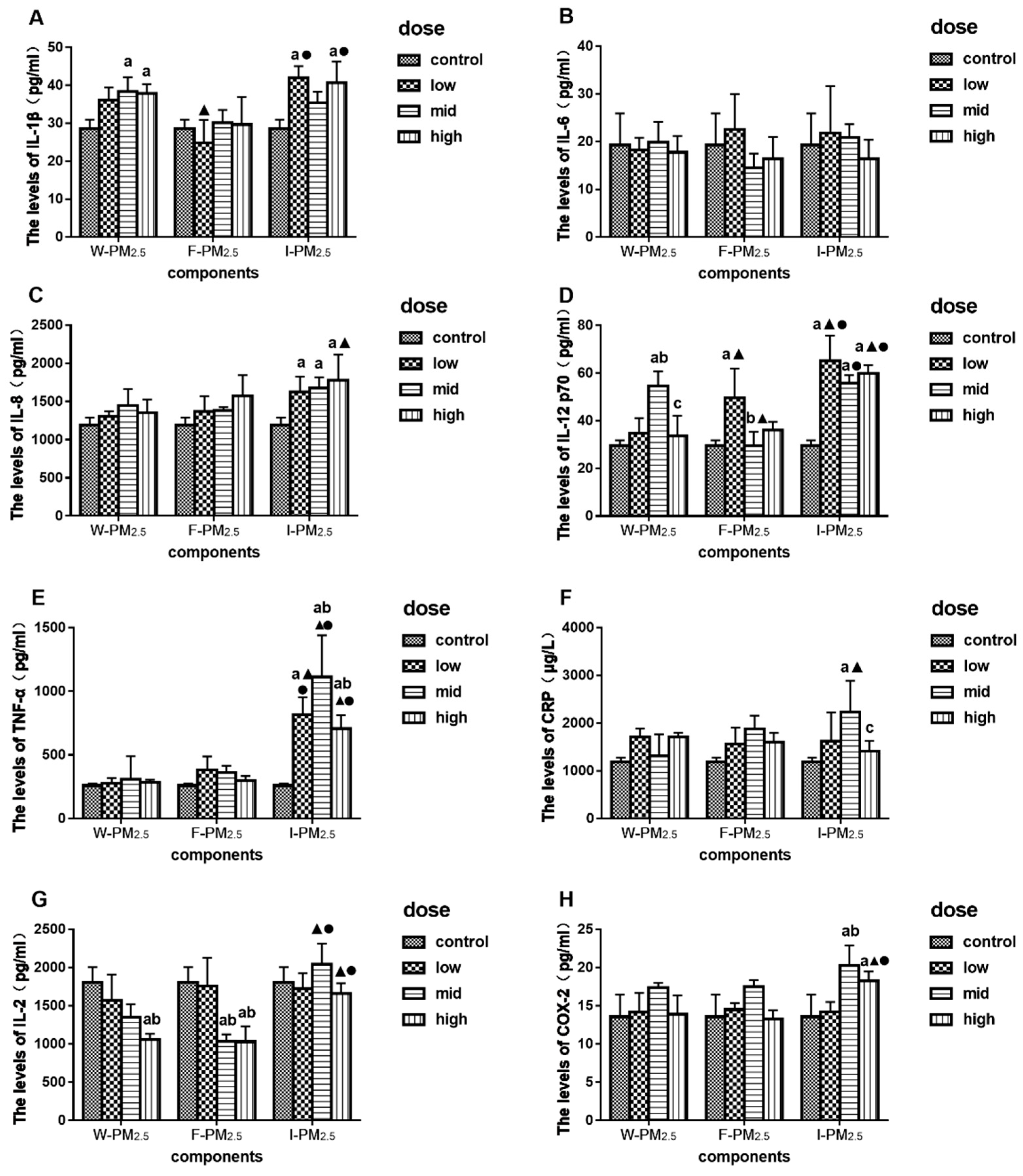
Figure 4A–F shows the effects of exposure to PM2.5 for 24 h on the levels of inflammatory cytokines. After exposure to W-PM2.5, the expression levels of IL-1β significantly increased at mid and high doses, and the levels of IL-12 p70 were significantly higher than that of control at mid dose (Figure 4A,D, p < 0.05). After exposure to F-PM2.5, the expression levels of IL-12 p70 in the low-dose group were significantly higher than control (Figure 4D, p < 0.05). After exposure to I-PM2.5, the expression levels of IL-8, IL-12, and TNF-α significantly increased (Figure 4C–E, p < 0.05). The levels of IL-1β increased significantly after exposure to the low and high dose of I-PM2.5 (Figure 4A, p < 0.05). The levels of CRP significantly increased for the mid dose of I-PM2.5 (Figure 4F, p < 0.05).
In addition, the expression levels of IL-2 and COX-2 in the cell supernatant were detected and recorded. As shown in Figure 4G, the levels of IL-2 decreased significantly after exposure to the high dose of W- and F-PM2.5 (p < 0.05). The expression levels of COX-2 were significantly higher in the mid- and high-dose group after exposure to I-PM2.5 than that of control (Figure 4H, p < 0.05).
After exposure to I-PM2.5, the levels of IL-12 p70 and TNF-α were significantly higher than that in W- and F-PM2.5 treatment groups (Figure 4D, p < 0.05). The expression levels of IL-1β, after exposure to low and high doses of I-PM2.5, were significantly higher than that for the same dose of F-PM2.5 (Figure 4A, p < 0.05). Moreover, the levels of IL-8, CRP, and COX-2 increased only after exposure to I-PM2.5 (Figure 4C, F and H, p < 0.05).

3.4. Effects of PM2.5 on the Relative mRNA Expression Levels of NF-KB Family Gene

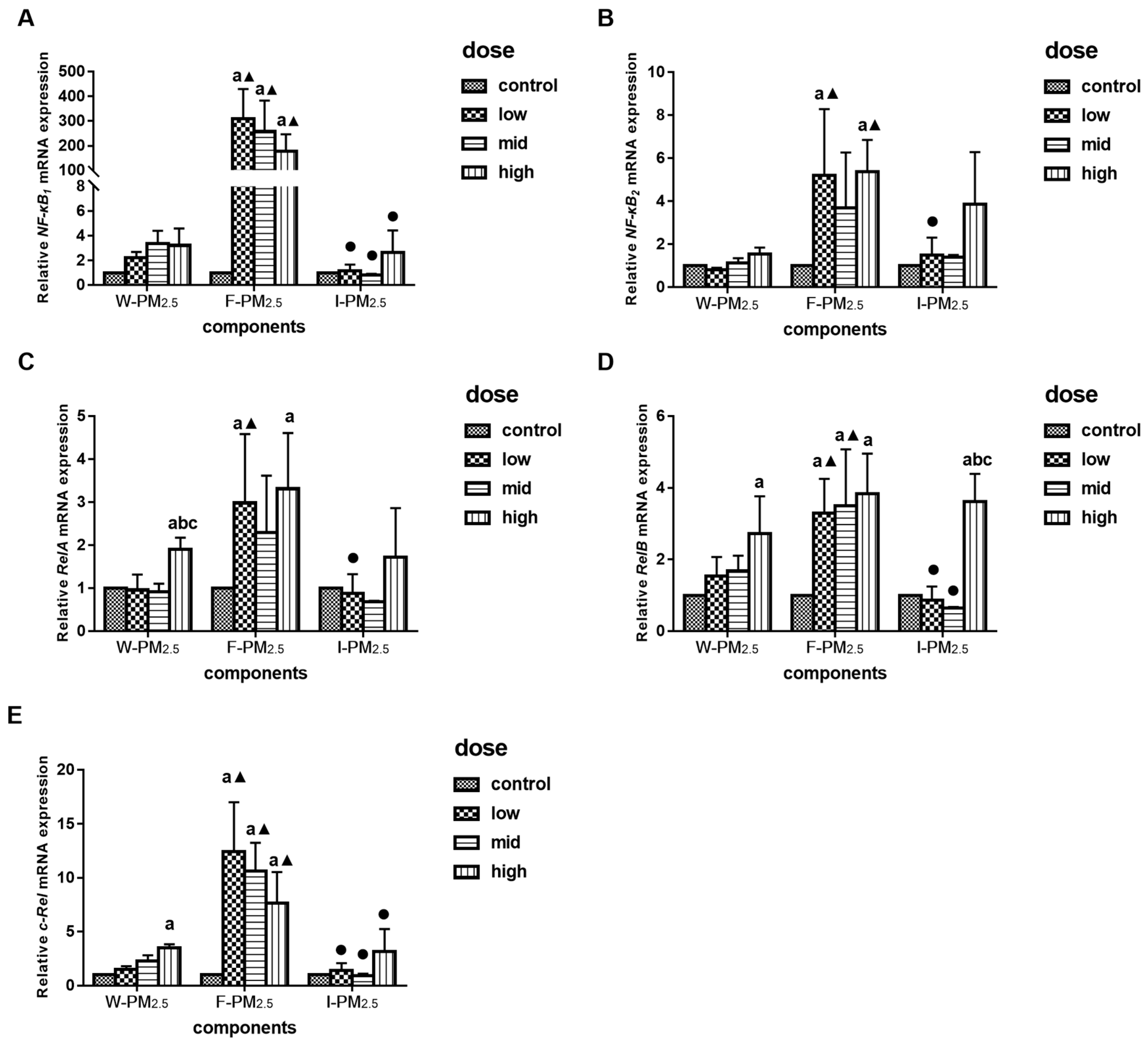
The relative mRNA expression levels of the NF-κB signaling pathway related genes were measured after exposure to PM2.5 for 24 h. As shown in Figure 5C–E, when exposed to a high dose of W-PM2.5, the relative mRNA levels of RelA, RelB, and c-Rel were significantly higher than that of control (p < 0.05). Exposure to high dose of I-PM2.5 only resulted in the high expression of RelB (p < 0.05, Figure 5D). The low and high doses of F-PM2.5 increased the relative expressions of NF-κB2 and RelA, whereas the relative levels of NF-κB1, RelB, and c-Rel increased at all concentrations (p < 0.05).
In conclusion, a high dose of W- and I-PM2.5 exposure increased the expression of several genes related to NF-κB signaling pathways, but low-dose F-PM2.5 increased the mRNA levels of NF-κB genes, especially NF-κB1 and RelA.

3.5. The Connection between the Expression of NF-κB mRNA and Inflammatory Molecules

The correlation of the expression levels between NF-κB signaling pathway mRNA and inflammatory molecules was analyzed to determine whether the exposure of different components of PM2.5 caused inflammation through the NF-κB signaling pathway. After exposure to F-PM2.5, the expression levels of IL-8 were negatively correlated with c-Rel, whereas IL-12 p70 was positively correlated with NF-κB2 and c-Rel. After treating the cells with I-PM2.5 for 24 h, the expression levels of IL-1 were positively correlated with NF-κB1, NF-κB2, and RelB, as well as IL-8 and NF-κB2. In addition, IL-6 was negatively correlated with the levels of NF-κB genes (Table 3).

4. Discussion

PM2.5 are one of the important causes of respiratory diseases, cardiovascular diseases, and premature death [6,27,28,29]. Due to small particle sizes, PM2.5 can be easily inhaled and deposited into alveolar cells in the respiratory tract, causing or exacerbating lung diseases [30]. The compositions of PM2.5 are complex, and the chemical compositions are pathogenic [31]. Park et al. found that different components of PM2.5 had different effects on airway epithelial cells, and the combination components could cause respiratory diseases such as asthma [7]. The present study divided PM2.5 into W-PM2.5, F-PM2.5, and I-PM2.5 to investigate the macrophage toxicity of PM2.5 and its effect on the levels of the NF-κB signaling pathway and inflammatory molecules, to analyze the mechanism of PM2.5-induced inflammation in respiratory diseases and to provide a new theoretical basis to prevent and treat the respiratory system diseases caused by atmospheric PM2.5.
Macrophages are the first barrier of the body’s immune system and the target cells for PM2.5 action. They can consume particles, secrete inflammatory molecules, and participate in various immune responses. In this study, THP-1-derived macrophages were selected as the research subjects according to the research objectives, and treatment was conducted with different PM2.5 components. Based on previous studies [32,33], we scaled the exposure doses. We chose higher doses of the total PM2.5 because we decomposed the total PM2.5.
Many studies have confirmed that PM2.5 has a toxic effect on cells [34,35,36]. The results of this study showed that all three components of PM2.5 led to a significant decrease in cell survival rates. The effect of I-PM2.5 was even more pronounced. The reason may be that I-PM2.5 leads to cell death through damage to the cell membrane and cell–particle interaction, whereas W-PM2.5 causes cell damage through the early response of oxidative stress. The former is more direct [26,37,38,39].
Water-soluble PM2.5 was mainly considered to comprise various inorganic salt ions and metal ions, including NO3−, SO42−, NH4+ arsenic, Fe, Cu, zinc, and lead, in which soluble metal ions are regarded as the main cause of oxidative stress and lung damage [40,41]. Transition metals in PM promote the production and release of ROS, which cause or exacerbate inflammation [42]. The results of this experiment indicate that exposure to W-PM2.5 for 24 h could promote the expression levels of IL-1β and IL-12 p70. The former (IL-1β) is one of the most representative inflammatory molecules, and its high expression indicates that W-PM2.5 caused inflammation of macrophages, but it may be easily cleared by cells to eliminate inflammation. The latter (IL-12 p70) is an important immune regulatory cytokine and a member of a small family of heterodimeric cytokines [43]. The increase of IL-12 p70 expression could promote the development of inflammation [44].
Fat-soluble PM2.5 mainly comprises organic compounds that are harmful to the human body, such as polycyclic aromatic hydrocarbons and dioxins [16]. Fuentes-Mattei et al. have suggested that organic extract of PM2.5 possibly suppresses the role of pregnant X receptor (PXR) and CYP3A5 on human epithelial cells, which triggers an inflammatory response [45]. Metal not only exists in the aqueous extract of PM2.5, but can also be found in organic extracts. It is interesting to note that, according to reports, heavy metal in PM2.5 organic extracts becomes redox-active, and when there is a metal-chelating agent in airway epithelial cells, organic extracts release IL-6 and IL-8. Therefore, the combination of polycyclic aromatic hydrocarbons (PAHs) or polycyclic aromatic hydrocarbons and organic extraction metals such as Fe and Cu may help to promote inflammatory responses through ROS, thereby exacerbating respiratory disease. The results of this study show that the levels of IL-12 p70 increased significantly after exposure to F-PM2.5 for 24 h. Furthermore, the small changes of the inflammatory molecules caused by F-PM2.5 may mainly be due to its low dose.
Insoluble PM2.5 was the main component of total PM2.5, accounting for more than 50% of the total weight of PM2.5 [46]. The concentrations of PAHs and heavy metals were much higher in I-PM2.5 than in others, due to their tendency to partition the solid [47]. Additionally, larger total quantities of catalytically and biologically active metals were likely to be associated with the insoluble fraction. Insoluble heavy metals and dust could also have adverse effects on the human body [41]. In addition, PM2.5 could also attach a large number of bacterial microbes. Previous toxicological studies proposed that particulate matter containing more insoluble components from incomplete combustion was more toxic than PM2.5 in macrophage and fibroblast cell lines [48,49]. Our experiment showed that, after exposure to PM2.5 for 12 h, the levels of IL-6, IL-12 p70, and TNF-α significantly increased in the high dose group of I-PM2.5. This indicated that a high dose of I-PM2.5 could cause inflammation in a relatively short time. After exposure to I-PM2.5, the expression levels of IL-1β, IL-8, IL-12 p70, TNF-α, and CRP increased significantly, and I-PM2.5 caused a drastic inflammatory response to macrophages. However, the levels of IL-1β and IL-12 p70 in the mid-dose group did not increase, which may be due to the effect of cell resistance to inflammation or the decrease in cell survival rates. An important inflammatory transmitter, TNF-α plays a crucial role in the initiation and maintenance of inflammation. Increased secretion of TNF-α could induce other pro-inflammatory molecules (such as IL-1, IL-6, and IL-8), amplify inflammatory signals, and cause cascade reactions. When the respiratory system is infected, the expression levels of TNF-α will be significantly increased. Insoluble PM2.5 led to high levels of these important inflammatory molecules, such as IL-1β, IL-8, IL-12 p70, and TNF-α, but there was no significant change in IL-6, which was a cause of doubt in this experiment. C-reactive protein has inflammatory marker properties and expresses when acute inflammation occurs. The effect of I-PM2.5 on CRP was also consistent with previous studies [50]. A cytokine secreted by macrophages and epithelial cells etc., IL-8 is considered to be a mediator of inflammatory responses [51]. Further, COX-2 is a proinflammatory enzyme that has been reported to be involved in inflammatory responses and cytotoxicity after PM stimulation from different sources [52,53]. Previous research has shown that urban dust or diesel exhaust induce IL-8 and COX-2 in an AhR-dependent manner in human U937-derived macrophages [54,55]. AhR is also considered to be an important receptor protein mediating the toxic responses of dioxin-like PAHs [56]. In our study, exposure to I-PM2.5 caused a significant increase in IL-8 and COX-2, and PAHs in I-PM2.5 could play a major role. PM2.5 caused the occurrence of inflammation and eliminated inflammation through the feedback mechanism. Previous studies have shown that I-PM2.5 can cause an increase of ROS at a lower concentration than W- and F-PM2.5 [25,57], which indicates that I-PM2.5 have a more obvious effect on cytotoxicity and inflammatory reactions. Exposure to PM2.5 led to the decrease of IL-2, suggesting that PM2.5 might not affect the levels of IL-2 and that changes in IL-2 might be due to the decrease of the cell survival rate.
In our previous study, we found that the overexpression of NF-κB mRNA levels was associated with inflammation and played a role in the development of chronic obstructive pulmonary disease (COPD) in a population experiment [58]. Therefore, we explored whether PM2.5 could affect the occurrence and development of respiratory inflammation through such effects in vitro. When cells are in a resting state, the NF-κB signaling pathway is activated by both classical and non-canonical pathways. During the activation of the NF-κB signaling pathway, dimeric protein is transcribed to generate corresponding mRNA. The cytoplasm enters the nucleus rapidly and is specifically bound to the κ B site, where the target gene is at the promoter site to regulate its expression [59]. Previous studies have reported that the NF-κB signal pathway could be activated when exposed to PM2.5, and it has been significantly correlated with the content of metal elements [34,60]. Moreover, NF-κB plays a dual role in the inflammatory response. Our results show that after exposure to the high dose of W-PM2.5, the mRNA expression levels of RelA, RelB, and c-Rel increased. The mRNA levels of the NF-κB signaling pathway genes increased significantly after exposure to F-PM2.5. This indicates that the NF-κB signaling pathway responded to the exposure of F-PM2.5 at a lower dose, and might lead to subsequent cascade reactions. However, exposure to I-PM2.5 had no obvious effect on NF-κB genes, possibly because the excessive accumulation of inflammation destroyed the regulation of the NF-κB signaling pathway.
Studies have shown that the NF-κB family gene is related to the levels of inflammatory molecules—TNF-α, for example, could induce the activation of NF-κB [32]. In addition, the macrophages knockout c-Rel and RelB lost the function of secreting TNF-α, but oversecreted IL-1β [61]. This indicates that NF-κB can also regulate the expression levels of inflammatory molecules. After exposure to F-PM2.5, expression levels of IL-12 p70 showed a positive correlation with NF-κB2 and c-Rel. After exposure to I-PM2.5, IL-1β showed a positive correlation with the mRNA levels of the NF-κB signaling pathway. However, we did not find a correlation between TNF-α and NF-κB, which may be due to the temporality of NF-κB activation.
The source of PM2.5 was very wide and its components were complex, which made it difficult to study. The toxic mechanism of macrophages induced by PM2.5 is uncertain, because of the interaction between different components. Our subsequent research will focus on the mechanisms of cytotoxicity induced by PM2.5.
Although we have done a lot of work, there are still many limitations to our experiment. We only measured the levels of cytokines at 12 and 24 h, and genes at 24 h; thus, some changes in levels could not be fully explained. We only studied changes of mRNA expression levels, and did not observe the expression of NF-κB protein, which is a defect of our study. Because there was no plan to inhibit NF-κB, it is difficult to describe the role of NF-κB genes on the levels of inflammatory molecules. The correlation analysis is different to the causal connection.

5. Conclusions

PM2.5 decreases the cell survival rate and up-regulates the relative mRNA levels of the NF-κB gene family and the levels of inflammatory molecules. The main toxic components of PM2.5 relating to inflammatory response in THP-1-derived macrophages were those of I-PM2.5. Exposure to F-PM2.5 significantly increased the mRNA expression levels of the NF-κB genes. In addition, the expression levels of inflammatory molecules were correlated with the mRNA expression levels of the NF-κB genes. These findings suggest that PM2.5 could up-regulate the relative mRNA levels of the NF-κB family gene and the levels of inflammatory molecules involved in the inflammatory response.

Author Contributions

Conceptualization, J.Z., L.Z. and L.Y.; Data curation, J.Z., Y.Z. (Yaming Zhao), C.L., L.Z., W.Q. and Y.Z. (Yuezhu Zhang); Formal analysis, Y.Z. (Yaming Zhao); Funding acquisition, L.Y.; Methodology, J.Z., Y.G., L.Z. and W.Q.; Project administration, W.Q. and L.Y.; Resources, J.Z. and L.Y.; Software, Y.Z. (Yaming Zhao); Supervision, L.Z. and L.Y.; Writing—original draft, J.Z., Y.Z. (Yaming Zhao), L.Z. and Y.Z. (Yuezhu Zhang); Writing—review & editing, Y.Z. (Yaming Zhao), Y.G., C.L., L.Z. and L.Y.

Funding

This work was sponsored by the Jilin Province Science and Technology Department (20150101208JC, 20160520159JH) and the Education Department of Jilin Province (JJKH20170874KJ).

Acknowledgments

We would like to acknowledge the Animal Resources Centre for animal technical support.

Conflicts of Interest

The authors declare no conflict of interest.

References

  1. Pruss-Ustun, A.; Neira, M. Preventing Disease through Healthy Environments: A Global Assessment of the Environmental Burden of Disease Abstract. Available online: https://apps.who.int/iris/bitstream/handle/10665/204585/9789241565196_eng.pdf (accessed on 5 April 2019).
  2. Lo, W.C.; Shie, R.H.; Chan, C.C.; Lin, H.H. Burden of disease attributable to ambient fine particulate matter exposure in Taiwan. J. Formos Med. Assoc. 2017, 116, 32–40. [Google Scholar] [CrossRef] [PubMed] [Green Version]
  3. Sanyal, S.; Rochereau, T.; Maesano, C.N.; Com-Ruelle, L.; Annesi-Maesano, I. Long-Term Effect of Outdoor Air Pollution on Mortality and Morbidity: A 12-Year Follow-Up Study for Metropolitan France. Int. J. Environ. Res. Public Health 2018, 15, 2487. [Google Scholar] [CrossRef] [PubMed]
  4. EEA. Ambient (Outdoor) Air Quality and Health. Available online: http://www.who.int/mediacentre/factsheets/fs313/en/ (accessed on 5 April 2019).
  5. Kim, K.H.; Kabir, E.; Kabir, S. A review on the human health impact of airborne particulate matter. Environ. Int. 2015, 74, 136–143. [Google Scholar] [CrossRef] [PubMed]
  6. Xing, Y.F.; Xu, Y.H.; Shi, M.H.; Lian, Y.X. The impact of PM2.5 on the human respiratory system. J. Thorac. Dis. 2016, 8, E69–E74. [Google Scholar] [PubMed]
  7. Park, E.J.; Roh, J.; Kim, Y.; Park, K.; Kim, D.S.; Yu, S.D. PM2.5 collected in a residential area induced Th1-type inflammatory responses with oxidative stress in mice. Environ. Res. 2011, 111, 348–355. [Google Scholar] [CrossRef] [PubMed]
  8. Ma, Q.Y.; Huang, D.Y.; Zhang, H.J.; Wang, S.; Chen, X.F. Exposure to particulate matter 2.5 (PM2.5) induced macrophage-dependent inflammation, characterized by increased Th1/Th17 cytokine secretion and cytotoxicity. Int. Immunopharmacol. 2017, 50, 139–145. [Google Scholar] [CrossRef] [PubMed]
  9. Wessels, A.; Birmili, W.; Albrecht, C.; Hellack, B.; Jermann, E.; Wick, G.; Harrison, R.M.; Schins, R.P.F. Oxidant Generation and Toxicity of Size-Fractionated Ambient Particles in Human Lung Epithelial Cells. Environ. Sci. Technol. 2010, 44, 3539–3545. [Google Scholar] [CrossRef]
  10. Zhou, Z.X.; Liu, Y.H.; Duan, F.K.; Qin, M.N.; Wu, F.C.; Sheng, W.; Yang, L.X.; Liu, J.G.; He, K.B. Transcriptomic Analyses of the Biological Effects of Airborne PM2.5 Exposure on Human Bronchial Epithelial Cells. PLoS ONE 2015, 10, e0138267. [Google Scholar] [CrossRef] [PubMed]
  11. Li, R.J.; Kou, X.J.; Geng, H.; Xie, J.F.; Yang, Z.H.; Zhang, Y.X.; Cai, Z.W.; Dong, C. Effect of Ambient PM2.5 on Lung Mitochondrial Damage and Fusion/Fission Gene Expression in Rats. Chem. Res. Toxicol. 2015, 28, 408–418. [Google Scholar] [CrossRef]
  12. Zhao, Q.J.; Chen, H.; Yang, T.; Rui, W.; Liu, F.; Zhang, F.; Zhao, Y.; Ding, W.J. Direct effects of airborne PM2.5 exposure on macrophage polarizations. Biochim. Biophys. Acta. Gen. Subj. 2016, 1860, 2835–2843. [Google Scholar] [CrossRef] [PubMed]
  13. Wang, J.; Huang, J.; Wang, L.; Chen, C.; Yang, D.; Jin, M.; Bai, C.; Song, Y. Urban particulate matter triggers lung inflammation via the ROS-MAPK-NF-kappaB signaling pathway. J. Thorac. Dis. 2017, 9, 4398–4412. [Google Scholar] [CrossRef] [PubMed]
  14. Rumelhard, M.; Ramgolam, K.; Hamel, R.; Marano, F.; Baeza-Squiban, A. Expression and role of EGFR ligands induced in airway cells by PM2.5 and its components. Eur. Respir. J. 2007, 30, 1064–1073. [Google Scholar] [CrossRef] [PubMed]
  15. Ge, C.X.; Qin, Y.T.; Lou, D.S.; Li, Q.; Li, Y.Y.; Wang, Z.M.; Yang, W.W.; Wang, M.; Liu, N.; Wang, Z.; et al. iRhom2 deficiency relieves TNF-alpha associated hepatic dyslipidemia in long-term PM2.5-exposed mice. Biochem. Biophys. Res. Commun. 2017, 493, 1402–1409. [Google Scholar] [CrossRef] [PubMed]
  16. He, M.; Ichinose, T.; Yoshida, S.; Ito, T.; He, C.Y.; Yoshida, Y.; Arashidani, K.; Takano, H.; Sun, G.F.; Shibamoto, T. PM2.5-induced lung inflammation in mice: Differences of inflammatory response in macrophages and type II alveolar cells. J. Appl. Toxicol. 2017, 37, 1203–1218. [Google Scholar] [CrossRef] [PubMed]
  17. Peng, H.; Zhao, X.H.; Bi, T.T.; Yuan, X.Y.; Guo, J.B.; Peng, S.Q. PM2.5 obtained from urban areas in Beijing induces apoptosis by activating nuclear factor-kappa B. Mil. Med. Res. 2017, 4, 27. [Google Scholar] [CrossRef] [PubMed]
  18. Ku, T.; Li, B.; Gao, R.; Zhang, Y.; Yan, W.; Ji, X.; Li, G.; Sang, N. NF-kappaB-regulated microRNA-574-5p underlies synaptic and cognitive impairment in response to atmospheric PM2.5 aspiration. Part. Fibre Toxicol. 2017, 14, 34. [Google Scholar] [CrossRef] [PubMed]
  19. Li, R.; Zhao, L.; Tong, J.; Yan, Y.; Xu, C. Fine Particulate Matter and Sulfur Dioxide Coexposures Induce Rat Lung Pathological Injury and Inflammatory Responses Via TLR4/p38/NF-kappaB Pathway. Int. J. Toxicol. 2017, 36, 165–173. [Google Scholar] [CrossRef] [PubMed]
  20. Dagher, Z.; Garcon, G.; Billet, S.; Verdin, A.; Ledoux, F.; Courcot, D.; Aboukais, A.; Shirali, P. Role of nuclear factor-kappa B activation in the adverse effects induced by air pollution particulate matter (PM2.5) in human epithelial lung cells (L132) in culture. J. Appl. Toxicol. 2007, 27, 284–290. [Google Scholar] [CrossRef] [PubMed]
  21. Stone, S.; Jamison, S.; Yue, Y.; Durose, W.; Schmidt-Ullrich, R.; Lin, W. NF-κB Activation Protects Oligodendrocytes against Inflammation. J. Neurosci. 2017, 37, 9332–9344. [Google Scholar] [CrossRef] [Green Version]
  22. Hoesel, B.; Schmid, J.A. The complexity of NF-κB signaling in inflammation and cancer. Mol. Cancer 2013, 12, 86. [Google Scholar] [CrossRef] [PubMed]
  23. Mitchell, S.; Vargas, J.; Hoffmann, A. Signaling via the NFκB system. Wiley Interdiscip. Rev. Syst. Biol. Med. 2016, 8, 227–241. [Google Scholar] [CrossRef] [PubMed]
  24. Cheng, Y.; He, K.B.; Du, Z.Y.; Zheng, M.; Duan, F.K.; Ma, Y.L. Humidity plays an important role in the PM2.5 pollution in Beijing. Environ. Pollut. 2015, 197, 68–75. [Google Scholar] [CrossRef] [PubMed]
  25. Wang, D.B.; Pakbin, P.; Shafer, M.M.; Antkiewicz, D.; Schauer, J.J.; Sioutas, C. Macrophage reactive oxygen species activity of water-soluble and water-insoluble fractions of ambient coarse, PM2.5 and ultrafine particulate matter (PM) in Los Angeles. Atmos. Environ. 2013, 77, 301–310. [Google Scholar] [CrossRef]
  26. Zou, Y.J.; Jin, C.Y.; Su, Y.; Li, J.R.; Zhu, B.S. Water soluble and insoluble components of urban PM2.5 and their cytotoxic effects on epithelial cells (A549) in vitro. Environ. Pollut. 2016, 212, 627–635. [Google Scholar] [CrossRef] [PubMed]
  27. Silva, R.A.; West, J.J.; Zhang, Y.Q.; Anenberg, S.C.; Lamarque, J.F.; Shindell, D.T.; Collins, W.J.; Dalsoren, S.; Faluvegi, G.; Folberth, G.; et al. Global premature mortality due to anthropogenic outdoor air pollution and the contribution of past climate change. Environ. Res. Lett. 2013, 8, 031002. [Google Scholar] [CrossRef]
  28. Zeng, Q.; Ni, Y.; Jiang, G.H.; Li, G.X.; Pan, X.C. The short term burden of ambient particulate matters on non-accidental mortality and years of life lost: A ten-year multi-district study in Tianjin, China. Environ. Pollut. 2017, 220, 713–719. [Google Scholar] [CrossRef] [PubMed]
  29. Turner, M.C.; Krewski, D.; Pope, C.A.; Chen, Y.; Gapstur, S.M.; Thun, M.J. Long-term Ambient Fine Particulate Matter Air Pollution and Lung Cancer in a Large Cohort of Never-Smokers. Am. J. Respir. Crit. Care 2011, 184, 1374–1381. [Google Scholar] [CrossRef]
  30. Ye, X.Q.; Hong, W.; Hao, B.W.; Peng, G.Y.; Huang, L.M.; Zhao, Z.X.; Zhou, Y.M.; Zheng, M.N.; Li, C.L.; Liang, C.X.; et al. PM2.5 promotes human bronchial smooth muscle cell migration via the sonic hedgehog signaling pathway. Respir. Res. 2018, 19, 37. [Google Scholar] [CrossRef] [PubMed]
  31. Shang, Y.; Zhu, T.; Lenz, A.G.; Frankenberger, B.; Tian, F.; Chen, C.Y.; Stoeger, T. Reduced in vitro toxicity of fine particulate matter collected during the 2008 summer Olympic Games in Beijing: The roles of chemical and biological components. Toxicol. In Vitro 2013, 27, 2084–2093. [Google Scholar] [CrossRef] [PubMed]
  32. Zhang, Y.Z.; Wang, S.Y.; Zhu, J.; Li, C.Y.; Zhang, T.R.; Liu, H.B.; Xu, Q.; Ye, X.F.; Zhou, L.T.; Ye, L. Effect of Atmospheric PM2.5 on Expression Levels of NF-kappa B Genes and Inflammatory Cytokines Regulated by NF-kappa B in Human Macrophage. Inflammation 2018, 41, 784–794. [Google Scholar] [CrossRef]
  33. Chen, S.; Wu, X.; Hu, J.; Dai, G.; Rong, A.; Guo, G. PM2.5 exposure decreases viability, migration and angiogenesis in human umbilical vein endothelial cells and human microvascular endothelial cells. Mol. Med. Rep. 2017, 16, 2425–2430. [Google Scholar] [CrossRef] [PubMed]
  34. Pavagadhi, S.; Betha, R.; Venkatesan, S.; Balasubramanian, R.; Hande, M.P. Physicochemical and toxicological characteristics of urban aerosols during a recent Indonesian biomass burning episode. Environ. Sci. Pollut. Res. 2013, 20, 2569–2578. [Google Scholar] [CrossRef] [PubMed]
  35. Steenhof, M.; Gosens, I.; Strak, M.; Godri, K.J.; Hoek, G.; Cassee, F.R.; Mudway, I.S.; Kelly, F.J.; Harrison, R.M.; Lebret, E.; et al. In vitro toxicity of particulate matter (PM) collected at different sites in the Netherlands is associated with PM composition, size fraction and oxidative potential—The RAPTES project. Part. Fibre Toxicol. 2011, 8, 26. [Google Scholar] [CrossRef]
  36. Cavanagh, J.A.E.; Trought, K.; Brown, L.; Duggan, S. Exploratory investigation of the chemical characteristics and relative toxicity of ambient air particulates from two New Zealand cities. Sci. Total Environ. 2009, 407, 5007–5018. [Google Scholar] [CrossRef] [PubMed]
  37. Deng, F.; Guo, X.; Liu, H.; Fang, X.; Yang, M.; Chen, W. Effects of dust storm PM2.5 on cell proliferation and cell cycle in human lung fibroblasts. Toxicol. In Vitro 2007, 21, 632–638. [Google Scholar] [CrossRef] [PubMed]
  38. Fotakis, G.; Timbrell, J.A. In vitro cytotoxicity assays: Comparison of LDH, neutral red, MTT and protein assay in hepatoma cell lines following exposure to cadmium chloride. Toxicol. Lett. 2006, 160, 171–177. [Google Scholar] [CrossRef] [PubMed]
  39. Jalava, P.I.; Hirvonen, M.R.; Sillanpaa, M.; Pennanen, A.S.; Happo, M.S.; Hillamo, R.; Cassee, F.R.; Gerlofs-Nijland, M.; Borm, P.J.A.; Schins, R.P.F.; et al. Associations of urban air particulate composition with inflammatory and cytotoxic responses in RAW 246.7 cell line. Inhal. Toxicol. 2009, 21, 994–1006. [Google Scholar] [CrossRef] [PubMed]
  40. Palleschi, S.; Rossi, B.; Armiento, G.; Montereali, M.R.; Nardi, E.; Tagliani, S.M.; Inglessis, M.; Gianfagna, A.; Silvestroni, L. Toxicity of the readily leachable fraction of urban PM2.5 to human lung epithelial cells: Role of soluble metals. Chemosphere 2018, 196, 35–44. [Google Scholar] [CrossRef]
  41. Zhang, Y.; Li, Y.; Shi, Z.; Wu, J.; Yang, X.; Feng, L.; Ren, L.; Duan, J.; Sun, Z. Metabolic impact induced by total, water soluble and insoluble components of PM2.5 acute exposure in mice. Chemosphere 2018, 207, 337–346. [Google Scholar] [CrossRef]
  42. Loxham, M.; Morgan-Walsh, R.J.; Cooper, M.J.; Blume, C.; Swindle, E.J.; Dennison, P.W.; Howarth, P.H.; Cassee, F.R.; Teagle, D.A.H.; Palmer, M.R.; et al. The Effects on Bronchial Epithelial Mucociliary Cultures of Coarse, Fine, and Ultrafine Particulate Matter from an Underground Railway Station. Toxicol. Sci. 2015, 145, 98–107. [Google Scholar] [CrossRef]
  43. Klinke, D.J. The ratio of P40 monomer to dimer is an important determinant of IL-12 bioactivity. J. Theor. Biol. 2006, 240, 323–335. [Google Scholar] [CrossRef] [PubMed]
  44. Tao, J.L.; Yang, M.H.; Wu, H.; Ma, T.; He, C.J.; Chai, M.L.; Zhang, X.S.; Zhang, J.L.; Ding, F.R.; Wang, S.T.; et al. Effects of AANAT overexpression on the inflammatory responses and autophagy activity in the cellular and transgenic animal levels. Autophagy 2018, 14, 1850–1869. [Google Scholar] [CrossRef] [PubMed]
  45. Fuentes-Mattei, E.; Rivera, E.; Gioda, A.; Sanchez-Rivera, D.; Roman-Velazquez, F.R.; Jimenez-Velez, B.D. Use of human bronchial epithelial cells (BEAS-2B) to study immunological markers resulting from exposure to PM2.5 organic extract from Puerto Rico. Toxicol. Appl. Pharm. 2010, 243, 381–389. [Google Scholar] [CrossRef] [PubMed]
  46. Ghio, A.J.; Stonehuerner, J.; Dailey, L.A.; Carter, J.D. Metals associated with both the water-soluble and insoluble fractions of an ambient air pollution particle catalyze an oxidative stress. Inhal. Toxicol. 1999, 11, 37–49. [Google Scholar] [CrossRef] [PubMed]
  47. Lau, S.L.; Stenstrom, M.K. Metals and PAHs adsorbed to street particles. Water Res. 2005, 39, 4083–4092. [Google Scholar] [CrossRef]
  48. Feng, L.; Yang, X.Z.; Asweto, C.O.; Wu, J.; Zhang, Y.N.; Hu, H.J.; Shi, Y.F.; Duan, J.C.; Sun, Z.W. Low-dose combined exposure of nanoparticles and heavy metal compared with PM2.5 in human myocardial AC16 cells. Environ. Sci. Pollut. Res. 2017, 24, 27767–27777. [Google Scholar] [CrossRef] [PubMed]
  49. Jiang, S.L.; Shang, M.T.; Mu, K.; Jiang, N.; Wen, H.Y.; Wang, R.; Wu, H.; Li, W.Y. In vitro and in vivo toxic effects and inflammatory responses induced by carboxylated black carbon-lead complex exposure. Ecotoxicol. Environ. Saf. 2018, 165, 484–494. [Google Scholar] [CrossRef] [PubMed]
  50. Pinto-Plata, V.M.; Mullerova, H.; Toso, J.F.; Feudjo-Tepie, M.; Soriano, J.B.; Vessey, R.S.; Celli, B.R. C-reactive protein in patients with COPD, control smokers and non-smokers. Thorax 2006, 61, 23–28. [Google Scholar] [CrossRef]
  51. Suwa, T.; Hogg, J.C.; Quinlan, K.B.; Ohgami, A.; Vincent, R.; van Eeden, S.F. Particulate air pollution induces progression of atherosclerosis. J. Am. Coll. Cardiol. 2002, 39, 935–942. [Google Scholar] [CrossRef] [Green Version]
  52. Bai, N.; Tranfield, E.M.; Kavanagh, T.J.; Kaufman, J.D.; Rosenfeld, M.E.; van Eeden, S.F. Exposure to diesel exhaust upregulates COX-2 expression in ApoE knockout mice. Inhal. Toxicol. 2012, 24, 518–527. [Google Scholar] [CrossRef] [Green Version]
  53. Zhao, Y.; Gorshkova, I.; He, D.; Usatyuk, P.; Kalari, S.; Pendyala, S.; Garcia, J.G.; Berdyshev, E.; Natarajan, V. Sphingosine-1-Phosphate Lyase Regulates Lps-Induced Interleukin-6 Secretion in Pulmonary Microvascular Endothelial Cells and a Murine Model of Acute Lung Injury. J. Investig. Med. 2009, 57, 554. [Google Scholar]
  54. Vogel, C.F.A.; Sciullo, E.; Wong, P.; Kuzmicky, P.; Kado, N.; Matsumura, F. Induction of proinflammatory cytokines and C-reactive protein in human macrophage cell line U937 exposed to air pollution particulates. Environ. Health Perspect. 2005, 113, 1536–1541. [Google Scholar] [CrossRef] [PubMed]
  55. Vogel, C.F.A.; Kado, S.Y.; Kobayashi, R.; Liu, X.X.; Wong, P.; Na, K.; Durbin, T.; Okamoto, R.A.; Kado, N.Y. Inflammatory marker and aryl hydrocarbon receptor-dependent responses in human macrophages exposed to emissions from biodiesel fuels. Chemosphere 2019, 220, 993–1002. [Google Scholar] [CrossRef]
  56. Esakky, P.; Hansen, D.A.; Drury, A.M.; Moley, K.H. Cigarette smoke-induced cell cycle arrest in spermatocytes [GC-2spd(ts)] is mediated through crosstalk between Ahr-Nrf2 pathway and MAPK signaling. J. Mol. Cell Biol. 2015, 7, 73–87. [Google Scholar] [CrossRef] [PubMed]
  57. Gao, Z.X.; Song, X.L.; Li, S.S.; Lai, X.R.; Yang, Y.L.; Yang, G.; Li, Z.J.; Cui, Y.H.; Pan, H.W. Assessment of DNA Damage and Cell Senescence in Corneal Epithelial Cells Exposed to Airborne Particulate Matter (PM2.5) Collected in Guangzhou, China. Investig. Ophthalmol. Vis. Sci. 2016, 57, 3093–3102. [Google Scholar] [CrossRef] [PubMed]
  58. Zhou, L.; Liu, Y.; Chen, X.; Wang, S.; Liu, H.; Zhang, T.; Zhang, Y.; Xu, Q.; Han, X.; Zhao, Y.; et al. Over-expression of nuclear factor-kappaB family genes and inflammatory molecules is related to chronic obstructive pulmonary disease. Int. J. Chronic Obstr. Pulm. Dis. 2018, 13, 2131–2138. [Google Scholar] [CrossRef] [PubMed]
  59. Natoli, G.; Chiocca, S. Nuclear ubiquitin ligases, NF-kappaB degradation, and the control of inflammation. Sci. Signal. 2008, 1. [Google Scholar] [CrossRef]
  60. Bourgeois, B.; Owens, J.W. The influence of Hurricanes Katrina and Rita on the inflammatory cytokine response and protein expression in A549 cells exposed to PM2.5 collected in the Baton Rouge-Port Allen industrial corridor of Southeastern Louisiana in 2005. Toxicol. Mech. Methods 2014, 24, 220–242. [Google Scholar] [CrossRef]
  61. Haddad, J.J. Redox and oxidant-mediated regulation of apoptosis signaling pathways: Immuno-pharmaco-redox conception of oxidative siege versus cell death commitment. Int. Immunopharmacol. 2004, 4, 475–493. [Google Scholar] [CrossRef]
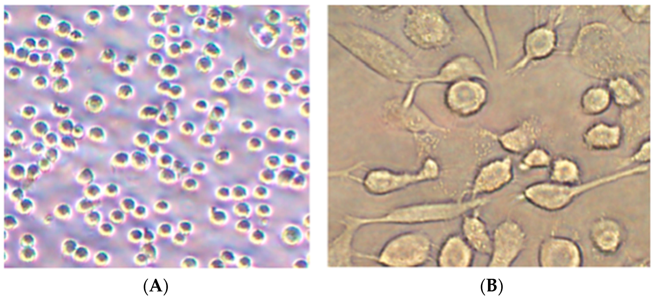
Figure 1. Morphology of THP-1 cells (A) before and (B) after differentiation.
Ijerph 16 01408 g001
Figure 2. Effects of PM2.5 on the cell survival rate of macrophages. (A) Effects of W-PM2.5 on cell survival rates; (B) effects of F-PM2.5 on cell survival rates; (C) effects of I-PM2.5 on cell survival rates; (D) effects of low-dose PM2.5 on cell survival rates; (E) effects of mid-dose PM2.5 on cell survival rates; (F) effects of high-dose PM2.5 on cell survival rates. Data are presented as means ± SD of three independent experiments. a p < 0.05 vs. control group at the same time; b p < 0.05 vs. low-dose group at the same time; c p < 0.05 vs. mid-dose group at the same time; # p < 0.05 vs. 12 h group at the same dose; * p < 0.05 vs. 24 h group at the same dose; 1 p < 0.05 vs. W-PM2.5 at the same dose; 2 p < 0.05 vs. F-PM2.5 at the same dose.
Ijerph 16 01408 g002
Figure 3. Effects of exposure to PM2.5 on the levels of IL-1β (A), IL-6 (B), IL-8 (C), IL-12 p70 (D), TNF-α (E), CRP (F), IL-2 (G), and COX-2 (H). Data are presented as means ± SD of three independent experiments. a p < 0.05 vs. control group; b p < 0.05 vs. low-dose group; c p < 0.05 vs. mid-dose group; p < 0.05 vs. W-PM2.5 at the same dose; p < 0.05 vs. F-PM2.5 at the same dose.
Ijerph 16 01408 g003
Figure 4. Effects of exposure to PM2.5 on the levels of IL-1β (A), IL-6 (B), IL-8 (C), IL-12 p70 (D), TNF-α (E), CRP (F), IL-2 (G), and COX-2 (H). Data are presented as means ± SD of three independent experiments. a p < 0.05 vs. control group; b p < 0.05 vs. low-dose group; c p < 0.05 vs. mid-dose group; p < 0.05 vs. W-PM2.5 at the same dose; p < 0.05 vs. F-PM2.5 at the same dose.
Ijerph 16 01408 g004
Figure 5. Effects of PM2.5 on the relative mRNA expression levels of NF-κB1 (A), NF-κB2 (B), RelA (C), RelB (D), and c-Rel (E). Data are presented as means ± SD of three independent experiments. a p < 0.05 vs. control group; b p < 0.05 vs. low-dose group; c p < 0.05 vs. mid-dose group; p < 0.05 vs. W-PM2.5 at the same dose; p < 0.05 vs. F-PM2.5 at the same dose.
Ijerph 16 01408 g005
Table 1. Concentration of different components of PM2.5.
DosesConcentration of PM2.5 (μg/mL)
W-PM2.5F-PM2.5I-PM2.5Total PM2.5
Low dose7525100200
Mid dose15050200400
High dose300100400800
Note: W-PM2.5 = water-soluble PM2.5, F-PM2.5 = fat-soluble PM2.5, I-PM2.5 = insoluble PM2.5.
Table 2. Primers used in real-time quantitative PCR.
GenesPrimerSequence
β-actinForward5′-CTGGAACGGTGAAGGTGACA-3′
Reverse5′-CGGCCACATTGTGAACTTTG-3′
NF-κB1Forward5′-CACAAGGCAGCAAATAGACGAG-3′
Reverse5′-TGGGGCATTTTGTTGAGAGTT-3′
NF-κB2Forward5′-GGCTGGTGCTGACATCCAT-3′
Reverse5′-CTGCTTCGGGTGTCCTTCTC-3′
RelAForward5′-CCCCAGCCCTATCCCTTTAC-3′
Reverse5′-TGCCCAGAAGGAAACACCA-3′
RelBForward5′-ATGAATGTGGTGAGGATCTGCTT-3′
Reverse5′-CTCTGATGTGTTTGTGGATTTCTTG-3′
c-RelForward5′-GACGACTGCTCTTCCTCCTGTT-3′
Reverse5′-TCATCTCCTCCTCTGACACTTCC-3′
Table 3. Correlation between NF-κB mRNA and inflammatory molecules of macrophage after PM2.5 exposure.
ComponentCytokinesIL-1βIL-6IL-8IL-12 p70CRPTNF-α
W-PM2.5NF-κB10.211−0.626−0.3880.2510.055−0.398
NF-κB20.460−0.335−0.068−0.221−0.081−0.189
RelA−0.020−0.330−0.369−0.468−0.2890.287
RelB0.500−0.512−0.374−0.259−0.495−0.014
c-Rel0.479−0.519−0.278−0.085−0.305−0.030
F-PM2.5NF-κB1−0.370−0.178−0.5840.5800.100−0.117
NF-κB2−0.440−0.212−0.0480.641 *−0.372−0.627
RelA−0.428−0.325−0.1640.445−0.466−0.325
RelB−0.332−0.487−0.1290.260−0.203−0.343
c-Rel−0.424−0.263−0.696 *0.634 *0.047−0.156
I-PM2.5NF-κB10.674 *−0.695 *0.605−0.173−0.131−0.424
NF-κB20.636 *−0.739 *0.641 *−0.256−0.043−0.356
RelA0.681 *−0.710 *0.577−0.167−0.148−0.405
RelB0.322−0.656 *0.212−0.093−0.175−0.444
c-Rel0.700 *−0.681 *0.609−0.149−0.139−0.436
* p < 0.05.