The Secretory Response of Rat Peritoneal Mast Cells on Exposure to Mineral Fibers


Abstract
1. Introduction
2. Materials and Methods
2.1. Reagents
2.2. Mineral Fibers
2.3. Characterization of Fibers by SEM—Energy Dispersive X-ray Spectrometry (EDX) Analysis
2.4. Animals
2.5. Rat Peritoneal Cell Preparation, Mast Cell Purification, and Cell Viability
2.6. RPMC Lysate Preparation and Treatment
2.7. RPMC Fiber Interaction
2.8. Release of Granule Components
2.9. MC Granule Preparation and Granule–Fiber Interaction
2.10. Optical and Ultrastructural Scanning Electron Microscope Analysis (SEM)
2.11. Transmission Electron Microscope Analysis (TEM) Fibers
2.12. Peroxidases
2.13. Isoelectric Point (IP)
2.14. Statistical Analysis
3. Results
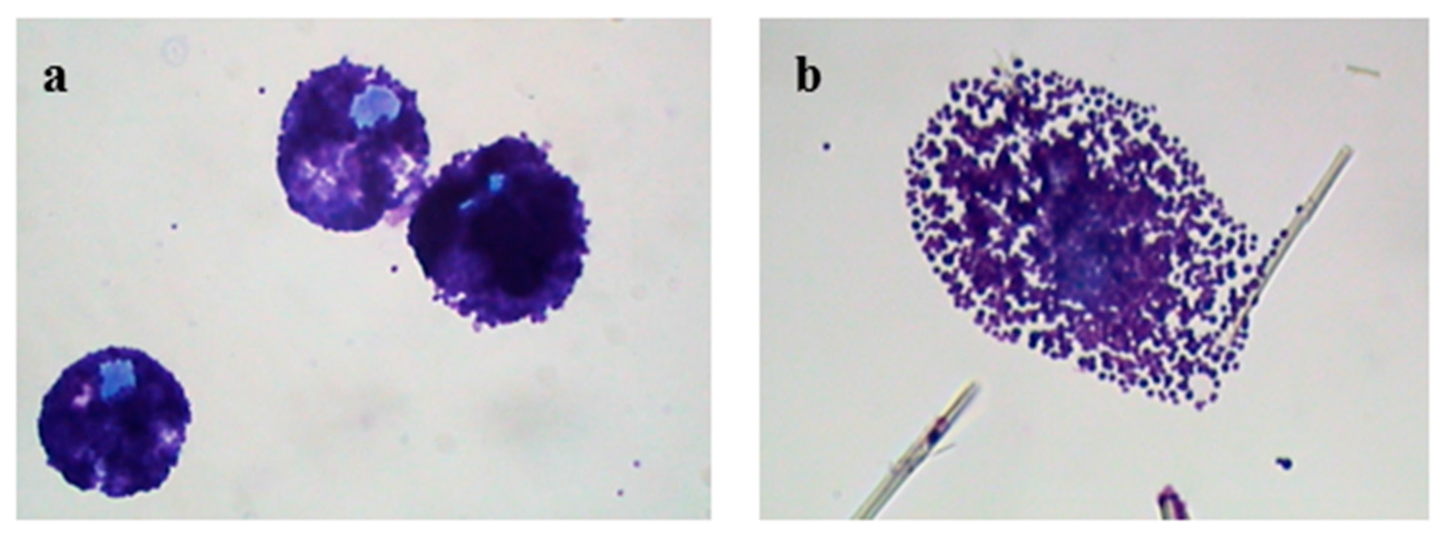
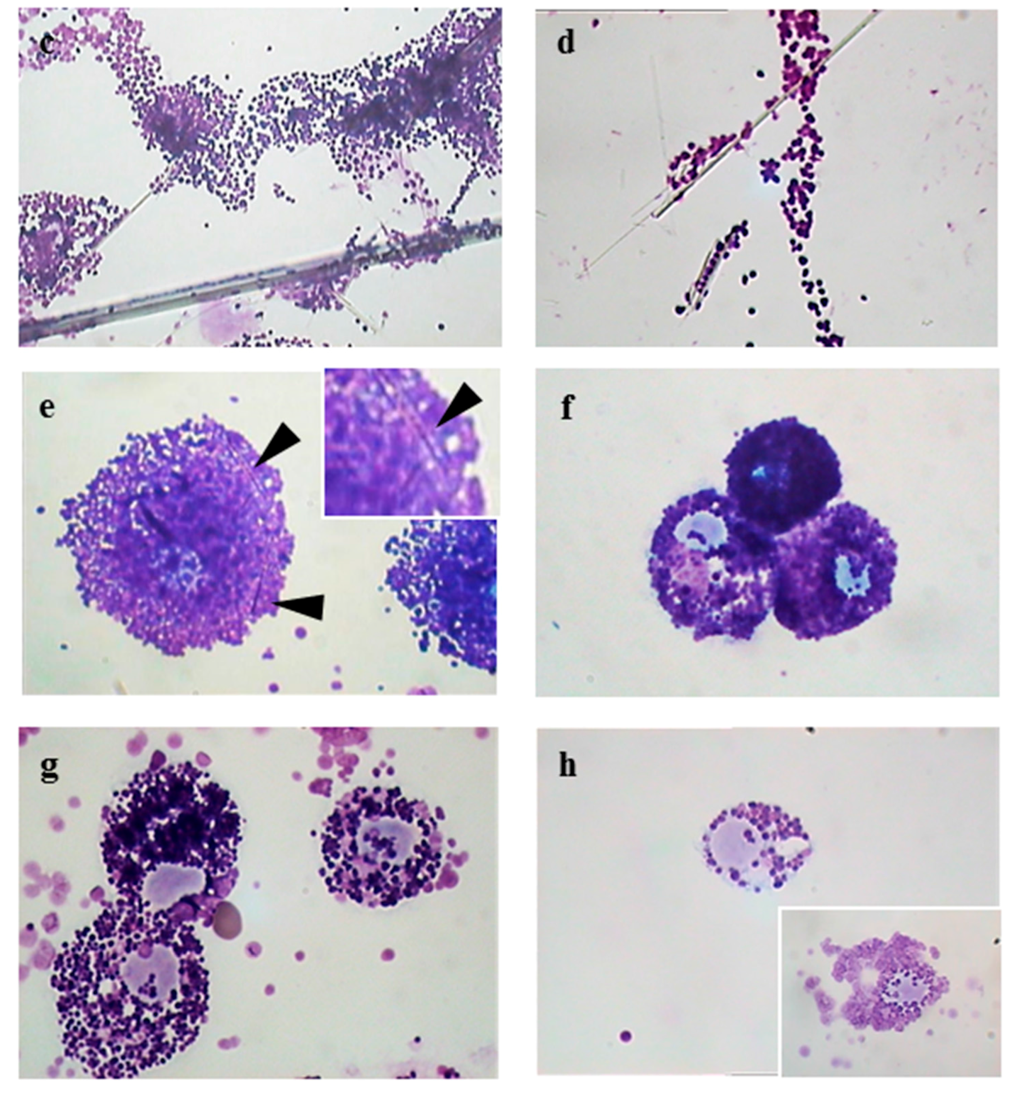
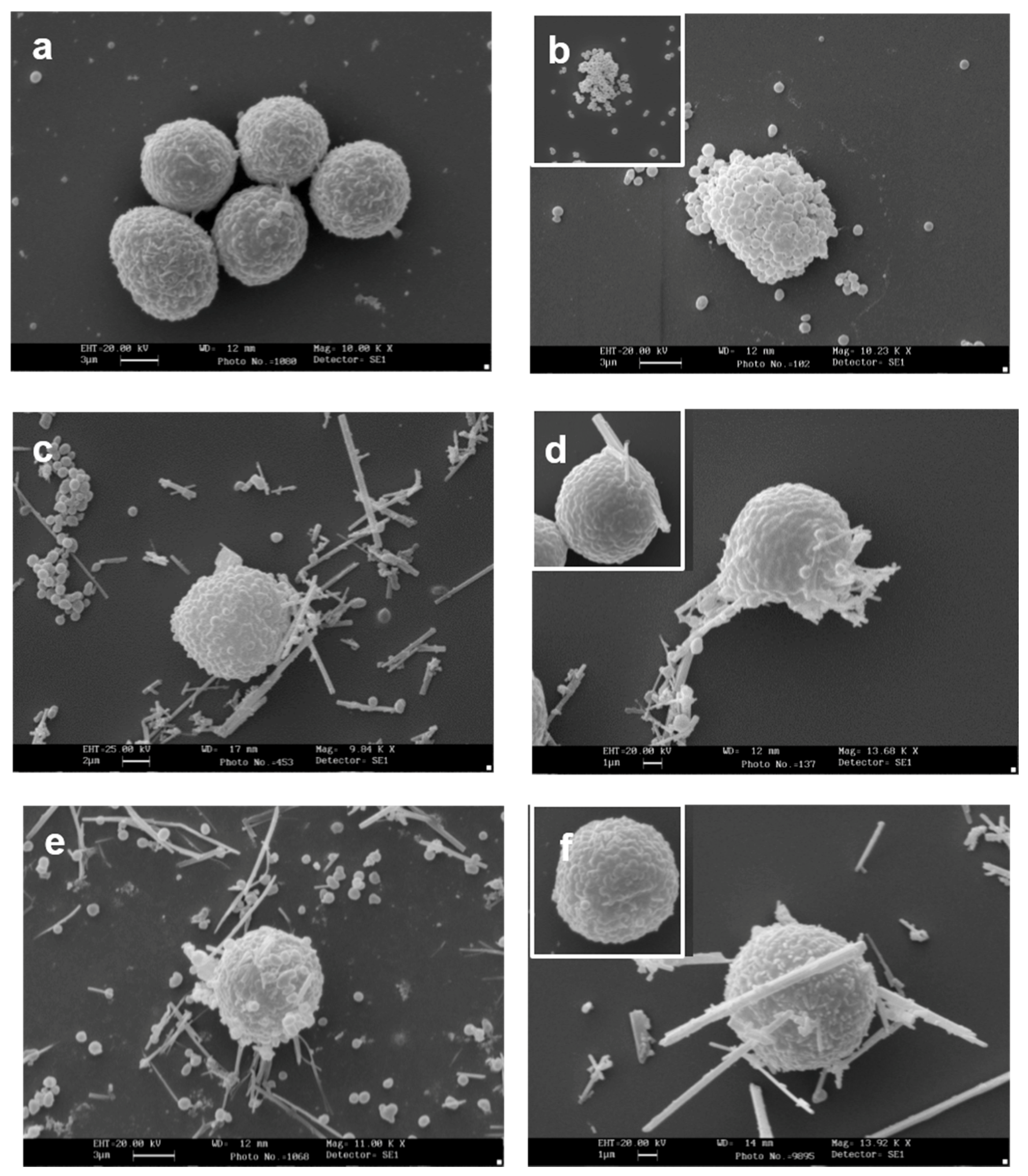
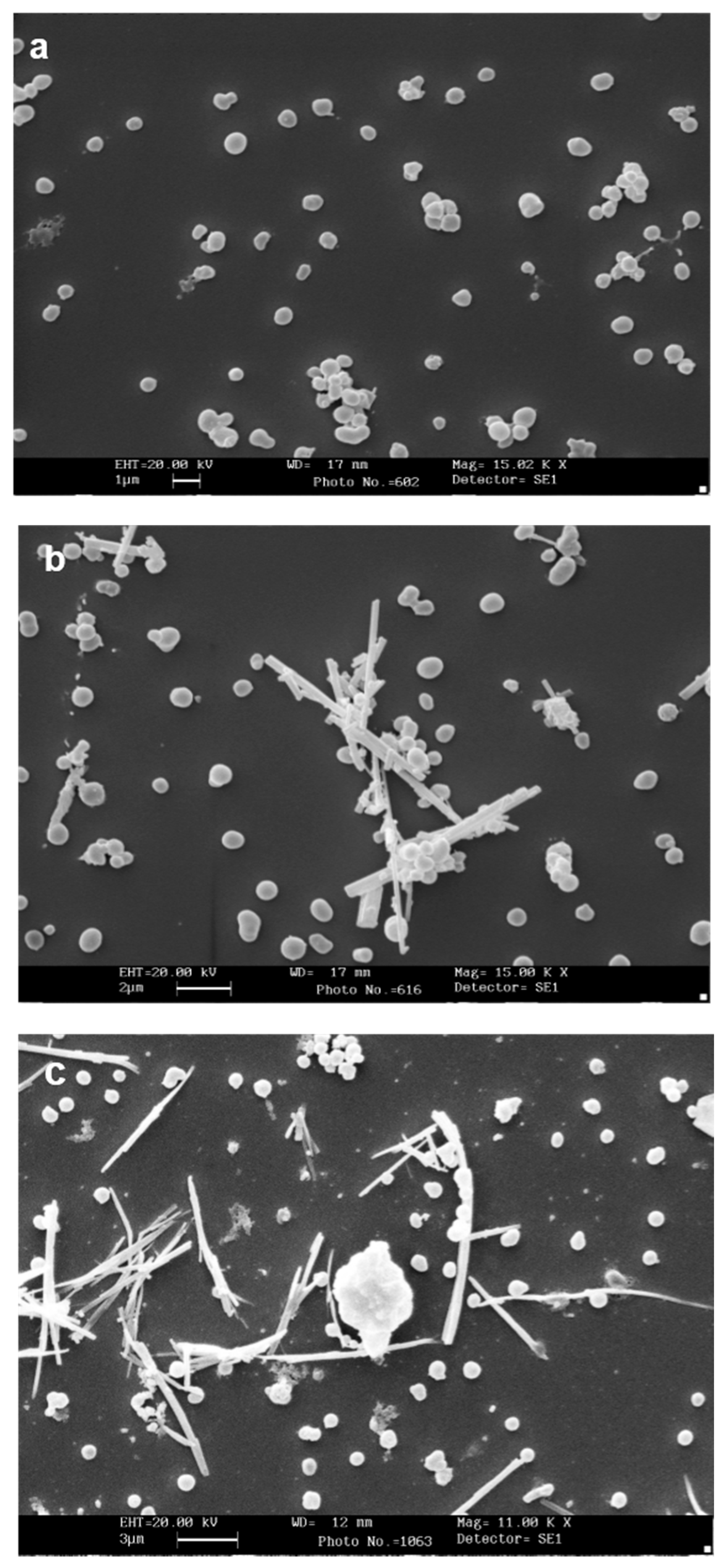
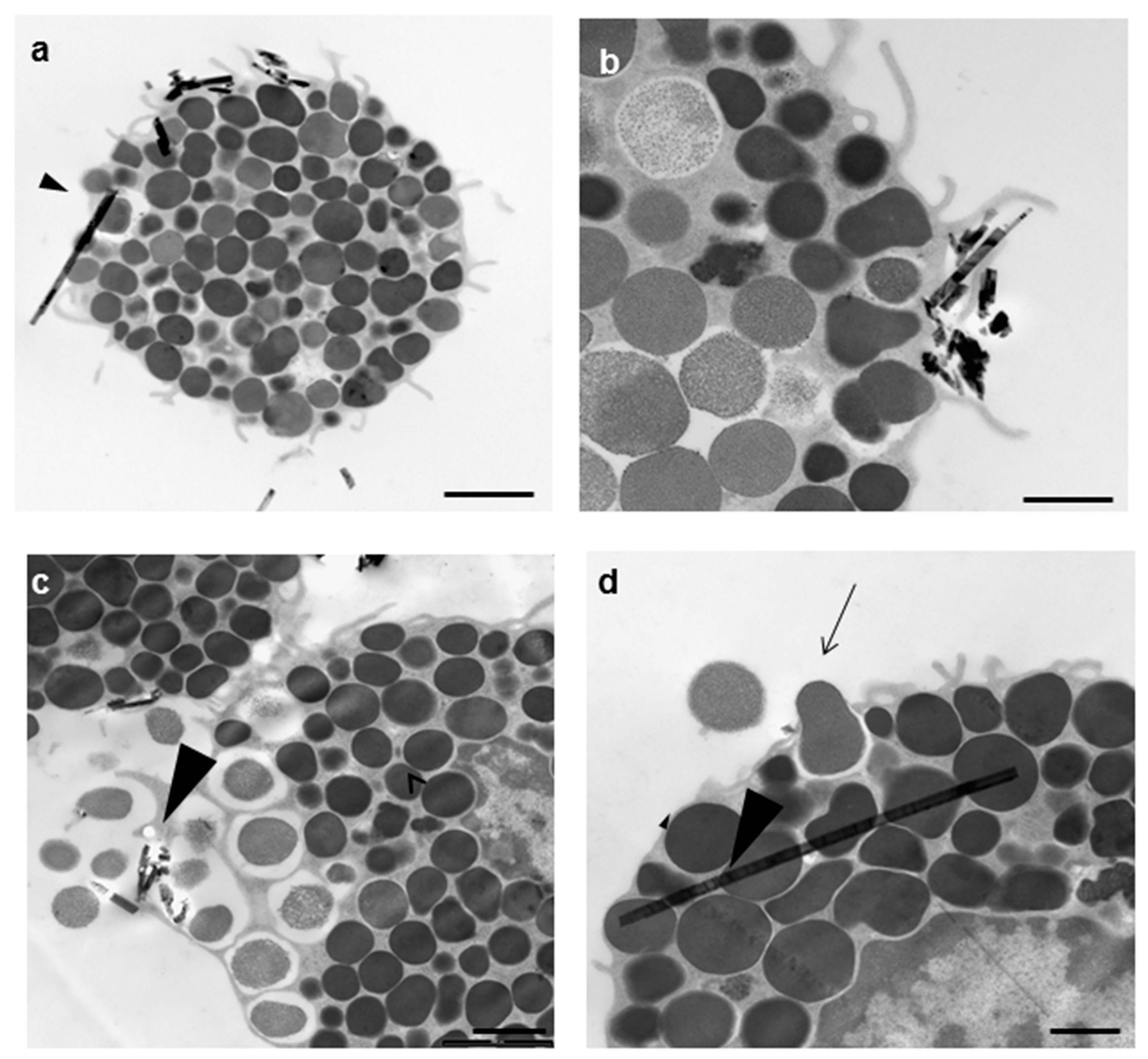
3.1. Morphological Analysis of Mineral Fiber–RPMC Interaction by Light and Electron Microscopy
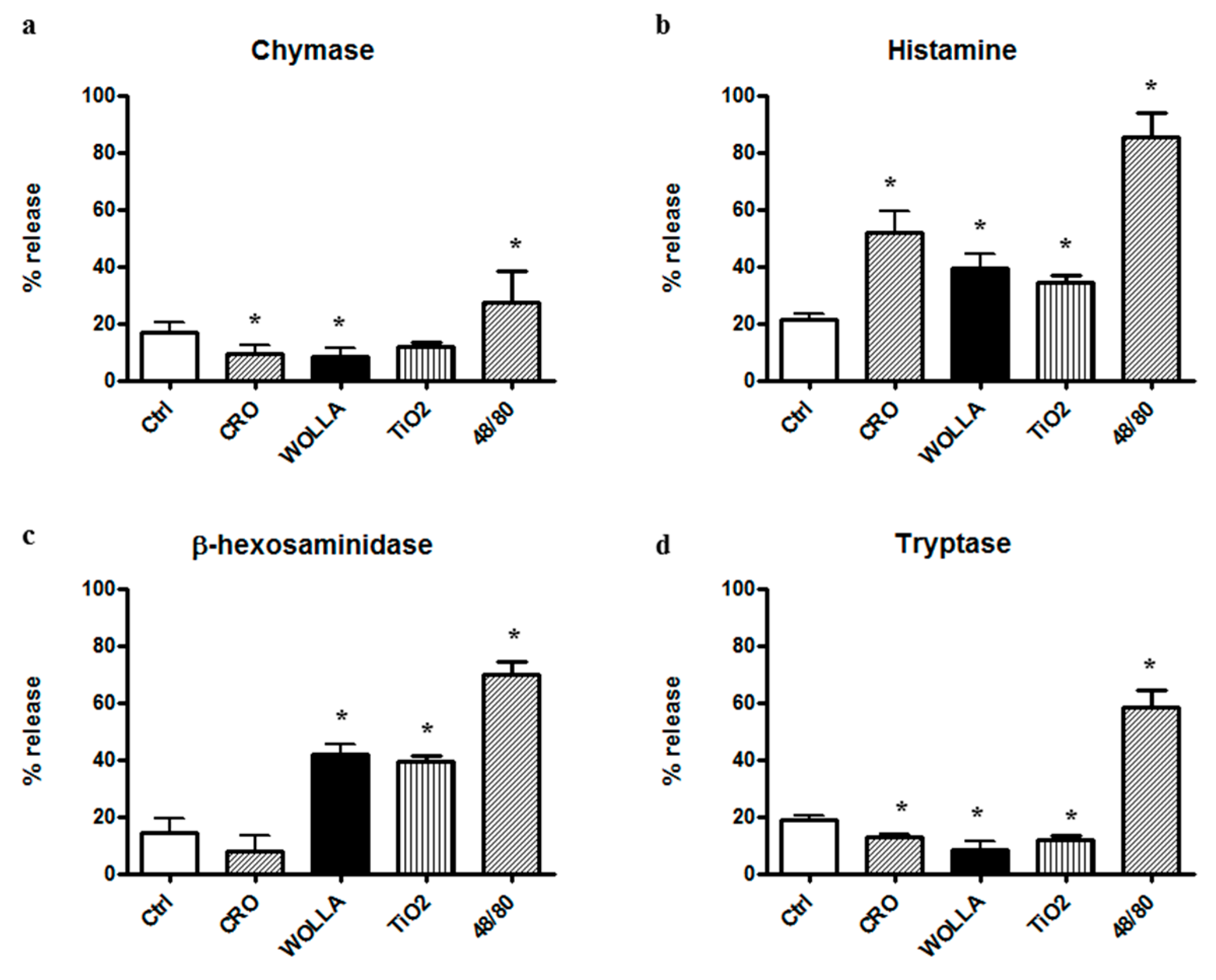
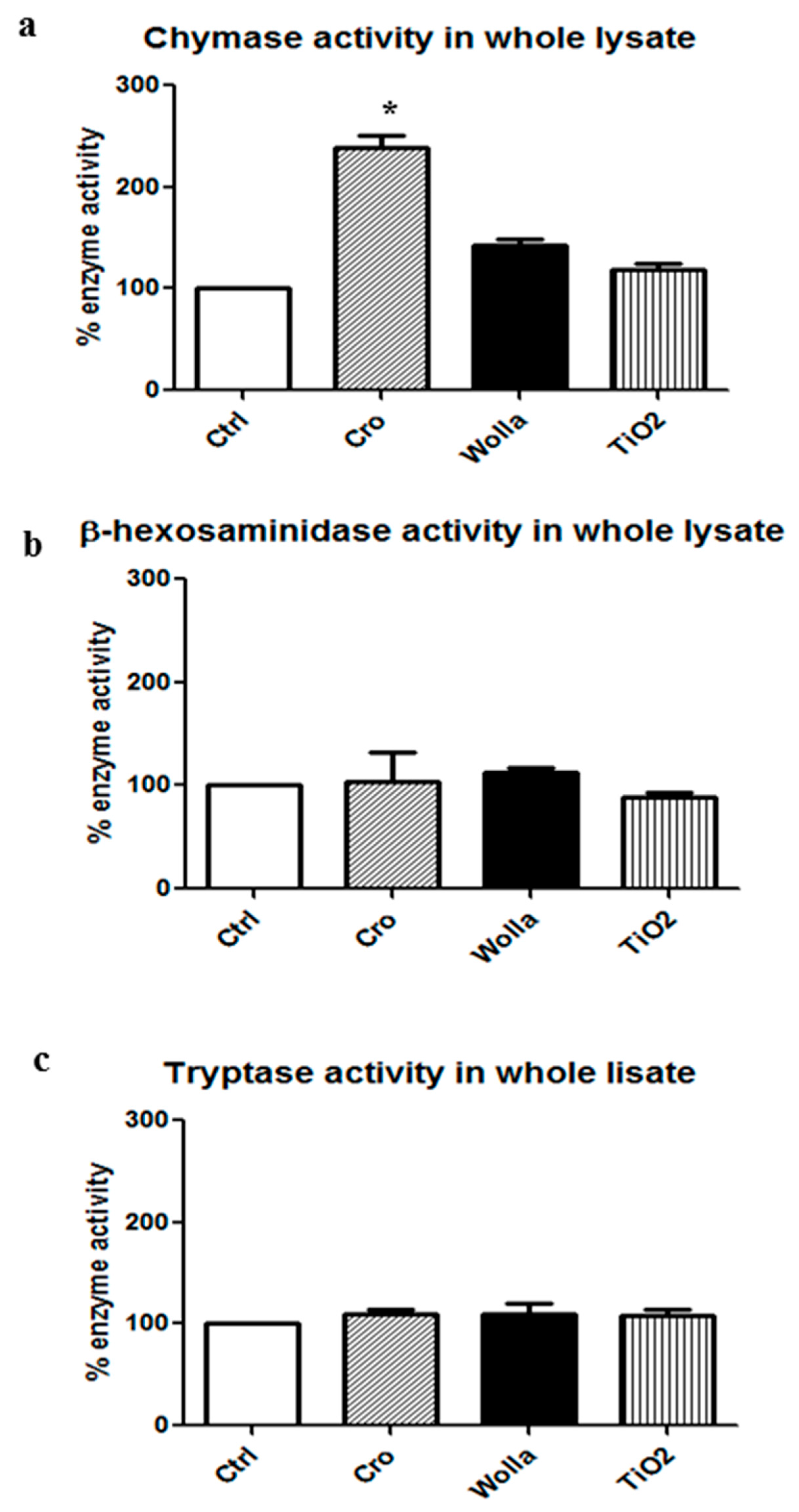
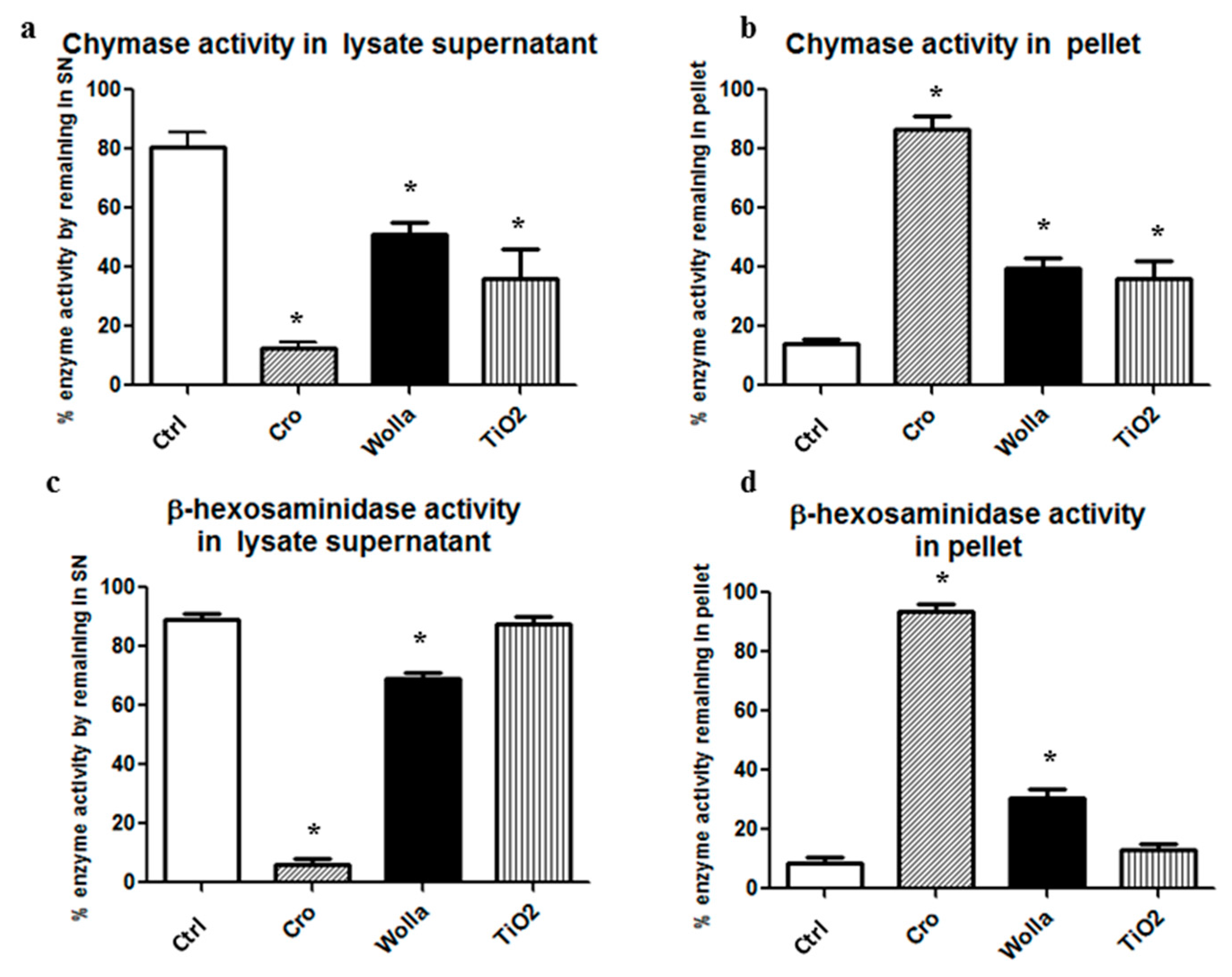
3.2. Quantization of Secretion of Granules Components Induced by Mineral Fibers
4. Discussion
5. Conclusions
- Mineral fibers are a potent stimulus for the mast cell secretory process, through both active (during membrane interaction) and passive (during cytosolic penetration) interactions. The rank order is: CRO > TiO2NW >> WOLLA. We demonstrate by serial sections at TEM analysis that the cytosolic inclusion of asbestos fibers is not an artifact, but that they are actually inside the cell. We therefore speculate that the presence of fibers inside the cytosol may be toxic and trigger cell rupture and granule expulsion. Of note, TiO2 nanoparticles were also found inside the cytosol without any surrounding membrane.
- Tissue-deposited fibers can aggregate large amounts of pro-inflammatory factors by binding free enzymes, intact granules, and granule remnants. Deposited fibers can continue to bind granules from MCs as cells are renewed and more fibers are inhaled, thereby creating a long-lasting pro-inflammatory environment.
- MCs could contribute to the inflammatory and toxic effects associated with some engineered nanomaterials, including TiO2NPs, which are widely used and are therefore a cause of increasing concern for their potential to harm humans.
Supplementary Materials
Acknowledgments
Author Contributions
Conflicts of Interest
Abbreviations
| BSA | bovine serum albumin |
| MCs | mast cells |
| RPMCs | rat peritoneal mast cells |
| β-hexo | β-hexosaminidase |
| CHY | chymase |
| TRY | tryptase |
| CRO | crocidolite |
| TiO2 | titanium oxide |
| NWs | nanowires |
| NPs | nanoparticles |
| WOLLA | wollastonite fibers |
| NLRP3 | NLR pyrin domain-containing 3 |
| PBS | phosphate buffered saline |
| SEM | scanning electron microscope |
| TEM | transmission electron microscope |
| IP | isoelectric point |
| hMPO | human myeloperoxidase |
| hEPO | eosinophil peroxidase |
| MCETs | mast cell extracellular traps |
| TMB | 3,3′,5,5′-Tetramethylbenzidine |
| TB | trypan blue |
| SN | supernatant |
| P | pellet |
| ROS | reactive oxygen species |
References
- Novello, S.; Pinto, C.; Torri, V.; Porcu, L.; Di Maio, M.; Tiseo, M.; Ceresoli, G.; Magnani, C.; Silvestri, S.; Veltri, A.; et al. The Third Italian Consensus Conference for Malignant Pleural Mesothelioma: State of the art and recommendations. Crit. Rev. Oncol. Hematol. 2016, 104, 9–20. [Google Scholar] [CrossRef] [PubMed]
- Ferin, J.; Oberdörster, G.; Penney, D.P. Pulmonary retention of ultrafine and fine particles in rats. Am. J. Respir. Cell Mol. Biol. 1992, 6, 535–542. [Google Scholar] [CrossRef] [PubMed]
- Magrez, A.; Horváth, L.; Smajda, R.; Salicio, V.; Pasquier, N.; Forró, L.; Schwaller, B. Cellular toxicity of TiO2-based nanofilaments. ACS Nano 2009, 3, 2274–2280. [Google Scholar] [CrossRef] [PubMed]
- Park, E.J.; Shim, H.W.; Lee, G.H.; Kim, J.H.; Kim, D.W. Comparison of toxicity between the different-type TiO2 nanowires in vivo and in vitro. Arch. Toxicol. 2013, 87, 1219–1230. [Google Scholar] [CrossRef] [PubMed]
- Wang, J.; Fan, Y. Lung Injury Induced by TiO2 Nanoparticles Depends on Their Structural Features: Size, Shape, Crystal Phases and Surface Coating. Int. J. Mol. Sci. 2014, 15, 22258–22278. [Google Scholar] [CrossRef] [PubMed]
- Hamilton, R.F.; Wu, N.; Porter, D.; Buford, M.; Wolfarth, M.; Holian, A. Particle length-dependent titanium dioxide nanomaterials toxicity and bioactivity. Part. Fiber Toxicol. 2009, 6, 35. [Google Scholar] [CrossRef] [PubMed]
- Vuong, N.Q.; Goegan, P.; Mohottalage, S.; Breznan, D.; Ariganello, M.; Williams, A.; Elisma, F.; Karthikeyan, S.; Vincent, R.; Kumarathasan, P. Proteomic changes in human lung epithelial cells (A549) in response to carbon black and titanium dioxide exposures. J. Proteom. 2016, 21, 53–63. [Google Scholar] [CrossRef] [PubMed]
- Allegri, M.; Bianchi, M.G.; Chiu, M.; Varet, J.; Costa, A.L.; Ortelli, S.; Blosi, M.; Bussolati, O.; Poland, C.A.; Bergamaschi, E. Shape-Related Toxicity of Titanium Dioxide Nanofibres. PLoS ONE 2016, 11, e0151365. [Google Scholar] [CrossRef] [PubMed]
- Ohyama, M.; Otake, T.; Morinaga, K. Effect of Size of Man-Made and Natural Mineral Fibers on Chemiluminescent Response in Human Monocyte-Derived Macrophages. Environ. Health Perspect. 2001, 109, 1033–1038. [Google Scholar] [CrossRef] [PubMed]
- Vergaro, V.; Aldieri, E.; Fenoglio, I.; Marucco, A.; Carlucci, C.; Ciccarella, G. Surface reactivity and in vitro toxicity on human bronchial epithelial cells (BEAS-2B) of nanomaterials intermediates of the production of titania-based composites. Toxicology 2016, 34, 171–178. [Google Scholar] [CrossRef] [PubMed]
- Watanabe, M.; Okada, M.; Kudo, Y.; Tonori, Y.; Niitsuya, M.; Sato, T.; Aizawa, Y.; Kotani, M. Differences in the effects of fibrous and particulate titanium dioxide on alveolar macrophages of Fischer 344 rats. J. Toxicol. Environ. Health A 2002, 65, 1047–1060. [Google Scholar] [CrossRef] [PubMed]
- Hong, J.; Wang, L.; Zhao, X.; Yu, X.; Sheng, L.; Xu, B.; Liu, D.; Zhu, Y.; Long, Y.; Hong, F. Th2 factors may be involved in TiO₂ NP-induced hepatic inflammation. J. Agric. Food Chem. 2014, 62, 6871–6878. [Google Scholar] [CrossRef] [PubMed]
- Eom, Y.; Song, J.S.; Lee, H.K.; Kang, B.; Kim, H.C.; Lee, H.K.; Kim, H.M. The Effect of Ambient Titanium Dioxide Microparticle Exposure to the Ocular Surface on the Expression of Inflammatory Cytokines in the Eye and Cervical Lymph Nodes. Investig. Ophthalmol. Vis. Sci. 2016, 57, 6580–6590. [Google Scholar] [CrossRef] [PubMed]
- Jain, A.K.; Senapati, V.A.; Singh, D.; Dubey, K.; Maurya, R.; Pandey, A.K. Impact of anatase titanium dioxide nanoparticles on mutagenic and genotoxic response in Chinese hamster lung fibroblast cells (V-79): The role of cellular uptake. Food Chem. Toxicol. 2017, 105, 127–139. [Google Scholar] [CrossRef] [PubMed]
- Fage, S.W.; Muris, J.; Jakobsen, S.S.; Thyssen, J.P. Titanium: A review on exposure, release, penetration, allergy, epidemiology, and clinical reactivity. Contact Dermat. 2016, 74, 323–345. [Google Scholar] [CrossRef] [PubMed]
- Pelclova, D.; Zdimal, V.; Kacer, P.; Fenclova, Z.; Vlckova, S.; Komarc, M.; Navratil, T.; Schwarz, J.; Zikova, N.; Makes, O.; et al. Leukotrienes in exhaled breath condensate and fractional exhaled nitric oxide in workers exposed to TiO2 nanoparticles. J. Breath Res. 2016, 10, 036004. [Google Scholar] [CrossRef] [PubMed]
- Pelclova, D.; Zdimal, V.; Kacer, P.; Zikova, F.; Komarc, M.; Fenclova, Z.; Vlckova, S.; Schwarz, J.; Makeš, O.; Syslova, K.; et al. Markers of lipid oxidative damage in the exhaled breath condensate of nano TiO2 production workers. Nanotechnology 2017, 11, 52–63. [Google Scholar] [CrossRef] [PubMed]
- Kang, C.M.; Jang, A.S.; Ahn, M.H.; Shin, J.A.; Kim, J.H.; Choi, Y.S.; Rhim, T.Y.; Park, C.S. Interleukin-25 and interleukin-13 production by alveolar macrophages in response to particles. Am. J. Respir. Cell Mol. Biol. 2005, 33, 290–296. [Google Scholar] [CrossRef] [PubMed]
- Östberg, A.K.; Dahlgren, U.; Sul, Y.T.; Johansson, C.B. Inflammatory cytokine release is affected by surface morphology and chemistry of titanium implants. J. Mater. Sci. Mater. Med. 2015, 26, 155. [Google Scholar] [CrossRef] [PubMed]
- Kumar, S.; Meena, R.; Paulraj, R. Role of Macrophage (M1 and M2) in Titanium-Dioxide Nanoparticle-Induced Oxidative Stress and Inflammatory Response in Rat. Appl. Biochem. Biotechnol. 2016, 180, 1257–1275. [Google Scholar] [CrossRef] [PubMed]
- Yanagisawa, R.; Takano, H.; Inoue, K.; Koike, E.; Kamachi, T.; Sadakane, K.; Ichinose, T. Titanium dioxide nanoparticles aggravate atopic dermatitis-like skin lesions in NC/Nga mice. Exp. Biol. Med. 2009, 234, 314–322. [Google Scholar] [CrossRef] [PubMed]
- Larsen, S.T.; Roursgaard, M.; Jensen, K.A.; Nielsen, G.D. Nano titanium dioxide particles promote allergic sensitization and lung inflammation in mice. Basic Clin. Pharmacol. Toxicol. 2010, 106, 114–117. [Google Scholar]
- Mishra, V.; Baranwal, V.; Mishra, R.K.; Sharma, S.; Paul, B.; Pandey, A.C. Titanium dioxide nanoparticles augment allergic airway inflammation and Socs3 expression via NF-κB pathway in murine model of asthma. Biomaterials 2016, 92, 90–102. [Google Scholar] [CrossRef] [PubMed]
- Ahn, M.H.; Kang, C.M.; Park, C.S.; Park, S.J.; Rhim, T.; Yoon, P.O.; Chang, H.S.; Kim, S.H.; Kyono, H.; Kim, K.C. Titanium dioxide particle-induced goblet cell hyperplasia: Association with mast cells and IL-13. Respir. Res. 2005, 13, 34. [Google Scholar] [CrossRef] [PubMed]
- Fu, Y.; Zhang, Y.; Chang, X.; Zhang, Y.; Ma, S.; Sui, J.; Yin, L.; Pu, Y.; Liang, G. Systemic immune effects of titanium dioxide nanoparticles after repeated intratracheal instillation in rat. Int. J. Mol. Sci. 2014, 15, 6961–6973. [Google Scholar] [CrossRef] [PubMed]
- Chang, X.; Fu, Y.; Zhang, Y.; Tang, M.; Wang, B. Effects of Th1 and Th2 cells balance in pulmonary injury induced by nano titanium dioxide. Environ. Toxicol. Pharmacol. 2014, 37, 275–283. [Google Scholar] [CrossRef] [PubMed]
- Huang, C.; Sun, M.; Yang, Y.; Wang, F.; Ma, X.; Li, J.; Wang, Y.; Ding, Q.; Ying, H.; Song, H.; et al. Titanium dioxide nanoparticles prime a specific activation state of macrophages. Nanotoxicology 2017, 15, 1–14. [Google Scholar] [CrossRef] [PubMed]
- Geiser, M.; Rothen-Rutishauser, B.; Kapp, N.; Schürch, S.; Kreyling, W.; Schulz, H.; Semmler, M.; Im Hof, V.; Heyder, J.; Gehr, P. Ultrafine particles cross cellular membranes by nonphagocytic mechanisms in lungs and in cultured cells. Environ. Health Perspect. 2005, 113, 1555–1560. [Google Scholar] [CrossRef] [PubMed]
- Singh, S.; Shi, T.; Duffin, R.; Albrecht, C.; van Berlo, D.; Höhr, D.; Fubini, B.; Martra, G.; Fenoglio, I.; Borm, P.J.; et al. Endocytosis, oxidative stress and IL-8 expression in human lung epithelial cells upon treatment with fine and ultrafine TiO2: Role of the specific surface area and of surface methylation of the particles. Toxicol. Appl. Pharmacol. 2007, 222, 141–151. [Google Scholar] [CrossRef] [PubMed]
- Malorni, W.; Iosi, F.; Falchi, M.; Donelli, G. On the mechanism of cell internalization of chrysotile fibers: An immunocytochemical and ultrastructural study. Environ. Res. 1990, 52, 164–177. [Google Scholar] [CrossRef]
- Andolfi, L.; Trevisan, E.; Zweyer, M.; Prato, S.; Troian, B.; Vita, F.; Borelli, V.; Soranzo, M.R.; Melato, M.; Zabucchi, G. The crocidolite fibers interaction with human mesothelial cells as investigated by combining electron microscopy, atomic force and scanning near-field optical microscopy. J. Microsc. 2013, 249, 173–183. [Google Scholar] [CrossRef] [PubMed]
- Bernareggi, A.; Ren, E.; Borelli, V.; Vita, F.; Constanti, A.; Zabucchi, G. Xenopus laevis Oocytes as a Model System for Studying the Interaction Between Asbestos Fibers and Cell Membranes. Toxicol. Sci. 2015, 45, 263–272. [Google Scholar] [CrossRef] [PubMed]
- Miles, S.E.; Sandrini, A.; Johnson, A.R.; Yates, D.H. Clinical consequences of asbestos-related diffuse pleural thickening: A review. J. Occup. Med. Toxicol. 2008, 3, 20. [Google Scholar] [CrossRef] [PubMed]
- Aust, A.E.; Cook, P.M.; Dodson, R.F. Morphological and chemical mechanisms of elongated mineral particle toxicities. J. Toxicol. Environ. Health B Crit. Rev. 2011, 14, 40–75. [Google Scholar] [CrossRef] [PubMed]
- Liu, G.; Cheresh, P.; Kamp, D.W. Molecular basis of asbestos-induced lung disease. Annu. Rev. Pathol. 2013, 24, 161–187. [Google Scholar] [CrossRef] [PubMed]
- Davis, J.M. Further observations on the ultrastructure and chemistry of the formation of asbestos bodies. Exp. Mol. Pathol. 1970, 13, 346–358. [Google Scholar] [CrossRef]
- Suzuki, Y. Interaction of asbestos with alveolar cells. Environ. Health Perspect. 1974, 9, 241–252. [Google Scholar] [CrossRef] [PubMed]
- Wright, A.; Donaldson, K.; Davis, J.M.G. Cytotoxic Effect of Asbestos on Macrophages in Different Activation States. Environ. Health Perspect. 1983, 5, 147–152. [Google Scholar] [CrossRef]
- Roney, P.L.; Holian, A. Possible mechanism of chrysotile asbestos-stimulated superoxide anion production in guinea pig alveolar macrophages. Toxicol. Appl. Pharmacol. 1989, 100, 132–144. [Google Scholar] [CrossRef]
- Koerten, H.K.; de Bruijn, J.D.; Daems, W.T. The formation of asbestos bodies by mouse peritoneal macrophages. An in vitro study. Am. J. Pathol. 1990, 137, 121–134. [Google Scholar] [PubMed]
- Perkins, R.C.; Scheule, R.K.; Holian, A. In vitro bioactivity of asbestos for the human alveolar macrophage and its modification by IgG. Am. J. Respir. Cell Mol. Biol. 1991, 4, 532–537. [Google Scholar] [CrossRef] [PubMed]
- Ando, T.G.T.; Verma, K.; Kagan, E. Asbestos fibers and interferon-gamma up-regulate nitric oxide production in rat alveolar macrophages. Am. J. Respir. Cell Mol. Biol. 1994, 11, 707–715. [Google Scholar]
- Hamilton, R.F.; Iyer, L.L.; Holian, A. Asbestos induces apoptosis in human alveolar macrophages. Am. J. Physiol. 1996, 271, L813–L819. [Google Scholar] [CrossRef] [PubMed]
- Holian, A.; Uthman, M.O.; Goltsova, T.; Brown, S.D.; Hamilton, R.F., Jr. Asbestos and silica-induced changes in human alveolar macrophage phenotype. Environ. Health Perspect. 1997, 105, 1139–1142. [Google Scholar] [CrossRef] [PubMed]
- Afaq, F.; Abidi, P.; Matin, R.; Rahman, Q. Activation of alveolar macrophages and peripheral red blood cells in rats exposed to fibers/particles. Toxicol. Lett. 1998, 99, 175–182. [Google Scholar] [CrossRef]
- Flaherty, D.M.; Monick, M.M.; Carter, A.B.; Peterson, M.W.; Hunninghake, G.W. Oxidant-mediated increases in redox factor-1 nuclear protein and activator protein-1 DNA binding in asbestos-treated macrophages. J. Immunol. 2002, 168, 5675–5681. [Google Scholar] [CrossRef] [PubMed]
- Blake, D.J.; Bolin, C.M.; Cox, D.P.; Cardozo-Pelaez, F.; Pfau, J.C. Internalization of Libby amphibole asbestos and 80 induction of oxidative stress in murine macrophages. Toxicol. Sci. 2007, 99, 277–288. [Google Scholar] [CrossRef] [PubMed]
- Rom, W.N. Role of oxidants in interstitial lung diseases: Pneumoconioses, constrictive bronchiolitis, and chronic tropical pulmonary eosinophilia. Mediat. Inflamm. 2011, 2011, 407657. [Google Scholar] [CrossRef] [PubMed]
- Kamp, D.W.; Liu, G.; Cheresh, P.; Kim, S.J.; Mueller, A.; Lam, A.P.; Trejo, H.; Williams, D.; Tulasiram, S.; Baker, M.; et al. Asbestos-induced alveolar epithelial cell apoptosis. The role of endoplasmic reticulum stress response. Am. J. Respir. Cell Mol. Biol. 2013, 49, 892–901. [Google Scholar] [CrossRef] [PubMed]
- Ryan, A.J.; Larson-Casey, J.L.; He, C.; Murthy, S.; Carter, A.B. Asbestos-induced disruption of calcium homeostasis induces endoplasmic reticulum stress in macrophages. J. Biol. Chem. 2014, 289, 33391–33403. [Google Scholar] [CrossRef] [PubMed]
- Yazdi, A.S.; Guarda, G.; Riteau, N.; Drexler, S.K.; Tardivel, A.; Couillin, I.; Tschopp, J. Nanoparticles activate the NLR pyrin domain containing 3 (Nlrp3) inflammasome and cause pulmonaryinflammation through release of IL-1α and IL-1β. Proc. Natl. Acad. Sci. USA 2010, 107, 19449–19454. [Google Scholar] [CrossRef] [PubMed]
- Dostert, C.; Pétrilli, V.; Van Bruggen, R.; Steele, C.; Mossman, B.T.; Tschopp, J. Innate immune activation through Nalp3 inflammasome sensing of asbestos and silica. Science 2008, 320, 674–677. [Google Scholar] [CrossRef] [PubMed]
- Rydman, E.M.; Ilves, M.; Vanhala, E.; Vippola, M.; Lehto, M.; Kinaret, P.A.; Pylkkänen, L.; Happo, M.; Hirvonen, M.R.; Greco, D.; et al. A single aspiration of rod-like carbon nanotubes induces asbestos-like pulmonary inflammation mediated in part by the IL-1R. Toxicol. Sci. 2015, 147, 140–155. [Google Scholar] [CrossRef] [PubMed]
- Maki, Y.; Nishimura, Y.; Toyooka, S.; Soh, J.; Tsukuda, K.; Shien, K.; Furukawa, M.; Muraoka, T.; Ueno, T.; Tanaka, N.; et al. The proliferative effects of asbestos-exposed peripheral blood mononuclear cells on mesothelial cells. Oncol. Lett. 2016, 11, 3308–3316. [Google Scholar] [CrossRef] [PubMed]
- Eagan, T.M.L.; Gulsvik, A.; Eide, G.E.; Bakke, P.S. Occupational Airborne Exposure and the Incidence of Respiratory Symptoms and Asthma. Am. J. Respir. Crit. Care Med. 2002, 166, 933–938. [Google Scholar] [CrossRef] [PubMed]
- Abraham, S.N.; Thankavel, K.; Malaviya, R. Mast cells as modulators of host defense in the lung. Front. Biosci. 1997, 15, 78–87. [Google Scholar] [CrossRef]
- Crivellato, E.; Ribatti, D. The mast cell: An evolutionary perspective. Biol. Rev. Camb. Philos. Soc. 2010, 85, 347–360. [Google Scholar] [CrossRef] [PubMed]
- Trevisan, E.; Vita, F.; Medic, N.; Soranzo, M.R.; Zabucchi, G.; Borelli, V. Mast cells kill Candida albicans in the extracellular environment but spare ingested fungi from death. Inflammation 2014, 37, 2174–2189. [Google Scholar] [CrossRef] [PubMed]
- Zabucchi, G.; Trevisan, E.; Vita, F.; Soranzo, M.R.; Borelli, V. NOD1 and NOD2 Interact with the Phagosome Cargo in Mast Cells: A Detailed Morphological Evidence. Inflammation 2015, 38, 1113–1125. [Google Scholar] [CrossRef] [PubMed]
- Puxeddu, I.; Piliponsky, A.M.; Bachelet, I.; Levi-Schaffer, F. Mast cells in allergy and beyond. Int. J. Biochem. Cell Biol. 2003, 35, 1601–1607. [Google Scholar] [CrossRef]
- Brown, E.J. Integrins, mast cells, and innate immunity. Blood 2004, 103, 1980–1981. [Google Scholar] [CrossRef]
- Frossi, B.; De Carli, M.; Pucillo, C. The mast cell: An antenna of the microenvironment that directs the immune response. J. Leukoc. Biol. 2004, 75, 579–585. [Google Scholar] [CrossRef] [PubMed]
- Metz, M.; Grimbaldeston, M.A.; Nakae, S.; Piliponsky, A.M.; Tsai, M.; Galli, S.J. Mast cells in the promotion and limitation of chronic inflammation. Immunol. Rev. 2007, 217, 304–328. [Google Scholar] [CrossRef] [PubMed]
- Hofmann, A.M.; Abraham, S.N. New roles for MC in modulating allergic reaction and immunity against phatogens. Curr. Opin. Immunol. 2009, 21, 679–686. [Google Scholar] [CrossRef] [PubMed]
- Fox, B.; Bull, T.B.; Guz, A. Mast cells in the human alveolar wall: An electronmicroscopic study. J. Clin. Pathol. 1981, 34, 1333–1342. [Google Scholar] [CrossRef] [PubMed]
- Choe, N.; Tanaka, S.; Xia, W.; Hemenway, D.R.; Roggli, V.L.; Kagan, E. Pleural macrophage recruitment and activation in asbestos-induced pleural injury. Environ. Health Perspect. 1997, 105, 1257–1260. [Google Scholar] [CrossRef] [PubMed]
- Andersson, C.K.; Mori, M.; Bjermer, L.; Löfdahl, C.G.; Erjefält, J.S. Novel site-specific mast cell subpopulations in the human lung. Thorax 2009, 64, 297–305. [Google Scholar] [CrossRef] [PubMed]
- Edwards, R.E.; Wagner, M.M.; Moncrieff, C.B. Cell population and histochemistry of asbestos related lesions of rat pleural cavity after injection of various inorganic dusts. Br. J. Ind. Med. 1984, 4, 506–513. [Google Scholar] [CrossRef]
- Wagner, M.M.; Edwards, R.E.; Moncrieff, C.B.; Wagner, J.C. Mast cells and inhalation of asbestos in rats. Thorax 1984, 39, 539–544. [Google Scholar] [CrossRef] [PubMed]
- Keith, I.; Day, R.; Lemaire, S.; Lemaire, I. Asbestos-induced fibrosis in rats: Increase in lung mast cells and autacoid contents. Exp. Lung Res. 1987, 13, 311–327. [Google Scholar] [CrossRef] [PubMed]
- Chen, H.; Xu, Y.; Yang, G.; Zhang, Q.; Huang, X.; Yu, L.; Dong, X. Mast cell chymase promotes hypertrophic scar fibroblast proliferation and collagen synthesis by activating TGF-β1/Smads signaling pathway. Exp. Ther. Med. 2017, 14, 4438–4442. [Google Scholar] [CrossRef] [PubMed]
- Smith, M.J.; Brown, J.M.; Zamboni, W.C.; Walker, N.J. From immunotoxicity to nanotherapy: The effects of nanomaterials on the immune system. Toxicol. Sci. 2014, 138, 249–255. [Google Scholar] [CrossRef] [PubMed]
- Xu, J.; Alexander, D.B.; Futakuchi, M.; Numano, T.; Fukamachi, K.; Suzui, M.; Omori, T.; Kanno, J.; Hirose, A.; Tsuda, H. Size- and shape-dependent pleural translocation, deposition, fibrogenesis, and mesothelial proliferation by multiwalled carbon nanotubes. Cancer Sci. 2014, 105, 763–769. [Google Scholar] [CrossRef] [PubMed]
- Rydman, E.M.; Ilves, M.; Koivisto, A.J.; Kinaret, P.A.S.; Fortino, V.; Savinko, T.S.; Lehto, M.T.; Pulkkinen, V.; Vippola, M.; Hämeri, K.J.; et al. Inhalation of rod-like carbon nanotubes causes unconventional allergic airway. Part. Fiber Toxicol. 2014, 11, 48. [Google Scholar] [CrossRef] [PubMed]
- Aldossari, A.A.; Shannahan, J.H.; Podila, R.; Brown, J.M. Influence of physicochemical properties of silver nanoparticles on mast cell activation and degranulation. Toxicology 2015, 29, 195–203. [Google Scholar] [CrossRef] [PubMed]
- Chen, E.Y.; Garnica, M.; Wang, Y.C.; Mintz, A.J.; Chen, C.S.; Chin, W.C. A mixture of anatase and rutile TiO2 nanoparticles induces histamine secretion in mast cells. Part. Fiber Toxicol. 2012, 9, 2. [Google Scholar] [CrossRef] [PubMed]
- Governa, M.; Camilucci, L.; Amati, M.; Visonà, I.; Valentino, M.; Botta, G.C.; Campopiano, A.; Fanizza, C. Wollastonite fibers in vitro generate reactive oxygen species able to lyse erythrocytes and activate the complement alternate pathway. Toxicol. Sci. 1998, 44, 32–38. [Google Scholar] [CrossRef] [PubMed]
- Kohyama, N.; Shinohara, Y.; Suzuki, Y. Mineral phases and some reexamined characteristics of the International Union against Cancer standard asbestos samples. Am. J. Ind. Med. 1996, 30, 515–528. [Google Scholar] [CrossRef]
- Kusiorowski, R.; Zaremba, T.; Gerle, A.; Piotrowski, J.; Simka, W.; Adamek, J. Study on the thermal decomposition of crocidolite asbestos. J. Therm. Anal. Calorim. 2015, 120, 1585–1595. [Google Scholar] [CrossRef]
- Sehati, S.; Entezari, M.H. Sono-intercalation of CdS nanoparticles into the layers of titanate facilitates the sunlight degradation of Congo red. J. Colloid. Interface Sci. 2016, 462, 130–139. [Google Scholar] [CrossRef] [PubMed]
- Medic, N.; Vita, F.; Abbate, R.; Soranzo, M.R.; Pacor, S.; Fabbretti, E.; Borelli, V.; Zabucchi, G. Mast cell activation by myelin through scavenger receptor. J. Neuroimmunol. 2008, 200, 27–40. [Google Scholar] [CrossRef] [PubMed]
- Shore, P.; Burkhalter, A.; Cohn, V., Jr. A method for the fluorometric assay of histamine in tissues. J. Pharmacol. Exp. Ther. 1959, 127, 182–186. [Google Scholar] [PubMed]
- Lindstedt, K.A.; Kovanen, P.T. Isolation of mast cell granules. Curr. Protoc. Cell Biol. 2006, 3, 16. [Google Scholar] [PubMed]
- Zabucchi, G.; Menegazzi, R.; Soranzo, M.R.; Patriarca, P. Uptake of human eosinophil peroxidase by human neutrophils. Am. J. Pathol. 1986, 124, 510–518. [Google Scholar] [PubMed]
- Menegazzi, R.; Zabucchi, G.; Knowles, A.; Cramer, R.; Patriarca, P. A new, one-step assay on whole cell suspensions.for peroxidase secretion by human neutrophils and eosinophils. J. Leukoc. Biol. 1992, 52, 619–624. [Google Scholar] [PubMed]
- Brody, A.R.; George, G.; Hill, L.H. Interactions of chrysotile and crocidolite asbestos with red blood cell membranes. Chrysotile binds to sialic acid. Lab. Investig. 1983, 49, 468–475. [Google Scholar]
- Brody, A.R.; Hill, L.H. Interactions of chrysotile asbestos with erythrocyte membranes. Environ. Health Perspect. 1983, 51, 85–89. [Google Scholar] [CrossRef] [PubMed]
- Iguchi, H.; Kojo, S. Possible generation of hydrogenperoxide and lipid peroxidation of erythrocyte membrane by asbestos: Cytotoxic mechanism of asbestos. Biochem. Int. 1989, 8, 981–990. [Google Scholar]
- Gendek, E.G.; Brody, A.R. Changes in lipid ordering of model phospholipid membranes treated with chrysotile and crocidolite asbestos. Environ. Res. 1990, 53, 152–167. [Google Scholar] [CrossRef]
- Elferink, J.G.; Kelters, I. Chrysotile asbestos-induced membrane damage in human erythrocytes. Res. Commun. Chem. Pathol. Pharmacol. 1991, 73, 355–365. [Google Scholar] [PubMed]
- Johnson, N.F.; Davies, R. Effect of asbestos on the P388D1 macrophage-like cell line: Preliminary ultrastructural observations. Environ. Health Perspect. 1983, 51, 109–117. [Google Scholar] [CrossRef] [PubMed]
- Palomaki, J.; Valimaki, E.; Sund, J.; Vippola, M.; Clausen, P.A.; Jensen, K.A.; Savolainen, K.; Matikainen, S.; Alenius, H. Long, needle-like carbon nanotubes and asbestos activate the Nlrp3 inflammasome through a similar mechanism. ACS Nano 2011, 5, 6861–6870. [Google Scholar] [CrossRef] [PubMed]
- Rajotte, D.; Stearns, C.D.; Alisa, K.K. Isolation of Mast Cell Secretory Lysosomes Using Flow Cytometry. Cytom. Part A 2003, 55, 94–101. [Google Scholar] [CrossRef] [PubMed]
- Lindstedt, K.A.; Kokkonen, J.O.; Kovanen, P.T. Soluble heparin proteoglycans released from stimulated mast cells induce uptake of low density lipoproteins by macrophages via scavenger receptor-mediated phagocytosis. J. Lipid Res. 1992, 33, 65–75. [Google Scholar] [PubMed]
- Wu, L.; Wu, S.; Xu, Z.; Qiu, Y.; Li, S.; Xu, H. Modified nanoporous titanium dioxide as a novel carrier for enzyme immobilization. Biosens. Bioelectron. 2016, 80, 59–66. [Google Scholar] [CrossRef] [PubMed]
- Lazarus, G.S. Mastocytosis: New understandings in cutaneous pathophysiology. J. Dermatol. 1996, 23, 769–772. [Google Scholar] [CrossRef] [PubMed]
- Lefrançais, E.; Duval, A.; Mirey, E.; Roga, S.; Espinosa, E.; Cayrol, C.; Girard, J.P. Central domain of IL-33 is cleaved by mast cell proteases for potent activation of group-2 innate lymphoid cells. Proc. Natl. Acad. Sci. USA 2014, 28, 15502–15507. [Google Scholar] [CrossRef] [PubMed]
- Waern, I.; Lundequist, A.; Pejler, G.; Wernersson, S. Mast cell chymase modulates IL-33 levels and controls allergic sensitization in dust-mite induced airway inflammation. Mucosal Immunol. 2013, 6, 911–920. [Google Scholar] [CrossRef] [PubMed]
- Dodson, R.F.; Williams, M.G., Jr.; Corn, C.J.; Brollo, A.; Bianchi, C. Asbestos content of lung tissue, lymph nodes, and pleural plaques from former shipyard workers. Am. Rev. Respir. Dis. 1990, 142, 843–847. [Google Scholar] [CrossRef] [PubMed]
- Abel, J.; Goldmann, O.; Ziegler, C.; Höltje, C.; Smeltzer, M.S.; Cheung, A.L.; Bruhn, D.; Rohde, M.; Medina, E. Staphylococcus aureus evades the extracellular antimicrobial activity of mast cells by promoting its own uptake. J. Innate Immun. 2011, 3, 495–507. [Google Scholar] [CrossRef] [PubMed]


| Isoelectric Point | Chymase Activity | β-Hexosaminidase Activity | Tryptase Activity | ||||||||||
|---|---|---|---|---|---|---|---|---|---|---|---|---|---|
| (IP) | 9.6 | 5.6–6.1 | 6.0–6.3 | ||||||||||
| CTRL | CRO | WOLLA | TiO2 | CTRL | CRO | WOLLA | TiO2 | CTRL | CRO | WOLLA | TiO2 | ||
| RPMC | % Release | 16.5 ± 3.8 | 9.4 ± 2.6 * | 8.2 ± 3.2 * | 12.00 ± 2.6 | 14.2 ± 5.2 | 8.1 ± 5.1 | 41.0 ± 3.6 * | 39.1 ± 2.9 * | 19 ± 3.0 | 13.0 ± 1.5 | 8.0 ± 2.0 | 10.5 ± 1.8 |
| Lysate | % increment | 100 | 238.5 ± 19.6 * | 143.0 ± 7.1 | 125.0 ± 7.1 | 100 | 103.3 ± 51.3 | 112.0 ± 7.0 | 88.00 ± 10.2 | 100 | 125 ± 6.0 | 110 ± 12.2 | 105 ± 5.0 |
| % pellet activity | 13.8 ± 2.5 | 86.5 ± 7.7 * | 39.7 ± 6.1 * | 36.2 ± 10.1 * | 8.6 ± 3.5 | 93.6 ± 4.1 * | 30.4 ± 5.5 * | 12.9 ± 4.0 | nd | nd | nd | nd | |
| Human pure enzymes | % increment | 100 | 224.9 ± 44.6 * | 244.6 ± 50.8 * | 244.7 ± 8.3 * | nd | nd | nd | nd | 100 | 160 ± 10.0 | 97.0 ± 12.0 | 110.0 ± 6.0 |
| % pellet activity | 8.1 ± 2.1 | 68.9 ± 11.7 * | 61.8 ± 1.9 * | 54.3 ± 9.1 * | nd | nd | nd | nd | nd | nd | nd | nd | |
© 2018 by the authors. Licensee MDPI, Basel, Switzerland. This article is an open access article distributed under the terms and conditions of the Creative Commons Attribution (CC BY) license (http://creativecommons.org/licenses/by/4.0/).
Share and Cite
Borelli, V.; Trevisan, E.; Francesca, V.; Zabucchi, G. The Secretory Response of Rat Peritoneal Mast Cells on Exposure to Mineral Fibers. Int. J. Environ. Res. Public Health 2018, 15, 104. https://doi.org/10.3390/ijerph15010104
Borelli V, Trevisan E, Francesca V, Zabucchi G. The Secretory Response of Rat Peritoneal Mast Cells on Exposure to Mineral Fibers. International Journal of Environmental Research and Public Health. 2018; 15(1):104. https://doi.org/10.3390/ijerph15010104
Chicago/Turabian StyleBorelli, Violetta, Elisa Trevisan, Vita Francesca, and Giuliano Zabucchi. 2018. "The Secretory Response of Rat Peritoneal Mast Cells on Exposure to Mineral Fibers" International Journal of Environmental Research and Public Health 15, no. 1: 104. https://doi.org/10.3390/ijerph15010104
APA StyleBorelli, V., Trevisan, E., Francesca, V., & Zabucchi, G. (2018). The Secretory Response of Rat Peritoneal Mast Cells on Exposure to Mineral Fibers. International Journal of Environmental Research and Public Health, 15(1), 104. https://doi.org/10.3390/ijerph15010104