Article

Protective Roles of Sodium Selenite against Aflatoxin B1-Induced Apoptosis of Jejunum in Broilers

Key Laboratory of Animal Diseases and Environmental Hazards of Sichuan Province, College of Veterinary Medicine, Sichuan Agricultural University, Ya'an 625014, China
*
Authors to whom correspondence should be addressed.
These authors contributed equally to this study.
Int. J. Environ. Res. Public Health 2014, 11(12), 13130-13143; https://doi.org/10.3390/ijerph111213130
Submission received: 16 July 2014 / Revised: 4 December 2014 / Accepted: 4 December 2014 / Published: 17 December 2014

Abstract

:
The effects of aflatoxin B1 (AFB1) exposure and sodium selenite supplementation on cell apoptosis of jejunum in broilers were studied. A total of 240 one-day-old male AA broilers were randomly assigned four dietary treatments containing 0 mg/kg of AFB1 (control), 0.3 mg/kg AFB1 (AFB1), 0.4 mg/kg supplement Se (+ Se) and 0.3 mg/kg AFB1 + 0.4 mg/kg supplement Se (AFB1 + Se), respectively. Compared with the control broilers, the number of apoptotic cells, the expression of Bax and Caspase-3 mRNA were significantly increased, while the expression of Bcl-2 mRNA and the Bcl-2/Bax ratio were significantly decreased in AFB1 broilers. The number of apoptotic cells and the expression of Caspase-3 mRNA in AFB1 + Se broilers were significantly higher than those in the control broilers, but significantly lower than those in AFB1 broilers. There were no significant changes in the expression of Bax mRNA between AFB1 + Se and control broilers; the expression of Bcl-2 mRNA and the Bcl-2/Bax ratio in AFB1 + Se broilers were significantly lower than those in the control broilers, but significantly higher than those in AFB1 broilers. In conclusion, 0.3 mg/kg AFB1 in the diet can increase cell apoptosis, decrease Bcl-2 mRNA expression, and increase of Bax and Caspase-3 mRNA expression in broiler’s jejunum. However, supplementation of dietary sodium selenite at the concentration of 0.4 mg/kg Se may ameliorate AFB1-induced apoptosis by increasing Bcl-2 mRNA expression, and decreasing Bax and Caspase-3 mRNA expression.

1. Introduction

Aflatoxin B1 (AFB1) is a well-known mycotoxin produced by different strains of Aspergillus flavus and Aspergillus parasiticum. In humans and various animal species, it has been reported to be a potent hepatotoxic and hepatocarcinogenic agent [1,2]. AFB1 is readily transported across the plasma membrane and interacts with nucleic acids and proteins, altering various cellular activities [3]. Previous research has shown that poultry are extremely sensitive to the toxic and carcinogenic action of AFB1, resulting in millions of dollars in annual losses to producers due to reduced growth rates, reduced egg production, increased susceptibility to disease, and other adverse effects [4,5,6,7,8,9].
Apoptosis is a specialized process of cell death that is part of the normal development of organs and tissue maintenance, but may also occur as a response to various environmental stimuli, indicating toxicity. Early research has shown that AFB1 is able to being a direct and indirect initiator as well as promoter of apoptotic process [10,11]. Several studies indicated that AFB1 induced apoptosis of different cells, such as hepatocyte [12], bone marrow cells [13], and bronchial epithelial cells [14]. Moreover, Chen et al. [15] reported that 0.3 mg/kg AFB1 in the broilers’ diet could induce the increase of apoptotic thymocytes by the up-regulation of Bax and Caspase-3 expression and the down-regulation of Bcl-2 expression. Similarly, Wang et al. [16] has demonstrated that AFB1 could increase the percentage of apoptotic splenocytes in broilers, which was closely related to oxidative stress. However, the effects of AFB1 on the apoptosis of jejunum were rarely reported. The gastrointestinal tract is the main site where conversion and absorption of food components takes place. As part of the small intestine, the jejunum is the major component of the gastrointestinal tract. Epithelium cells in the small intestine have a high turnover and as it is essential to maintain normal balance, apoptosis is crucial for maintenance of normal morphology and function [17,18,19]. Since poultry are extremely sensitive to the toxic and carcinogenic action of AFB1, studies on AFB1-related apoptosis in the jejunum in broilers are very important.
As an important micronutrient for humans and animals, Selenium (Se) plays a vital role in biological systems, such as chemopreventive [20], antioxidant, detoxification [21] and anticancer effects [22], and effects on both the innate and acquired immune system [23,24]. Furthermore, Se plays a key role in cell apoptosis [22]. At nutritional doses, Se is an essential component of selenocysteine (SeCys) in selenoproteins, and it promotes cell cycle progression and prevents cell death [22]. Previous studies have shown that Se could counteract the adverse effects of AFB1 in poultry [4,15,16,24,25]. For example, Se may ameliorate AFB1-induced lesions of the thymus and accordingly improve the impaired cellular immune function in broilers [15]. Similarly, Se may exhibit protective effects on AFB1-induced splenic toxicity by inhibiting oxidative stress and excessive apoptosis [16]. Recently, our study has demonstrated that supplementation of dietary sodium selenite at the concentration of 0.4 mg/kg Se protected the jejunum from the developmental retardation, decreased proliferation, and G2/M phase arrest caused by AFB1 [26]. However, the effects of Se against AFB1-induced jejunal cell apoptosis have yet not been reported. In the present research, experiments were conducted to examine the effect of AFB1 exposure and sodium selenite supplementation on the cell apoptosis of broilers’ jejunum by TUNEL assay and quantitative real-time PCR.

2. Materials and Methods

2.1. Animals and Diets

Two hundred forty 1-day-old healthy male AA broilers were obtained from a commercial rearing farm (Wenjiang poultry farm, Sichuan Province). Chickens were randomly assigned four dietary treatments containing 0 mg/kg of AFB1 (control), 0.3 mg/kg AFB1 (AFB1), 0.4 mg/kg supplement Se (+ Se) and 0.3 mg/kg AFB1 + 0.4 mg/kg supplement Se (AFB1 + Se), respectively. Our earlier studies have demonstrated that 0.3 mg/kg AFB1 in diet had obvious adverse effects on broilers, and an appropriate level of Se supplied in the diet (0.4 mg/kg) could provide optimal protective effects against AFB1-induced toxicity in broilers [15,16]. Based on this information, an appropriate toxin concentration (0.3 mg/kg AFB1) and dietary Se level (0.4 mg/kg) were chosen. 1% Feed-grade sodium selenite was mixed into the control diet to formulate + Se and AFB1 + Se diets containing 0.4 mg/kg Se supplement by a stepwise dilution method. AFB1 was obtained from Pribolab Pte. Ltd (Singapore). The AFB1-contaminated diets were made up according to the method described by Kaoud [27]. Briefly, 3 mg AFB1 was completely dissolved in 30 mL methanol, and then the 30 mL mixture was mixed into a 10 kg corn-soybean basal diet to formulate the AFB1 and AFB1 + Se diets, respectively. An equivalent amount of methanol was mixed into corn-soybean basal diet to produce the control diet. Then the methanol of the diets was evaporated at 98 °F (37 °C)(the concentration of dietary AFB1 was not detected in this experiment, but it can be assured that any possible back-ground contamination was evenly distributed among the experimental groups throughout the trial because the same lot of basal diet was used for formulating experimental diets). The content of Se (0.332 mg/kg) in the control diet was analyzed by hydride-generation atomic absorption spectroscopy. Broilers were housed in cages with electrically heated units and were provided with water as well as the aforementioned diets ad libitum for 21 days. The basal diets were formulated according to National Research Council (NRC, 1994) and Chinese Feeding Standard of Chicken (NY/T33-2004) recommendations to meet the nutrient requirements of broilers from 1 to 21 days. The composition of the basal diets is presented in Table 1. All procedures of the experiment were performed in compliance with the laws and guidelines of Sichuan Agricultural University Animal Care and Use Committee.

2.2. Clinical Signs and Body Weight

The clinical symptoms were observed each day. At 7, 14 and 21 days of age during the experiment, the body weight of chicken in each group was measured.
Table 1. Composition of the Basal Diet.
CompositionContent (%)NutrientContent (%)
corn51.95crude protein (CP)21.50
soybean39.50Methionine (Met)0.50
rapeseed oil4.10calcium (Ca)1.00
D,L-metionine0.20all phosphorus (P)0.70
calcium hydrogen phosphate1.85Methionine + cysteine (Met+Cys)0.84
calcium carbonate1.30lysine (Lys)1.15
sodium chloride0.40Threonine (Thr)0.83
trace element premix a0.50metabolizable energy (ME) (MJ/Kg)29.90
choline0.17
multivitamins b0.03
total100
Note: a trace element premix (mg/kg): FeSO4·7H2O, 530; CuSO4·5H2O, 30; MnSO4·H2O, 400; ZnSO4·7H2O, 470; KI, 18; NaSeO3, 0.3. b Multivitamins: Vitamin A, 13500 IU/kg; Vitamin D, 3000 IU/kg; Vitamin E, 24 IU/kg; Vitamin K3, 3 mg/kg; pantothenic acid, 15 mg/kg; folic acid,1.05 mg/kg; nicotinamide, 30 mg/kg; biotin, 0.14 mg/kg.

2.3. TUNEL Immunohistochemistry

At the end of 7, 14, and 21 days, six chickens in each treatment were euthanized, and the jejunum (the midpoint between the bile duct entry and Meckel’s diverticulum) were immediately fixed in 4% paraformaldehyde. After fixation for 24 h, tissues were dehydrated, paraffin-embedded, sectioned into 5 μm slices. Sections were stained with TUNEL immunohistochemistry assay, which was performed using apoptosis detection kit (QIA33, Merck, Darmstadt, Germany) according to the manufacturer’s instructions, as described by Peng et al [28]. The number of TUNEL-positive cells was evaluated in the apical region of villi using Image-Pro Plus 5.1 (Media Cybernetics, Silver Spring, MD, USA) image analysis software. For each sample, five random fields of 0.064 mm2 were quantified (corresponding approximately to five fields at ×400 magnification), respectively. Results were expressed as the average of TUNEL-positive cells per 0.064 mm2 area.

2.4. Quantitative Real-Time PCR (qRT-PCR)

Quantitative real-time PCR (qRT-PCR) assay was carried out as reported by Chen et al. [15]. Briefly, the jejunal mucosae from six chickens in each treatment at 7, 14, and 21 days of the experiment were stored in liquid nitrogen, respectively. Adding liquid nitrogen, the jejunal mucosae were crushed with pestle to homogenize until powdery, respectively. Total RNA was extracted from the powdery of jejunal mucosae using RNAiso Plus (9108/9109, Takara, Otsu, Japan). The mRNA was then reverse transcribed into cDNA using PrimScriptTM RT reagent Kit with gDNA Eraser (RR047A, Takara, Otsu, Japan). The cDNA was used as a template for quantitative real-time PCR analysis.
For qRT-PCR reactions, 25 μL mixtures were made by using SYBR® Premix Ex TaqTM II (DRR820A, Takara , Otsu, Japan), containing 12.5 μL Tli RNaseH Plus, 1.0 μL of forward and 1.0 μL of reverse primer, 8.5 μL RNAase-free water and 2 μL cDNA. Reaction conditions were set to 3 min at 95 °C (first segment, one cycle), 10 s at 95 °C and 30 s at Tm of a specific primer pair (second segment, 44 cycles) followed by 10 s at 95 °C, and 72 °C for 10 s (dissociation curve segment) using Thermal Cycler (C1000, BIO RAD, CA, USA). The mRNA expression of Bax, Bcl-2 and Caspase-3 was analyzed, and β-actin was used as an internal control gene. Sequence of primers was obtained from GenBank of NCBI. Primers were designed with Primer 5, and synthesized by BGI Tech (Shenzhen, China). The oligonucleotides used as primers in RT-qPCR analysis of Bax, Bcl-2, Caspase-3 and β-actin were determined according to the references [29,30,31]. The control broilers responses (mRNA amount) were been as reference values for between treatments comparisons within the same control day in each week, respectively. The results were analyzed with 2 Δ Δ C T calculation method [32].

2.5. Statistical Analysis

The results were shown as means ± standard error (M ± SE). Statistical analyses were performed using one-way analysis of variance, and Dunnett’s test was employed for multiple comparisons. A value of p < 0.05 was considered significant.

3. Results

3.1. Clinical Signs and Body Weight

There were no evident clinical symptoms among four treatments. The body weight of broilers showed no significant differences between treatments at 1, 7, and 14 days of age (p > 0.05) (Table 2). At 21 days of age, the body weight in AFB1 broilers was significantly lower than that in control broilers (p < 0.05), but no significant differences occurred among control, +Se and AFB1+Se broilers (p > 0.05) (Table 2).
Table 2. The Body Weights of Broilers (g).
TimeTreatments
ControlAFB1+SeAFB1 + Se
1 day (n = 60)41.56 ± 3.3641.81 ± 3.2341.59 ± 3.2641.86 ± 3.02
7day (n = 60)121.81 ± 7.45120.66 ± 7.38120.56 ± 8.77121.93 ± 6.84
14day (n = 42)328.62 ± 17.36323.30 ± 22.26324.51 ± 20.14327.49 ± 20.70
21day (n = 24)695.71 ± 37.77 a672.25 ± 37.95 b681.80 ± 26.84694.44 ± 41.85
Note: data are presented with the means ± standard error. Figures marked with the different small superscript letters are significantly different (a,b p < 0.05) between treatment and control within the time of exposure.

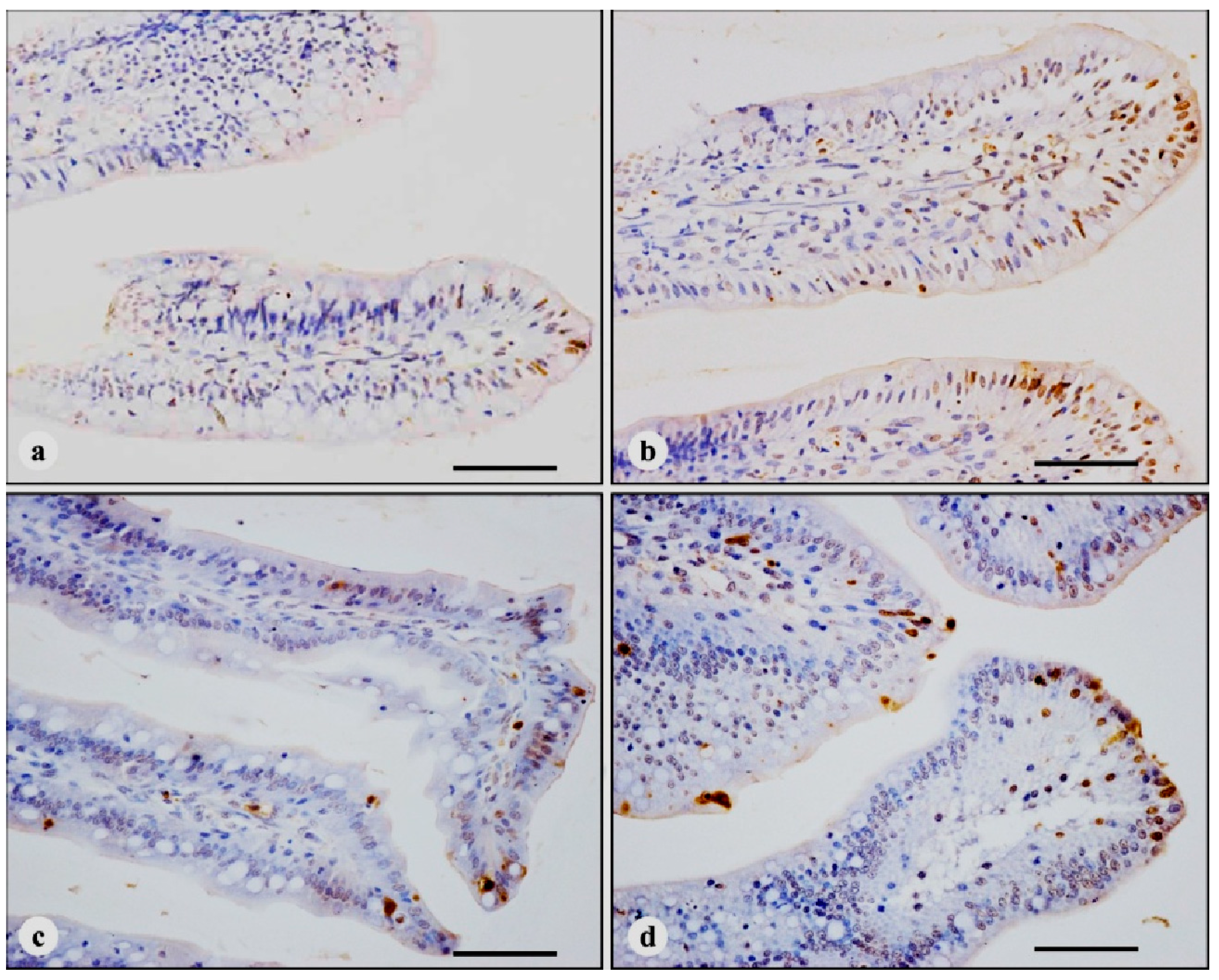
3.2. TUNEL Immunohistochemistry

In four treatments, the nuclei of TUNEL-positive cells were stained brown. TUNEL-positive cells were mainly distributed in the apical region of villi (Figure 1), with a few scattered positive cells in the middle and basal regions of villi and the crypt (Figure 2). Compared with control broilers on days 7, 14 and 21, the number of TUNEL-positive cells in AFB1 broilers was significantly increased (p < 0.01), however, the number of TUNEL positive cells in + Se broilers showed no significant changes (p > 0.05). In addition, the number of TUNEL-positive cells in AFB1 + Se broilers was significantly higher than that in control broilers (p < 0.01), but, significantly lower (p < 0.01) than that in AFB1 broilers during the experiment. The number of TUNEL-positive cells in the apical region of villi is shown in Table 3.
Figure 1. TUNEL-positive cells in the apical regions of jejunal villi at 14 days of age, immunohistochemistry stain. (a) in control broiler; (b) in AFB1 broiler, the number of TUNEL-positive cells is increased, compared with control broiler; (c) in + Se broiler, the number of TUNEL-positive cells is not obviously changed compared with control broiler; (d) in AFB1 + Se broiler, the number of TUNEL-positive cells is increased compared with control broiler, but, decreased compared with AFB1 broiler. Bars = 50 μm.
Ijerph 11 13130 g001
Figure 2. TUNEL immunohistochemistry staining in the crypts and in the middle regions of jejunal villi at 14 days of age. There are a few scattered TUNEL-positive cells in the crypts and in the middle regions of villi in all treatments. (a) The crypts in control broiler; (b) the middle regions of villi in control broiler; (c) the crypts in AFB1 broiler; (d) the middle regions of villi in AFB1 broiler. Bars = 50 μm.
Ijerph 11 13130 g002
Table 3. The Number of TUNEL-positive Cells in the Apical Region of Jejunal Villi (number/0.064 mm2).
TimeTreatments
controlAFB1+ SeAFB1 + Se
7 day5.200 ± 0.227 C18.900 ± 0.273 A B5.533 ± 0.184 C11.767 ± 0.243 B B
14 day5.600 ± 0.212 C18.833 ± 0.322 A B5.267 ± 0.33 C13.300 ± 0.437 B A
21 day5.100 ± 0.175 C19.767 ± 0.493 A A5.567 ± 0.233 C12.100 ± 0.175 B A
Note: data are presented with the means ± standard error (n = 6). Figures marked with the different capital letters are significantly different (p < 0.01). A–C p < 0.01 between treatment and control within the time of exposure; A,B p < 0.01 between times of exposure within the control or treatment.

3.3. Quantitative Real-Time PCR (qRT-PCR)

The mRNA expressions of Bax, Bcl-2 and Caspase-3 and the Bcl-2/Bax ratio in jejunal mucosa are shown in Table 4. The expressions of Bax and Caspase-3 mRNA in AFB1 broilers were significantly higher than those in control broilers at 7, 14 and 21 days of age (p < 0.01). There were no significant changes in the expression of Bax mRNA between AFB1 + Se broilers and control broilers at 7, 14 and 21 days of age. In addition, the expression of Caspase-3 mRNA in AFB1 + Se broilers was significantly higher (p < 0.01) than that in control broilers, but significantly lower (p < 0.05) than that in AFB1 broilers during the experiment, except for at 14 days of age (Table 4). Compared with control broilers, the expression of Bcl-2 mRNA and the Bcl-2/Bax ratio in AFB1 broilers significantly decreased on days 7, 14 and 21 (p < 0.01). The expression of Bcl-2 mRNA and the Bcl-2/Bax ratio in AFB1 + Se broilers were significantly lower than those in control broilers (p < 0.01 or p < 0.05), but significantly higher than those in AFB1 broilers (p < 0.01 or p < 0.05) (Table 4).
Table 4. The mRNA Expression of Bax, Bcl-2 and Caspase-3 and Bcl-2/Bax Ratio in Jejunal Mucosa.
ItemTimeTreatments
ControlAFB1+ SeAFB1 + Se
Bax7 day1.003 ± 0.054 B1.218 ± 0.028 A b0.936 ± 0.035 B1.024 ± 0.019 B
14 day1.001 ± 0.037 B1.235 ± 0.020 A b0.948 ± 0.019 Bb1.040 ± 0.014 Ba b
21 day1.003 ± 0.054 B1.302 ± 0.026 A a0.954 ± 0.030 B0.993 ± 0.013 B a
Bcl-27 day1.001 ± 0.027 Aa0.748 ± 0.009 B0.940 ± 0.013 Ab0.923 ± 0.014 Ab a
14 day1.001 ± 0.027 Aa0.770 ± 0.020 B0.972 ± 0.025 A0.894 ± 0.024 Ab
21 day1.001 ± 0.039 A0.757 ± 0.018 Bc0.976 ± 0.016 ACa0.857 ± 0.031 BCb b
Bcl-2/Bax7 day1.001 ± 0.030 Aa0.615 ± 0.013 B1.007 ± 0.050 Aa0.902 ± 0.006 Ab
14 day1.003 ± 0.054 Aa0.623 ± 0.008 D1.025 ± 0.027 AC0.860 ± 0.029 ABb
21 day1.002 ± 0.042 Aa0.582 ± 0.026 B1.025 ± 0.031 Aa0.863 ± 0.042 Ab
Caspase-37 day1.000 ± 0.016 B1.247 ± 0.023 Aa b1.029 ± 0.027 B1.166 ± 0.017 Ab b
14 day1.003 ± 0.054 C1.302 ± 0.009 A a1.036 ± 0.050 BCb1.195 ± 0.014 ABa
21 day1.002 ± 0.040 B1.305 ± 0.010 Aa a0.995 ± 0.021 B1.214 ± 0.016 Ab a
Note: data are presented with the means ± standard error (n = 6). The data are expressed as relative responses with respect to the control. Figures marked with the different capital letters are significantly different (p < 0.01); figures marked with the different small letters are significantly different (p < 0.05). A–D p < 0.01 or a–c p < 0.05 between treatment and control within the time of exposure; a,b p < 0.05 between times of exposure within the control or treatment.

4. Discussions

The results of clinical signs and body weight observed in this study showed that 0.4 mg/kg supplemented dietary Se could be safe for chickens, which is in agreement with Cai et al.’s report [33]. AFB1 (0.3 mg/kg) did not induce evident clinical symptoms, but significantly decreased body weight of broiler at 21 days of age was observed. It is thus suggested that 0.3 mg/kg AFB1 may retard the growth of the broiler. As reported in a review paper, broiler’s performance may be affected, when the concentration of dietary AFB1 is about 0.5 mg/kg [34].
Tissue homeostasis depends on both cell proliferation and cell death. The small intestinal epithelium is a rapidly renewing tissue, in which cells are lost from the villus into the gut lumen and are generally replaced at an equal rate by the proliferation of cells in the crypts [18]. Early researches indicate that apoptosis is responsible for controlling the majority of intestinal epithelial cell loss, and apoptosis is occurs predominantly in the villus tip cells [18], which is supported by the following observation (1) high levels of the pro-apoptotic protein, Bax, have been detected in these terminally differentiated cells [18]; (2) the expression of a possible apoptotic endonuclease (DNase I) also increases towards the villus tip [35]; (3) possible increased expression of transforming growth factor-β and evidence for reduced adhesion may also lend support to this hypothesis [36,37,38]; (4) The presence of large numbers of macrophages and lymphocytes at the villus tip is consistent with apoptosis of terminally differentiated cells [39]. TUNEL assay can identify DNA fragmentation and examine the topographic distribution of apoptotic cells [15,40,41]. Similar to previous reports in other animals’ intestine [42,43], in the present study, TUNEL-positive cells in all treatments were predominantly distributed in the apical region of the villus. Therefore, TUNEL-positive cells in the villi tip were counted as the number of apoptotic cells in the jejunal mucosa.
Several studies have indicated that AFB1 was able to induce apoptosis in hepatocytes, lung and bone marrow cells, bronchial epithelial cells, thymocytes and splenocytes [12,13,14,15,16]. Our result shows that the number of TUNEL-positive cells was increased in AFB1 broilers when compared with control broilers. This result indicates that AFB1 could induce excessive apoptosis of broilers’ jejunum. Our early study has revealed that 0.3 mg/kg AFB1 in the diet can induce pathological lesions (shedding) and reduce cellular proliferation of broilers’ jejunum [26]. Epithelial cells of the intestine experience permanent renewal that includes cell proliferation, migration, differentiation, apoptosis, and cell shedding into the intestinal lumen [18,44]. Homeostasis of these activities is essential for structural and functional properties of intestine [45,46]. Also, both decreased proliferation and/or increased cell death may reduce cell number, whereas increased proliferation and/or decreased death may increase cell number [18]. Therefore, the increased apoptosis observed in this study, the decreased proliferation [26] and pathological shedding in the villus tips in broilers’ jejunum induced by AFB1 [26] may lead to the decreased enterocytes which may be followed by the declined function of this organ.
Previous researches have demonstrated that AFB1 could lead to cellular apoptosis via mitochondrial or cell death receptor pathways [47,48,49,50]. In the present study, the mRNA expression of Bax, Bcl-2 and Caspase-3 was determined for evaluating whether AFB1-induced apoptosis of jejunum is related to the mitochondrial pathway. The results showed that the mRNA expression of Bax and Caspase-3 significantly increased, while the mRNA expression of Bcl-2 and the ratio of Bcl-2/Bax significantly decreased in AFB1 broilers, when compared with those in control broilers. These results suggest that the excessive apoptosis of jejunum induced by AFB1 was onset by the mitochondrial signaling pathway, which is accord with the previous research in human bronchial epithelial cells [14] and broiler thymocytes [15]. Future studies should focus on whether AFB1-induced apoptosis in jejunal cells is triggered by a cell death receptor pathway. Previous studies have revealed that AFB1 might induce oxidative stress by the formation of Reactive Oxygen Species (ROS) and the decrease of the activity and the gene expression of antioxidant enzymes [51,52,53,54,55,56]. As an important physiological effector of apoptosis [57], ROS induces the disruption of the mitochondrial membrane potential (MMP) and formation of mitochondrial apoptosis-induced channel promoted by Bax, by which cytochrome c releases from mitochondria [58]. Cytochrome c compounds in cytoplasm can activate caspase-9 followed by activation of Caspase-3 promoting the apoptosis process [59].
In the present study, the number of apoptotic cells had no significant difference between + Se and control broilers from 7 to 21 days of age. In addition, no significant difference was observed in the ratio of Bcl-2/Bax between + Se and control broilers during the experiment. The result suggests that 0.4 mg/kg Se supplied in the diet had almost no obvious effects on the apoptosis of broilers’ jejunum.
Recent studies have shown that Se has protective action against cell apoptosis induced by AFB1 in poultry [15,16]. In the present research, the number of apoptotic cells and the expression of Caspase-3 mRNA in AFB1 + Se broilers were significantly lower than those in AFB1 broilers during the experiment, and the expression of Bcl-2 mRNA and the Bcl-2/Bax ratio was significantly higher than those in AFB1 broilers. These results indicate that the diet supplemented with 0.4 mg/kg Se might have protective roles against AFB1-induced jejunal apoptosis of broiler by the upregulation of the Bcl-2/Bax ratio. Similar results were also reported in broilers’s thymus and spleen by Chen and Wang [15,16]. Previous researches showed that AFB1-induced apoptosis could be caused by lipid peroxidation and oxidative DNA damage [60,61]. However, selenium could repress ROS-mediate apoptosis by inhibiting the apoptosis due to ROS and mitochondrial dysfunction [62,63], which could be related to the antioxidant effects of Se [64]. Our results suggest that appropriate dietary Se could inhibit AFB1-induced apoptosis, which may be related to its anti-oxidant function. In comparison with those in the control group, the Bcl-2 expression and the Bcl-2/Bax ratio were significantly decreased, but the Bax expression showed substantially the same extent. This result suggested that supplemented Se could protect AFB1-induced apoptosis in some extent, but cannot restore it to the normal level as in the control group, because the Bcl-2 family member can be activated or suppressed by complex factors [65].The mechanism(s) of this observed action require further investigation.

5. Conclusions

In conclusion, 0.3 mg/kg AFB1 in the diet can induce an increase of cell apoptosis, a decrease of Bcl-2 mRNA expression, and an increase of Bax and Caspase-3 mRNA expression in broilers’ jejunum. However, supplementation of dietary sodium selenite at the concentration of 0.4 mg/kg Se may ameliorate AFB1-induced apoptosis by increasing Bcl-2 mRNA expression, and decreasing Bax and Caspase-3 mRNA expression.

Acknowledgments

This work was supported by the Program for Changjiang Scholars and University Innovative Research Teams (IRT 0848) and the Education Department of Sichuan Province (2012FZ0066).

Author Contributions

Xi Peng and Shengqiang Zhang conducted the research, analyzed the data, wrote and revised the paper. Jing Fang and Hengmin Cui designed the research and corrected the paper. Zhicai Zuo and Junliang Deng helped with qRT-PCR portions of the research.

Conflicts of Interest

The authors declare no conflict of interest.

References

  1. Massey, T.E.; Stewart, R.K.; Daniels, J.M.; Liu, L. Biochemical and molecular aspects of mammalian susceptibility to aflatoxin B1 carcinogenicity. Exp. Biol. Med. 1995, 208, 213–227. [Google Scholar] [CrossRef]
  2. McLean, M.; Dutton, M.F. Cellular interactions and metabolism of aflatoxin: An update. Pharmacol. Ther. 1995, 65, 163–192. [Google Scholar] [CrossRef] [PubMed]
  3. Ricordy, R.; Gensabella, G.; Cacci, E.; Augusti-Tocco, G. Impairment of cell cycle progression by aflatoxin B1 in human cell lines. Mutagenesis 2002, 17, 241–249. [Google Scholar] [CrossRef] [PubMed]
  4. Jakhar, K.K.; Sadana, J.R. Sequential pathology of experimental aflatoxicosis in quail and the effect of selenium supplementation in modifying the disease process. Mycopathologia 2004, 157, 99–109. [Google Scholar] [CrossRef] [PubMed]
  5. Rawal, S.; Kim, J.E.; Coulombe, R., Jr. Aflatoxin B1 in poultry: Toxicology, metabolism and prevention. Res. Vet. Sci. 2010, 89, 325–331. [Google Scholar] [CrossRef]
  6. Yunus, A.W.; Ghareeb, K.; Abd-El-Fattah, A.A.M.; Twaruzek, M.; Böhm, J. Gross intestinal adaptations in relation to broiler performance during chronic aflatoxin exposure. Poult. Sci. 2011, 90, 1683–1689. [Google Scholar] [CrossRef]
  7. Guo, S.; Shi, D.; Liao, S.; Su, R.; Lin, Y.; Pan, J.; Tang, Z. Influence of selenium on body weights and immune organ indexes in ducklings intoxicated with aflatoxin B1. Biol. Trace Elem. Res. 2012, 146, 167–170. [Google Scholar] [CrossRef] [PubMed]
  8. Shi, D.; Guo, S.; Liao, S.; Su, R.; Pan, J.; Lin, Y.; Tang, Z. Influence of selenium on hepatic mitochondrial antioxidant capacity in ducklings intoxicated with aflatoxin B1. Biol. Trace Elem. Res. 2012, 145, 325–329. [Google Scholar] [CrossRef] [PubMed]
  9. Wan, X.L.; Yang, Z.B.; Yang, W.R.; Jiang, S.Z.; Zhang, G.G.; Johnston, S.L.; Chi, F. Toxicity of increasing aflatoxin B1 concentrations from contaminated corn with or without clay adsorbent supplementation in ducklings. Poult. Sci. 2013, 92, 1244–1253. [Google Scholar] [CrossRef] [PubMed]
  10. Ribeiro, D.H.; Ferreira, F.L.; Da Silva, V.N.; Aquino, S.; Corrêa, B. Effects of aflatoxin B1 and fumonisin B1 on the viability and induction of apoptosis in rat primary hepatocytes. Int. J. Mol. Sci. 2010, 11, 1944–1955. [Google Scholar] [CrossRef]
  11. Brahmi, D.; Bouaziz, C.; Ayed, Y.; Ben Mansour, H.; Zourgui, L.; Bacha, H. Chemopreventive effect of cactus Opuntia ficus indica on oxidative stress and genotoxicity of aflatoxin B1. Nutr. MeTab. (Lond) 2011, 8. [Google Scholar] [CrossRef]
  12. Deng, S.X.; Tian, L.X.; Liu, F.J.; Jin, S.J.; Liang, G.Y.; Yang, H.J.; Du, Z.Y.; Liu, Y.J. Toxic effects and residue of aflatoxin B1 in tilapia (Oreochromis niloticus× O. aureus) during long-term dietary exposure. Aquaculture 2010, 307, 233–240. [Google Scholar] [CrossRef]
  13. Raj, H.G.; Kohli, E.; Rohil, V.; Dwarakanath, B.S.; Parmar, V.S.; Malik, S.; Adhikari, J.S.; Tyagi, Y.K.; Goel, S.; Gupta, K.; et al. Acetoxy-4-methylcoumarins confer differential protection from aflatoxin B1-induced micronuclei and apoptosis in lung and bone marrow cells. Mutat. Res. 2001, 494, 31–40. [Google Scholar] [CrossRef] [PubMed]
  14. Yang, X.J.; Lu, H.Y.; Li, Z.Y.; Bian, Q.; Qiu, L.L.; Li, Z.; Liu, Q.; Li, J.; Wang, X.; Wang, S.L. Cytochrome P450 2A13 mediates aflatoxin B1-induced cytotoxicity and apoptosis in human bronchial epithelial cells. Toxicology 2012, 300, 138–148. [Google Scholar] [CrossRef] [PubMed]
  15. Chen, K.J.; Shu, G.; Peng, X.; Fang, J.; Cui, H.M.; Chen, J.; Wang, F.Y.; Chen, Z.L.; Zuo, Z.C.; Deng, J.L.; et al. Protective role of sodium selenite on histopathological lesions, decreased T-cell subsets and increased apoptosis of thymus in broilers intoxicated with aflatoxin B1. Food Chem. Toxicol. 2013, 59, 446–454. [Google Scholar] [CrossRef] [PubMed]
  16. Wang, F.Y.; Shu, G.; Peng, X.; Fang, J.; Chen, K.J.; Cui, H.M.; Chen, Z.L.; Zuo, Z.C.; Deng, J.L.; Geng, Y.; Lai, W.M. Protective effects of sodium selenite against aflatoxin B1-induced oxidative stress and apoptosis in broiler spleen. Int. J. Environ. Res. Public Health 2013, 10, 2834–2844. [Google Scholar] [CrossRef] [PubMed]
  17. De Conto, C.; Oevermann, A.; Burgener, I.A.; Doherr, M.G.; Blum, J.W. Gastrointestinal tract mucosal histomorphometry and epithelial cell proliferation and apoptosis in neonatal and adult dogs. J. Anim. Sci. 2010, 88, 2255–2264. [Google Scholar] [CrossRef] [PubMed]
  18. Potten, C.S.; Wilson, J.W.; Booth, C. Regulation and significance of apoptosis in the stem cells of the gastrointestinal epithelium. Stem. Cells 1997, 15, 82–93. [Google Scholar] [CrossRef] [PubMed]
  19. Jones, B.A.; Gores, G.J. Physiology and pathophysiology of apoptosis in epithelial cells of the liver, pancreas, and intestine. Am. J. Physiol. 1997, 273, G1174–G1188. [Google Scholar] [PubMed]
  20. Combs, G.F.; Clark, L.C.; Turnbull, B.W. An analysis of cancer prevention by selenium. Biofactors 2001, 14, 153–159. [Google Scholar] [CrossRef] [PubMed]
  21. Allan, C.B.; Lacourciere, G.M.; Stadtman, T.C. Responsiveness of selenoproteins to dietary selenium 1, 2. Annu. Rev. Nutr. 1999, 19, 1–16. [Google Scholar] [CrossRef] [PubMed]
  22. Zeng, H.; Combs, G.F., Jr. Selenium as an anticancer nutrient: Roles in cell proliferation and tumor cell invasion. J. Nutr. Biochem. 2008, 19, 1–7. [Google Scholar] [CrossRef]
  23. McKenzie, R.C.; Rafferty, T.S.; Beckett, G.J. Selenium: An essential element for immune function. Immunol. Today 1998, 19, 342–345. [Google Scholar] [CrossRef] [PubMed]
  24. Hoffmann, P.R.; Berry, M.J. The influence of selenium on immune responses. Mol. Nutr. Food Res. 2008, 52, 1273–1280. [Google Scholar] [CrossRef] [PubMed]
  25. Perozo, F.; Rivera, S. Effect of Aflatoxin B1 exposure and selenium supplementation on immune response in broilers. Indian Vet. J. 2003, 80, 1218–1221. [Google Scholar]
  26. Zhang, S.Q.; Peng, X.; Fang, J.; Cui, H.M.; Zuo, Z.C.; Chen, Z.L. Effects of aflatoxin B1 exposure and sodium selenite supplementation on the histology, cell proliferation and cell cycle of jejunum in broilers. Biol. Trace Elem. Res. 2014, 160, 32–40. [Google Scholar] [CrossRef]
  27. Kaoud, H.A. Innovative methods for the amelioration of aflatoxin (afb1) effect in broiler chicks. Sci. J. Appl. Res. 2013, 1, 15–19. [Google Scholar]
  28. Peng, X.; Cui, Y.; Cui, W.; Deng, J.; Cui, H. The decrease of relative weight, lesions, and apoptosis of bursa of Fabricius induced by excess dietary selenium in chickens. Biol. Trace Elem. Res. 2009, 131, 33–42. [Google Scholar] [CrossRef] [PubMed]
  29. Hamidu, J.A.; Uddin, Z.; Li, M.; Fasenko, G.M.; Guan, L.L.; Barreda, D.R. Broiler egg storage induces cell death and influences embryo quality. Poult. Sci. 2011, 90, 1749–1757. [Google Scholar] [CrossRef] [PubMed]
  30. Dusanic, D.; Bencina, D.; Oven, I.; Cizelj, I.; Bencina, M.; Narat, M. Mycoplasma synoviae induces upregulation of apoptotic genes, secretion of nitric oxide and appearance of an apoptotic phenotype in infected chicken chondrocytes. Vet. Res. 2012, 43. [Google Scholar] [CrossRef]
  31. Iqbal, M.; Philbin, V.J.; Smith, A.L. Expression patterns of chicken Toll-like receptor mRNA in tissues, immune cell subsets and cell lines. Vet. Immunol. Immunopathol. 2005, 104, 117–127. [Google Scholar] [CrossRef] [PubMed]
  32. Livak, K.J.; Schmittgen, T.D. Analysis of relative gene expression data using real-time quantitative PCR and the 2-ΔΔCT method. Methods 2001, 25, 402–408. [Google Scholar] [CrossRef] [PubMed]
  33. Cai, S.J.; Wu, C.X.; Gong, L.M.; Song, T.; Wu, H.; Zhang, L.Y. Effects of nano-selenium on performance, meat quality, immune function, oxidation resistance, and tissue selenium content in broilers. Poultry Sci. 2012, 91, 2532–2539. [Google Scholar] [CrossRef]
  34. Yunus, A.W.; Razzazi-Fazeli, E.; Bohm, J. Aflatoxin B1 in affecting broiler’s performance, immunity, and gastrointestinal tract: A review of history and contemporary issues. Toxins 2011, 3, 566–590. [Google Scholar] [CrossRef] [PubMed]
  35. Polzar, B.; Zanotti, S.; Stephan, H.; Rauch, F.; Peitsch, M.C.; Irmler, M.; Tschopp, J.; Mannherz, H.G. Distribution of deoxyribonuclease I in rat tissues and its correlation to cellular turnover and apoptosis (programmed cell death). Eur. J. Cell. Biol. 1994, 64, 200–210. [Google Scholar] [PubMed]
  36. Barnard, J.A.; Warwick, G.J.; Gold, L.I. Localisation of transforming growth factor beta isoforms in the normal murine small intestine and colon. Gastroenterology 1993, 105, 67–73. [Google Scholar]
  37. Probstmeier, R.; Martini, R.; Tacke, R.; Schachner, M. Expression of the adhesion molecules L1, N-CAM and J1/tenascin during development of the murine small intestine. Differentiation 1990, 44, 42–55. [Google Scholar] [CrossRef] [PubMed]
  38. Probstmeier, R.; Martini, R.; Schachner, M. Expression of J1/tenascin in the crypt-villus unit of adult mouse small intestine: Implications for its role in epithelial cell shedding. Development 1990, 109, 313–321. [Google Scholar] [PubMed]
  39. Shibahara, T.; Sato, N.; Waguri, S.; Iwanaga, T.; Nakahara, A.; Fukutomi, H.; Uchiyama, Y. The fate of effete epithelial cells at the villus tip of the human small intestine. Arch. Histol. Cytol. 1995, 58, 205–219. [Google Scholar] [CrossRef] [PubMed]
  40. Negoescu, A.; Guillermet, C.; Lorimier, P.; Brambilla, E.; Labat-Moleur, F. Importance of DNA fragmentation in apoptosis with regard to TUNEL specificity. Biomed. Pharmacother. 1998, 52, 252–258. [Google Scholar] [CrossRef] [PubMed]
  41. Huang, J.; Cui, H.; Peng, X.; Fang, J.; Zuo, Z.; Deng, J.; Wu, B. The association between splenocyte apoptosis and alterations of Bax, Bcl-2, Caspase-3 mRNA expression, and oxidative stress induced by dietary nickel chloride in broilers. Int. J. Environ. Res. Public Health 2013, 10, 7310–7326. [Google Scholar] [CrossRef] [PubMed]
  42. Benard, O.; Madesh, M.; Anup, R.; Balasubramanian, K.A. Apoptotic process in the monkey small intestinal epithelium: 1. Association with glutathione level and its efflux. Free Radic. Biol. Med. 1999, 26, 245–252. [Google Scholar] [CrossRef] [PubMed]
  43. Westcarr, S.; Farshori, P.; Wyche, J.; Anderson, W.A. Apoptosis and differentiation in the crypt-villus unit of the rat small intestine. J. Submicrosc. Cytol. Pathol. 1999, 31, 15–30. [Google Scholar] [PubMed]
  44. Bjerknes, M.; Cheng, H. Gastrointestinal stem cells. II. Intestinal stem cells. Am. J. Physiol. Gastrointest Liver Physiol. 2005, 289, G381–G387. [Google Scholar] [CrossRef] [PubMed]
  45. Hall, P.A.; Coates, P.J.; Ansari, B.; Hopwood, D. Regulation of cell number in the mammalian gastrointestinal tract: The importance of apoptosis. J. Cell. Sci. 1994, 107, 3569–3577. [Google Scholar] [PubMed]
  46. Karan, S.M. Lineage commitment and maturation of epithelial cells in the gut. Front. Biosci. 1999, 4, D286–D298. [Google Scholar] [CrossRef] [PubMed]
  47. Duan, X.X.; Ou, J.S.; Li, Y.; Su, J.J.; Ou, C.; Yang, C.; Yue, H.F.; Ban, K.C. Dynamic expression of apoptosis-related genes during development of laboratory hepatocellular carcinoma and its relation to apoptosis. World J. Gastroenterol. 2005, 11, 4740–4744. [Google Scholar] [PubMed]
  48. Van Vleet, T.R.; Watterson, T.L.; Klein, P.J.; Coulombe, R.A., Jr. Aflatoxin B1 alters the expression of p53 in cytochrome P450-expressing human lung cells. Toxicol. Sci. 2006, 89, 399–407. [Google Scholar] [CrossRef] [PubMed]
  49. Reddy, L.; Odhav, B.; Bhoola, K. Aflatoxin B1-induced toxicity in HepG2 cells inhibited by carotenoids: Morphology, apoptosis and DNA damage. Biol. Chem. 2006, 387, 87–93. [Google Scholar] [CrossRef] [PubMed]
  50. Hassan, A.M.; Abdel-Aziem, S.H.; Abdel-Wahhab, M.A. Dietary supplementation with whey protein and ginseng extract counteracts oxidative stress and DNA damage in rats fed an aflatoxin-contaminated diet. Mutat. Res. 2011, 723, 65–71. [Google Scholar] [CrossRef] [PubMed]
  51. Alpsoy, L.; Yildirim, A.; Agar, G. The antioxidant effects of vitamin A, C, and E on aflatoxin B1-induced oxidative stress in human lymphocytes. Toxicol. Ind. Health 2009, 25, 121–127. [Google Scholar] [CrossRef] [PubMed]
  52. Kanbur, M.; Eraslan, G.; Sarıca, Z.S.; Aslan, Ö. The effects of evening primrose oil on lipid peroxidation induced by subacute aflatoxin exposure in mice. Food Chem. Toxicol. 2011, 49, 1960–1964. [Google Scholar] [CrossRef] [PubMed]
  53. Halliwell, B. Mechanisms involved in the generation of free radicals. Pathol. Biol. 1996, 44, 6–13. [Google Scholar] [PubMed]
  54. Choi, K.C.; Chung, W.T.; Kwon, J.K.; Yu, J.Y.; Jang, Y.S.; Park, S.M.; Lee, S.Y.; Lee, J.C. Inhibitory effects of quercetin on aflatoxin B1-induced hepatic damage in mice. Food Chem. Toxicol. 2010, 48, 2747–2753. [Google Scholar] [CrossRef] [PubMed]
  55. Yener, Z.; Celik, I.; Ilhan, F.; Bal, R. Effects of Urtica dioica L. seed on lipid peroxidation, antioxidants and liver pathology in aflatoxin-induced tissue injury in rats. Food Chem. Toxicol. 2009, 47, 418–424. [Google Scholar] [CrossRef] [PubMed]
  56. Brenneisen, P.; Steinbrenner, H.; Sies, H. Selenium, oxidative stress, and health aspects. Mol. Aspects. Med. 2005, 26, 256–267. [Google Scholar] [CrossRef] [PubMed]
  57. Ramachandran, A.; Madesh, M.; Balasubramanian, K.A. Apoptosis in the intestinal epithelium: Its relevance in normal and pathophysiological conditions. J. Gastroenterol. Hepatol. 2000, 15, 109–120. [Google Scholar] [CrossRef] [PubMed]
  58. Dejean, L.M.; Martinez-Caballero, S.; Manon, S.; Kinnally, K.W. Regulation of the mitochondrial apoptosis-induced channel, MAC, by Bcl-2 family proteins. Biochim Biophys Acta. 2006, 1762, 191–201. [Google Scholar] [CrossRef] [PubMed]
  59. Antonsson, B. Mitochondria and the Bcl-2 family proteins in apoptosis signaling pathways. Mol. Cell. Biochem. 2004, 256–257, 141–155. [Google Scholar] [CrossRef] [PubMed]
  60. Chandra, J.; Samali, A.; Orrenius, S. Triggering and modulation of apoptosis by oxidative stress. Free Radic. Biol. Med. 2000, 29, 323–333. [Google Scholar] [CrossRef] [PubMed]
  61. Souza, M.F.; Tome, A.R.; Rao, V.S. Inhibition by the bioflavonoid ternatin of Aflatoxin B1-induced lipid peroxidation in rat liver. J. Pharm. Pharmacol. 1999, 51, 125–129. [Google Scholar] [CrossRef] [PubMed]
  62. Ebert, R.; Ulmer, M.; Zeck, S.; Meissner-Weigl, J.; Schneider, D.; Stopper, H.; Schupp, N.; Kassem, M.; Jakob, F. Selenium supplementation restores the antioxidative capacity and prevents cell damage in bone marrow stromal cells in vitro. Stem. Cells 2006, 24, 1226–1235. [Google Scholar] [CrossRef] [PubMed]
  63. Zhou, Y.J.; Zhang, S.P.; Liu, C.W.; Cai, Y.Q. The protection of selenium on ROS mediated-apoptosis by mitochondria dysfunction in cadmium-induced LLC-PK1 cells. Toxicol. In Vitro 2009, 23, 288–294. [Google Scholar] [CrossRef] [PubMed]
  64. Rotruck, J.T.; Pope, A.L.; Ganther, H.E.; Swanson, A.B.; Hafeman, D.G.; Hoekstra, W.G. Selenium: Biochemical role as a component of glutathione peroxidase. Science 1973, 179, 588–590. [Google Scholar] [CrossRef] [PubMed]
  65. Martinou, J.C.; Youle, R.J. Mitochondria in apoptosis: Bcl-2 family members and mitochondrial dynamics. Dev. Cell 2011, 21, 92–101. [Google Scholar] [CrossRef] [PubMed]

