Article

Anthelmintic Potential of Agelasine Alkaloids from the Australian Marine Sponge Agelas axifera

1
Institute for Biomedicine and Glycomics, Griffith University, Brisbane, QLD 4111, Australia
2
School of Environment and Science, Griffith University, Brisbane, QLD 4111, Australia
3
Department of Veterinary Biosciences, Melbourne Veterinary School, Faculty of Science, The University of Melbourne, Parkville, VIC 3010, Australia
4
School of Molecular Sciences, The University of Western Australia, Crawley, WA 6009, Australia
5
School of Environment and Science, Griffith University, Gold Coast, QLD 4222, Australia
6
Biodiversity and Geosciences, Queensland Museum, South Brisbane BC, QLD 4101, Australia
7
NatureBank, Griffith University, Brisbane, QLD 4111, Australia
*
Authors to whom correspondence should be addressed.
Mar. Drugs 2025, 23(7), 276; https://doi.org/10.3390/md23070276
Submission received: 6 June 2025 / Revised: 25 June 2025 / Accepted: 26 June 2025 / Published: 1 July 2025
(This article belongs to the Section Structural Studies on Marine Natural Products)

Abstract

A recent high-throughput screening of the NatureBank marine extract library (7616 samples) identified an extract from the Australian marine sponge Agelas axifera with in vitro activity against an economically important parasitic nematode, Haemonchus contortus (barber’s pole worm). The bioassay-guided fractionation of the CH2Cl2/MeOH extract from A. axifera led to the purification of a new diterpene alkaloid, agelasine Z (1), together with two known compounds agelasine B (2) and oxoagelasine B (3). Brominated compounds (–)-mukanadin C (4) and 4-bromopyrrole-2-carboxylic acid (5) were also isolated from neighbouring UV-active fractions. All compounds, together with agelasine D (6) from NatureBank’s pure compound library, were tested for in vitro anthelmintic activity against exsheathed third-stage (xL3s) and fourth-stage larvae (L4s) of H. contortus and young adult Caenorhabditis elegans. Compounds 1, 2 and 6 induced an abnormal “skinny” phenotype, while compounds 2 and 6 also reduced the motility of H. contortus L4s by 50.5% and 51.8% at 100 µM, respectively. The minimal activity of agelasines against C. elegans young adults suggests a possible species-specific mechanism warranting further investigation. For the first time, the unexpected lability of agelasine H-8′ was explored using kinetic studies, revealing rapid deuterium exchange in MeOH-d4 at room temperature.

Graphical Abstract

1. Introduction

Gastrointestinal nematodes pose a significant global health burden by causing severe diseases in livestock, significantly affecting productivity, and are considered one of the most important production-limiting diseases of grazing animals worldwide [1]. Among the nematodes of the order Strongylida, Haemonchus contortus, commonly known as barber’s pole worm, stands out as one of the most pathogenic parasites of small ruminants [2]. The control of these parasites has primarily relied on anthelmintic drugs, which have been crucial in combatting nematode infections. However, the effectiveness of these drugs is increasingly threatened by the emergence of anthelmintic resistance. Notably, H. contortus was the first species to develop resistance to commercial anthelmintic drugs [3]; more concerning is the emergence of resistance to monepantel, a next-generation drug typically reserved for resistant infections, emphasizing the need for new treatment options [4]. In light of this challenge, recent investigations have identified H. contortus as a valuable model for the identification and evaluation of anthelmintic natural products [5,6,7]. This approach not only addresses the immediate concern of drug resistance in H. contortus but also paves the way for discovering novel treatments against other parasitic nematodes.
As part of a drug discovery effort aimed at identifying new anthelmintic leads from natural resources, we recently screened the NatureBank [8] marine extract library collection (n = 7616) against larvae of H. contortus in a high-throughput screening (HTS) bioassay [9]. This effort revealed 58 hit extracts [10,11] one of which was derived from the Australian marine sponge Agelas axifera. This extract inhibited larval motility by 96% (90 h) and larval development by >80% (168 h) and induced a curved (Cur) phenotype in 80% of larvae [9].
Marine sponges from the genus Agelas (class Demospongiae, family Agelasidae) are widely distributed and are known for their structurally diverse diterpene alkaloids and bromopyrroles [12,13]. In the present study, we report the bioassay-guided fractionation and purification of an A. axifera extract with inhibitory activity against H. contortus larvae, yielding a new diterpene alkaloid, which we named agelasine Z (1), as well as two known compounds agelasine B (2) [14] and oxoagelasine B (3) [15]. Moreover, two other known pyrrole alkaloids (–)-mukanadin C (4) [16] and 4-bromopyrrole-2-carboxylic acid (5) [17] were also isolated. All of the isolated compounds (15), along with agelasine D (6) [14] from the NatureBank pure compound library, were individually tested in vitro for nematocidal or nematostatic activity against H. contortus and Caenorhabditis elegans.

2. Results and Discussion

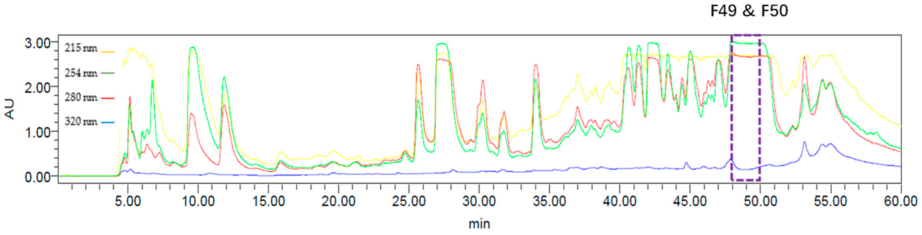
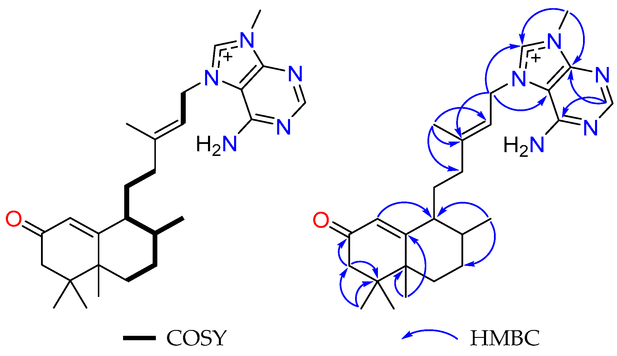
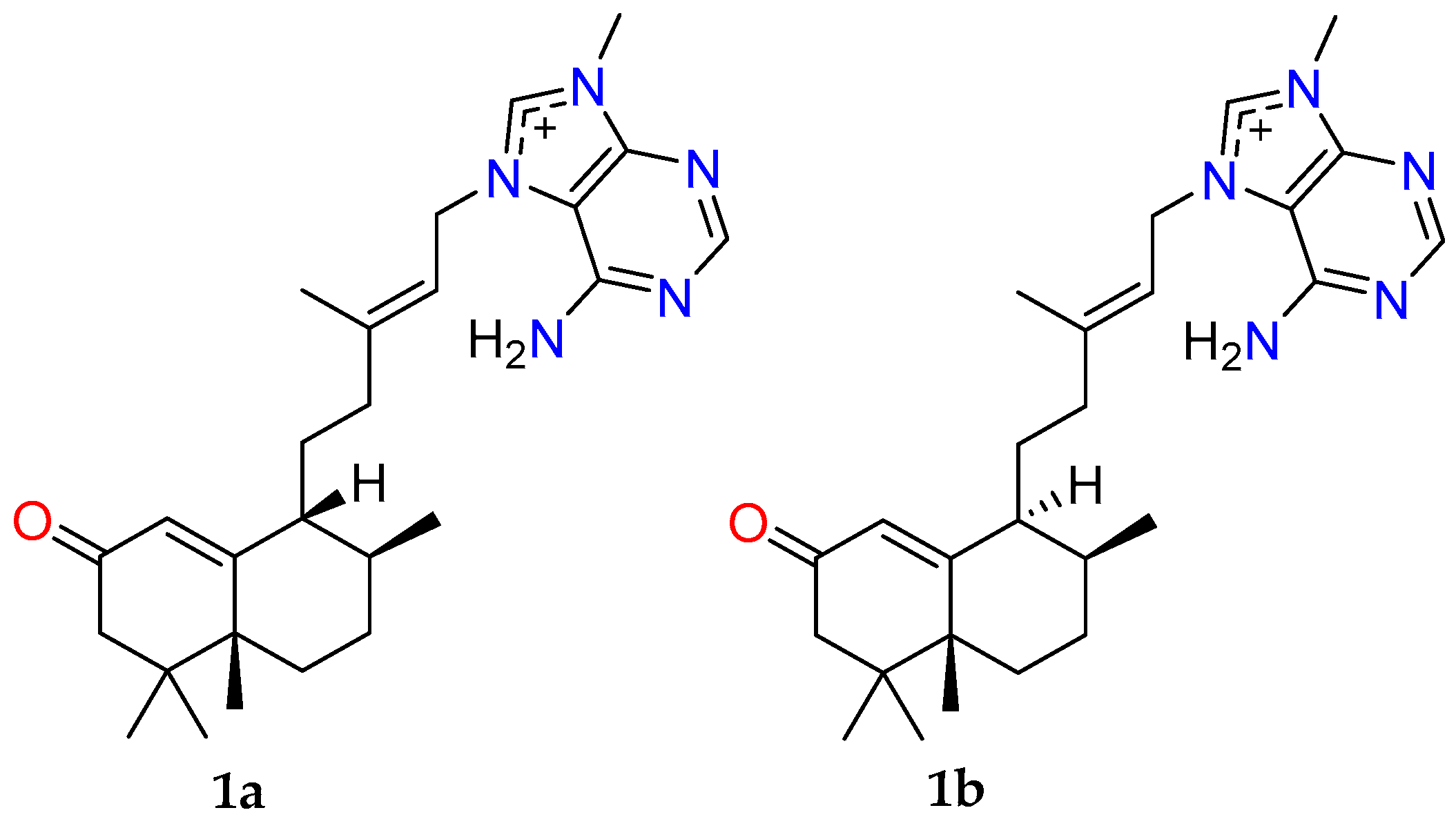
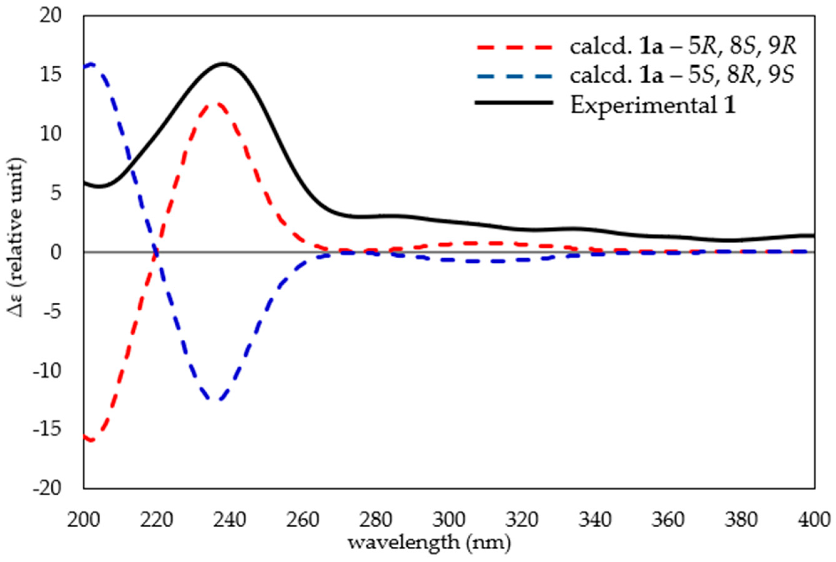
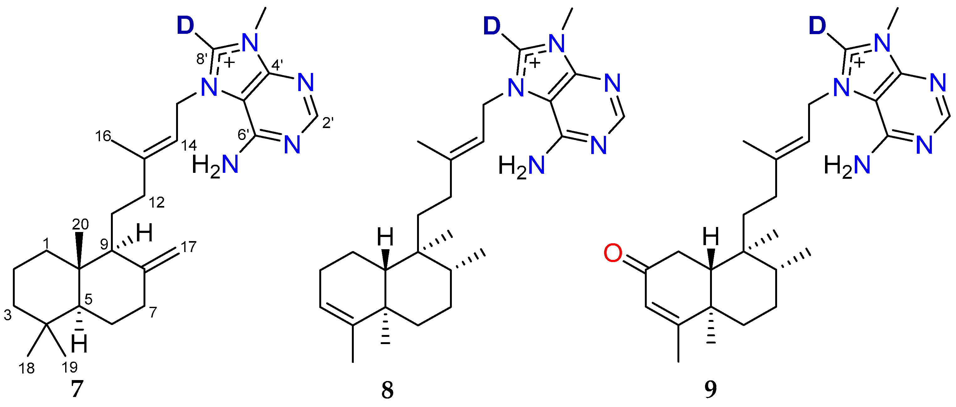
2.1. Bioassay-Guided Fractionation of Compounds from A. axifera

A freeze-dried and ground sample of A. axifera was extracted exhaustively with n-hexane, CH2Cl2 and MeOH; the CH2Cl2 and MeOH extracts were combined, and a portion of this material was subjected to reversed-phase HPLC (RP-HPLC, MeOH/H2O/0.1% TFA) (Figure 1) to produce 60 fractions that were then evaluated for anthelmintic activity using an established xL3 motility assay [11]. Fractions 49 (F49) and 50 (F50) showed moderate motility reduction (37.9% and 26.3%, respectively) at 168 h despite the strong activity observed for the original sponge extract. However, these fractions were able to induce an evisceration (Evi) phenotype in 19.6% and 12.5% of larvae, respectively. The Evi phenotype is lethal and displays an anterior protrusion in the affected larvae, as previously described [18], relative to the untreated control. Consequently, F49 and F50 fractions were selected for further purification and characterization studies.
An analysis of the 1H NMR data for F49 and F50 suggested that they contained a mixture of agelasine-type diterpenoid alkaloids [15]. Therefore, the two fractions were combined and subjected to purification by RP-HPLC using a different gradient system to yield one new compound agelasine Z (1) and two known alkaloids, agelasine B (2) and oxoagelasine B (3). An analysis of other UV-active HPLC fractions enabled the identification of the previously reported marine natural products (–)-mukanadin C (4) and 4-bromopyrrole-2-carboxylic acid (5). The known compounds were confirmed following a comparison of experimental NMR, MS and specific rotation data with their respective literature values [14,16,17,19]. Agelasine D (6) was obtained from the NatureBank pure compound library (Figure 2) following a substructure search of the NatureBank database [8].
The HRESIMS of compound 1 revealed an ion at m/z 436.3060 [M]+ (calcd for C26H38N5O, 436.3071) that allowed a molecular formula of C26H38N5O+ to be assigned. A comparison of the 1H and 13C NMR data of 1 with the previously reported agelasine alkaloid nakamusine C showed a high degree of similarity but was not identical, suggesting that 1 was an isomer of nakamusine C [20]. The 1H NMR data for 1 in MeOH-d4 (Table 1 and Table S1) exhibited resonances for two protons at δH 9.33 (H-8′) and 8.47 (H-2′) that were characteristic for N9-adeninium alkaloids [15].
A further analysis of the 1H NMR data showed that 1 contained two olefinic protons (δH 5.87 and 5.55), an allylic methylene unit (δH 5.21), an N-methyl group (δH 3.98), two methines (δH 2.19 and 1.48), five methylene units (δH 2.42/1.98, 2.11/1.43, 2.10/1.78, 2.08/1.50, 1.64), an olefinic methyl group (δH 1.88) and four upfield methyl groups (δH 1.20, 1.04, 1.01, 0.98). The 13C NMR data (Table 1) contained 26 carbon resonances, including six sp2 non-protonated carbons (δC 202.7, 176.4, 154.1, 150.9, 149.1, 111.2), four sp2 methines (δC 157.1, 141.9, 122.6, 115.8), two sp3 quaternary carbons (δC 46.1, 35.4), two sp3 methines (δC 47.5, 47.2), six sp3 methylenes (δC 48.6, 47.8, 35.2, 31.6, 31.0, 31.0) and six methyl groups, one of which was an N-methyl (δC 32.0, 28.8, 27.8, 23.2, 17.0, 16.4).
The 1H-1H COSY data established two spin systems, which included -CH=CH2- and -CH2-CH2-CH(CH3)-CH-CH2-CH2-. Moreover, key HMBC correlations of H-1 to C9; H2-3 to C-2 and C-4; H2-15 to C-13, C-5’ and C-8’; H3-16 to C-12, C-13 and C-14; H3-17 to C-7 and C-9; H3-18 and H3-19 to C-3, C-4 and C-5; H3-20 to C-5, C-7 and C-10; H-2’ to C-4’ and C-6’; and H3-9’-Me to C-4’ and C-8’ (Figure 3) aided in the establishment of the planar structure of agelasine Z (1). Following a careful analysis of the 2D NMR data of 1, it was identified that the C-20 methyl group of nakamusine C has been switched to C-5 in agelasine Z.
To determine the relative configuration of 1, ROESY NMR data were acquired in MeOH-d4. However, poor signal dispersion led to the overlap of key proton resonances associated with the decalin core, thereby preventing the definitive assignment of the configuration at C-9. As a result, two potential diastereomers were proposed for agelasine Z (1a and 1b, Figure 4).
With the aim of improving the 1H NMR spectrum to facilitate more definitive ROESY-based relative configuration assignments, we undertook additional investigations of 1 in other deuterated NMR solvents (benzene-d6 and MeCN-d3). While compound 1 showed poor solubility in benzene-d6, it dissolved readily in MeCN-d3, with the resulting 1H NMR spectrum displaying superior signal dispersion compared to MeOH-d4 (Figure S9). Hence, COSY and ROESY NMR data were recorded in MeCN-d3, and all proton chemical shifts were reassigned based on the analysis of the new 2D NMR data (Figures S10 and S11). Fortuitously, the ROESY NMR data in MeCN-d3 showed clear and strong correlations associated with protons situated around the decalin subunit that ultimately allowed us to resolve the relative configuration for 1. Key ROESY correlations from methyl H3-17 (δH 0.95) to H-9 (δH 2.15) and methyl H3-20 (δH 1.16) suggested that they must reside on the same face of the decalin ring system. In addition, methyl H3-20 displayed further ROESY correlations with the alpha keto methine H-1 (δH 5.79), methylene proton H-6a (δH 2.04) and methyl H3-18 (δH 0.98), indicating that these protons also resided on the same face of 1. These data allowed the relative configuration of the decalin moiety in 1 to be assigned as 5R*, 8S* and 9R* (Figure 5), consistent with candidate 1a in MeOH-d4. Furthermore, the ∆13 geometry was assigned as E based on ROESY correlations between the olefinic methyl (H3-16, δH 1.80) and the methylene group (H2-15, δH 5.08).
Quantum chemical NMR and time-dependent density functional theory (TDDFT) ECD calculations have found increasing utility in informing the structure elucidation of complex natural products [21,22,23]. Notably, density functional theory (DFT) techniques have been used to aid the absolute configurational assignment of related agelasines, isoagelasine C [13] and nakamusine A [20]. To support the experimentally assigned relative configuration of agelasine Z (1), gauge including atomic orbital (GIAO) DFT NMR chemical shift calculations were performed on truncated candidate diastereomers, 1a and 1b (Tables S4–S10). After DFT geometry optimization, the NMR shielding tensors for 1a and 1b were calculated at the mPW1PW91/6-311+G(d,p) level of theory and evaluated using DP4+ [21]. The DP4+ analysis returned 100% probabilistic support for the 5R*, 8S* and 9R* diastereomer (1a, Table S4), in excellent agreement with our experimentally ascribed relative configuration. Next, TDDFT ECD calculations were undertaken to examine the absolute configuration of 1. The TDDFT-calculated ECD spectra for 1a’s 5R, 8S and 9R enantiomer was found to match the experimental CD spectrum for agelasine Z (1, Figure 6), thereby allowing the absolute configuration of 1 to be assigned.
It is noteworthy that agelasines exhibit variability in their chiral centres, with multiple configurations observed within the same sponge material [15,24]. For example, agelasines B (2) and D (6), originally isolated from Agelas sp., have been reported with the absolute configurations 5R, 8R, 9S, 10R and 5S, 9S, 10S, respectively [14]. Given these observations, and the fact that oxoagelasine B (3) is a C-2 oxidized derivative of agelasine B (2) with matching optical rotation, it is likely that both alkaloids share the same absolute configuration.

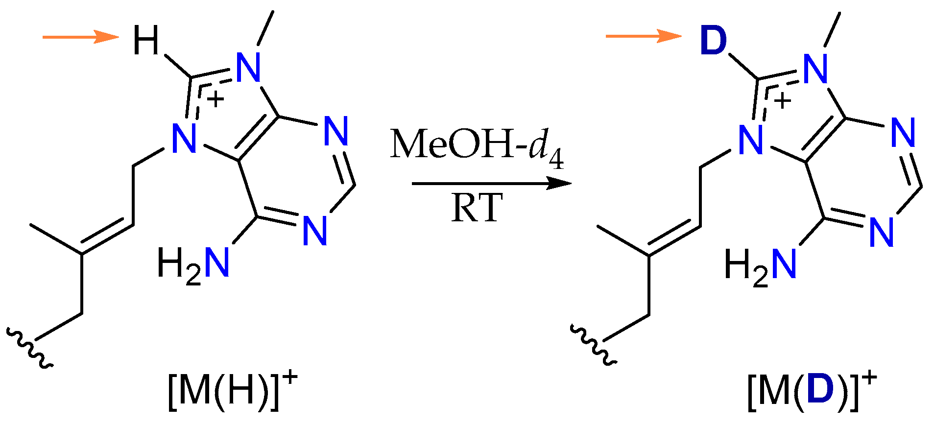
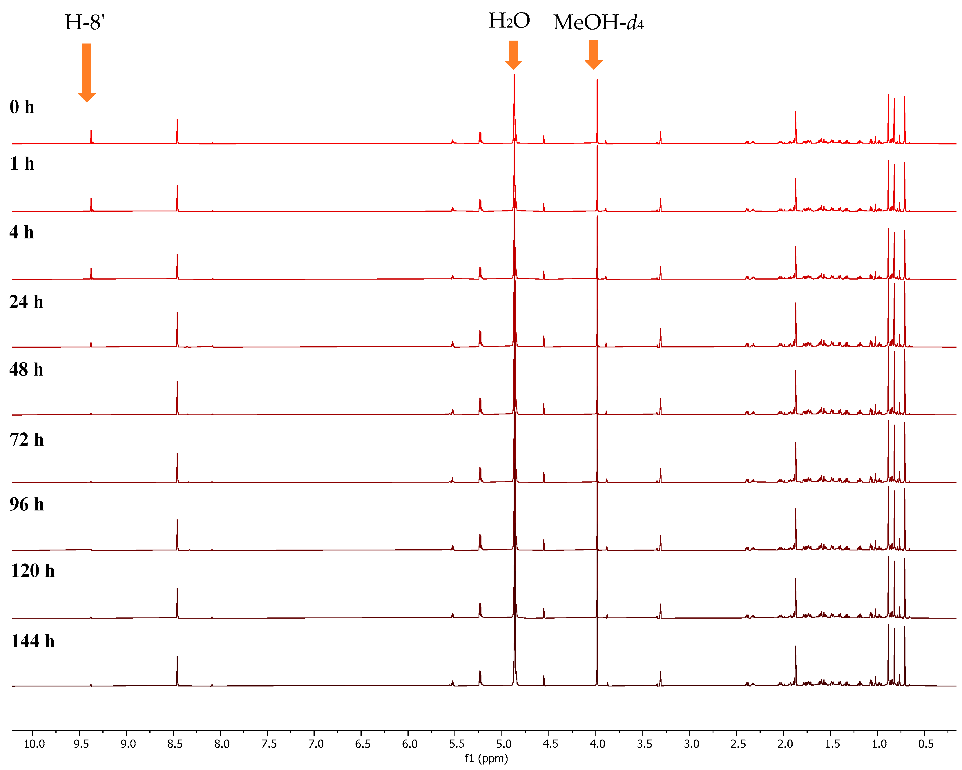
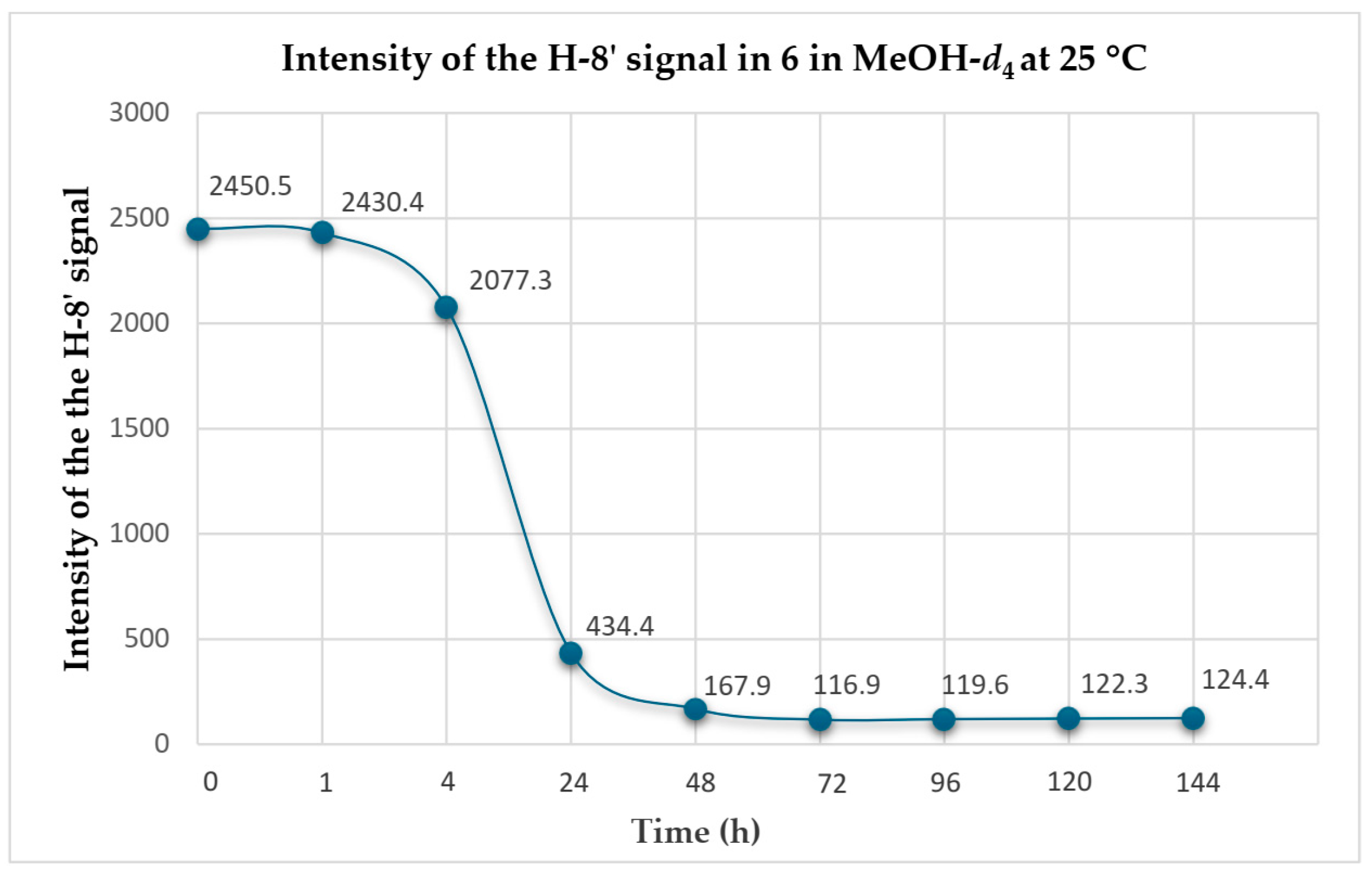
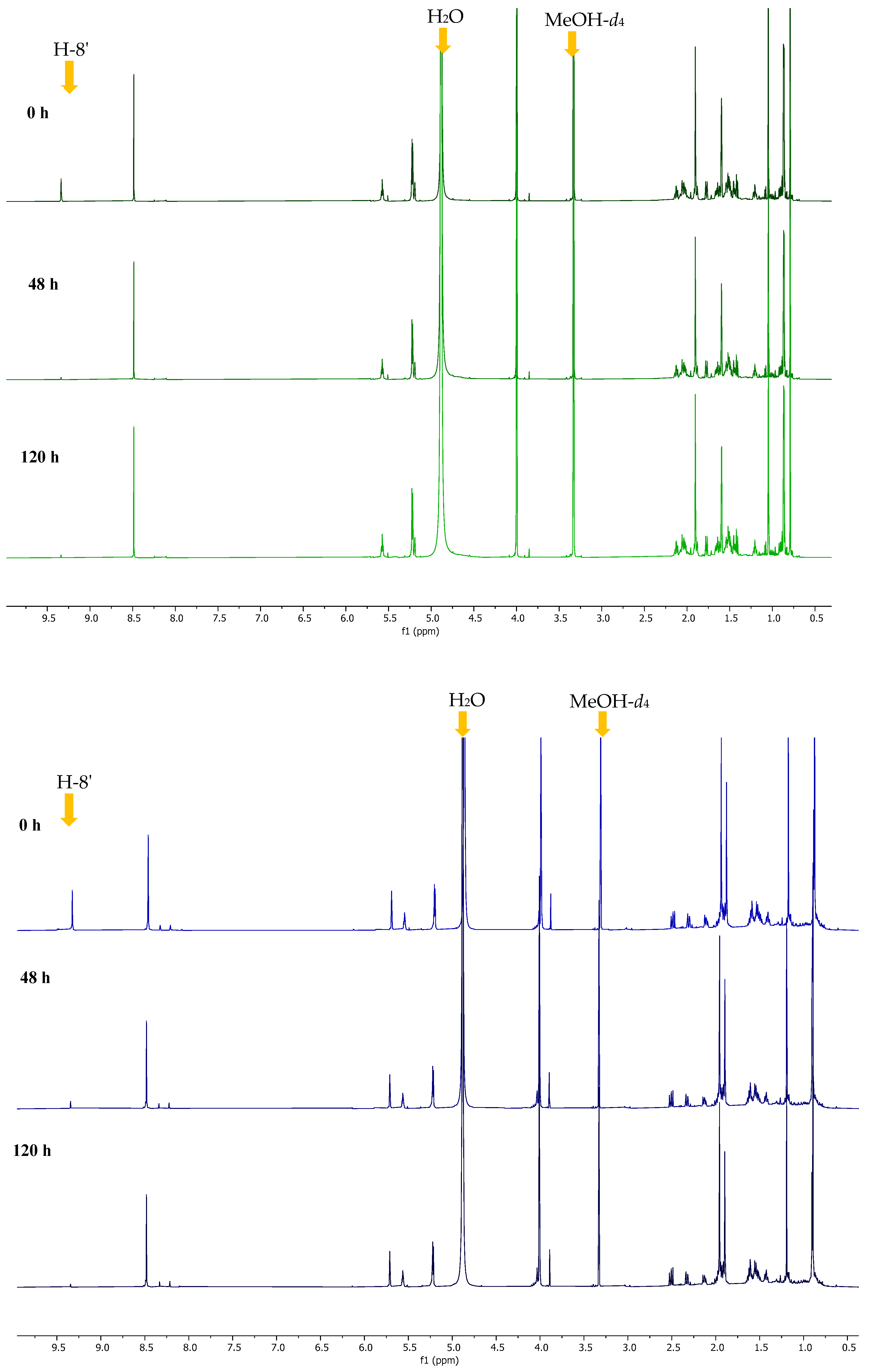
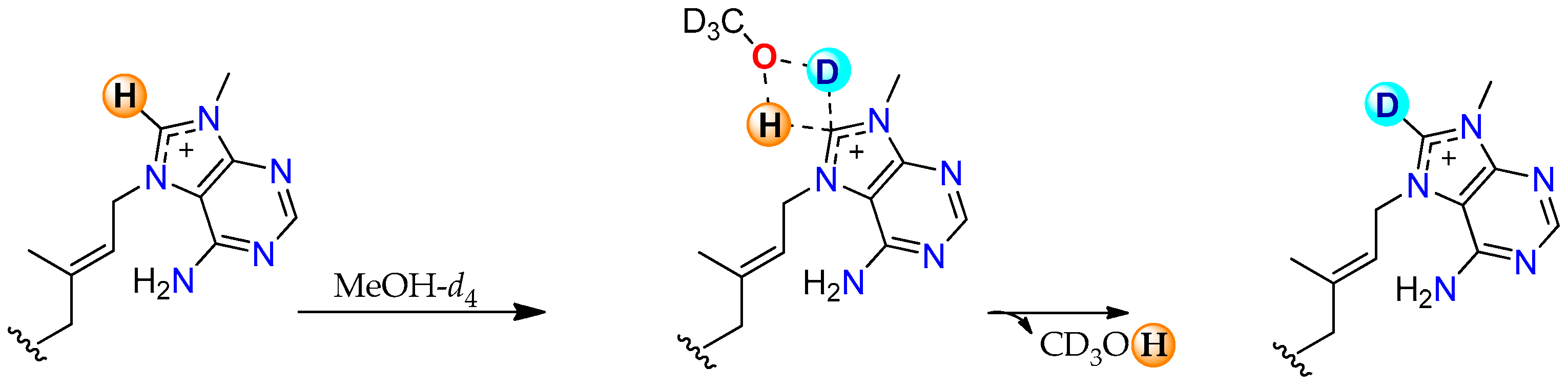
2.2. Conversion of H-8’ to D-8’ of Agelasines in MeOH-d4

Deuteration is the process of replacing hydrogen atoms with their heavier isotope, deuterium (D), and has recently gained significant attention due to its potential to alter the physiochemical and biological properties of molecules [25]. Many deuteration methods involve metal catalysts [26], while in some instances, deuterated solvents trigger the reaction [27]. During this investigation, we observed a gradual decrease in the intensity of the adenine proton resonance H-8’ in agelasines 13 and 6 when NMR samples were stored in MeOH-d4 at room temperature while awaiting access to the NMR spectrometer for 1D/2D NMR data acquisition. Supported by LC-MS analysis, these data suggest that agelasines undergo deuterium exchange at H-8’ (Figure 7), with replacement by D occurring under mild storage conditions and within short timeframes (<16 h). To the best of our knowledge, this is the first report of D exchange for agelasine-type alkaloids, which is surprising given that the majority of these marine natural products have had their NMR data recorded in MeOH-d4.
To gain a better understanding of the reaction, especially the kinetics of this conversion, we undertook more detailed studies on 6, the most abundant agelasine available in our studies. The process began by dissolving 6 (10 mg) in MeOH-d4 (180 µL) in a 3 mm NMR tube. The 1H NMR spectrum of 6 was recorded at 0, 1, 4, 24, 48, 72, 96, 120 and 144 h (Figure 8), and the intensity of the H-8’ resonance was measured at each time point.
These detailed kinetic studies indicated that deuteration takes place rapidly with an almost full conversion of H-8’ to D-8’ during the first 24 h (96%), after which time there was no significant change (Figure 9).
Moreover, the addition of deuterium to agelasine D to produce compound 7 was further supported by 1D/2D NMR (Table 2) and LRESIMS data (Figure S32).
A similar trend was also observed in the deuteration of the two known compounds agelasine B (2) and oxoagelasine B (3) with the 1H NMR spectra in MeOH-d4 and H-8’ intensity for both compounds recorded at the 0, 48 and 120 h time points (Figure 10). Unfortunately, deuteration kinetic studies could not be performed on the new compound 1 due to the small quantities available.
The deuteration of 2 and 3 was further confirmed by LRESIMS data with the respective deuterated molecular ion observed (Figure S35 and Figure S38, respectively). The chemical structures of deuterated agelasine D (7), agelasine B (8) and oxoagelasine B (9) are shown below (Figure 11).
Based on these results, we decided to explore the mechanism by which this exchange takes place. After literature analysis, we proposed that the H-8’ bearing ring of agelasines may undergo H/D exchange as this atom is labile enough to undergo deprotonation with MeOH-d4 acting as the deuterium source [28]. There are a few research reports on the H/D exchange, and the mechanism involves a carbene intermediate [29]. Recently, Cerro et al. reported a mechanism of H/D exchange through a concerted and carbene-free intermediate supported by DFT calculations [30]. In agelasines, we hypothesize that H/D exchange occurs through an analogous transition state, mediated by H-bonded MeOH-d4 molecules (Figure 12).
In order to investigate the stability of the deuterated alkaloids, compound 7 was dissolved in MeOH-d3, and 1H NMR spectra were recorded at the 0, 1, 4 and 24 h time points. The lability of the proton (or deuteron) on the C-8’ carbon was apparent as over 95% D-8’ to H-8’ conversion was observed in the first 1 h. We suggest that this facile deuteration process is a general one for agelasines, and it is noteworthy that it does not appear to have been reported previously.

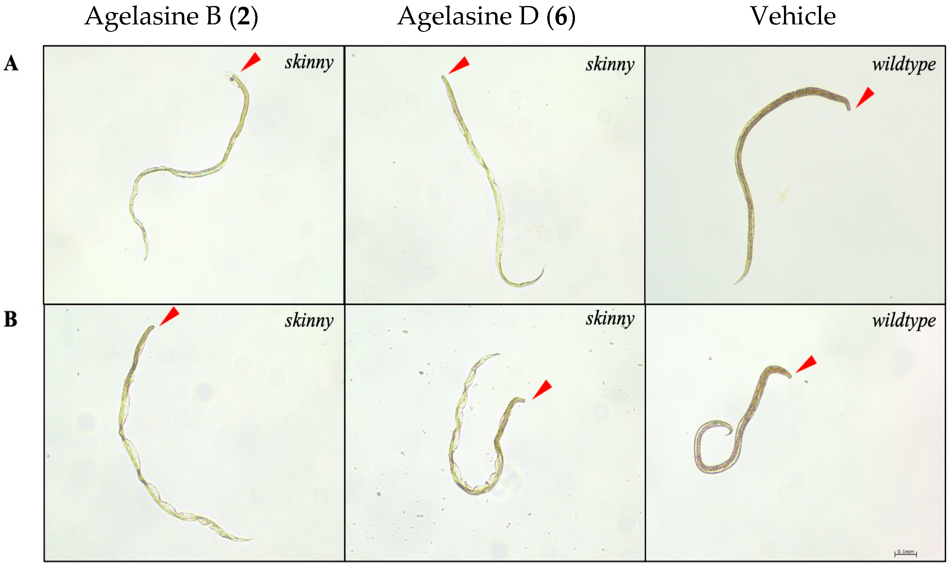
2.3. Anthelmintic Activity of Compounds 16

Compounds 16 were tested for their anthelmintic effect on H. contortus xL3s (infective) and on in vitro-raised H. contortus L4s (parasitic), using an established phenotypic in vitro bioassay at 100 µM [31,32]. The activity of each compound was assessed based on the reduction in larval motility (xL3 and L4) and/or the induction of an abnormal phenotype.
First, compounds were tested for activity on xL3s. None of the compounds (16) significantly reduced xL3 motility at 100 µM at 168 h. The lack of motility reduction observed for compounds 13, which were isolated from active fractions, was anticipated since these fractions did not show a reduction in xL3 motility. Agelasine B (2) and agelasine D (6) induced a skinny (Ski) phenotype in 4% and 34% of affected larvae at 168 h, respectively (Figure 13A). Compounds that induce a Ski phenotype often exhibit effects on L4s [6,10,11,33]. Therefore, we proceeded to evaluate the activity of these compounds on the in vitro-raised L4s of H. contortus. Compounds 2 and 6 reduced L4 motility by 50.5% and 51.8%, respectively, at 100 µM after 90 h of incubation. Furthermore, compounds 2 and 6 induced 54% and 50% of a Ski phenotype in L4s (Figure 13B). While compound 1 did not reduce L4 motility, it induced a Ski phenotype (25%) after 90 h. Compounds 3, 4 and 5 showed no effects on larval (xL3 or L4) motility or phenotypes.
Secondary metabolites from the genus Agelas exhibit antimicrobial [34,35], cytotoxic [17,36] and antiplasmodial activities [37,38]. In the literature, several agelasines have been reported to exhibit promising antiparasitic activities. Notably, agelasine D (6) has been shown to exhibit significant antiparasitic activity against Plasmodium falciparum, Trypanosoma brucei, Leishmania infantum and Trypanosoma cruzi [39,40]. Recently, agelasine B (2) isolated from a NatureBank Agelas sp. sample showed nematocidal activity against the model and free-living nematode C. elegans [41]. Thus, we assessed the activity of all six compounds (16) against C. elegans L4/young adults in an established bioassay [32]. Despite earlier findings suggesting the nematocidal activity of agelasine B (2) against C. elegans, as reported by Risi et al. in 2024 [41], the recent assessment of four compounds, including 2, did not show them to have significant activity in reducing the motility of L4/young adult C. elegans after 40 h of incubation.
The observed discrepancy in the C. elegans assay results may be attributed to variations in experimental conditions, including differences in assay protocols, the C. elegans strains utilized or the range of compound concentrations tested. Such factors can significantly influence the outcomes of bioactivity studies and underscore the importance of standardized methodologies in comparative analyses.

3. Materials and Methods

3.1. General Experimental Procedures

Specific rotations were recorded using a JASCO P-2000 polarimeter (JASCO, Tokyo, Japan). ECD spectra were recorded on a JASCO J-1500 CD spectrophotometer (JASCO, Tokyo, Japan). NMR spectra were recorded at 25 °C on a Bruker AVANCE III 800 MHz NMR spectrometer (Bruker Corporation, Billerica, MA, USA). equipped with a cryoprobe. The 1H and 13C NMR chemical shifts were referenced to solvent peaks for MeOH-d4 at δH 3.31/δC 49.00 and MeCN-d3 at δH 3.31/δC 118.26. LRESIMS data was recorded on an Ultimate 3000 RS UHPLC (Thermo Fisher Scientific, Waltham, MA, USA) connected with a Thermo Scientific Accurose C18-bonded silica column (2.6 μm, 80 Å, 150 × 2.1 mm) coupled to a Thermo Fisher Scientific ISQEC single quadruple ESI mass spectrometer (Thermo Fisher Scientific, Waltham, MA, USA). HRESIMS data were acquired on a Bruker maXis II ETD ESI-qTOF mass spectrometer (Bruker Corporation, Billerica, MA, USA). GRACE Davisil (35–70 µm, 60 Å) C18-bonded silica was used for pre-adsorption work before reversed-phase (RP) HPLC separations. The pre-adsorbed material was subsequently packed into a GRACE stainless steel guard cartridge (10 × 30 mm) and then attached to an HPLC column prior to fractionation. A Waters 600 pump fitted with a Waters 996 photodiode array detector (Milford, MA, USA) fitted with a Gilson 717-plus autosampler was used for RP-HPLC separations (Middleton, WI, USA). Thermo Electron Betasil C18-bonded silica (5 μm, 100 Å, 150 × 21.2 mm) or Phenomenex Luna C18-bonded silica (5 μm, 100 Å, 250 × 10 mm) columns were used for RP-HPLC separations. Frozen marine biota was dried using a Dynamic FD12 freeze dryer (Buchi, Vineyard, NSW, AUS) and ground using a Fritsch Universal Cutting Mill Pulverisette 19 (FRITSCH Milling, Pittsboro, NC, USA). Solvents were removed with a Buchi R-144 rotary evaporator (Buchi, Billerica, MA, USA) and from HPLC fractions using a GeneVac XL4 centrifugal evaporator (Biopharma, Ipswich, UK). All solvents used for chromatography, UV, MS, ECD and [α]D were Honeywell Burdick & Jackson or Lab-Scan HPLC-grade. H2O was filtered using a Sartorius Stedium Arium® Pro VF ultrapure water system (Sartorius, Göttingen, Germany). All NMR spectra were processed using MestReNova version 14.3.0 (Mestrelab Research, Santiago de Compostela, Spain) software. All chemical structures were drawn using ChemDraw version 23.1.1.3 software (Revvity, Waltham, MA, USA). All HPLC and LC-MS results were analyzed by Waters Millennium32 version 4.0 and Chromeleon™ 7.2 software, respectively.

3.2. Animal Material

The sponge Agelas axifera was collected on the 25th of January 1999 from Stanley Reef, Great Barrier Reef, Australia. A voucher specimen (G314522) was deposited at the Queensland Museum, South Brisbane, Queensland, Australia.

3.3. Extraction and Isolation

For initial anthelmintic bioassay-guided fractionation, the freeze-dried and ground specimen of A. axifera (1 g) was extracted sequentially with n-hexane (21 mL), CH2Cl2:MeOH (8:2, 21 mL) and MeOH (39 mL); the n-hexane extract was discarded as it only contained highly lipophilic fatty acid-derived material, while the CH2Cl2 and MeOH extracts were combined and dried under reduced pressure to produce a dark brown gum-like extract (226.1 mg). A portion of this extract (203 mg) was pre-adsorbed to C18-bonded silica (1 g), then packed into a guard cartridge and subjected to semipreparative HPLC using a Betasil C18-bonded silica HPLC column. Isocratic solvent conditions of 90% H2O (0.1% TFA)/10% MeOH (0.1% TFA) were initially employed for the first 10 min; then a linear gradient to 100% MeOH (0.1% TFA) was run over 40 min, followed by isocratic conditions of 100% MeOH (0.1% TFA) for a further 10 min, all at a flow rate of 9 mL/min. Sixty fractions were collected from 0 to 60 min (60 × 1 min fractions), concentrated in a centrifugal evaporator and prepared for anthelmintic screening. Fractions 49 (F49) and 50 (F50) that displayed eviscerated phenotypes in xL3 were combined (16.3 mg) and subsequently purified via semipreparative HPLC using a Luna C18-bonded silica HPLC column. Isocratic solvent conditions of 50% H2O (0.1% TFA)/50% MeOH (0.1% TFA) were initially employed for the first 10 min; then a linear gradient to 100% MeOH (0.1% TFA) was run over 40 min, followed by isocratic conditions of 100% MeOH (0.1% TFA) for a further 10 min, all at a flow rate of 4 mL/min to afford the TFA salts of agelasine Z (1, 0.9 mg, tR 19–20 min, 0.009% dry wt), oxoagelasine B (3, 1.0 mg, tR 22–23 min, 0.01% dry wt) and agelasine B (2, 1.5 mg, tR 35–36 min, 0.015% dry wt).
In order to obtain more of the agelasine-type compounds and potentially other pure compounds, an additional 5 g of freeze-dried sponge sample was extracted with CH2Cl2:MeOH (80:20, 125 mL, 2 h) and MeOH (125 mL, 2 h). Both extracts were combined and dried under reduced pressure to give a dark brown gum extract (1.74 g). The crude extract was subjected to RP-HPLC using Betasil C18-HPLC with isocratic solvent conditions of 90% H2O (0.1% TFA)/10% MeOH (0.1% TFA) for the first 10 min, then a linear gradient to 100% MeOH (0.1% TFA) over 40 min, followed by isocratic conditions of 100% MeOH (0.1% TFA) for a further 10 min, all at a flow rate of 9 mL/min to afford 60 fractions. (–)-Mukanadin C (4, 6.1 mg, 0.12% dry wt) and 4-bromopyrrole-2-carboxylic acid (5, 14.5 mg, 0.29% dry wt) were isolated at tR 25–26 min and tR 28–29 min, respectively. Fractions containing targeted agelasines (F49 and F50, 118.8 mg) were combined and repurified using Betasil C18-bonded silica HPLC using 50% H2O (0.1% TFA)/50% MeOH (0.1% TFA) the first 10 min followed by a linear gradient to 100% MeOH (0.1% TFA) over 40 min and then isocratic conditions of 100% MeOH (0.1% TFA) for a further 10 min, all at a flow rate of 9 mL/min to afford additional agelasine Z (1, 2.6 mg, tR 21–22 min, 0.052% dry wt), oxoagelasine B (3, 16.7 mg, tR 25–27 min, 0.33% dry wt) and agelasine B (2, 41.2 mg, tR 35–40 min, 0.82% dry wt).
Agelasine Z (1): pale yellow gum; [ α ] D 26 –16.5 (c 0.09, MeOH); UV (MeOH) λmax (log ε) 208 (0.30), 274 (0.08) nm; ECD (MeOH) λext 242 nm (Δε +15.8), 291 (Δε +2.9); 1H and 13C NMR data, see Table 1 and Table S1; LRESIMS m/z 436 [M]+; HRESIMS m/z 436.3060 [M]+ (calcd for C26H38N5O, 436.3071).
Agelasine B (2): white amorphous solid; [ α ] D 26 –11.6 (c 0.06, MeOH), lit. [ α ] D 20 –21.5 (c 0.1, MeOH) [14]; ECD (MeOH) λext 288 nm (Δε +6.8); ); 1H and 13C NMR spectra, see Figures S12 and S13; LRESIMS m/z 422 [M]+.
Oxoagelasine B (3): pale yellow gum; [ α ] D 26 –11.6 (c 0.01, MeOH), lit. [ α ] D 20 —8.7 (c 0.3, MeOH) [15]; ECD (MeOH) λext 220 nm (Δε +11.7), 248 (Δε −6.1), 272 nm (Δε +1.7), 289 nm (Δε −0.5), 326 nm (Δε +4.0); 1H and 13C NMR spectra, see Figures S14 and S15; LRESIMS m/z 436 [M]+.
(–)-Mukanadin C (4): amorphous white solid; [ α ] D 26 −10.7 (c 0.1, MeOH), lit. [ α ] D 20 —19 (c 1.9, MeOH) [16]; 1H and 13C NMR spectra, see Figures S19 and S20; LRESIMS m/z 231 [M–79Br]+ and 233 [M–81Br]+.
4-Bromopyrrole-2-carboxylic acid (5): amorphous white solid; 1H and 13C NMR spectra, see Figures S21 and S22; LRESIMS m/z 189 [M–79Br]+ and 191 [M–81Br]+.
Agelasine D (6): amorphous white solid; [ α ] D 26 +12.9 (c 0.2, MeOH), lit. [ α ] D 20 + 15.4 (c 0.02, MeOH)15; 1H and 13C NMR spectra, see Figures S16 and S17; LRESIMS m/z 422 [M]+.
Deuterated agelasine D (7): pale yellow gum; 1H and 13C NMR data, see Table 2 and Table S3; LRESIMS m/z 423 [M(D)]+.
Deuterated agelasine B (8): pale yellow gum; 1H and 13C NMR spectra, see Figures S33 and S34; LRESIMS m/z 423 [M(D)]+.
Deuterated oxoagelasine B (9): pale brown gum; 1H and 13C NMR spectra, see Figures S36 and S37; LRESIMS m/z 437 [M(D)]+.

3.4. Production and Maintenance of H. contortus for Bioassays

The Haecon-5 strain of H. contortus was maintained in experimental sheep according to established protocols [42,43], in compliance with institutional animal ethics guidelines (permit no. 23983; University of Melbourne) and Australian regulations. Helminth-free Merino sheep (six months old; male) were orally inoculated with 7000 third-stage larvae (L3s) of H. contortus. Fecal samples, containing H. contortus eggs, were collected daily from sheep with patent infection, commencing four weeks post-infection. These eggs were incubated at 27 °C and >90% relative humidity for seven days to develop into L3 larvae. The developed larvae were then collected, suspended in tap water and filtered through two layers of nylon mesh (pore size: 20 µm; Sefar, Australia) to eliminate debris or dead larvae. Filtered larvae were stored in 0.9% NaCl saline solution (at a density of 2000 L3s per mL) at 11 °C for up to six months [33]. Immediately prior to use, L3s were exsheathed and sterilized by incubation in 0.15% (v/v) sodium hypochlorite (NaClO) at 38 °C for 20 min with gentle shaking [43]. Post-treatment, exsheathed L3s (xL3s) were immediately washed five times in 50 mL of sterile saline by centrifugation at 800× g (5 min) at room temperature (22–24 °C). Following the final wash, xL3s were suspended in sterile lysogeny broth (LB) [44] supplemented with 100 IU/mL of penicillin, 100 µg/mL of streptomycin and 0.25 µg/mL of amphotericin B (Fungizone®, Thermo Fisher Scientific, Waltham, MA, USA)—referred to as LB*. To develop in vitro fourth-stage larvae (L4) of H. contortus, xL3s were incubated at 38 °C, 10% (v/v) CO2 and >90% relative humidity for 168 h prior to incubation with each compound [6].

3.5. Production and Synchronization of C. elegans for Bioassays

The N2 strain (Bristol; wildtype) of C. elegans was cultured under standard laboratory conditions at 20 °C on nematode growth medium (NGM) agar plates seeded with Escherichia coli OP50 as a food source [45,46]. Gravid adult worms were harvested from NGM plates, washed with sterile M9 buffer and then treated with a sterile 0.4% (v/v) NaOCl solution with 170 mM sodium hydroxide for 4–8 min at room temperature (22–24 °C) to release eggs, with intermittent shaking [46,47]. Upon release, eggs were immediately collected and washed five times in 15 mL of sterile M9 buffer by centrifugation at 500× g for 2 min. Washed eggs were suspended in 8 mL of sterile M9 buffer and incubated in a 15 mL tube with gentle agitation for 24 h at room temperature (22–24 °C), allowing first-stage larvae (L1s) to hatch and enter diapause. Approximately 36 h before setting up assays, synchronized L1s were transferred to NGM agar plates (10 cm) previously seeded with 500 µL of E. coli OP50 (~3000 larvae per plate) and allowed to synchronously develop to fourth-stage larvae (L4s) at 20 °C. On the day of assay, L4s were collected from NGM agar plates and washed twice with sterile M9 buffer by centrifugation at 500× g (2 min) to remove E. coli OP50 and then resuspended in LB*.

3.6. Bioassay for Assessment of Anthelmintic Activity of A. axifera-Derived HPLC Fractions

Individual HPLC fractions (n = 60) were tested for their anthelmintic effect on the larvae (xL3s) of H. contortus using an established bioassay [11]. The assay was performed in triplicate. In brief, fractions were diluted to 12 μge/μL in LB*, and 40 μL of diluted fractions was dispensed into the wells of sterile 368-well flat-bottom microtiter plates (cat. no. 3680; Corning, Corning, NY, USA) containing 80 xL3s; quadruplicate wells with no compound (LB* + 0.6% DMSO; negative control) or monepantel (Zolvix™; Elanco, Greenfield, IN, USA), moxidectin (Cydectin®; Virbac, Carros, France) and compound MIPS-0018666 (abbreviated here as M-666) [48] were used as positive controls (20 µM). The motility of xL3s was measured at 90 h, and the development and phenotypic alterations of xL3s were assessed at 168 h. At 168 h, larvae in individual wells were fixed with 40 µL of 1% iodine and microscopically examined (using an M80 light microscope; Leica, Wetzlar, Germany) at 60-times magnification to assess their development morphology (phenotype) [9,32].

3.7. Bioassay for Evaluation of Anthelmintic Activity of Pure Compounds Using Larvae of H. contortus or C. elegans

These evaluations were carried out for compounds 16 against H. contortus xL3s/L4 or C. elegans L4s with reference to the two positive control compounds (monepantel and moxidectin) using established assays [6,9]. H. contortus xL3s/L4s or C. elegans L4s were dispensed in 96-well plates (cat. no. 3596; Corning, USA) at a density of 300 H. contortus xL3s/L4s or 100 C. elegans L4s per well, respectively, with individual, serially diluted compounds (9 points; 2-fold serial dilution in LB*; 100 µM to 0.39 nM), in a final volume of 100 µL of LB*. Plates containing H. contortus xL3s/L4s were incubated at 38 °C, 10% (v/v) CO2 and >90% relative humidity. Plates containing C. elegans L4s were incubated at 20 °C. Compounds were tested by three independent experiments in triplicate. The motility of H. contortus xL3s and L4s was measured following 168 h and 90 h of incubation with the compound, respectively. The motility of C. elegans young adults was measured following 40 h of incubation with the compound. A one-way analysis of variance (ANOVA) with Tukey’s multiple comparison test or an unpaired t-test was used to establish statistically significant differences in larval motility.

3.8. Computational Methods

Conformer searches for GIAO NMR and TDDFT ECD calculations were performed using the Schrödinger Macromodel software (version 10.7) software suite, Monte Carlo Minimum method (MCMM) molecular mechanics, MMFF forcefield and a 21.0 kJ/mol energy window. The step count for establishing accurate conformer representation was set so that all low-energy conformers were found at least 10 times. For GIAO DFT NMR calculations, each conformer sets for candidate structures 1a and 1b underwent an initial gas-phase geometry optimization (GO) at the B3LYP/6-31G* level of theory using Gaussian 16 (Revision C.01) [49]. The resulting GO sets were filtered for duplicate and high-energy conformers (>3.0 kcal/mol above the energy minimum removed, Tables S5–S8). 1H and 13C NMR chemical shifts were calculated at the mPW1PW91/6-311+G(d,p) level of theory with the PCM solvent model for methanol [50]. The Boltzmann-averaged NMR data for each candidate stereoisomer were then analyzed using the Bayesian probabilistic DP4+ method [21]. For TDDFT ECD calculations, two DFT geometry optimization (GO) calculations were performed on 1 using Gaussian 16 software. The first GO was calculated at the B3LYP-31G(d) level of theory in the gas phase, while the second GO and frequency calculation were performed at the higher B3LYP/6-311G(d,p) level of theory with the PCM solvent model for methanol. The second GO set was filtered for imaginary frequencies, redundant conformers and energies > 3.0 kcal/mol (Tables S9 and S10). Following this, electronic transition and rotational strength calculations were performed using the TDDFT method at the CAM-B3LYP/6-311+G(d,p) level of theory (inclusive of the PCM solvent model for methanol) within Gaussian 16. The resultant TDDFT-calculated UV and ECD spectra for 1 were Boltzmann-weighted and visualized using the freely available SpecDis software (version 1.71) [51]. TDDFT-calculated and experimental UV spectra for 1 were matched to accurately align experimental and TDDFT-calculated ECD spectra. A Gaussian band shape and sigma gamma value of 0.29 eV and a +5nm UV correction were applied to experimental spectra for 1. Automated processes with the HPC were performed using a Windows 11 PC with modified Python scripts originally sourced from the Willoughby protocol [52]. SpectroIBIS (v1.01) was used to scale NMR shielding tensors and to generate tabulated DFT energies and output data [53].

4. Conclusions

In summary, the bioassay-guided fractionation of an A. axifera extract afforded one new and two known agelasine alkaloids. Despite the initial activity observed in the crude extract, neither the fractions nor the isolated compounds demonstrated significant activity in subsequent assays. A challenge associated with the isolation of natural products from bioactive extracts is the loss of activity or inability to isolate the bioactive compounds during bioassay-guided fractionation. Several factors may potentially contribute to this issue, including the degradation of the bioactive constituents during purification, insufficient amounts of compounds for effective separation and/or the observed activity actually being a result of the synergistic interaction of several different compounds [54]. The current findings from our research suggest that bioactivity may be attributed to the synergistic effects of the compounds within the fractions. Whilst additional studies could explore potential synergistic interactions and optimize the isolation process to identify other minor and active constituents, limited sponge material currently prevents such investigations. Finally, it is interesting to note that the H-8’ of the adenine moiety for the three agelasines studied here was unexpectedly labile and underwent a facile deuterium exchange reaction in MeOH-d4 at room temperature.

Supplementary Materials

The following supporting information can be downloaded at https://www.mdpi.com/article/10.3390/md23070276/s1, Tables S1–S3: NMR data of compounds 1 and 7; Table S4: DP4+ output for 1; Tables S5–S10: Computational data for 1; Figures S1–S38: NMR, LC-MS, HRESIMS and ECD spectra of compounds 19.

Author Contributions

Conceptualization, R.A.D. and R.B.G.; methodology, K.W., A.C.T., J.J.B., R.B.G. and R.A.D.; software, K.W., D.C.H., A.C.T. and J.J.B.; formal analysis, K.W., A.C.T., J.J.B. and D.C.H.; validation, A.C.T., J.J.B., D.C.H., I.D.J., R.B.G. and R.A.D.; investigation, K.W., A.C.T. and J.J.B.; resources, R.A.D., R.B.G. and M.G.E.; data curation, K.W., A.C.T., J.J.B., D.C.H., R.B.G. and R.A.D.; writing—original draft preparation, K.W. and R.A.D.; writing—review and editing, A.C.T., J.J.B., D.C.H., I.D.J., M.G.E., A.R.C., R.B.G. and R.A.D.; visualization, A.C.T., J.J.B. and D.C.H.; supervision, R.A.D. and A.R.C.; project administration, R.B.G. and R.A.D.; funding acquisition, R.B.G. and R.A.D. All authors have read and agreed to the published version of the manuscript.

Funding

This research was funded by the Australian Research Council (ARC) NMR and MS equipment (grants LE0668477, LE140100119 and LE0237908). Research at The University of Melbourne was supported by the ARC (LP220200614 and LP180101085), PhylumTECH and Oz Omics Pty Ltd.

Institutional Review Board Statement

The study was approved by the Institutional Animal Ethics Committee of The University of Melbourne (permit no. 23983; approval date: 5 May 2022), following Australian regulations.

Data Availability Statement

The original data presented in the study are included in the article/Supplementary Materials; further inquiries can be directed to the corresponding author.

Acknowledgments

We acknowledge the Benefit Sharing Agreement between the Queensland Government and Griffith University under the Biodiscovery Act 2004 (QLD), enabling equitable benefit sharing from biodiscovery using Queensland-sourced Native Biological Material. We thank NatureBank for the supply of the sponge sample (Agelas axifera) that the natural products detailed in this paper were isolated from. D.C.H. thanks Griffith University and the University of Western Australia for access to the Gowonda and Kaya HPCs for quantum chemical computations. K.W. acknowledges Griffith University for scholarship support.

Conflicts of Interest

The authors declare no conflicts of interest.

References

  1. Charlier, J.; Bartley, D.; Sotiraki, S.; Martinez-Valladares, M.; Claerebout, E.; von Samson-Himmelstjerna, G.; Thamsborg, S.; Hoste, H.; Morgan, E.; Rinaldi, L. Anthelmintic resistance in ruminants: Challenges and solutions. Adv. Parasitol. 2022, 115, 171–227. [Google Scholar] [PubMed]
  2. Charlier, J.; Rinaldi, L.; Musella, V.; Ploeger, H.W.; Chartier, C.; Vineer, H.R.; Hinney, B.; von Samson-Himmelstjerna, G.; Băcescu, B.; Mickiewicz, M.; et al. Initial assessment of the economic burden of major parasitic helminth infections to the ruminant livestock industry in Europe. Prev. Vet. Med. 2020, 182, 105103. [Google Scholar] [CrossRef]
  3. Sangster, N.C.; Cowling, A.; Woodgate, R.G. Ten events that defined anthelmintic resistance research. Trends Parasitol. 2018, 34, 553–563. [Google Scholar] [CrossRef]
  4. Kotze, A.; Prichard, R. Anthelmintic resistance in Haemonchus contortus: History, mechanisms and diagnosis. Adv. Parasitol. 2016, 93, 397–428. [Google Scholar] [PubMed]
  5. Herath, H.D.; Preston, S.; Jabbar, A.; Garcia-Bustos, J.; Taki, A.C.; Addison, R.S.; Hayes, S.; Beattie, K.D.; McGee, S.L.; Martin, S.D. Identification of fromiamycalin and halaminol A from Australian marine sponge extracts with anthelmintic activity against Haemonchus contortus. Mar. Drugs 2019, 17, 598. [Google Scholar] [CrossRef]
  6. Taki, A.C.; Brkljača, R.; Wang, T.; Koehler, A.V.; Ma, G.; Danne, J.; Ellis, S.; Hofmann, A.; Chang, B.C.H.; Jabbar, A.; et al. Natural Compounds from the Marine Brown Alga Caulocystis cephalornithos with Potent In Vitro-Activity against the Parasitic Nematode Haemonchus contortus. Pathogens 2020, 9, 550. [Google Scholar] [CrossRef] [PubMed]
  7. Herath, H.D.; Taki, A.C.; Sleebs, B.E.; Hofmann, A.; Nguyen, N.; Preston, S.; Davis, R.A.; Jabbar, A.; Gasser, R.B. Advances in the discovery and development of anthelmintics by harnessing natural product scaffolds. Adv. Parasitol. 2021, 111, 203–251. [Google Scholar]
  8. NatureBank. Available online: https://www.griffith.edu.au/institute-drug-discovery/unique-resources/naturebank (accessed on 31 March 2025).
  9. Taki, A.C.; Byrne, J.J.; Jabbar, A.; Lum, K.Y.; Hayes, S.; Addison, R.S.; Ramage, K.S.; Hofmann, A.; Ekins, M.G.; Wang, T. High Throughput Screening of the NatureBank ‘Marine Collection’ in a Haemonchus Bioassay Identifies Anthelmintic Activity in Extracts from a Range of Sponges from Australian Waters. Molecules 2021, 26, 5846. [Google Scholar] [CrossRef]
  10. Hayes, S.; Taki, A.C.; Lum, K.Y.; Byrne, J.J.; White, J.M.; Ekins, M.G.; Gasser, R.B.; Davis, R.A. Identification of Anthelmintic Bishomoscalarane Sesterterpenes from the Australian Marine Sponge Phyllospongia bergquistae and Structure Revision of Phyllolactones A–D. J. Nat. Prod. 2022, 85, 1723–1729. [Google Scholar] [CrossRef]
  11. Ramage, K.S.; Taki, A.C.; Lum, K.Y.; Hayes, S.; Byrne, J.J.; Wang, T.; Hofmann, A.; Ekins, M.G.; White, J.M.; Jabbar, A. Dysidenin from the Marine Sponge Citronia sp. Affects the Motility and Morphology of Haemonchus contortus Larvae In Vitro. Mar. Drugs 2021, 19, 698. [Google Scholar] [CrossRef]
  12. Chu, M.-J.; Li, M.; Ma, H.; Li, P.-L.; Li, G.-Q. Secondary metabolites from marine sponges of the genus Agelas: A comprehensive update insight on structural diversity and bioactivity. RSC Adv. 2022, 12, 7789–7820. [Google Scholar] [CrossRef]
  13. Chu, M.J.; Tang, X.L.; Qin, G.F.; Sun, Y.T.; Li, L.; de Voogd, N.J.; Li, P.L.; Li, G.Q. Pyrrole Derivatives and Diterpene Alkaloids from the South China Sea Sponge Agelas nakamurai. Chem. Biodivers. 2017, 14, e1600446. [Google Scholar] [CrossRef]
  14. Nakamura, H.; Wu, H.; Ohizumi, Y.; Hirata, Y. Agelasine-A, -B, -C and -D, novel bicyclic diterpenoids with a 9-methyladeninium unit possessing inhibitory effects on Na, K-ATPase from the Okinawa sea sponge Agelas sp. Tetrahedron Lett. 1984, 25, 2989–2992. [Google Scholar] [CrossRef]
  15. Abdjul, D.B.; Yamazaki, H.; Kanno, S.-i.; Takahashi, O.; Kirikoshi, R.; Ukai, K.; Namikoshi, M. Structures and biological evaluations of agelasines isolated from the Okinawan marine sponge Agelas nakamurai. J. Nat. Prod. 2015, 78, 1428–1433. [Google Scholar] [CrossRef]
  16. Lin, Y.-C.; Chao, C.-H.; Fu, C.-W.; Chiou, S.-F.; Huang, T.-Y.; Yang, Y.-J.; Wu, S.-H.; Chen, S.-L.; Wang, H.-C.; Yu, M.-C. Computationally assisted structure elucidation of new 2-guanidinoethanesulfonyl sesquiterpenoid alkaloids: Agelasidines G–I from the marine sponge Agelas nakamurai. Tetrahedron 2022, 126, 133077. [Google Scholar] [CrossRef]
  17. Regalado, E.L.; Laguna, A.; Mendiola, J.; Thomas, O.P.; Nogueiras, C. Bromopyrrole alkaloids from the Caribbean sponge Agelas cerebrum. Quím. Nova 2011, 34, 289–291. [Google Scholar] [CrossRef]
  18. Jiao, Y.; Preston, S.; Garcia-Bustos, J.F.; Baell, J.B.; Ventura, S.; Le, T.; McNamara, N.; Nguyen, N.; Botteon, A.; Skinner, C. Tetrahydroquinoxalines induce a lethal evisceration phenotype in Haemonchus contortus in vitro. Int. J. Parasitol. Drugs Drug Resist. 2019, 9, 59–71. [Google Scholar] [CrossRef]
  19. Gundersen, L.-L. Synthesis and biological activities of marine terpene-adenine hybrids and synthetic analogs. Phytochem. Rev. 2013, 12, 467–486. [Google Scholar] [CrossRef]
  20. Fu, C.-W.; Chiang, L.; Chao, C.-H.; Huang, Y.-L.; Chiou, S.-F.; Wang, L.-C.; Chang, H.-W.; Chen, S.-L.; Wang, H.-C.; Yu, M.-C.; et al. Nakamusines A−C, new 9-methyladeninium diterpenoid alkaloids from a Formosan marine sponge Agelas nakamurai. Tetrahedron 2023, 149, 133745. [Google Scholar] [CrossRef]
  21. Grimblat, N.; Zanardi, M.M.; Sarotti, A.M. Beyond DP4: An improved probability for the stereochemical assignment of isomeric compounds using quantum chemical calculations of NMR shifts. J. Org. Chem. 2015, 80, 12526–12534. [Google Scholar] [CrossRef] [PubMed]
  22. Zanardi, M.M.; Biglione, F.A.; Sortino, M.A.; Sarotti, A.M. General quantum-based NMR method for the assignment of absolute configuration by single or double derivatization: Scope and limitations. J. Org. Chem. 2018, 83, 11839–11849. [Google Scholar] [CrossRef]
  23. Holland, D.C.; Carroll, A.R. Structure Revision of Formyl Phloroglucinol Meroterpenoids: A Unified Approach Using NMR Fingerprinting and DFT NMR and ECD Analyses. Molecules 2024, 29, 594. [Google Scholar] [CrossRef]
  24. Pech-Puch, D.; Forero, A.M.; Fuentes-Monteverde, J.C.; Lasarte-Monterrubio, C.; Martinez-Guitian, M.; González-Salas, C.; Guillén-Hernández, S.; Villegas-Hernández, H.; Beceiro, A.; Griesinger, C. Antimicrobial Diterpene Alkaloids from an Agelas citrina Sponge Collected in the Yucatán Peninsula. Mar. Drugs 2022, 20, 298. [Google Scholar] [CrossRef]
  25. Atzrodt, J.; Derdau, V.; Kerr, W.J.; Reid, M. Deuterium and tritium labelled compounds: Applications in the life sciences. Angew. Chem. Int. Ed. 2018, 57, 1758–1784. [Google Scholar] [CrossRef]
  26. Yang, H.; Hesk, D. Base metal catalyzed hydrogen isotope exchange. J. Labelled Compd. Radiopharm. 2020, 63, 296–307. [Google Scholar] [CrossRef]
  27. Zhang, J.; Chen, J.; Chen, J.; Luo, Y.; Xia, Y. Solvent as photoreductant for dehalogenation of α-haloketones under catalyst-free conditions. Tetrahedron Lett. 2022, 98, 153835. [Google Scholar] [CrossRef]
  28. Kumar, V.; Rai, R.; Pandey, S. Controlling excited-state prototropism via the acidity of ionic liquids. RSC Adv. 2013, 3, 11621–11627. [Google Scholar] [CrossRef]
  29. Herrmann, W.A.; Köcher, C. N-Heterocyclic carbenes. Angew. Chem. Int. Ed. Engl. 1997, 36, 2162–2187. [Google Scholar] [CrossRef]
  30. Rico del Cerro, D.; Mera-Adasme, R.; King, A.W.; Perea-Buceta, J.E.; Heikkinen, S.; Hase, T.; Sundholm, D.; Wähälä, K. On the Mechanism of the Reactivity of 1, 3-Dialkylimidazolium Salts under Basic to Acidic Conditions: A Combined Kinetic and Computational Study. Angew. Chem. Int. Ed. 2018, 57, 11613–11617. [Google Scholar] [CrossRef]
  31. Wijesekera, K.; Taki, A.C.; Byrne, J.J.; White, J.M.; Carroll, A.R.; Gasser, R.B.; Davis, R.A. Anthelmintic activity of selected neolignans and semisynthetic derivatives from Styrax suberifolius. Tetrahedron 2025, 169, 134366. [Google Scholar] [CrossRef]
  32. Taki, A.C.; Byrne, J.J.; Wang, T.; Sleebs, B.E.; Nguyen, N.; Hall, R.S.; Korhonen, P.K.; Chang, B.C.; Jackson, P.; Jabbar, A. High-throughput phenotypic assay to screen for anthelmintic activity on Haemonchus contortus. Pharmaceuticals 2021, 14, 616. [Google Scholar] [CrossRef]
  33. Preston, S.; Jabbar, A.; Nowell, C.; Joachim, A.; Ruttkowski, B.; Baell, J.; Cardno, T.; Korhonen, P.K.; Piedrafita, D.; Ansell, B.R. Low cost whole-organism screening of compounds for anthelmintic activity. Int. J. Parasitol. 2015, 45, 333–343. [Google Scholar] [CrossRef]
  34. Kusama, T.; Tanaka, N.; Takahashi-Nakaguchi, A.; Gonoi, T.; Fromont, J.; Kobayashi, J.i. Bromopyrrole Alkaloids from a Marine Sponge Agelas sp. Chem. Pharm. Bull. 2014, 62, 499–503. [Google Scholar] [CrossRef]
  35. Youssef, D.T.A.; Alqarni, A.S.; Almohammadi, A.M.; Abujamel, T.; Shaala, L.A. Marmaricines A-C: Antimicrobial Brominated Pyrrole Alkaloids from the Red Sea Marine Sponge Agelas sp. aff. marmarica. Mar. Drugs 2025, 23, 80. [Google Scholar] [CrossRef]
  36. Pettit, G.R.; Tang, Y.; Zhang, Q.; Bourne, G.T.; Arm, C.A.; Leet, J.E.; Knight, J.C.; Pettit, R.K.; Chapuis, J.-C.; Doubek, D.L. Isolation and structures of axistatins 1–3 from the Republic of Palau marine sponge Agelas axifera Hentschel. J. Nat. Prod. 2013, 76, 420–424. [Google Scholar] [CrossRef]
  37. Appenzeller, J.; Mihci, G.; Martin, M.T.; Gallard, J.F.; Menou, J.L.; Boury-Esnault, N.; Hooper, J.; Petek, S.; Chevalley, S.; Valentin, A. Agelasines J, K, and L from the Solomon Islands marine sponge Agelas cf. mauritiana. J. Nat. Prod. 2008, 71, 1451–1454. [Google Scholar] [CrossRef]
  38. Ueoka, R.; Nakao, Y.; Kawatsu, S.; Yaegashi, J.; Matsumoto, Y.; Matsunaga, S.; Furihata, K.; Van Soest, R.W.; Fusetani, N. Gracilioethers A− C, antimalarial metabolites from the marine sponge Agelas gracilis. J. Org. Chem. 2009, 74, 4203–4207. [Google Scholar] [CrossRef]
  39. Gordaliza, M. Terpenyl-Purines from the Sea. Mar. Drugs 2009, 7, 833–849. [Google Scholar] [CrossRef]
  40. Vik, A.; Proszenyák, Á.; Vermeersch, M.; Cos, P.; Maes, L.; Gundersen, L.L. Screening of Agelasine D and Analogs for Inhibitory Activity against Pathogenic Protozoa; Identification of Hits for Visceral Leishmaniasis and Chagas Disease. Molecules 2009, 14, 279–288. [Google Scholar] [CrossRef]
  41. Risi, G.; Liu, M.; Vairoletti, F.; Quinn, R.J.; Salinas, G. A Screening of 10,240 NatureBank Fractions Identifies Nematicidal Activity in Agelasine-Containing Extracts from Sponges. J. Nat. Prod. 2024, 87, 1532–1539. [Google Scholar] [CrossRef]
  42. Schwarz, E.M.; Korhonen, P.K.; Campbell, B.E.; Young, N.D.; Jex, A.R.; Jabbar, A.; Hall, R.S.; Mondal, A.; Howe, A.C.; Pell, J. The genome and developmental transcriptome of the strongylid nematode Haemonchus contortus. Genome Biol. 2013, 14, R89. [Google Scholar] [CrossRef] [PubMed]
  43. Taki, A.C.; Byrne, J.J.; Boag, P.R.; Jabbar, A.; Gasser, R.B. Practical high-throughput method to screen compounds for anthelmintic activity against Caenorhabditis elegans. Molecules 2021, 26, 4156. [Google Scholar] [CrossRef] [PubMed]
  44. Bertani, G. Studies on lysogenesis I: The mode of phage liberation by lysogenic Escherichia coli. J. Bacteriol. 1951, 62, 293–300. [Google Scholar] [CrossRef] [PubMed]
  45. Brenner, S. The genetics of Caenorhabditis elegans. Genetics 1974, 77, 71–94. [Google Scholar] [CrossRef]
  46. Stiernagle, T. Maintenance of C. elegans. In WormBook: The Online Review of C. elegans Biology; The C. elegans Research Community: Pasadena, CA, USA, 2006. [Google Scholar]
  47. Porta-de-la-Riva, M.; Fontrodona, L.; Villanueva, A.; Cerón, J. Basic Caenorhabditis elegans methods: Synchronization and observation. J. Vis. Exp. 2012, 64, e4019. [Google Scholar]
  48. Le, T.G.; Kundu, A.; Ghoshal, A.; Nguyen, N.H.; Preston, S.; Jiao, Y.; Ruan, B.; Xue, L.; Huang, F.; Keiser, J. Optimization of novel 1-methyl-1H-pyrazole-5-carboxamides leads to high potency larval development inhibitors of the barber’s pole worm. J. Med. Chem. 2018, 61, 10875–10894. [Google Scholar] [CrossRef]
  49. Frisch, M.; Trucks, G.; Schlegel, H.; Scuseria, G.; Robb, M.; Cheeseman, J.; Scalmani, G.; Barone, V.; Petersson, G.; Nakatsuji, H. Gaussian 16, Revision C. 01; Gaussian, Inc.: Wallingford, CT, USA, 2016. [Google Scholar]
  50. Tomasi, J.; Mennucci, B.; Cammi, R. Quantum Mechanical Continuum Solvation Models. Chem. Rev. 2005, 105, 2999–3094. [Google Scholar] [CrossRef]
  51. Bruhn, T.; Schaumlöffel, A.; Hemberger, Y.; Bringmann, G. SpecDis: Quantifying the Comparison of Calculated and Experimental Electronic Circular Dichroism Spectra. Chirality 2013, 25, 243–249. [Google Scholar] [CrossRef]
  52. Willoughby, P.H.; Jansma, M.J.; Hoye, T.R. A guide to small-molecule structure assignment through computation of (1H and 13C) NMR chemical shifts. Nat. Protoc. 2014, 9, 643–660. [Google Scholar] [CrossRef]
  53. Bulcock, B.W.; Chooi, Y.-H.; Flematti, G.R. SpectroIBIS: Automated Data Processing for Multiconformer Quantum Chemical Spectroscopic Calculations. J. Nat. Prod. 2025, 88, 495–501. [Google Scholar] [CrossRef]
  54. Nothias, L.-F.; Nothias-Esposito, M.; da Silva, R.; Wang, M.; Protsyuk, I.; Zhang, Z.; Sarvepalli, A.; Leyssen, P.; Touboul, D.; Costa, J.; et al. Bioactivity-Based Molecular Networking for the Discovery of Drug Leads in Natural Product Bioassay-Guided Fractionation. J. Nat. Prod. 2018, 81, 758–767. [Google Scholar] [CrossRef] [PubMed]
Figure 1. RP-HPLC (MeOH/H2O/0.1% TFA) chromatogram of CH2Cl2/MeOH extract of Agelas axifera; F49 and F50 displayed anthelmintic activity.
Marinedrugs 23 00276 g001
Figure 2. Chemical structures of agelasine Z (1), agelasine B (2), oxoagelasine B (3), (–)-mukanadin C (4), 4-bromopyrrole-2-carboxylic acid (5) and agelasine D (6). All alkaloids were isolated as their TFA salts.
Marinedrugs 23 00276 g002
Figure 3. Key COSY and HMBC correlations of 1.
Marinedrugs 23 00276 g003
Figure 4. Two possible configurations for agelasine Z (1) following MeOH-d4 ROESY NMR analysis; (1a) 5R*, 8S*, 9R* and (1b) 5R*, 8S*, 9S*.
Marinedrugs 23 00276 g004
Figure 5. Key ROESY NMR correlations for 1 in MeCN-d3.
Marinedrugs 23 00276 g005
Figure 6. Comparative TDDFT-calculated (red dashed line 1a—5R, 8S and 9R; blue dashed line 1a—5S, 8R and 9S) and experimental ECD spectra for agelasine Z (1).
Marinedrugs 23 00276 g006
Figure 7. Conversion of H-8’ to D-8’ of the adenine moiety of compounds 13 and 6 during storage in MeOH-d4 at room temperature.
Marinedrugs 23 00276 g007
Figure 8. Stacked 1H NMR spectra of agelasine D (6) in MeOH-d4 at 25 °C recorded at 0, 1, 4, 24, 48, 72, 96, 120 and 144 h.
Marinedrugs 23 00276 g008
Figure 9. Conversion of H-8’ to D-8’ for agelasine D (6) in MeOH-d4 at 25 °C.
Marinedrugs 23 00276 g009
Figure 10. Stacked 1H NMR spectra of agelasine B (2, top spectra) and oxoagelasine B (3, bottom spectra) in MeOH-d4 at 25 °C recorded at 0, 48 and 120 h.
Marinedrugs 23 00276 g010
Figure 11. Chemical structures of deuterated compounds 79.
Marinedrugs 23 00276 g011
Figure 12. Proposed mechanism for H/D exchange of agelasines in MeOH-d4.
Marinedrugs 23 00276 g012
Figure 13. Representative images of H. contortus L4s following incubation with agelasine B (2) and agelasine D (6) at 100 µM displaying skinny (Ski) phenotype—compared with untreated control with wildtype phenotype (vehicle, containing 0.25% DMSO; right). (A) xL3s treated with compound for 168 h, (B) in vitro-raised L4s treated with compound for 90 h. Red arrow indicates anterior end of larvae. Scale bar represents 0.1 mm.
Marinedrugs 23 00276 g013
Table 1. 1H (800 MHz) and 13C (200 MHz) NMR data for the TFA salt of agelasine Z (1) in MeOH-d4.
PositionδC, TypeδH, mult., (J in Hz)
1122.6, CH5.87, s
2202.7, C-
3a47.8, CH22.42, d (16.1)
3b1.98, d (16.1)
435.4, C-
546.1, C-
6a31.0, CH22.11, m
6b1.43, m
7a31.6, CH21.64, m
7b
847.5, CH1.48, m
947.2, CH2.19, dd (12.8, 3.7)
10176.4, C-
11a31.0, CH22.08, m
11b1.50, m
12a35.2, CH22.10, m
12b1.78, m
13149.1, C-
14115.8, CH5.55, t (7.0)
15a48.6, CH25.21, d (7.0)
15b
1617.0, CH31.88, s
1716.4, CH30.98, d (6.8)
1827.8, CH31.01, s
1928.8, CH31.04, s
2023.2, CH31.20, s
2’157.1, CH8.47, s
4’150.9, C-
5’111.2, C-
6’154.1, C-
8’141.9, CH9.33, s
NH2-n.d.
9-NCH332.0, CH33.98, s
n.d.: not detected.
Table 2. 1H (800 MHz) and 13C (200 MHz) NMR data of deuterated agelasine D (7) in MeOH-d4.
PositionδC, TypeδH, mult., (J in Hz)
1a40.3, CH21.78, m
1b0.98, td (12.8, 3.5)
2a20.4, CH21.74, m
2b1.57, m
3a43.3, CH21.40, m
3b1.19, td (13.5, 3.7)
434.5, C-
557.4, CH1.06, dd (12.6, 2.8)
6a25.6, CH21.75, m
6b1.33, m
7a39.4, CH22.39, m
7b1.93, m
8149.7, C-
956.9, CH1.60, m
1040.7, C-
11a22.6, CH21.71, m
11b1.55, m
12a39.4, CH22.33, m
12b2.04, m
13149.5, C-
14115.7, CH5.52, t (7.2)
15a48.6, CH25.23, d (7.2)
15b
1617.0, CH31.87, s
17a107.0, CH24.88, m
17b4.59, brs
1834.1, CH30.87, s
1922.1, CH30.82, s
2015.0, CH30.71, s
2’157.2, CH8.47, s
4’150.9, C-
5’111.1, C-
6’145.4, C-
8’142.0, CD-
NH2-n.d.
9-NCH332.0, CH33.98, s
n.d.: not detected.
Disclaimer/Publisher’s Note: The statements, opinions and data contained in all publications are solely those of the individual author(s) and contributor(s) and not of MDPI and/or the editor(s). MDPI and/or the editor(s) disclaim responsibility for any injury to people or property resulting from any ideas, methods, instructions or products referred to in the content.

