Previous Article in Journal
Lipotrichaibol A and Trichoderpeptides A–D: Five New Peptaibiotics from a Sponge-Derived Trichoderma sp. GXIMD 01001
Previous Article in Special Issue
Composition of Triterpene Glycosides of the Far Eastern Sea Cucumber Cucumaria conicospermium Levin et Stepanov; Structure Elucidation of Five Minor Conicospermiumosides A3-1, A3-2, A3-3, A7-1, and A7-2; Cytotoxicity of the Glycosides Against Human Breast Cancer Cell Lines; Structure–Activity Relationships
 
 
Article

Cladolosides of Groups S and T: Triterpene Glycosides from the Sea Cucumber Cladolabes schmeltzii with Unique Sulfation; Human Breast Cancer Cytotoxicity and QSAR

by
Alexandra S. Silchenko
*,
Elena A. Zelepuga
,
Ekaterina A. Chingizova
,
Ekaterina S. Menchinskaya
,
Kseniya M. Tabakmakher
,
Anatoly I. Kalinovsky
,
Sergey A. Avilov
,
Roman S. Popov
,
Pavel S. Dmitrenok
and
Vladimir I. Kalinin
*
G.B. Elyakov Pacific Institute of Bioorganic Chemistry, Far Eastern Branch of the Russian Academy of Sciences, Pr. 100-letya Vladivostoka 159, 690022 Vladivostok, Russia
*
Authors to whom correspondence should be addressed.
Mar. Drugs 2025, 23(7), 265; https://doi.org/10.3390/md23070265
Submission received: 16 May 2025 / Revised: 17 June 2025 / Accepted: 23 June 2025 / Published: 25 June 2025
(This article belongs to the Special Issue Novel Biomaterials and Active Compounds from Sea Cucumbers)

Abstract

Four new minor monosulfated triterpene penta- and hexaosides, cladolosides S (1), S1 (2), T (3), and T1 (4), were isolated from the Vietnamese sea cucumber Cladolabes schmeltzii (Sclerodactylidae, Dendrochirotida). The structures of the compounds were established based on extensive analysis of 1D and 2D NMR spectra as well as HR-ESI-MS data. Cladodosides S (1), S1 (2) and T (3), T1 (4) are two pairs of dehydrogenated/hydrogenated compounds that share identical carbohydrate chains. The oligosaccharide chain of cladolosides of the group S is new for the sea cucumber glycosides due to the presence of xylose residue attached to C-4 Xyl1 in combination with a sulfate group at C-6 MeGlc4. The oligosaccharide moiety of cladolosides of the group T is unique because of the position of the sulfate group at C-3 of the terminal sugar residue instead of the 3-O-Me group. This suggests that the enzymatic processes of sulfation and O-methylation that occur during the biosynthesis of glycosides can compete with each other. This can presumably occur due to the high level of expression or activity of the enzymes that biosynthesize glycosides. The mosaicism of glycoside biosynthesis (time shifting or dropping out of some biosynthetic stages) may indicate a lack of compartmentalization inside the cells of organism producers, leading to a certain degree of randomness in enzymatic reactions; however, this also offers the advantage of providing chemical diversity of the glycosides. Analysis of the hemolytic activity of a series of 26 glycosides from C. schmeltzii revealed some patterns of structure–activity relationships: the presence or absence of 3-O-methyl groups has no significant impact, hexaosides, which are the final products of biosynthesis and predominant compounds of the glycosidic fraction of C. schmeltzii, are more active than their precursors, pentaosides, and the minor tetraosides, cladolosides of the group A, are weak membranolytics and therefore are not synthesized in large quantities. Two glycosides from C. schmeltzii, cladolosides D (18) and H1 (26), display selectivity of cytotoxic action toward triple-negative breast cancer cells MDA-MB-231, while remaining non-toxic in relation to normal mammary cells MCF-10A. Quantitative structure–activity relationships (QSAR) were calculated based on the correlational analysis of the physicochemical properties and structural features of the glycosides and their hemolytic and cytotoxic activities against healthy MCF-10A cells and cancer MCF-7 and MDA-MB-231 cell lines. QSAR highlighted the complexity of the relationships as the cumulative effect of many minor contributions from individual descriptors can have a significant impact. Furthermore, many structural elements were found to have different effects on the activity of the glycosides against different cell lines. The opposing effects were especially pronounced in relation to hormone-dependent breast cancer cells MCF-7 and triple-negative MDA-MB-231 cells.

Graphical Abstract

1. Introduction

Sea cucumbers (Holothuroidea, Echinodermata) are marine invertebrates that have become a focus of extensive chemical investigations because they produce pharmacologically relevant triterpene glycosides, making them a valuable source of novel bioactive compounds. Despite the long history of investigations of the triterpene glycosides of different species of sea cucumbers, they continue to attract the attention of scientists due to their intriguing chemical diversity and various biological activities [1,2]. Some of the recent investigations of the glycosidic composition of sea cucumbers are based on mass-spectrometric metabolomic profiling [3,4,5]. However, this approach should be used in conjunction with the isolation and structure elucidation of individual native glycosides, since it does not allow the unambiguous determination of the spatial structure of substances and the distinction of isomers, especially given that modern techniques and technologies allow the separation of complex mixtures of polar native glycosides and isolation of minor compounds [6]. Nevertheless, metabolomic studies of sea cucumbers provide useful data on the chemical diversity of glycosides and their content inside the different internal parts of animals, as well as ecological functions [7,8,9,10].
In addition, glycosides have promising and diverse biological activities [5,11,12,13,14]. Recently, a selective cytotoxic effect of glycosides against triple-negative breast cancer cells (MDA-MB-231) was discovered [15,16,17], suggesting their potential use in targeted therapy of breast cancer.
The main patterns of structure–activity relationships (SAR) of these metabolites were deduced earlier [18,19], but the application of correlational analysis for quantitative structure–activity relationship (QSAR) calculations [16,20] based on a broadened combinatorial library will provide further opportunities to predict the bioactivity of glycosides.
Previously, 40 glycosides (cladolosides of groups A–R (544, Figure S33)) characterized by extreme variability of carbohydrate chains were isolated from the sea cucumber Cladolabes schmeltzii (Sclerodactylidae, Dendrochirotida) [21,22,23,24,25,26]. The anticancer action of 14 cladolosides against colorectal adenocarcinoma HT-29 was tested, revealing varying effects on the inhibition of colony formation. Some glycosides demonstrated a synergistic effect with radioactive irradiation, increasing the inhibitory effect of radiation against cancer cells [26].
The minor glycosides, cladolosides S (1), S1 (2), T (3), and T1 (4), were isolated in the final study on the glycosidic composition of the sea cucumber C. schmeltzii. Their structures were established by analyzing 1H, 13C NMR, 1D TOCSY, and 2D NMR (1H,1H COSY, HMBC, HSQC, ROESY), and HR-ESI mass spectra. All original spectra are provided in Figures S1–S32 in the Supplementary Data. The hemolytic activity against human erythrocytes and the cytotoxicity against human breast cancer cell lines MCF-7, T-47D, and triple-negative MDA-MB-231, as well as the non-tumor mammary epithelial cell line MCF-10A, for the series of 26 cladolosides, including isolated earlier and novel compounds, were tested. QSAR analysis was conducted for the same series of compounds.

2. Results and Discussion

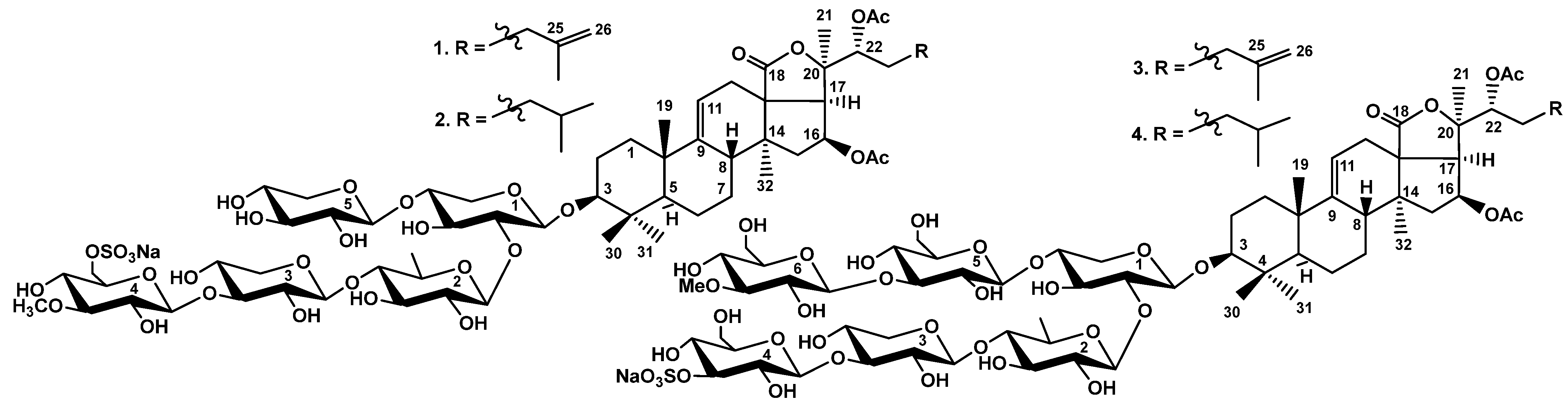
2.1. Structure Elucidation of Glycosides

Specimens of C. schmeltzii were collected from the Nha Trang Gulf in the East Sea of Vietnam. Glycosides were isolated from the concentrated ethanol extract using a previously described methodology [21]. Silica gel column chromatography with different ratios of solvents CHCl3/EtOH/H2O as the mobile phase gave several main fractions with varying polarity, as well as two minor fractions weighing 20 and 14 mg. The isolation of individual compounds was achieved by subsequent HPLC of the latter fractions on reversed-phase and silica-based columns to give four glycosides 14 (Figure 1).
Absolute configurations of all monosaccharide residues (xylose, quinovose, glucose, and 3-O-methylglucose) comprising the sugar chains of the glycosides of C. schmeltzii were assigned earlier as D using acid hydrolysis of the total glycosidic fraction with TFA, followed by alcoholysis with (R)-(−)-2-octanol and subsequent acetylation and GLC analysis (see Supplementary S1 for methodology and GLC data of modified monosaccharides) [23].
The molecular formula of cladoloside S (1) was determined to be C62H95O31SNa from the [MNa − Na] ion peak at m/z 1367.5580 (calc. 1367.5584) and [MNa − Na − H]2− ion peak at m/z 683.2766 (calc. 683.2755) in (−)HR-ESI-MS (Figure S8). As deduced from extensive analysis of the NMR spectra, the aglycone of 1 was typical for the major part of the glycosides from C. schmeltzii [21,23,24,25,26,27,28,29] and contained 18(20)-lactone, 9(11)- and 25(26)-double bonds, and 16S- and 22R-O-acetic groups (Table 1, Figures S1–S6).
The analysis of the 1H, 13C NMR, and HSQC spectra of the carbohydrate moiety of cladoloside S (1) (Table 2; Figures S1–S7) revealed the presence of five doublets of the anomeric protons at δH 4.66–5.27 (J = 6.8–8.3 Hz) and the signals of the corresponding anomeric carbons at δC 102.2–104.8. These data indicate that the pentasaccharide chain with β-glycosidic bonds is inherent to glycoside 1. Starting from the NOE-correlation of H-3 of the aglycone (δH 3.25 (H-3, dd, J = 12.0; 3.9 Hz) (typical position of sugar chain attachment) with the anomeric proton of the first sugar unit at δC 4.66 (H-1 Xyl1, d, J = 6.9 Hz), observed in the ROESY spectrum, followed by analysis of the 1H, 1H COSY, 1D TOCSY, and HSQC spectra of each monosaccharide residue, the structure of the carbohydrate moiety of 1 was established. From this analysis, it was concluded that the oligosaccharide moiety of 1 consisted of quinovose (as the second unit in the chain), three xylose units (as the first, third, and fifth units), and 3-O-methylglucose as the terminal unit. ROESY and HMBC correlations allowed the determination of the positions of glycosidic bonds: correlations were observed between the H-1 Xyl1 and H-3 (C-3) of the aglycone, H-1 Qui2 and H-2 (C-2) Xyl1, H-1 Xyl3 and H-4 (C-4) Qui2, H-1 MeGlc4 and H-3 (C-3) Xyl3, and H-1 Xyl5 and H-4 (C-4) Xyl1 (Table 2). The single sulfate group was attached to C-6 of 3-O-methylglucose, causing an α-shifting effect of its signal to δC 66.6, and a β-shifting effect of the signal of C-5 MeGlc4 to δC 76.3 [24].
The (−)HR-ESI-MS/MS of 1 (Figure S8) demonstrated the fragmentation of the [MNa − Na] ion at m/z 1367.5 leading to the fragment ions appearance at m/z 1307.5 [MNa − Na − CH3COOH], 1247.5 [MNa − Na − 2CH3COOH], 1235.5 [MNa − Na − Xyl], 1175.5 [MNa − Na − Xyl − CH3COOH], 797.2 [MNa − Na − Agl − H], 533.1 [MNa − Na − Agl − 2Xyl], and 387.0 [MNa − Na − Agl − 2Xyl − Qui], corroborating the structure of 1 elucidated by analysis of NMR spectra.
These data indicate that cladoloside S (1) is 3β-O-{6-O-sodium sulfate-3-O-methyl-β-D-glucopyranosyl-(1→3)-β-D-xylopyranosyl-(1→4)-β-D-quinovopyranosyl-(1→2)-[(1→4)-β-D-xylopyranosyl]-β-D-xylopyranosyl}-16S,22R-diacetoxyholosta-9,25-diene.
The molecular formula of cladoloside S1 (2) was determined to be C62H97O31SNa based on the [MNa − Na] ion peak at m/z 1369.5735 (calc. 1369.5740), and the [MNa − Na − H]2− ion peak at m/z 684.2849 (calc. 684.2834) in (−)HR-ESI-MS (Figure S16). The aglycone of cladoloside S1 (2) differed from that of 1 only in the absence of a terminal double bond in its side chain. Hence, the signals of polycyclic systems in the 13C NMR spectra of 1 and 2 were almost identical. In the spectrum of 2 (Table 3, Figures S9–S15), the signals of geminal methyl groups C-26 and C-27 and tertiary C-25 were observed at δC 22.5, 22.8, and 28.1, respectively. This aglycone is also characteristic of the glycosides of C. schmeltzii [21,23,25,26,27].
The carbohydrate chain of cladoloside S1 (2) was identical to that of cladoloside S (1), as confirmed by its 2D NMR spectra (Table S1, Figures S9–S15).
The fragment ion peaks in the (−)HR-ESI-MS/MS of 2 (Figure S16) were observed at m/z 1309.5 [MNa − Na − CH3COOH], 1249.5 [MNa − Na − 2CH3COOH], 1177.5 [MNa − Na − Xyl − CH3COOH], differing from the corresponding ion-peaks observed in the spectrum of cladoloside S (1) by 2 amu due to the presence of hydrogenated side chain. The series of fragment ion peaks: 797.2 [MNa − Na − Agl − H], 533.1 [MNa − Na − Agl − 2Xyl], and 387.0 [MNa − Na − Agl − 2Xyl − Qui] are characterized by the same masses as in the spectrum of 1, which corroborates the identity of carbohydrate chains of cladolosides of the group S.
Thus, cladoloside S1 (2) is 3β-O-{6-O-sodium sulfate-3-O-methyl-β-D-glucopyranosyl-(1→3)-β-D-xylopyranosyl-(1→4)-β-D-quinovopyranosyl-(1→2)-[(1→4)-β-D-xylopyranosyl]-β-D-xylopyranosyl}-16S,22R-diacetoxyholost-9-en.
Thus, cladodosides S (1) and S1 (2) are a pair of dehydrogenated/hydrogenated compounds sharing the identical carbohydrate chain, which is characteristic of C. schmeltzii. The sugar moiety of 1 and 2 is new for sea cucumber glycosides due to the presence of xylose attached to C-4 Xyl1, which is rather rare for this group of metabolites. Glycosides with a xylose residue that branches the oligosaccharide chain at C-4Xyl1 were first discovered in this species of sea cucumber. These are cladolosides of groups D (1820), R (44) (with hexasaccharide chains) [21,26] and E (21, 22) (with pentasaccharide chains) [23] (Figure S33). Cladolosides of the group E are desulfated derivatives of cladolosides of the group S (1, 2) because the latter pair contains a sulfate group at C-6 MeGlc4.
The molecular formula of cladoloside T (3) was determined to be C69H107O37SNa from the [MNa − Na] ion peak at m/z 1559.6233 (calc. 1559.6217) and [MNa − Na − H]2− ion peak at m/z 779.3096 (calc. 779.3072) in (−)HR-ESI-MS (Figure S24). The aglycone of 3 was identical to that of 1, which was deduced from the identity of 13C NMR spectra of their aglycone parts (Table S2, Figures S17–S23).
In 1H and 13C NMR spectra of the carbohydrate moiety of cladoloside T (3) (Table 4; Figures S17–S23) six doublets of the anomeric protons at δH 4.66–5.27 (J = 6.8–8.3 Hz) and the signals of the corresponding anomeric carbons at δC 102.2–104.8 were observed indicating the presence of hexasaccharide chain with β-glycosidic bonds. The same algorithm was applied for structure elucidation as described above. In the monosaccharide composition of 3 two xylose, one quinovose, two glucose, and one 3-O-methylglucose residues were found. The positions of the glycosidic bonds deduced from the ROESY and HMBC correlations were typical for all glycosides of holothuroids. Xylose and glucose residues were third and fifth units, correspondingly, in the chain of 3. Analysis of the chemical shifts of the signals of the terminal residue attached to C-3 Glc5 showed that it was a 3-O-methylated glucose because the HMBC correlations OMe (δH 3.80, s)/C-3 MeGlc6 (δC 86.9) and H-3 MeGlc6 (δH 3.67 (t, J=9.1 Hz)/OMe (δC 60.7) were observed. Hence, the single OMe group was attached to the upper semi-chain, and the terminal (glucose) residue in the bottom semi-chain was non-methylated. However, the signal of C-3 Glc4 was deshielded to δC 84.2 as well as the signal of the corresponding proton (δH-3 Glc4 5.03, t, J = 8.3 Hz). Taking into consideration the presence of the sulfate group deduced from the mass spectra of 3, as well as the absence of sulfation of the hydroxymethylene group (C-6 Glc4) due to its signal being observed at δC 61.7, the position of the sulfate group was assigned as C-3 Glc4 [24].
The (−)HR-ESI-MS/MS of 3 (Figure S24) demonstrated the fragmentation of the [MNa − Na] ion at m/z 1559.6 that resulted in the fragment ions appearance at m/z 1499.6 [MNa − Na − CH3COOH], 1439.6 [MNa − Na − 2CH3COOH], 1383.6 [MNa − Na − MeGlc], 1323.5 [MNa − Na − MeGlc − CH3COOH], 1161.5 [MNa − Na − CH3COOH − MeGlc − Glc], 1101.5 [MNa − Na − 2CH3COOH − MeGlc − Glc], 1007.3 [MNa − Na − Agl − H], 519.1 [MNa − Na − Agl − MeGlc − Glc − Xyl], confirming the sequence of monosaccharide residues as well as aglycone structure of 3.
These data indicate that cladoloside T (3) is 3β-O-{3-O-sodium sulfate-β-D-glucopyranosyl-(1→3)-β-D-xylopyranosyl-(1→4)-β-D-quinovopyranosyl-(1→2)-[3-O-methyl-β-D-glucopyranosyl-(1→3)-β-D-glucopyranosyl-(1→4)]-β-D-xylopyranosyl}-16S,22R-diacetoxyholosta-9,25-diene.
Cladoloside T1 (4) was determined to be a hydrogenated derivative of 3 from the molecular formula C69H109O37SNa, deduced from the [MNa − Na] ion peak at m/z 1561.6377 (calc. 1561.6374) in (−)HR-ESI-MS (Figure S32). Therefore, the aglycone of 4 was the same as that of cladoloside S1 (2), which was confirmed by the coincidence of the 13C NMR spectra of its aglycone parts (Table S3, Figures S25–S30). The carbohydrate chain of cladoloside T1 (4) (Table S4; Figures S25–S31) was identical to that of cladoloside T (3); hence, close proximity of the signals in the 13C NMR spectra of their oligosaccharide moieties was observed.
The (−)HR-ESI-MS/MS spectrum of 4 (Figure S32) demonstrated fragmentation of the [MNa − Na] ion at m/z 1561.6. Thus, the fragment ions were observed at m/z 1501.6 [MNa − Na − CH3COOH], 1367.6 [MNa − Na − MeGlc − H], 1325.5 [MNa − Na − MeGlc − CH3COOH], 1163.5 [MNa − Na − CH3COOH − MeGlc − Glc ], 1103.5 [MNa − Na − 2CH3COOH − MeGlc − Glc], 1007.3 [MNa − Na − Agl − H], 519.1 [MNa − Na − Agl − MeGlc − Glc − Xyl], confirming the structure of 4.
These data indicate that cladoloside T1 (4) is 3β-O-{3-O-sodium sulfate-β-D-glucopyranosyl-(1→3)-β-D-xylopyranosyl-(1→4)-β-D-quinovopyranosyl-(1→2)-[3-O-methyl-β-D-glucopyranosyl-(1→3)-β-D-glucopyranosyl-(1→4)]-β-D-xylopyranosyl}-16S,22R-diacetoxyholost-9-en.
Cladolosides T (3) and T1 (4) are another pair of compounds that differ in the presence or absence of a terminal double bond in their side chain and have identical to each other carbohydrate chains. The oligosaccharide moiety of cladolosides of the group T is unique because of the position of the sulfate group at C-3 of the terminal sugar residue. This suggests that the processes of sulfation and O-methylation occurring during the biosynthesis of glycosides can compete with each other.
Sulfation is generally considered the final stage of sugar moiety biosynthesis. In fact, cladolosides of the groups I, J, K, L (2833) [25] (Figure S33), and S (1, 2) are penta- and hexaosides with the sulphate groups attached to the terminal 3-O-methylated monosaccharide residues [25], while 3-O-methylation is a signal of termination, which stops further elongation of the biosynthesizing carbohydrate chain. However, the incorporation of 3-O-methyl groups into hexasaccharide chains does not occur simultaneously: first, it is attached to one semi-chain (upper or bottom), while another semi-chain could be subsequently elongated and then 3-O-methylated. The time shift (delay) of these stages in relation to each other, which is characteristic of the mosaic type of biosynthesis [6], leads to the formation of carbohydrate chains with 3-O-methylated or non-methylated terminal residues. This is well illustrated by the structures of the cladolosides of groups D (1820) [21], P (3942), Q (43), and R (44) [26,27] (Figure S33), and kurilosides of groups A, D, and E − glycosides from the sea cucumber Thyonidium kurilensis [28].
In highly polar glycosides, such as psolusosides—compounds found in the sea cucumber Psolus fabricii [29] and bearing up to four sulfate groups, two sulfate groups can bond to one monosaccharide residue. This broadened the number of known possible positions of introducing the sulfate group. As a result, they are not only attached to the C-6 of hexose residues, which is common for glycosides, but also to the C-2 and C-4 positions. However, sulfation did not compete with 3-O-methylation. Some glycosides of the sea cucumber Paracaudina chilensis contain a sulfate group at C-3 Glc3, which prevents further elongation of the carbohydrate chain during biosynthesis. This suggests the following assumption: sulfatase competes with glycosyltransferases. A similar situation was observed in the glycosides of C. schmeltzii, but another pair of enzymes rival each other: 3-O-methylase and sulfatase. This can presumably be explained by the high level of expression or activity of the enzymes involved in glycoside biosynthesis. Therefore, these enzymes can compete with each other. The sea cucumber species C. shmeltzii, P. fabricii [29], and P. chilensis [30], which are characterized by a great variety of carbohydrate parts of their glycosides, are good examples of this phenomenon.
Triterpene glycosides of the sea cucumbers are the products of the mosaic type of biosynthesis, when the parts of one molecule, aglycones and carbohydrate chains, are biosynthesized simultaneously and independently from each other [6]. The different stages of biosynthesis can occur at different times relative to each other, or even be skipped, resulting in significant structural diversity in glycosides. The mosaicism of glycoside biosynthesis may indicate a lack of compartmentalization inside the cells of organisms–producers that leads to a certain degree of randomness in enzymatic reactions. However, this offers the advantage of increasing the chemical diversity of the glycosides.

2.2. Biological Activity of the Glycosides

The hemolytic activity of 26 glycosides (4 novel and 22 known compounds, available in our collection) isolated from C. schmeltzii was tested against human erythrocytes (Table 5). Cladolosides T (3) and T1 (4) were among the most active compounds (ED50 0.39 and 0.12 μM, respectively). This suggests that the sulfate group attached instead of the 3-O-Me group to the terminal sugar residue does not decrease membranolytic activity. Cladolosides S (1) and S1 (2) were only moderately active in relation to erythrocytes. Their activity is comparable to cladolosides I2 (29) and I1 (28) (Figure S33), which have the same aglycones and sulfate group positions. Compounds 28, 29 differ from 1 and 2 in that they contain glucose as the fifth sugar in the chain instead of xylose. Thus, the sulfate group attached to C-6 of the terminal monosaccharide slightly reduces the glycosides’ membranolytic action, which aligns with earlier observations [20].
Surprisingly, none of the cladolosides of group A, having a linear tetrasaccharide chain and holostane aglycones, which commonly provide high activity, were effective hemolytics. Among the most active compounds were cladolosides of the group B (pentaosides with one glucose residue in upper semi-chain, ED50 0.55 μM (cladoloside B1 (11)) and 0.31 μM (cladoloside B2 (12)) and group C (hexaosides with glucose and 3-O-methyl glucose in the upper semi-chain, ED50 0.44 μM (cladoloside C (13)) and 0.15 μM (cladoloside C1 (14)) (Figure S33). There were no significant differences in activity between hydrogenated/dehydrogenated pairs of glycosides. Cladoloside D (18), hexaoside, having non-methylated terminal residue in the upper semi-chain was also highly active (ED50 0.29 μM). Its 3-O-methylated analog, cladoloside G (25), is also highly hemolytic (ED50 0.35 μM) (Figure S33). These facts indicate that the 3-OMe groups do not significantly influence the hemolytic activity of hexaosides.
Two pairs of glycosides, differing in the presence/absence of a 25(26)-double bond within each pair, demonstrated that hydrogenated compounds were more active than their dehydrogenated analogs. This pattern was observed for sulfated pentaosides, cladolosides of the group I (ED50 1.72 μM (cladoloside I1 (28) and 4.24 μM (cladoloside I2 (29)), and also for sulfated hexaosides, cladolosides of the group K (ED50 0.31 μM (cladoloside K1 (31) and 4.63 μM (cladoloside K2 (32)) (Figure S33).
Cladolosides L1 (33), M1 (35), and P2 (41) (Figure S33), having the same aglycones (holotoxinogenin, with 16-oxo group and 25(26)-double bond) and differing by monosaccharide composition of hexasaccharide chains, were all strong hemolytics (Table 5).
The weak activities of cladoloside C3 (16) (ED50 12.10 μM) was caused by the presence of 16-OH group instead of common for the majority of glycosides 16-OAc, of cladoloside A3 (7) (ED50 7.58 μM) − by the absence of 22-OAc, of cladoloside A5 (9) (ED50 7.19 μM) − by the presence of 25-OH-group. The weakest effect from the series was demonstrated by cladoloside A4 (8) (Figure S33), with a 24(25)-double bond and 16-oxo-group instead of 16β-OAc in the other cladolosides of group A, in combination with the tetrasaccharide chain. This is unusual because these features are not known to reduce the hemolytic activity of glycosides [20].
The cytotoxic activities of the same series of compounds were studied against three types of human breast cancer cells (MCF-7, T-47D, and triple-negative MDA-MB-231) as well as the non-tumor mammary epithelial cell line MCF-10A. Cisplatin and cucumarioside A0-1 [15] were used as positive controls (Table 5).
MDA-MB-231 cells were most susceptible to the cytotoxic action of the glycosides, whereas MCF-7 and T-47D cell lines were equally more viable. The most promising compounds were those that showed no toxicity to normal cells, MCF-10A, but were highly active against triple-negative cancer cells, MDA-MB-231. Cladolosides B2 (12), D (18), G (25), H1 (26), and I1 (28) demonstrated such selectivity (expressed as the selectivity index (SI), Table 6). The strongest selectivity against the MDA-MB-231 cell line was demonstrated by cladolosides H1 (26) (SI 8.1) and D (18) (SI 6.7) with hexasaccharide chains without sulfate groups and differing from each other by two monosaccharides in the upper semi-chain and by the presence/absence of a 25(26)-double bond (Figure S33).

2.3. Correlational Analysis and QSAR Model

The quantitative structure–activity relationship (QSAR) approach was applied to identify the statistically significant dependence between the hemolytic activity values and the structural peculiarities of the 26 glycosides isolated from C. schmeltzii. First, three-dimensional models were created for the glycosides, protonated at pH 7.4, and then energy was minimized. A search for dominant glycoside conformations was performed by LowModeMD Search method using the Molecular Operating Environment software (MOE, version 2020.09, Chemical Computing Group ULC, Montreal, QC, Canada) as well as some properties for 49 dominant glycoside conformers were estimated (SI, clad conformer properties.xlsx and coordinates for cladoloside conformers conf.xyz). For the analysis, the set of various 2D and 3D descriptors (total number—315) reflecting the physicochemical properties, energy values and topological indexes expressing the geometric properties of molecular structures, was calculated and studied using the QuaSAR-Descriptor tool of the MOE 2020.0901 CCG software (version 2020.09, Chemical Computing Group ULC, Montreal, QC, Canada).
It is important to emphasize that selecting specific descriptors is crucial because no single, universal set of descriptors has been found that can adequately describe all types of activities and properties of the compounds being analyzed. Therefore, suitable descriptors must be carefully selected for each type of activity and compound being studied. So, in addition to the descriptors characterizing the physicochemical properties of the glycosides (polarizability, refractive index, surface charge distribution, dipole moment, hydrogen bonds’ potential strength (donors and acceptors), hydrophobic volume, surface area, atomic valence connectivity index, etc.), following descriptors as the carbohydrate chain length and branching, type of the third and fifth sugar residue (xylose, glucose or quinovose), the sulfate groups’ positions were added to the descriptors set provided by the MOE-QuaSAR-Descriptor software (MOE, version 2020.09, Chemical Computing Group ULC, Montreal, QC, Canada). The correlational analysis revealed a direct positive correlation between the hemolytic activities of the tested compounds in vitro and such descriptors as the molecular refractivity, number of rings, atomic connectivity index [31], and partial charges distribution on the van der Waals surface, principal moment of inertia describing the different aspects of molecular shape. In contrast, the hydrophilic integy moment, lowest hydrophobic energy, critical packing parameter, and approximation of the sum of the VDW surface areas of hydrophobic atoms (Å2) were found to correlate negatively.
Principal component analysis (PCA) reduced the number of descriptors to those that made a significant contribution. As a result, glycosides were divided into two groups (Figure 2), indicating that the choice of descriptors was correct. Next, a PLS linear QSAR model was created using the QuaSAR-Model tool of the MOE (version 2020.09, Chemical Computing Group ULC, Montreal, QC, Canada) using these descriptors. The resulting model demonstrated excellent agreement with the experimental results for glycoside hemolytic activities, with a correlation coefficient r2 = 0.9854 and RMSE = 0.0813 (Figure S34). The quality check of the model confirmed its reliability, with a cross-validation correlation coefficient of r2cros = 0.7286 and RMSEcros = 0.2676. The QSAR model includes 107 terms, of which 70 have the maximum weight; however, a reduction in the number of descriptors below a given critical level leads to a decrease in the accuracy of the created model. All these data once again highlighted the complexity of the relationships between the structural characteristics of glycosides and their membranolytic action, due to the summative effect of the many different contributions of individual descriptors that have a significant influence in a multifactor model.
Results of the molecular modeling demonstrate that the membranolytic action of the glycosides containing 18(20)-lactone, 9(11)-double bond, and normal non-shortened side chain in the aglycones (features critically important for high hemolytic activity) also depends on the presence/absence/position of the side chain double bonds, the presence/absence and type of substituent at C-22 (OAc or OH groups), and the type of substituent at C-16 (OAc, OH, or oxo-groups). In addition, the length, branching, and monosaccharide composition of the carbohydrate chain, as well as the localization of sulfate groups, have a significant effect on activity.
The constructed QSAR model confirmed the fundamental importance of the presence of the 25(26)-double bond in the aglycone side chain and the O-acetic or oxo-group at C-22 and C-16, respectively, for the hemolytic activity of the studied glycosides. In contrast, the presence of a hydroxyl group at C-16, C-22, or C-25, as well as 23(24)- or 24(25)-double bonds, contributes negatively to the hemolytic activity of glycosides. Additionally, the total number of oxygen atoms, as well as donors and acceptors of hydrogen bonds, significantly contributes to the activity of glycosides, whereas the presence of OH-groups in the aglycones has a strong negative effect.
The observed structure–activity relationships based on hemolytic activity demonstrated that the most active compound was cladoloside M1 (35) from the tested series. In addition to the above-mentioned structural features (such as 18(20)-lactone and 25(26)-double bond), it has a keto-group at C-16 and a side chain without any functional groups at C-22 in combination with a hexasaccharide chain with glucose as the third and fifth residues. As shown by correlation analysis, these elements were strongly positively correlated with hemolytic activity.
The large number of parameters in the constructed QSAR model reflects the varying influence of structural elements on activity, depending on their location within the molecule. Thus, the distribution of polar and hydrophobic regions on the surface of the molecule, as well as the direction and magnitude of the dipole moment and the ratio of the components of the moment of inertia, are positively correlated with the hemolytic activity of glycosides. This is consistent with the high hemolytic activity of compounds containing bulky penta- and hexasaccharide chains and is illustrated by the high activity of pentaoside cladoloside B2 (12) and hexaosides, cladolosides C (13), D (18), and G (25) in comparison with the moderate activity of cladoloside A2 (6), which has the same aglycone but a tetrasaccharide chain. However, not only the architecture of carbohydrate chains but also the combination of monosaccharides and variability of their sequence lead to slightly different orientations of monosaccharide residues relative to each other, resulting in differences in their molecular packing density, which ultimately affects the activity.
The dual nature of the correlations of molecular volume and shape with hemolytic activity probably indicates the importance of the shape of the molecule as a whole, revealing the role of its individual parts. For example, the combination of a bulky hexasaccharide chain with two glucose residues (in the third and fifth positions) and a less bulky aglycone, due to the absence of OAc-groups in the highly active cladoloside M1 (35), makes the final volume and shape of the molecule optimal for membranotropic action. Additionally, the low hemolytic activity of cladoloside A3 (7) is probably due to the non-optimal molecular volume and shape due to the presence of a tetrasaccharide chain with xylose as a third residue and the aglycone without any substituents in the side chain, in contrast to the more active and bulkier tetraoside cladoloside A6 (10), which has a third glucose in the sugar chain and a 22-OAc- group. This observation is also consistent with the QSAR model, indicating the importance of charge distribution on the surface of the molecule and the significance of the density of packing of the carbohydrate moiety for the membranotropic action of glycosides.
Previously, it was shown that sulfate groups have an ambiguous effect on glycoside activity [16,20]. In this case, it was possible to compare the activities of monosulfated and non-sulfated penta- and hexaosides. The QSAR model demonstrated a significant contribution of the sulfate group at the fourth residue of the carbohydrate chain. The presence of a sulfate group at C-3 of this residue was shown to have a positive effect on the activity, as demonstrated by cladolosides T (3) and T1 (4). In general, the sulfate groups at the fourth sugar did not negatively impact the hemolytic activity of penta- or hexaosides.
SAR and QSAR analyses revealed significant differences in the effects of triterpene glycosides on different cell lines. Interestingly, the hemolytic activity of the studied glycosides was slightly correlated with cytotoxicity against healthy cells (MCF-10A) and significantly correlated with cytotoxicity against TNBC cells of the MDA-MB-231 line, which is reflected in the selectivity index (Table 6).
An extensive analysis of the main descriptors’ (43 variables) significance and influence in the correlational matrix (Figure S35) revealed that a number of structural features have an opposite influence, mainly in relation to the estrogen-dependent MCF-7 cell line and the triple-negative MDA-MB-231 cells. This is probably due to different membrane interactions of glycosides. The key drivers of activity in different cell lines were shown to be the OAc-groups in the aglycones, sulfate groups, and glucose residues in the third and fifth positions in the sugar chains. The most pronounced antagonistic effects against MCF-7 and MDA-MB-231 cells were caused by the following descriptors: O-acetate or keto-group at C-16 of the aglycones, glucose residues in the fifth and sixth positions, the presence of 3-O-methyl groups at the fourth and sixth monosaccharide residues in carbohydrate chains, the presence of sulfate groups, and the localization of the sulfate group at C-3 of the fourth sugar unit.
These results indicate that numerous structural features of glycosides have different effects on cytotoxicity against hormone-dependent breast cancer cells MCF-7 and triple-negative MDA-MB-231 cells, suggesting that the glycosides target different membrane components in these tumor cell lines.

3. Materials and Methods

3.1. General Experimental Procedures

PerkinElmer 343 Polarimeter (PerkinElmer, Waltham, MA, USA) was used for specific rotation measuring; NMR spectra were registered on Avance III 700 Bruker FT-NMR spectrometer (Bruker BioSpin GmbH, Rheinstetten, Germany) (700.13/176.04 MHz (1H/13C, 30 °C, δC 148.9 resonance of C5D5N for 13C and δH 7.21 resonance of C5D5N for 1H used as the references, BBO probe)); ESI MS (negative ion mode) spectra were obtained on Agilent 6510 Q-TOF apparatus (Agilent Technology, Santa Clara, CA, USA), sample concentration 0.01 mg/mL; HPLC was conducted on Agilent 1260 Infinity II equipped with a differential refractometer (Agilent Technology, Santa Clara, CA, USA); columns were used: Supelco Discovery HS F5-5 (10 × 250 mm, 5 μM) (flow rate of 1.5 mL/min) and Supelcosil LC-Si (4.6 × 150 mm, 5 μM) (flow rate of 1.0 mL/min) (Supelco, Bellefonte, PA, USA).

3.2. Animals and Cells

Specimens of the sea cucumber Cladolabes schmeltzii (family Sclerodactylidae; order Dendrochirotida) were collected in the Nha Trang Gulf of the East Sea of Vietnam. Sampling was performed in May 2012 (collector I.Yu. Dolmatov) at a depth of 4−6 m. Sea cucumbers were identified taxonomically by I.Yu. Dolmatov; voucher specimens are preserved in the collection of the Museum of A.V. Zhirmunsky National Scientific Center of Marine Biology, Vladivostok, Russia.
Normal and cancer human cells were purchased from the Station of Blood Transfusion, Vladivostok, Russia (erythrocytes), ATCC (Manassas, VA, USA) (MCF-10A CRL-10317, T-47D HTB-133, MCF-7 HTB-22, and MDA-MB-231 CRM-HTB-26). Culturing conditions: RPMI-1640 medium with 1% penicillin/streptomycin and 10% fetal bovine serum (FBS) (Biolot, St. Petersburg, Russia) for T-47D cell line; Minimum Essential Medium (MEM) with 1% penicillin/streptomycin sulfate, and FBS (Biolot, St. Petersburg, Russia) to a final concentration of 10% for MCF-7 and MDA-MB-231 cells; DMEM/F12 medium with 20% FBS, 20 ng/mL EGF, 0.5 mg/mL hydrocortisone, 100 ng/mL cholera toxin, 10 μg/mL insulin, and 1% penicillin/streptomycin (Bioinnlabs, Rostov-on-Don, Russia) for MCF-10A cell line.

3.3. Extraction and Isolation

Sea cucumbers were minced and extracted twice with refluxing 60% EtOH. The dry weight of the residue is approximately 12.4 g. The combined extracts were concentrated to dryness in vacuo, dissolved in H2O, and chromatographed on a Polychrom-1 column (powdered Teflon, Biolar, Latvia) to eliminate inorganic salts and impurities. After this step 1.06 g of crude glycoside fraction was obtained, which was subsequently submitted to silica gel column chromatography using stepped gradient of solvent systems with different ratios of CHCl3/EtOH/H2O (from 25:12.5:1 to 2:3:1). This resulted in isolation of several glycosidic subfractions, including minor ones, weighing in 20 and 14 mg, and isolated with the most polar system (CHCl3/EtOH/H2O, 2:3:1). They were further separated by HPLC.
High-pressure liquid chromatography (HPLC) of the first subfraction (20 mg) on a reversed-phase column Supelco Discovery HS F5-5 (10 × 250 mm) with CH3CN/H2O/NH4OAc (1M water solution) in a ratio of (45/54/1) as the mobile phase yielded two fractions (1 and 2). Each of them was re-chromatographed on the same column but with ratio of the solvents (38/60/2) resulted in the isolation of cladoloside S1 (2) (3.5 mg) from fraction 1; and cladoloside S (1) (3 mg) from fraction 2. The second subfraction (14 mg) was subjected to HPLC on the same column with CH3CN/H2O/NH4OAc (1M water solution) (42/56/2) as the mobile phase and gave cladoloside T (3) (8 mg) and another fraction, which was subsequently subjected to HPLC on silica-based column Supelcosil LC-Si (4.6 × 150 mm) column with CHCl3/MeOH/H2O (85/20/1) as mobile phase to give individual cladoloside T1 (4) (3.2 mg).

3.3.1. Cladoloside S (1)

Colorless powder; [α]D20−25° (c 0.1, H2O), mp 195 °C. Data of NMR: Table 1 and Table 2, Figures S1–S8. (−)HR-ESI-MS m/z: 1367.5580 (calc. 1367.5584) [MNa − Na], 683.2766 (calc. 683.2755) [MNa − Na − H]2−; (−)ESI-MS/MS m/z: 1307.5 [MNa − Na − CH3COOH], 1247.5 [MNa − Na − 2CH3COOH], 1235.5 [MNa − Na − Xyl (C5H8O4)], 1175.5 [MNa − Na − Xyl (C5H8O4) − CH3COOH], 797.2 [MNa − Na − Agl (C34H49O7) − H], 533.1 [MNa − Na − Agl (C34H49O7) − 2Xyl (2C5H8O4)], and 387.0 [MNa − Na − Agl (C34H49O7) − 2Xyl (2C5H8O4) − Qui (C6H10O4)].

3.3.2. Cladoloside S1 (2)

Colorless powder; [α]D20−20° (c 0.1, H2O), mp 202 °C. Data of NMR: Table 3 and Table S1, Figures S9–S16. (−)HR-ESI-MS m/z: 1369.5735 (calc. 1369.5740) [MNa − Na], 684.2849 (calc. 684.2834) [MNa − Na − H]2−; (−)ESI-MS/MS m/z: 1309.5 [MNa − Na − CH3COOH], 1249.5 [MNa − Na − 2CH3COOH], 1177.5 [MNa − Na − Xyl − CH3COOH], 797.2 [MNa − Na − Agl (C34H51O7) − H], 533.1 [MNa − Na − Agl (C34H51O7) − 2Xyl (2C5H8O4)], and 387.0 [MNa − Na − Agl (C34H51O7) − 2Xyl (2C5H8O4) − Qui (C6H10O4)].

3.3.3. Cladoloside T (3)

Colorless powder; [α]D20−28° (c 0.1, H2O), mp 215 °C. Data of NMR: Table 4 and Table S2, Figures S17–S24. (−)HR-ESI-MS m/z: 1559.6233 (calc. 1559.6217) [MNa − Na]; 779.3096 (calc. 779.3072) [MNa− Na − H]2−; (−)ESI-MS/MS m/z: 1499.6 [MNa − Na − CH3COOH], 1439.6 [MNa − Na − 2CH3COOH], 1383.6 [MNa − Na − MeGlc (C7H12O5)], 1323.5 [MNa − Na − MeGlc (C7H12O5) − CH3COOH], 1161.5 [MNa − Na − CH3COOH − MeGlc (C7H12O5) − Glc (C6H10O5)], 1101.5 [MNa − Na − 2CH3COOH − MeGlc (C7H12O5) − Glc (C6H10O5)], 1007.3 [MNa − Na − Agl (C34H49O6) + H], 519.1 [MNa − Na − Agl (C34H49O6) − MeGlc (C7H13O6) − Glc (C6H10O5) − Xyl (C5H8O4)].

3.3.4. Cladoloside T1 (4)

Colorless powder; [α]D20−24° (c 0.1, H2O), mp 206 °C. Data of NMR: Tables S2 and S3, Figures S25–S32. (−)HR-ESI-MS m/z: (−)HR-ESI-MS m/z: 1561.6377 (calc. 1561.6377) [MNa − Na]; (−)ESI-MS/MS m/z: 1501.6 [MNa − Na − CH3COOH], 1367.6 [MNa − Na − MeGlc (C7H13O6) − H], 1325.5 [MNa − Na − MeGlc (C7H12O5) − CH3COOH], 1163.5 [MNa − Na − CH3COOH − MeGlc (C7H12O5) − Glc (C6H10O5)], 1103.5 [MNa − Na − 2CH3COOH − MeGlc (C7H12O5) − Glc (C6H10O5)], 1007.3 [MNa − Na − Agl (C34H51O6) − H], 519.1 [MNa − Na − Agl (C34H49O6) − MeGlc (C7H13O6) − Glc (C6H10O5) − Xyl (C5H8O4)].

3.4. Cytotoxic Activity (MTT Assay)

Solutions (20 µL) of the tested substances at different concentrations (0.1–20 µM) or positive controls—cisplatin and cucumarioside A0-1 [15] and the cell suspension (180 µL) (MCF-10A, MCF-7, T-47D, and MDA-MB-231—7 × 10³ cells per well) were added to the wells of 96-well plates and incubated overnight at 37 °C and 5% CO2, then the cells were precipitated by centrifugation, the medium was stowed and 100 µL of fresh medium and 10 µL of 3-(4,5-dimethylthiazol-2-yl)-2,5-diphenyltetrazolium bromide (MTT) (PanReac, AppliChem, Darmstadt, Germany) stock solution (5 mg/mL) were added into each well. After 4 h of incubation and addition of 100 µL of SDS-HCl, cells were subsequently incubated at 37 °C for 18 h. A Multiskan FC microplate photometer (Thermo Fisher Scientific, Waltham, MA, USA) was used to measure the absorbance of the converted dye formazan at 570 nm. The cytotoxic activity of each glycoside was expressed as the concentration that caused 50% inhibition of cell metabolic activity (IC50). The experiments were conducted in triplicate, p < 0.05.

3.5. Hemolytic Activity

A total of 450 g of human blood (B(III) Rh+) was used to obtain erythrocytes by centrifuging three times for 5 min with phosphate-buffered saline (PBS) (pH 7.4) at 4 °C on a centrifuge LABOFUGE 400R (Heraeus, Hanau, Germany). Ice-cold PBS (pH 7.4) was used for resuspension of erythrocyte residue to a final optical density of 1.5 at 700 nm. Then, 20 µL of tested compound solutions or control (cucumarioside A0-1 [15]) were added to 180 µL of erythrocyte suspension in V-bottom 96-well plates and incubated for 1 h at 37 °C. After centrifugation (900 g, 10 min, laboratory centrifuge LMC-3000 (Biosan, Riga, Latvia)), 100 µL of supernatant was transferred into new flat plates. The values of erythrocyte lysis were measured on microplate photometer Multiskan FC (Thermo Fisher Scientific, Waltham, MA, USA) at λ = 570 nm as hemoglobin concentration in supernatant. The effective dose, causing lysis of 50% erythrocytes (ED50), was calculated with SigmaPlot software (version 14, Grafiti LLC, Palo Alto, CA, USA). All the experiments were carried out in triple repetitions, p < 0.05.

3.6. Building a QSAR Model

QSAR model for the set of 26 glycosides was built using the QuaSAR-Descriptor and QuaSAR-Model tools of MOE (version 2020.09, Chemical Computing Group ULC, Montreal, QC, Canada). The order of stages of Preliminary Data Processing was as follows: the charge distribution calculation, optimization of the glycoside geometric structure, search for preferred spatial configurations of glycosides by LowModeMD Search method [32], which generated conformations using a short ~1 ps run of Molecular Dynamics (MD) with velocities initialized to low-frequency vibrational modes at constant temperature [33], followed by an all-atom energy minimization and determination of numerical characteristics reflecting the physicochemical properties of these glycoside molecules—2D and 3D descriptors calculation. The next stage of the Data Analysis involves the study of statistical relationships between the calculated descriptors and biological indicators of glycoside activity expressed as pED50, the PCA method application to reduce the number of independent variables, while maintaining the maximum dispersion of activity, elimination of multicollinear descriptors, building a QSAR model and the assessment of model’s reliability (cross-validation). Then, the Model Optimization and Final Testing were performed by removing the descriptors not contributing to the model, and model checking by making a graph showing the correlation between the model-predicted value and the experimental activity value expressed as pED50.

4. Conclusions

As a result of the investigation of the glycosidic composition of the sea cucumber Cladolabes schmeltzii, 44 glycosides, cladolosides of different groups, consisting of 21 types of carbohydrate chains and 9 diverse aglycones, and constituting a combinatorial library, were isolated and characterized.
Structurally biogenetic analysis of the carbohydrate chains comprising the cladolosides of groups A–T (144) showed that sulfation in most cases is a final stage of biosynthesis. Based on the structures of the recently discovered cladolosides T (3) and T1 (4), it can be assumed that the enzymatic processes of sulfation and O-methylation compete with each other. This may be due to the high level of expression of the enzymes and lack of compartmentalization inside the cells of organisms–producers that leads to a certain degree of randomness in enzymatic reactions, while providing an advantage in terms of chemical diversity of the glycosides.
Analysis of the membranolytic activity of cladolosides of different groups showed that hexaosides, which are the final products of biosynthesis, exhibited higher cytotoxic activity than their precursors, pentaosides. Cladolosides of the group A (tetraosides) are weak cytotoxins and, therefore, are not synthesized in large quantities.
Analysis of structure–activity relationships based on the hemolytic activity of glycosides showed that the sulfate group attached instead of the 3-O-Me group to the terminal sugar residue does not decrease activity. Two glycosides from C. schmeltzii display selectivity of action toward triple-negative breast cancer cells MDA-MB-231, being non-toxic in relation to normal mammary cells MCF-10A.
QSAR analysis of C. schmeltzii glycosides confirmed the complex nature of the revealed patterns due to the combined influence of a huge number of descriptors on the membranolytic activity of glycosides. Moreover, numerous structural features of glycosides are related in different ways to cytotoxicity against hormone-dependent breast cancer cells MCF-7 and triple-negative MDA-MB-231 cells, suggesting that the glycosides target different membrane components in these tumor cell lines. Therefore, the application of QSAR for the prediction or modeling of the biological activity of triterpene glycosides in sea cucumbers is prospective and useful.

Supplementary Materials

The following supporting information can be downloaded at https://www.mdpi.com/article/10.3390/md23070265/s1. Figures S1–S35: The original spectral data of compounds 14; Tables S1–S4: 13C and 1H NMR chemical shifts and HMBC and ROESY correlations of aglycones and carbohydrate moieties or the aglycones of glycosides 2, 3, 4; the PLS QSAR model correlation plot, correlational matrix.

Author Contributions

Conceptualization, A.S.S. and V.I.K.; investigation, A.S.S., S.A.A., A.I.K., R.S.P., P.S.D., E.A.C., E.S.M., K.M.T. and E.A.Z.; methodology, A.S.S., E.A.C., E.S.M. and E.A.Z.; writing—original draft, A.S.S. and E.A.Z.; review and editing, V.I.K. All authors have read and agreed to the published version of the manuscript.

Funding

The investigation was conducted with the financial support of a grant from the Russian Science Foundation, no. 23-13-00078.

Institutional Review Board Statement

The specimens of sea cucumbers were collected along with the special quota for scientific research obtained by the Institute of Oceanology of the Vietnam National Academy of Science (Nha Trang, Vietnam).

Informed Consent Statement

Not applicable.

Data Availability Statement

The original contributions presented in this study are included in the article. Further inquiries can be directed to the corresponding author.

Acknowledgments

This study was carried out on the equipment of the Collective Facilities Center “The Far Eastern Center for Structural Molecular Research (NMR/MS) PIBOC FEB RAS”; the computer simulation and conformational search for glycosides were performed using the cluster CCU “Far Eastern computing resource” FEB RAS (Vladivostok).

Conflicts of Interest

The authors declare no conflicts of interest.

References

  1. Kalinin, V.I.; Silchenko, A.S.; Avilov, S.A.; Stonik, V.A. Progress in the studies of triterpene glycosides from sea cucumbers (Holothuroidea, Echinodermata) between 2017 and 2021. Nat. Prod. Commun. 2021, 16, 10. [Google Scholar] [CrossRef]
  2. Kamyab, E.; Kellermann, M.Y.; Kunzmann, A.; Schupp, P.J. Chemical biodiversity and bioactivities of saponins in Echinodermata with an emphasis on sea cucumbers (Holothuroidea). In YOUMARES 9–The Oceans: Our Research, Our Future; Jungblut, S., Liebich, V., Bode-Dalby, M., Eds.; Springer: Cham, Switzerland, 2020; pp. 121–157. [Google Scholar] [CrossRef]
  3. Popov, R.S.; Ivanchina, N.V.; Silchenko, A.S.; Avilov, S.A.; Kalinin, V.I.; Dolmatov, I.Y.; Stonik, V.A.; Dmitrenok, P.S. Metabolite profiling of triterpene glycosides of the Far Eastern sea cucumber Eupentacta fraudatrix and their distribution in various body components using LC-ESI QTOF-MS. Mar. Drugs 2017, 15, 302. [Google Scholar] [CrossRef] [PubMed]
  4. Khodja, I.; Mezali, K.; Savarino, P.; Gerbaux, P.; Flammang, P.; Caulier, G. Structural characterization and profiles of saponins from Two Algerian Sea Cucumbers. Molecules 2024, 29, 5346. [Google Scholar] [CrossRef] [PubMed]
  5. Puspitasari, Y.E.; Tuenter, E.; Foubert, K.; Herawati, H.; Hariati, A.M.; Aulanni’am, A.; Pieters, L.; De Bruyne, T.; Hermans, N. Saponin and fatty acid profiling of the sea cucumber Holothuria atra, α-glucosidase inhibitory activity and the identification of a novel triterpene glycoside. Nutrients 2023, 15, 1033. [Google Scholar] [CrossRef]
  6. Silchenko, A.S.; Avilov, S.A.; Kalinin, V.I. Separation procedures for complicated mixtures of sea cucumber triterpene glycosides with isolation of individual glycosides, their comparison with HPLC/MS metabolomic approach, and biosynthetic interpretation of the obtained structural data. In Studies in Natural Products Chemistry; Rahman, A., Ed.; Elsevier Science B.V.: Amsterdam, The Netherlands, 2022; Volume 72, pp. 103–146. [Google Scholar] [CrossRef]
  7. Van Dyck, S.; Flammang, P.; Meriaux, C.; Bonnel, D.; Salzet, M.; Fourmier, I.; Wisztorski, M. Localization of secondary metabolites in marine invertebrates: Contribution of MALDI MSI for the study of saponins in Cuvierian tubules of H. forskali. PLoS ONE 2010, 5, 11. [Google Scholar] [CrossRef]
  8. Van Dyck, S.; Caulier, G.; Todeso, M.; Gebraux, P.; Fournier, I.; Wisztorsky, M.; Flammang, P. The triterpene glycosides of Holohuria forskali: Usefulness and efficiency as a chemical defense mechanism against predatory fish. J. Exp. Biol. 2011, 214, 1347–1356. [Google Scholar] [CrossRef]
  9. Van Dyck, S.; Gerbaux, P.; Flammang, P. Quialitative and quantitative saponin contents in five sea cucumbers from Indian Ocean. Mar. Drugs 2010, 8, 173–189. [Google Scholar] [CrossRef]
  10. Bahrami, Y.; Zhang, W.; Franco, C.M.M. Distribution of saponins in the sea cucumber Holothuria lessoni; the body wall versus the viscera, and their biological activities. Mar. Drugs 2018, 16, 423. [Google Scholar] [CrossRef]
  11. Aminin, D.L.; Menchinskaya, E.S.; Pisliagin, E.A.; Silchenko, A.S.; Avilov, S.A.; Kalinin, V.I. Sea cucumber triterpene glycosides as anticancer agents. In Studies in Natural Product Chemistry; Rahman, A., Ed.; Elsevier B.V.: Amsterdam, The Netherlands, 2016; Volume 49, pp. 55–105. [Google Scholar]
  12. Careaga, V.P.; Maier, M.S. Cytotoxic triterpene glycosides from sea cucumbers. In Handbook of Anticancer Drugs from Marine Origin; Kim, S.-K., Ed.; Springer International Publishing: Cham, Switzerland, 2015; pp. 515–528. [Google Scholar]
  13. Pislyagin, E.A.; Manzhulo, I.V.; Gorpenchenko, T.Y.; Dmitrenok, P.S.; Avilov, S.A.; Silchenko, A.S.; Wang, Y.-M.; Aminin, D.L. Cucumarioside A2-2 causes macrophage activation in mouse spleen. Mar. Drugs 2017, 15, 341. [Google Scholar] [CrossRef]
  14. Zhao, Y.C.; Xue, C.H.; Zhang, T.T.; Wang, Y.M. Saponins from sea cucumber and their biological activities. J. Agric. Food Chem. 2018, 66, 7222–7237. [Google Scholar] [CrossRef]
  15. Silchenko, A.S.; Kalinovsky, A.I.; Avilov, S.A.; Popov, R.S.; Dmitrenok, P.S.; Chingizova, E.A.; Menchinskaya, E.S.; Panina, E.G.; Stepanov, V.G.; Kalinin, V.I.; et al. Djakonoviosides A, A1, A2, B1–B4—Triterpene monosulfated tetra- and pentaosides from the sea cucumber Cucumaria djakonovi: The first finding of a hemiketal fragment in the aglycones; Activity against human breast Cancer Cell Lines. Int. J. Mol. Sci. 2023, 24, 11128. [Google Scholar] [CrossRef] [PubMed]
  16. Silchenko, A.S.; Kalinovsky, A.I.; Avilov, S.A.; Popov, R.S.; Chingizova, E.A.; Menchinskaya, E.S.; Zelepuga, E.A.; Panina, E.G.; Stepanov, V.G.; Kalinin, V.I.; et al. Sulfated triterpene glycosides from the Far Eastern sea cucumber Cucumaria djakonovi: Djakonoviosides C1, D1, E1, and F1; cytotoxicity against human breast cancer cell lines; quantitative structure–activity relationships. Mar. Drugs 2023, 21, 602. [Google Scholar] [CrossRef] [PubMed]
  17. Menchinskaya, E.S.; Chingizova, E.A.; Pislyagin, E.A.; Yurchenko, E.A.; Klimovich, A.A.; Zelepuga, E.A.; Aminin, D.L.; Avilov, S.A.; Silchenko, A.S. Mechanisms of Action of Sea Cucumber Triterpene Glycosides Cucumarioside A0-1 and Djakonovioside A Against Human Triple-Negative Breast Cancer. Mar. Drugs 2024, 22, 474. [Google Scholar] [CrossRef]
  18. Kalinin, V.I. System-theoretical (holistic) approach to the modelling of structural-functional relationships of Biomolecules and their evolution: An example of triterpene glycosides from sea cucumbers (Echinodermata, Holothurioidea). J. Theor. Biol. 2000, 206, 151–168. [Google Scholar] [CrossRef]
  19. Park, J.-I.; Bae, H.-R.; Kim, C.G.; Stonik, V.A.; Kwak, J.Y. Relationships between chemical structures and functions of triterpene glycosides isolated from sea cucumbers. Front. Chem. 2014, 2, 77. [Google Scholar] [CrossRef]
  20. Zelepuga, E.A.; Silchenko, A.S.; Avilov, S.A.; Kalinin, V.I. Structure-activity relationships of holothuroid’s triterpene glycosides and some in silico insights obtained by molecular dynamics study on the mechanisms of their membranolytic action. Mar. Drugs 2021, 19, 604. [Google Scholar] [CrossRef]
  21. Silchenko, A.S.; Kalinovsky, A.I.; Avilov, S.A.; Andryjascchenko, P.V.; Dmitrenok, P.S.; Yurchenko, E.A.; Dolmatov, I.Y.; Kalinin, V.I.; Stonik, V.A. Structure and biological action of cladolosides B1, B2, C, C1, C2 and D, six new triterpene glycosides from the sea cucumber Cladolabes schmeltzii. Nat. Prod. Commun. 2013, 8, 1527–1534. [Google Scholar] [CrossRef]
  22. Silchenko, A.S.; Kalinovsky, A.I.; Avilov, S.A.; Andryjascchenko, P.V.; Dmitrenok, P.S.; Yurchenko, E.A.; Dolmatov, I.Y.; Savchenko, A.M.; Kalinin, V.I. Triterpene glycosides from the sea cucumber Cladolabes schmeltzii II. Structure and Biological Action of Cladolosides A1–A6. Nat. Prod. Commun. 2014, 9, 1421–1428. [Google Scholar] [CrossRef]
  23. Silchenko, A.S.; Kalinovsky, A.I.; Avilov, S.A.; Andryjascchenko, P.V.; Dmitrenok, P.S.; Yurchenko, E.A.; Dolmatov, I.Y.; Kalinin, V.I. Structures and biological activities of cladolosides C3, E1, E2, F1, F2, G, H1 and H2, eight triterpene glycosides from the sea cucumber Cladolabes schmeltzii with one known and four new carbohydrate chains. Carb. Res. 2015, 414, 22–31. [Google Scholar] [CrossRef]
  24. Shashkov, A.S.; Chizhov, O.S. C-13-NMR spectroscopy in chemistry of carbohydrates and related compounds. Bioorg. Khim. 1976, 2, 437–497. [Google Scholar]
  25. Silchenko, A.S.; Kalinovsky, A.I.; Avilov, S.A.; Andryjascchenko, P.V.; Dmitrenok, P.S.; Chingizova, E.A.; Dolmatov, I.Y.; Kalinin, V.I. Cladolosides I1, I2, J1, K1 and L1, monosulfated triterpene glycosides with new carbohydrate chains from the sea cucumber Cladolabes schmeltzii. Carb. Res. 2017, 445, 80–87. [Google Scholar] [CrossRef] [PubMed]
  26. Silchenko, A.S.; Kalinovsky, A.I.; Avilov, S.A.; Andryjascchenko, P.V.; Dmitrenok, P.S.; Yurchenko, E.A.; Ermakova, S.P.; Malyarenko, O.S.; Dolmatov, I.Y.; Kalinin, V.I. Cladolosides C4, D1, D2, M, M1, M2, N and Q, new triterpene glycosides with diverse carbohydrate chains from sea cucumber Cladolabes schmeltzii. An uncommon 20,21,22,23,24,25,26,27-Okta-nor-lanostane aglycone. The synergism of inhibitory action of non-toxic dose of the glycosides and radioactive irradiation on colony formation of HT-29 cancer cells. Carb. Res. 2018, 468, 36–44. [Google Scholar]
  27. Silchenko, A.S.; Kalinovsky, A.I.; Avilov, S.A.; Andryjascchenko, P.V.; Dmitrenok, P.S.; Yurchenko, E.A.; Ermakova, S.P.; Malyarenko, O.S.; Dolmatov, I.Y.; Kalinin, V.I. Cladolosides O, P, P1–P3 and R, triterpene glycosides with two novel types of carbohydrate chains from the sea cucumber Cladolabes schmeltzii. Inhibition of cancer cells colony formation and its synergy with radioactive irradiation. Carb. Res. 2018, 468, 73–79. [Google Scholar] [CrossRef]
  28. Silchenko, A.S.; Kalinovsky, A.I.; Avilov, S.A.; Andrijaschenko, P.V.; Popov, R.S.; Dmitrenok, P.S.; Chingizova, E.A.; Kalinin, V.I. Kurilosides A1, A2, C1, D, E and F—Triterpene glycosides from the Far Eastern sea sucumber Thyonidium (=Duasmodactyla) kurilensis (Levin): Structures with unusual non-holostane aglycones and cytotoxicities. Mar. Drugs 2020, 18, 551. [Google Scholar] [CrossRef]
  29. Silchenko, A.S.; Kalinovsky, A.I.; Avilov, S.A.; Kalinin, V.I.; Andrijaschenko, P.V.; Dmitrenok, P.S.; Popov, R.S.; Chingizova, E.A. Structures and bioactivities of psolusosides B1, B2, J, K, L, M, N, O, P, and Q from the sea cucumber Psolus fabricii. The first finding of tetrasulfated marine low molecular weight metabolites. Mar. Drugs 2019, 17, 631. [Google Scholar] [CrossRef]
  30. Silchenko, A.S.; Avilov, S.A.; Andrijaschenko, P.V.; Popov, R.S.; Chingizova, E.A.; Grebnev, B.B.; Rasin, A.B.; Kalinin, V.I. The isolation, structure elucidation and bioactivity study of chilensosides A, A1, B, C, and D, holostane triterpene di-, tri- and tetrasulfated pentaosides from the sea cucumber Paracaudina chilensis (Caudinidae, Molpadida). Molecules 2022, 27, 7655. [Google Scholar] [CrossRef]
  31. Hall, L.H.; Kier, L.B. The molecular connectivity chi indices and kappa shape indices in structure-property modeling. In Reviwes in Computational Chemistry; Lipkowitz, K.B., Boyd, D.B., Eds.; Wiley-VCH, Inc.: Hoboken, NJ, USA, 1991; Volume 2, Chapter 9; pp. 367–422. [Google Scholar] [CrossRef]
  32. Labute, P. LowModeMD—Implicit Low Mode Velocity Filtering Applied to Conformational Search of Macrocycles and Protein Loops. J. Chem. Inf. Model. 2010, 50, 792–800. [Google Scholar] [CrossRef]
  33. Allen, M.P.; Tildesley, D.J. Computer Simulation of Liquids; Oxford University Press: Oxford, UK, 1987; pp. 231–232. [Google Scholar]
Figure 1. Chemical structures of the glycosides from Cladolabes schmeltzii: 1—cladoloside S; 2—cladoloside S1; 3—cladoloside T; 4—cladoloside T1.
Marinedrugs 23 00265 g001
Figure 2. Three-dimensional plot of hemolytic activity (pED50) depending on the principal component values (PCA1—PCA3) calculated for 26 glycosides. The glycosides that demonstrated hemolytic activity with ED50 ≤ 10 µM are outlined as active and are marked in red, while the rest are marked in violet.
Marinedrugs 23 00265 g002
Table 1. One- and two-dimensional NMR data of the aglycone moiety of cladoloside S (1).
PositionδC, mult. aδH, mult. (J in Hz) bHMBCROESY
136.1, CH21.81, m H-11
1.44, m H-3, H-5, H-11
226.9, CH22.22, m
1.97, m H-19, H-30
388.6, CH3.25, dd (12.0; 3.9)C: 4, 30, 31, C:1 Xyl1H-1, H-5, H-31, H1-Xyl1
439.8, C
552.7, CH0.92, mC: 6, 19, 30, 31H-1, H-3, H-31
620.7, CH21.71, m
1.53, m H-8, H-19, H-30
727.8, CH21.64, m H-15, H-32
1.24, m
839.5, CH3.25, brd (13.0) H-6, H-15, H-19
9150.9, C
1039.3, C
11110.4, CH5.23, mC: 10, 13
1233.9, CH22.53, brdd (17.2; 6.1)C: 9, 11, 14, 18H-17, H-32
2.50, brd (17.2)C: 9, 11, 14, 18H-21
1359.0, C
1443.6, C
1544.4, CH22.40, dd (12.1; 6.1)C: 13, 14, 16, 17, 32H-7, H-32
1.40, brdd (12.1; 9.1)C: 14, 16, 32H-8
1675.2, CH5.90, brq (9.1)C: 13, 20, OAc-16
1753.6, CH2.91, d (9.1)C: 12, 13, 15, 16, 18, 20, 21H-12, H-21, H-32
18176.0, C
1922.0, CH31.35, sC: 1, 5, 9, 10H-1, H-2, H-6, H-8, H-30
2085.8, C
2122.4, CH31.64, sC: 17, 20, 22H-12, H-17
2274.7, CH6.66, d (11.1)C: 20, 21, OAc-22
2329.3, CH21.78, m
2434.3, CH22.26, mC: 25
2.16, m H-22
25144.9, C
26110.6, CH24.84, brsC: 24, 25, 27
4.83, brsC: 24, 25, 27
2722.2, CH31.74, sC: 24, 25, 26H-26
3016.5, CH31.11, sC: 3, 4, 5, 31H-2, H-6, H-19, H-31
3127.9, CH31.29, sC: 3, 4, 5, 30H-3, H-5, H-6, H-30, H-1Xyl1
3221.0, CH30.91, sC: 8, 13, 14, 15H-7, H-12, H-15, H-16, H-17
OAc-16169.4, C
21.4, CH32.28, sC: 16H-22
OAc-22170.3, C
20.7, CH32.08, sC: 22
a Recorded at 176.04 MHz in C5D5N/D2O (4/1). b Recorded at 700.13 MHz in C5D5N/D2O (4/1). The original spectra of 1 are provided in Figures S1–S7.
Table 2. One- and two-dimensional NMR data of the carbohydrate chain of cladoloside S (1).
AtomδC, mult. a,b,cδH, mult. (J in Hz) dHMBCROESY
Xyl1 (1→C-3)
1105.1, CH4.76, d (7.5)C-3H-3; H-3, 5 Xyl1
283.4, CH4.09, t (8.8)C: 1 Xyl1, 1 Qui2H-1 Qui2
375.3, CH4.22, t (8.8)C: 2 Xyl 1
476.2, CH4.29, mC: 1 Xyl5
564.0, CH24.40, dd (11.9; 4.4)C: 1 Xyl1
3.63, brdd (11.9; 9.4) H-1, 3 Xyl1
Qui2 (1→2Xyl1)
1105.4, CH5.17, d (7.5)C: 2 Xyl1H-2 Xyl1, H-3, 5 Qui2
276.4, CH4.05, t (9.2)C: 1, 3 Qui2
375.1, CH4.12, t (9.2)C: 2 Qui2H-1 Qui2
485.6, CH3.67, t (9.2)C: 3, 5 Qui2, 1 Xyl3H-1 Xyl3, H-2 Qui2
571.6, CH3.82, dd (9.2; 5.9) H-1 Qui2
618.1, CH31.76, d (5.9)C: 4, 5 Qui2
Xyl3 (1→4Qui2)
1104.6, CH4.87, d (7.9)C: 4 Qui2H-4 Qui2, H-3, 5 Xyl3
273.5, CH3.97, t (8.9)C: 1, 3 Xyl3
386.6, CH4.14, t (8.9)C: 2, 4 Xyl3; 1 MeGlc4H-1 MeGlc4, H-1, 5 Xyl3
468.7, CH4.05, m
566.3, CH24.20, dd (11.6; 4.8)C: 3, 4 Xyl3
3.64, brt (11.6)C: 1 Xyl3H-1 Xyl3
MeGlc4 (1→3Xyl3)
1105.0, CH5.28, d (8.5)C: 3 Xyl3H-3 Xyl3, H-3, 5 MeGlc4
274.4, CH3.85, t (8.5)C: 1 MeGlc4
387.1, CH3.64, t (8.5)C: 2, 4 MeGlc4, OMe
470.0, CH4.19, t (8.5)
576.3, CH4.01, m H-1 MeGlc4
666.6, CH25.01, mC: 4, 5 MeGlc4
OMe60.5, CH33.81, sC: 3 MeGlc4H-3 MeGlc4
Xyl5 (1→4Xyl1)
1103.5, CH4.91, d (8.0)C: 4 Xyl1H-4 Xyl1, H-3, 5 Xyl5
273.7, CH4.00, t (8.6)C: 1, 3 Xyl5
377.8, CH4.12, t (8.6)C: 2, 4 Xyl5H-1 Xyl5
470.7, CH4.18, m
567.2, CH24.33, dd (11.7; 4.9)C: 1, 3, 4 Xyl5
3.68, t (11.7)C: 1, 3, 4 Xyl5
a Recorded at 176.04 MHz in C5D5N/D2O. b Bold—interglycosidic positions. c Italics—sulfate position. d Recorded at 700.13 MHz in C5D5N/D2O. Multiplicity by 1D TOCSY. The original spectra of 1 are provided in Figures S1–S7.
Table 3. One- and two-dimensional NMR data of the aglycone moiety of cladoloside S1 (2).
PositionδC, mult. aδH, mult. (J in Hz) bHMBCROESY
136.1, CH21.70, m H-19
1.31, m H-3, H-5
226.7, CH22.07, m
1.85, m H-19, H-30
388.8, CH3.14, brd (11.7)C:1 Xyl1H-1, H-5, H-31, H1-Xyl1
439.7, C
552.7, CH0.78, brd (11.7) H-3, H-31
621.0, CH21.57, m
1.42, m
727.7, CH21.60, m
1.15, m H-5, H-32
839.5, CH3.12, brd (16.0) H-15
9150.6, C
1039.2, C
11110.7, CH5.18, m H-1
1233.9, CH22.62, brd (16.8)
2.52, dd (16.8; 6.0)C: 17H-21
1359.2, C
1443.6, C
1544.4, CH22.35, dd (12.0; 7.2)C: 13, 17, 32H-32
1.28, brd (12.0) H-8
1675.5, CH5.83, brq (9.6) H-32
1753.4, CH2.97, d (9.6)C: 12, 13, 18, 21H-12, H-21, H-32
18176.9, C
1922.0, CH31.23, sC: 1, 5, 9, 10H-1, H-30
2086.5, C
2122.5, CH31.65, sC: 17, 20, 22
2275.4, CH6.48, d (10.8)
2328.5, CH21.52, m
2435.1, CH21.29, m
1.15, m
2528.1, CH1.52, m
2622.5, CH30.83, sC: 24, 25, 27
2722.8, CH30.82, sC: 24, 25, 26H-31
3016.5, CH30.98, sC: 3, 4, 5, 31H-2, H-6, H-19, H-30,
3127.9, CH31.16, sC: 3, 4, 5, 30H-1 Xyl1
3221.0, CH30.91, sC: 8, 13, 14, 15H-12, H-15, H-17, H-24
OAc-16171.4, C
20.9, CH32.07, s
OAc-22170.5, C
21.6, CH32.25, s H-22, H-24, H-26, H-27
a Recorded at 176.04 MHz in C5D5N/D2O. b Recorded at 700.13 MHz in C5D5N/D2O. The original spectra of 2 are provided in Figures S9–S15.
Table 4. One- and two-dimensional NMR data of the carbohydrate moiety of cladoloside T (3).
AtomδC, mult. a,b,cδH, mult. (J in Hz) dHMBCROESY
Xyl1 (1→C-3)
1104.8, CH4.66, d (6.9)C-3H-3; H-3, 5 Xyl1
282.5, CH3.96, t (7.6)C: 1,3 Xyl1, C: 1 Qui2H-1 Qui2; H-4 Xyl1
375.1, CH4.16, t (7.6)C: 2,4 Xyl 1H-1 Xyl1
477.1, CH4.22, mC: 3 Xyl1, C: 1 Glc5H-1 Glc5
563.5, CH24.39, mC: 1, 3 Xyl1
3.63, m H-1 Xyl1
Qui2 (1→2Xyl1)
1104.7, CH5.02, d (7.7)C: 2 Xyl1H-2 Xyl1, H-3, 5 Qui2
275.7, CH3.87, t (8.7)C: 1, 3 Qui2H-4 Qui2
374.8, CH3.94, t (8.7)C: 2, 4 Qui2H-1 Qui2
485.7, CH3.49, t (8.7)C: 3, 5 Qui2, 1 Xyl3H-1 Xyl3, H-2 Qui2
571.5, CH3.67, dd (8.7; 6.8) H-1 Qui2
617.8, CH31.63, d (6.8)C: 4, 5 Qui2
Xyl3 (1→4Qui2)
1104.4, CH4.76, d (8.3)C: 4 Qui2H-4 Qui2, H-3, 5 Xyl3
273.6, CH3.88, t (8.3)C: 1, 3 Xyl3
386.5, CH4.12, t (8.3)C: 2, 4 Xyl3; 1 Glc4H-1 Glc4, H-1 Xyl3
468.6, CH3.94, m
565.9, CH24.09, dd (10.8; 5.0)C: 1, 3, 4 Xyl3
3.60, t (10.8)C: 1, 3, 4 Xyl3H-1, 3 Xyl3
Glc4 (1→3Xyl3)
1104.0, CH5.27, d (8.3)C: 3 Xyl3H-3 Xyl3, H-3, 5 Glc4
273.6, CH3.98, t (8.3)C: 1, 3 Glc4
384.2, CH5.03, t (8.3)C: 2, 4 Glc4H-1, 5 Glc4
470.0, CH3.95, t (8.3)C: 3, 5 Glc4H-6 Glc4
577.1, CH3.85, t (8.3)C: 4 Glc4H-1, 3 Glc4
661.7, CH24.33, dd (11.6; 2.1) C: 4 Glc4
3.99, mC: 5 Glc4
Glc5 (1→4Xyl1)
1102.2, CH4.92, d (6.8)C: 4 Xyl1H-4 Xyl1, H-3, 5 Glc5
273.3, CH3.92, t (7.9)C: 1 Glc5
386.9, CH4.19, t (7.9)C: 2, 4 Glc5, C: 1 MeGlc6H-1 MeGlc6; H-1, 5 Glc5
469.4, CH3.87, t (7.9)C: 3, 5 Glc5
577.5, CH3.87, t (7.9)C: 6 Glc5H-3 Glcl5
661.7, CH24.29, brd (11.7)
4.02, brd (11.7)C: 5 Glc5
MeGlc6 (1→3Glc5)
1104.5, CH5.19, d (7.5)C: 3 Glc5H-3 Glc5; H-3, 5 MeGlc6
274.5, CH3.85, t (9.1)C: 1, 3 MeGlc6
386.9, CH3.67, t (9.1)C: 2, 4 MeGlc6; OMeH-1 MeGlc6
470.3, CH3.87, t (9.1)C: 3, 5 MeGlc6
577.5, CH3.93, t (9.1)C: 6 MeGlc6H-1 MeGlc6
661.7, CH24.37, brd (11.9)C: 4 MeGlc6H-4 MeGlc6
4.04, dd (11.9; 6.0)C: 5 MeGlc6
OMe60.7, CH33.80, sC: 3 MeGlc6
a Recorded at 176.04 MHz in C5D5N/D2O. b Bold—interglycosidic positions. c Italics—sulfate position. d Recorded at 700.13 MHz in C5D5N/D2O. Multiplicity by 1D TOCSY. The original spectra of 3 are provided in Figures S17–S23.
Table 5. Cytotoxic activities of glycosides 14 and 26 known glycosides from C. schmeltzii, as well as cucumarioside A0-1 and cisplatin as positive controls, against human erythrocytes and MCF-10A, MCF-7, T-47D, and MDA-MB-231 human cell lines.
Glycosides (Cladolosides)ED50, µM, ErythrocytesCytotoxicity, IC50 µM
MCF-10AMCF-7T-47DMDA-MB-231
S (1)3.40 ± 0.25>20.00>20.0014.85 ± 0.7113.24 ± 1.08
S1 (2)4.78 ± 0.30>20.00>20.00>20.00>20.00
T (3)0.39 ± 0.038.23 ± 0.528.99 ± 0.618.21 ± 0.723.41 ± 0.33
T1 (4)0.12 ± 0.028.41 ± 0.407.78 ± 0.627.41 ± 0.692.94 ± 0.21
A1 (5)6.17 ± 0.51>20.00>20.00>20.00>20.00
A2 (6)2.78 ± 0.32>20.00>20.00>20.00>20.00
A3 (7)7.58 ± 0.69>20.00>20.00>20.00>20.00
A4 (8)13.60 ± 0.70>20.00>20.00>20.00>20.00
A5 (9)7.19 ± 0.53>20.00>20.00>20.00>20.00
A6 (10)3.43 ± 0.22>20.00>20.00>20.00>20.00
B1 (11)0.55 ± 0.04>20.00>20.0012.99 ± 1.0710.12 ± 1.00
B2 (12)0.31 ± 0.0212.23 ± 1.2014.52 ± 0.4812.50 ±0.963.92 ± 0.35
C (13)0.44 ± 0.05>20.00>20.0012.39 ± 0.6613.01 ± 0.80
C1 (14)0.15 ± 0.02>20.00>20.00>20.0015.27 ± 0.71
C3 (16)12.10 ± 0.58>20.00>20.00>20.00>20.00
D (18)0.29 ± 0.03>20.0015.31 ± 0.9414.93 ± 0.852.98 ± 0.24
G (25)0.35 ± 0.04>20.00> 20.00>20.003.95 ± 0.38
H1 (26)0.21 ± 0.02>20.0015.93 ± 1.1413.20 ± 0.722.47 ± 0.32
I1 (28)1.72 ± 0.1013.44 ± 0.9211.23 ± 1.029.91 ± 0.683.02 ± 0.28
I2 (29)4.24 ± 0.31 >20.00>20.00>20.00>20.00
J1 (30)1.10 ± 0.10>20.00>20.00>20.006.18 ± 0.54
K1 (31)0.31 ± 0.02>20.00>20.0012.13 ± 1.005.83 ± 0.41
K2 (32)4.63 ± 0.4114.45 ± 1.07>20.0014.08 ± 0.916.81 ± 0.73
L1 (33)0.20 ± 0.0217.88 ± 1.1212.70 ± 1.019.03 ± 0.8113.36 ± 1.08
M1 (35)0.08 ± 0.019.81 ± 0.86>20.0017.14 ± 1.525.05 ± 0.38
P2 (41)0.17 ± 0.0211.10 ± 0.7416.32 ± 1.0310.59 ± 0.616.92 ± 0.44
Cuc A0-11.63 ± 0.217.51 ± 0.6311.33 ± 1.008.46 ± 0.533.53 ± 0.54
cysplatin-82.33 ± 5.21152.00 ± 8.02≥160.00>160.00
Table 6. Selectivity index of the glycosides to different cancer cells (SI; a ratio of IC50 calculated for healthy (MCF-10A) and cancer cells).
GlycosideSelectivity Index (SI)
MCF-7T-47DMDA-MB-231
Cladoloside S (1)->1.35>1.51
Cladoloside T (3)0.921.002.41
Cladoloside T1 (4)1.081.132.86
Cladoloside B1 (11)->1.54>1.98
Cladoloside B2 (12)0.840.983.12
Cladoloside C (13)->1.61>1.54
Cladoloside C1 (14)-->1.13
Cladoloside D (18)>1.31>1.34>6.7
Cladoloside G (25)-->5.06
Cladoloside H1 (26)1.261.518.10
Cladoloside I1 (28)1.201.364.45
Cladoloside J1 (30)-->3.24
Cladoloside K1 (31)->1.65>3.83
Cladoloside K2 (32)-1.032.17
Cladoloside L1 (33)1.411.981.34
Cladoloside M1 (35)-0.571.94
Cladoloside P2 (41)0.681.051.60
Cucumarioside A0-10.660.892.13
Disclaimer/Publisher’s Note: The statements, opinions and data contained in all publications are solely those of the individual author(s) and contributor(s) and not of MDPI and/or the editor(s). MDPI and/or the editor(s) disclaim responsibility for any injury to people or property resulting from any ideas, methods, instructions or products referred to in the content.

