Article

The Impact of Chitinase Binding Domain Truncation on the Properties of CaChi18B from Chitinilyticum aquatile CSC-1

1
Guangxi Key Laboratory for Polysaccharide Materials and Modifications, School of Marine Sciences and Biotechnology, Guangxi Minzu University, Nanning 530008, China
2
National Key Laboratory of Non-Food Biomass Energy Technology, Guangxi Key Laboratory of Marine Natural Products and Combinatorial Biosynthesis Chemistry, Guangxi Academy of Marine Sciences, Guangxi Academy of Sciences, Nanning 530007, China
3
Institute of Biology, Guangxi Academy of Sciences, Nanning 530007, China
*
Authors to whom correspondence should be addressed.
These authors contributed equally to this work.
Mar. Drugs 2025, 23(3), 93; https://doi.org/10.3390/md23030093
Submission received: 26 January 2025 / Revised: 18 February 2025 / Accepted: 20 February 2025 / Published: 20 February 2025
(This article belongs to the Section Biomaterials of Marine Origin)

Abstract

:
The chitinase binding domain (ChBD) plays a crucial role in the properties of enzymes. To assess its impact, we cloned a truncated mutant of the chitinase gene CaChi18B from the novel chitinase-producing facultative anaerobic bacterium Chitinilyticum aquatile CSC-1, designated as CaChi18B_ΔChBDs. The recombinant chitinase was successfully expressed and purified, exhibiting a specific activity of 3.48 U/mg on colloidal chitin, with optimal conditions at 45 °C and pH 6.0, and retaining over 80% activity at temperatures up to 40 °C. Kinetic analysis revealed that the Km value was 1.159 mg mL−1 and the Vmax was 10.37 μM min−1 mg−1. Compared to CaChi18B_ΔChBD1, which has only the first ChBD truncated at the N-terminus, CaChi18B_ΔChBDs exhibited minor changes in the optimal temperature and pH, while the Km and Vmax values increased significantly. CaChi18B_ΔChBDs exhibited tolerance to various metal ions, with K+ and NH4+ enhancing activity, while Cu2+ significantly inhibited it. Most organic reagents had minimal impact, except for formic acid, which severely reduced activity. The primary hydrolysis product in the initial phase was GlcNAc, contrasting with (GlcNAc)2 for CaChi18B_ΔChBD1. These findings indicated that the ChBD influences the enzyme’s Km, Vmax, and product distribution, enhancing our understanding of ChBD’s roles and advancing chitin utilization.

1. Introduction

Chitin is a high-molecular-weight polysaccharide composed of N-acetyl-D-glucosamine (GlcNAc) molecules linked by β-1,4-glycosidic bonds. It is the second most abundant natural organic polymer in nature, surpassed only by cellulose [1]. As the largest ecosystem on Earth, the ocean harbors chitin as one of the most abundant organic carbon sources. The degradation and metabolism of chitin by microorganisms play crucial roles in driving marine material cycles [2]. However, the staggering amount of chitin waste derived from marine sources, reaching up to 1012 to 1014 tons annually [3,4], poses significant challenges. Improper disposal of this waste not only results in resource wastage but also poses potential environmental threats. Therefore, the efficient utilization of chitin resources has become a focal point of current research.
Chitin’s poor solubility in water, dilute acids, and dilute alkalis limits its direct application. However, the degradation products of chitin, particularly chito-oligosaccharides (NCOS) and N-acetyl-D-glucosamine (GlcNAc), exhibit excellent water solubility, biocompatibility, and bioactivity [5]. NCOS possesses a wide range of biological activities, including anti-tumor [6], antioxidant [7], antibacterial [8], immunostimulatory [9], and gut microbiota-regulating properties [10]. Meanwhile, GlcNAc demonstrates anti-inflammatory [11,12] and antiviral properties [13], and serves as a glucosamine supplement [14]. Consequently, these degradation products hold broad application prospects and significant market potential in fields such as biomedicine, food processing, agricultural production, and environmental protection [15,16].
Chitinase (EC 3.2.1.14) is a class of glycoside hydrolases capable of hydrolyzing the β-1,4-glycosidic bonds in chitin. Based on their mechanisms of action, chitinases can be classified into endochitinases and exochitinases [17,18]. Endochitinases randomly cleave the β-1,4-glycosidic bonds within the chitin molecule, producing NCOS and GlcNAc [18,19], while exochitinases are further divided into chitobiosidases and β-N-acetylglucosaminidases (GlcNAcase, EC 3.2.1.52). Chitobiosidases act on the reducing or non-reducing ends of chitin, sequentially releasing (GlcNAc)2, whereas β-N-acetylglucosaminidases hydrolyze (GlcNAc)2 into GlcNAc [20].
These enzymes are widely distributed in bacteria, fungi, plants, animals, and even humans [21]. Bacterial chitinases have garnered significant attention due to their high efficiency and ease of genetic manipulation [22]. To date, various chitinases have been isolated and characterized from bacteria such as Bacillus cereus [23], Chitinibacter sp. GC72 [24], Laceyella putida [25], and Massilia timona [26]. Furthermore, numerous chitinase genes have been successfully cloned and heterologously expressed in Escherichia coli, demonstrating high expression levels and an ease of purification [27,28].
In biotechnology and industrial production, chitinase holds significant potential for the degradation of marine chitin waste, such as shrimp and crab shells, as well as for the production of its derivatives, including NCOS and GlcNAc. Some exochitinases are bifunctional enzymes that exhibit both chitobiosidase and β-N-acetylglucosaminidase activity. Due to their ability to produce a more uniform product, GlcNAc, these bi-functional enzymes are often more appealing for industrial applications than single-function chitinases, as they can simplify the processes of separation and purification. For example, the bi-functional enzyme derived from Aeromonas caviae CHZ306 produces over 90% GlcNAc from powdered chitin within 6 h [29]. Similarly, the chitinase from Paenibacillus sp. TKU052 possesses multiple catalytic functions, exhibiting exochitinase, endochitinase, and β-N-acetylglucosaminidase activities. This enzyme can efficiently convert colloidal chitin into GlcNAc, achieving a yield of 94.35 to 98.60% within 12 to 24 h [30]. As a result, these bi-functional and multi-functional chitinases are increasingly prominent in industrial applications.
Based on amino acid sequence similarity, chitinases are primarily classified into glycoside hydrolase families GH18 and GH19 in the carbohydrate-active enzymes (CAZy) database [31]. Notably, bacterial chitinases predominantly belong to the GH18 family [32]. Chitinases in the GH18 family adopt the classic (β/α)8 TIM barrel catalytic structure, consisting of eight parallel β-strands surrounded by eight α-helices [15]. In addition to the core catalytic domain (CD), GH18 chitinases often contain auxiliary domains such as chitin-binding domains (ChBDs) and fibronectin type III (FN-III) domains [33,34,35]. Studies have shown that ChBDs not only enhance the ability of chitinases to degrade crystalline chitin but also significantly influence their thermal stability [34]. For instance, a heterologous ChBD from Pyrococcus furiosus has been shown to markedly improve the thermal stability of other chitinases [36]. Therefore, investigating the role of ChBDs in chitinase function not only aids in optimizing enzymatic performance but also provides theoretical support for the development of highly efficient chitinases [37].
In our previous work [38], we successfully expressed a soluble chitinase, CaChi18B_ΔChBD1, by truncating the ChBD1 domain of the full-length chitinase CaChi18B. This enzyme exhibited a specific activity of 3.05 U/mg on colloidal chitin, with optimal activity at 50 °C and pH 7.0. It also demonstrated bi-functional enzymatic activity, possessing both chitobiosidase and β-N-acetylglucosaminidase activity [38]. To further investigate the role of ChBD2 in chitinase function, we constructed the CaChi18B_ΔChBDs variant by truncating the ChBD2 domain of CaChi18B_ΔChBD1 and heterologously expressed it in E. coli BL21-CodonPlus (DE3)-RIPL. Through chitin-binding assays and enzymatic property analyses, we systematically studied the effects of ChBD2 on enzyme activity, thermal stability, pH adaptability, kinetic parameters, and product distribution. This research aims to provide a theoretical foundation for the functional optimization and application development of chitinases.

2. Results

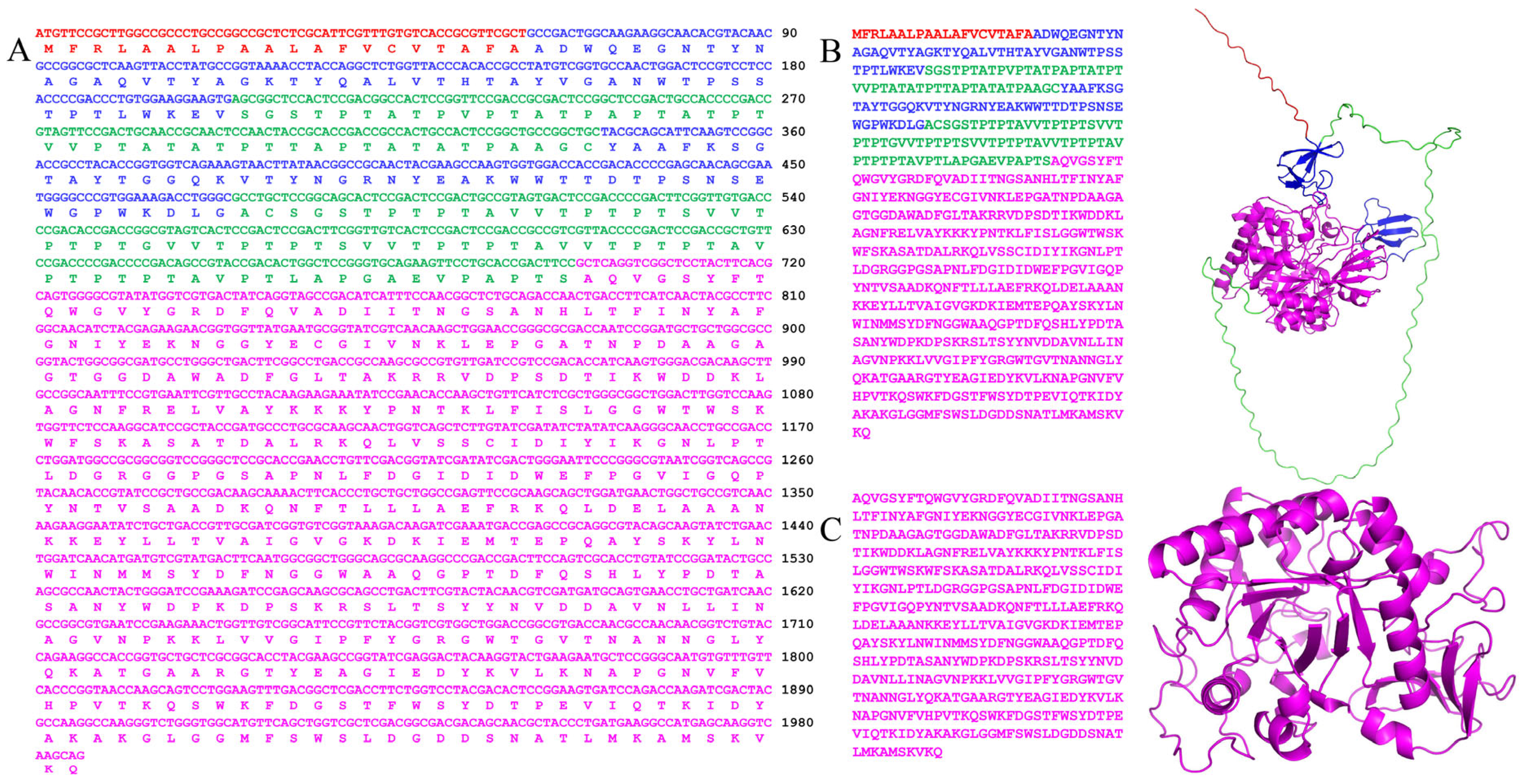
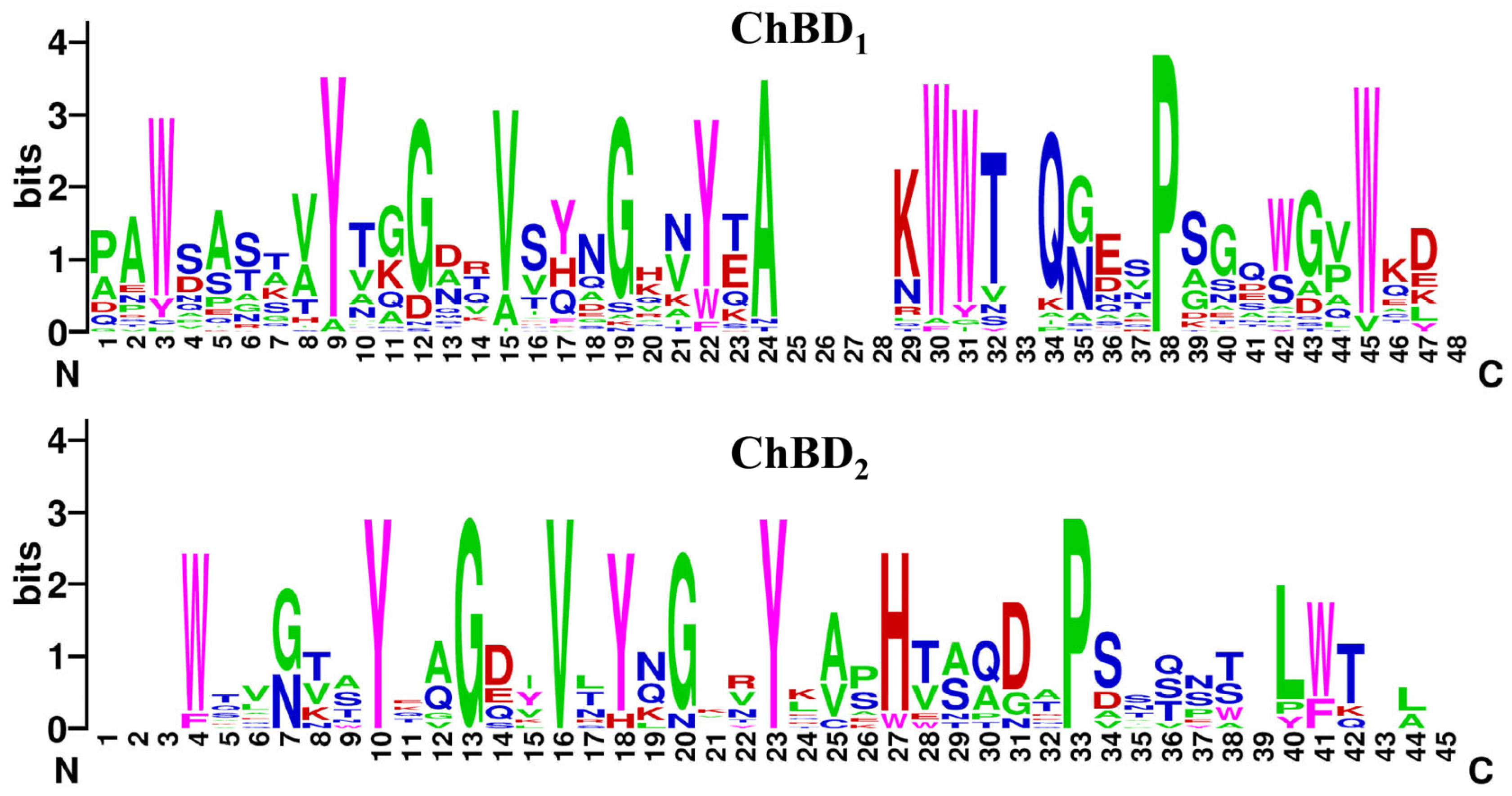
2.1. Bioinformatics Analysis

According to previous bioinformatics analyses [38], phylogenetic tree analysis indicated that the chitinase CaChi18B belongs to the GH18 family (Figure 1). Additionally, sequence alignment revealed that CaChi18B exhibits low similarity with other reported GH18 family chitinases. The highest similarity was observed with the multi-functional chitinase ATN39892.1 from Chitinolyticbacter meiyuanensis SYBC-H1, at 69.83% [39], followed by the chitinase CaChi18A from C. aquatile CSC-1, with a similarity of 67.08% [40]. In contrast, the similarity with the chitinase ACU59676.1 from Chitinophaga pinensis DSM 2588 was significantly lower, at only 26.21% [41]. ChBD1 was located between amino acids 20 and 68 (Ala20 to Val68), ChBD2 was found between amino acids 114 and 157 (Tyr114 to Leu157), and the catalytic domain (CD) spanned amino acids 233 to 647 (Gln233 to Asp647) (Figure 2). Therefore, the open reading frame (ORF) of the constructed chitinase CaChi18B_ΔChBDs was 1242 bp, encoding 414 amino acids, with a theoretical molecular weight of 47 kDa and a theoretical isoelectric point (pI) of 5.85.
Figure 1. Sequence analysis of the recombinant chitinase CaChi18B.
Marinedrugs 23 00093 g001
Figure 2. Sequences and three-dimensional structural model of CaChi18B. (A) Nucleotide sequence and amino acid sequence of CaChi18B. (B) and (C) show the amino acid sequences and predicted three-dimensional structural models of CaChi18B and CaChi18B_ΔChBDs, respectively. Red represents the signal sequence, blue represents the chitinase binding domains (ChBDs), and magenta represents the core catalytic domain (CD).Multiple sequence alignment analyses of the amino acid sequences of the two ChBD domains in CaChi18B revealed that one proline (P38) in ChBD1 was highly conserved. Additionally, four aromatic amino acid residues (W3, Y9, Y22, and W45) and five uncharged non-polar amino acid residues (G12, V15, G19, A24, and P38) were also highly conserved (Figure 3). In ChBD2, five amino acids were highly conserved (Y10, G13, V16, Y23, and P33). Additionally, four aromatic amino acid residues (Y10, Y18, Y23, and W41) and five uncharged non-polar amino acid residues (G13, V16, G20, A25, and P33) were also highly conserved.
Marinedrugs 23 00093 g002
Figure 3. The conserved amino acid residues within the ChBD domains of CaChi18B are depicted using sequence logos for ChBD1, ChBD2, and the sequence of CaChi18B itself. These logos visually represent the amino acid composition and conservation at each position. Specifically, magenta represents aromatic amino acids, green indicates uncharged non-polar amino acids, red signifies charged polar amino acids, and blue represents uncharged polar amino acids.
Marinedrugs 23 00093 g003

2.2. Induction, Expression, and Purification of Recombinant Chitinase

The target gene was ligated to the expression vector pET-22b (+), and the positive recombinant plasmid was transformed into the expression host E. coli BL21-CodonPlus (DE3)-RIPL. After purification using nickel affinity chromatography, SDS-PAGE analysis revealed a single protein band with a molecular weight of approximately 47 kDa (Figure 4), which was consistent with the theoretical molecular weight. The recombinant chitinase CaChi18B_ΔChBDs was obtained, exhibiting an enzyme activity of 3.48 U/mg. This indicated that the protein was suitable for further studies on its enzymatic properties.

2.3. Analysis of the Enzymatic Properties of Recombinant Chitinase

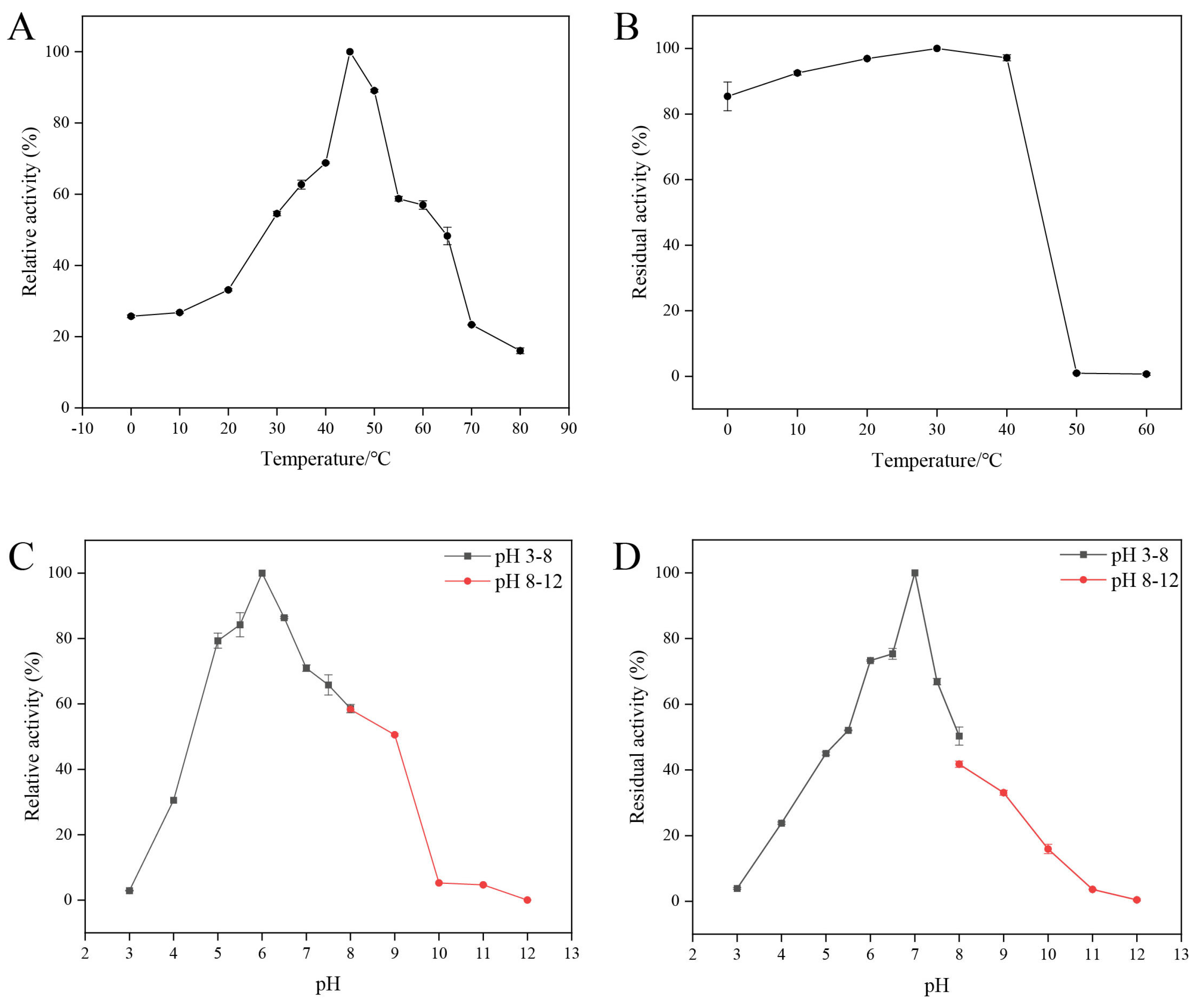
2.3.1. The Effects of Temperature and pH on the Activity of Recombinant Enzyme CaChi18B_ΔChBDs

Before assessing the effects of temperature and pH on the chitinase CaChi18B_ΔChBDs, we conducted elemental analysis on α-Chitin and colloidal chitin to determine their degree of acetylation based on the C/N ratio. It was found that the C/N ratio of colloidal chitin (C/N = 3.4156) was slightly higher than that of α-Chitin (C/N = 3.3493), indicating that acid hydrolysis does not significantly alter the degree of acetylation of chitin. Please refer to Figure S2 and Table S2 for further details.
The optimal temperature for the recombinant chitinase CaChi18B_ΔChBDs was 45 °C, as shown in Figure 5A. After incubation at temperatures ranging from 0 to 40 °C for 30 min, over 80% of the enzyme activity remained. However, at 50 °C, there was a sharp decline in residual enzyme activity.
The optimal pH was found to be 6.0, as illustrated in Figure 5C. The chitinase CaChi18B_ΔChBDs retained more than 60% of its residual activity within the pH range of 6.0 to 7.5.
The optimal reaction temperature for CaChi18B_ΔChBDs was 45 °C, and the optimal pH was 6.0, demonstrating slight variations in both the optimal temperature and pH compared to another truncated variant, CaChi18B_ΔChBD1 [38]; for instance, the chitinase Tc-ChiD from Thermococcus chitonophagus and its truncated variant Tc-ChiD (ΔCBD) exhibited a shift in temperature from 80 °C to 90 °C after truncation of the ChBD [42]. However, most ChBDs do not influence the optimal temperature and pH of chitinases. For example, the optimal temperature and pH of the chitinase CaChi18A and its mutant CaChi18A_ChBDs, which originate from the same source, remain unchanged [40]. Similarly, the thermophilic chitinase ActChi retained an optimal temperature of 80 °C and a pH of 6.0 even after the addition of a ChBD [43]. In terms of stability, CaChi18B_ΔChBD1 exhibited greater temperature and pH stability than CaChi18B_ΔChBDs [38].

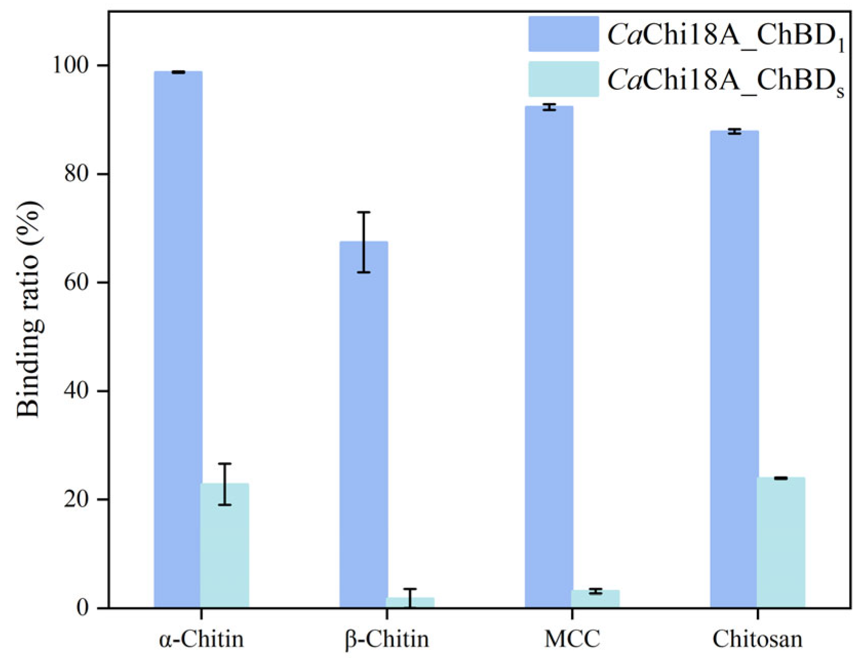
2.3.2. Determination of Enzyme Binding Ability

Both CaChi18B_ΔChBD1 and its mutant CaChi18B_ΔChBDs possess the ability to hydrolyze colloidal chitin into GlcNAc monomers [38]. Furthermore, this study also validated their binding potential with insoluble substrates, as clearly illustrated in Figure 6. The experimental results indicate that the presence of the ChBD significantly enhanced the binding affinity of chitinase for insoluble substrates. Specifically, when using α-Chitin, β-Chitin, microcrystalline cellulose (MCC), and chitosan as substrates, the binding rates of CaChi18B_ΔChBD1 were 98.88%, 71.32%, 98.14%, and 92.73%, respectively. In contrast, the binding rates of CaChi18B_ΔChBDs for these substrates were only 25.52%, 2.3%, 3.3%, and 24.02%. Compared to CaChi18A_ΔChBD1, the binding rates were significantly reduced, demonstrating the critical role of ChBD in promoting the effective binding of chitinase to various substrates, in line with previous literature reports [30,31].

2.3.3. Substrate Specificity and Kinetic Parameters of Recombinant Chitinase CaChi18B_ΔChBDs

To investigate the substrate specificity of CaChi18B_ΔChBDs, colloidal chitin, αChitin, β-Chitin, sodium carboxymethyl cellulose (CMC-Na), chitosan, and microcrystalline cellulose (MCC) were selected as substrates for the study, with the results summarized in Table 1. The data indicate that CaChi18B_ΔChBDs exhibited the highest enzymatic activity on colloidal chitin, reaching 3.48 U/mg, followed by β-Chitin. However, when hydrolyzing insoluble substrates, CaChi18B_ΔChBDs exhibited only 1.7% of its activity towards colloidal chitin on α-Chitin, whereas CaChi18B_ΔChBD1 achieved nearly double that at 3%. This aligned with the characteristics of many reported ChBDs, such as the fusion of the ChBD from Bacillus subtilis with the C-terminus of the chitinase Chit46, which enhanced the activity of the chimeric enzyme towards insoluble substrates by 219% [44]. Additionally, the chitinase Chi1 derived from A. caviae CB101 also exhibited reduced activity against MCC following truncation [45].
Using colloidal chitin as the substrate, the kinetic parameters of CaChi18B_ΔChBDs were determined. Fitting calculations performed with GraphPad software yielded a Michaelis constant (Km) of 1.159 mg mL−1 and a maximum reaction rate (Vmax) of 10.37 μM min−1 mg−1. The catalytic constant (kcat) was found to be as high as 103.7 s−1. The Km value of CaChi18B_ΔChBDs was 1.159 mg mL−1, whereas the Km value of CaChi18B_ΔChBD1 for colloidal chitin was 0.6626 mg mL−1. In comparison, CaChi18B_ΔChBD1 exhibited a lower Km value, indicating a higher affinity for the substrate [38]. It exhibited similarities to chitinases found in other sources, which demonstrated higher hydrolysis ability towards colloidal chitin but weaker hydrolysis ability towards powdered chitin [46,47,48]. Of interest, the enzyme also exhibited some activity when chitosan was used as a substrate, resembling the chitinases from Orpinomyces and Anaeromyces [49], but acting differently from the chitinases from Streptomyces roseolus [50] and Paenicibacillus barengoltzii [48].

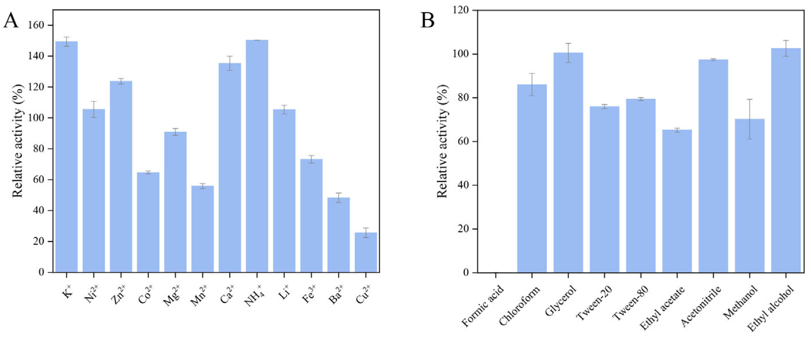
2.3.4. Tolerance of Recombinant Chitinase CaChi18B_ΔChBDs to Metal Ions and Chemical Reagents

Metal ions such as K+, Ni2+, Zn2+, Ca2+, NH4+, and Li+ exhibited a promoting effect on the hydrolytic activity of CaChi18B_ΔChBDs, with K+ and NH4+ showing the most significant enhancement, increasing activity by up to 150% (Figure 7A). In contrast, metal ions such as Co2+, Mn2+, Fe3+, Ba2+, and Cu2+ had an inhibitory effect on CaChi18B_ΔChBDs, with Cu2+ demonstrating the most pronounced inhibition, resulting in only 23% of the enzyme’s activity remaining. Generally, Cu2+ is often utilized as an inhibitor for most chitinases derived from bacteria [51,52,53,54]. Recent research has revealed that Cu2+ may coordinate with the thiol side chain of cysteine residues within the active site of chitinases, thereby inducing conformational changes in the enzyme molecule and ultimately leading to the reduction or loss of enzymatic activity [51,55]. Research has found that a certain concentration of Co2+ can inhibit the activity of chitinases. Similarly, Co2+ also exhibited a strong inhibitory effect on chitinases from Micrococcus sp. AG84 [56] and S. roseolus [50].
Regarding organic reagents, while most had minimal impact on the enzyme activity of CaChi18B_ΔChBDs, formic acid was a notable exception, severely inhibiting the enzyme’s activity. This observation indicated that while the recombinant enzyme CaChi18B_ΔChBDs behaves similarly to CaChi18B_ΔChBD1 [38], the activity of chitinase CaChi18B_ΔChBDs is largely unaffected by most chemical reagents, with the exception of formic acid. Chloroform, Tween-20, Tween-80, glycerol, and ethyl alcohol have been shown to inhibit chitinase activity in most cases [51,57,58]. Interestingly, ethyl alcohol and glycerol do not affect the activity of chitinase CaChi18B_ΔChBDs, which also distinguishes it from other chitinases [57,58]. Lee et al. [53] initially reported an organic solvent-tolerant chitinase, named MtCh509, from the bacterium Microbulbifer thermotolerans. Notably, the presence of organic solvents, including benzene, dimethyl sulfoxide, hexane, isoamyl alcohol, isopropanol, and toluene (10–20%, v/v), significantly enhanced the reactivity of MtCh509.

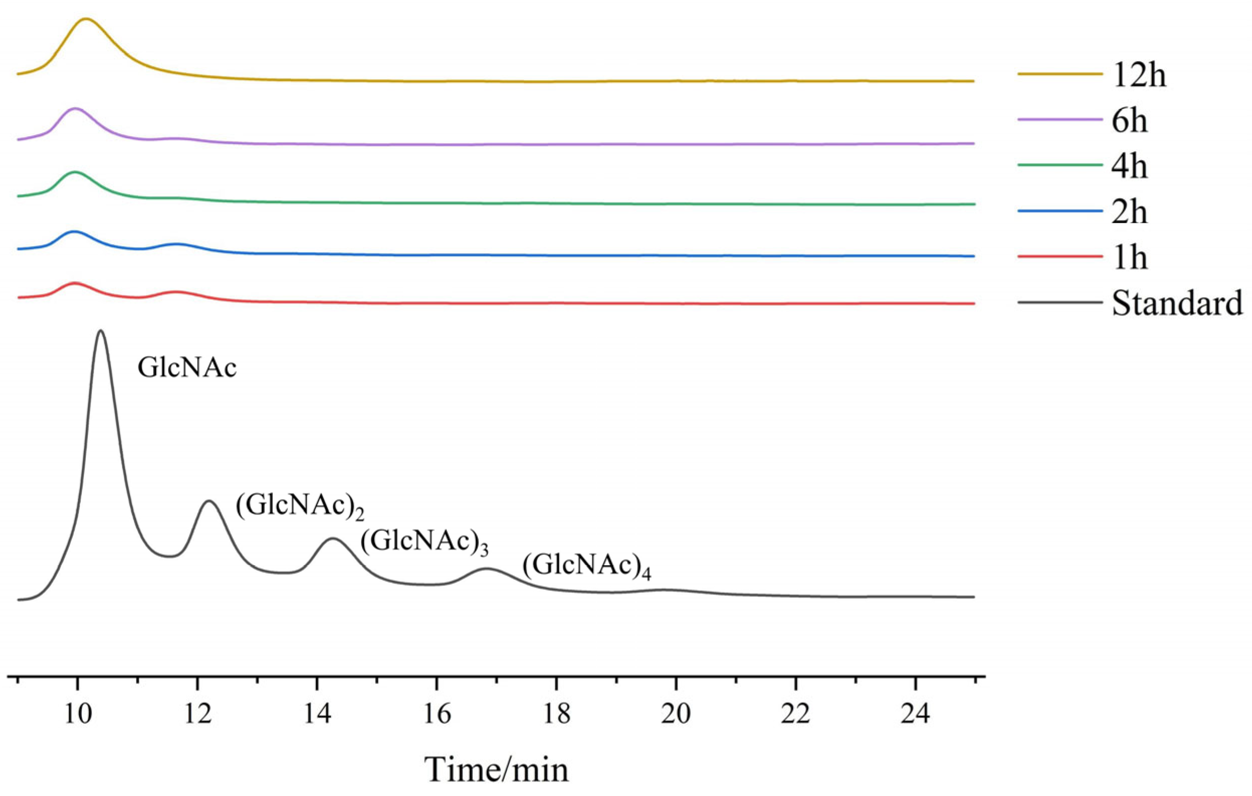
2.3.5. Analysis of Hydrolysis Products

High-performance liquid chromatography (HPLC) was employed to analyze the products generated by the recombinant enzyme CaChi18B_ΔChBDs during the hydrolysis of colloidal chitin, as illustrated in Figure 8. The analysis of the hydrolysis products revealed that the primary products during the initial stages of the reaction were GlcNAc and (GlcNAc)2. However, as the reaction progressed, the products gradually shifted to GlcNAc alone, suggesting that CaChi18B_ΔChBDs possesses both chitobiosidase activity and β-N-acetylglucosaminidase activity. In our research, we have discovered that the primary product of chitinase CaChi18B_ΔChBD1 during the initial stages of colloidal chitin hydrolysis was (GlcNAc)2, whereas the truncated variant CaChi18B_ΔChBDs yielded GlcNAc as its initial product, potentially attributed to the truncated ChBD. A similar phenomenon was also observed in the chitinase BmChi, derived from Bombyx mori, and its truncated mutant BmChi∆C [59]. When hydrolyzing (GlcNAc)5, the product of chitinase BmChi was GlcNAc and (GlcNAc)2, whereas the product of BmChi∆C was (GlcNAc)2 and (GlcNAc)3. Additionally, when hydrolyzing (GlcNAc)3, the product of chitinase BmChi was GlcNAc and (GlcNAc)2, whereas the product of BmChi∆C, when hydrolyzing (GlcNAc)5, remained as (GlcNAc)2 and (GlcNAc)3 [60]. The removal of the C-terminal chitin-binding domain (ChBD) had a negative impact on the hydrolytic ability of BmChi towards trisaccharide substrates. This proves that ChBD has an influence on the distribution of the hydrolytic products of chitinase. Oligomers can pose a significant challenge in terms of separating the desired product and can create difficulties in their application during large-scale production [39,61]. This property may enable the potential application of CaChi18B and CaChi18A_ΔChBDs in the field of green GlcNAc production.

3. Materials and Methods

3.1. Material and Reagents

3.1.1. Strain and Plasmid

C. aquatile CSC-1, the expression vector pET-22b (+), E. coli DH5α, and E. coli BL21-CodonPlus (DE3)-RIPL were all preserved in the laboratory.

3.1.2. Main Reagents and Instruments

The restriction enzymes Nco I and Hind III were purchased from Takara Biomedical Technology (Tokyo, Japan) Co., Ltd. The Star Marker D2000, plasmid mini-prep kit, and DNA purification kit were obtained from Genstar (Suzhou, China). The 2× ClonExpress Mix and 2× Phanta Max Master Mix were sourced from Vazyme (Nanjing, China), while the Ni-NTA Beads 6FF for protein purification were acquired from Smart-Life Sciences (Changzhou, China) Co., Ltd.
The LB liquid culture medium consisted of 5 g/L yeast extract, 10 g/L peptone, and 10 g/L NaCl, sterilized at 121 °C for 20 min. The LB solid medium contained 5 g/L yeast extract, 10 g/L peptone, 10 g/L NaCl, and 15 g/L agar powder, also sterilized at 121 °C for 20 min. The PCR instrument used was the Biometra TOne, and the gel imaging system used was the Gel Doc™ XR+ from Bio-Rad (Hercules, CA, USA).

3.1.3. Software and Websites

The physicochemical properties of the protein were analyzed using ExPASy [62] (https://web.expasy.org/compute_pi/, accessed on 6 July 2024). Signal peptide prediction was conducted using SignalP 6.0. [63] (https://services.healthtech.dtu.dk/services/SignalP-6.0, accessed on 1 July 2024). Domain analysis was performed using SMART (http://smart.embl.de/, accessed on 1 July 2024) [64]. MEGA 11 software was used to construct the phylogenetic tree based on the sequences [65]. ChiPlot was employed to beautify the phylogenetic tree (https://chiplot.online/, accessed on15 February 2025) [66]. WebLogo (http://weblogo.berkeley.edu/; WebLogo: A Sequence Logo Generator, accessed on 11 November 2024) was utilized to analyze the results of the multiple sequence alignments [67]. The three-dimensional structure was analyzed through the PyMOL program (The PyMOL Molecular Graphics System, accessed on 15 February 2025, Version 3.0 Schrödinger, LLC, New York, NY, USA).

3.2. Experimental Methods

3.2.1. Preparation of Colloidal Chitin

Colloidal chitin was prepared according to previously established methods in the laboratory [40]. Ten grams of powdered chitin were mixed with 100 mL of concentrated hydrochloric acid and stirred until a homogeneous solution was achieved. The mixture was then allowed to stand at 4 °C for 24 h. Meanwhile, 2 L of industrial alcohol was pre-cooled. After 24 h, the chitin solution was combined with the industrial alcohol and stirred until homogeneous. The mixture was then allowed to stand at 4 °C for another 24 h, followed by centrifugation at 8000 rpm for 10 min to collect the precipitate, which was washed repeatedly with distilled water until neutralized.

3.2.2. Gene Cloning and Construction of Expression Vectors for CaChi18B_ΔChBDs

Using the pET-22b (+) vector, primers for CaChi18B_ΔChBDs-F and CaChi18B_ΔChBDs-R were designed based on the gene sequence of CaChi18B_ΔChBDs. The primers included recognition sites for the restriction endonucleases Nco I (ccatgg) and Hind III (aagctt), as shown in Table 2.
The PCR conditions were as follows: initial denaturation at 95 °C for 5 min, denaturation at 95 °C for 30 s, annealing at 65 °C for 30 s, and extension at 72 °C for 90 s, followed by 35 cycles of these steps. A final extension was performed at 72 °C for 10 min. The amplified DNA fragments were purified from the gel and then ligated into the pET-22b (+) plasmid, which had been double-digested with Nco I and Hind III and subsequently purified. The ligation mixture was transformed into competent E. coli DH5α cells and sent for sequencing. The recombinant plasmid was designated as pET-22b (+)-CaChi18B_ΔChBDs/DH5α.

3.2.3. Protein Expression and Purification of CaChi18B_ΔChBDs

The correctly sequenced recombinant plasmid was introduced into the expression host E. coli BL21-CodonPlus (DE3)-RIPL.
After culturing the above strain for induction, the cells were collected by centrifugation at 8000 rpm for 10 min. The pellet was then resuspended in Tris-NaCl buffer and subjected to ultrasonic cell disruption to obtain the cell lysate. The lysate was centrifuged at 12,000 rpm for 30 min at 4 °C to collect the supernatant. Subsequently, affinity chromatography was performed using Ni-NTA Beads 6FF, and the elution fraction contained the recombinant enzyme.

3.2.4. Enzymatic Property Analysis of CaChi18B_ΔChBDs

The colloidal chitin was frozen at −80 °C for 24 h, followed by lyophilization using a freeze dryer (YAMATO DC801). The α-Chitin was dried in an oven at 80 °C for 24 h. The dried materials were then analyzed using an elemental analyzer (Thermo Scientific Flash TY001, Waltham, MA, USA) to determine the C/N ratio, which was used to assess the degree of acetylation of the materials.
The activity of the chitinase was measured using the DNS method, with slight modifications based on the previous literature [40]. The reaction system consisted of 400 μL of 2% colloidal chitin (pH 7.0) and 100 μL of the enzyme solution at a concentration of 0.1 mg mL−1. The mixture was incubated at 50 °C for 10 min, followed by treatment in a boiling water bath for 10 min to terminate the reaction. The mixture was then cooled to room temperature in an ice-water bath. Subsequently, 500 μL of DNS solution was added to each tube, and the mixture was heated in a boiling water bath for 5 min to develop color, after which it was cooled to room temperature. The mixture was centrifuged at 12,000 rpm for 5 min, and 200 μL of the supernatant was transferred to a microplate for absorbance measurements at 540 nm. The blank control consisted of 100 μL of citrate-phosphate buffer and 400 μL of 2% colloidal chitin (pH 7.0) under the same reaction conditions. The yield of the reducing sugars was calculated based on the difference in absorbance between the experimental and control groups, and the chitinase activity was determined using a standard curve. Each experimental group was repeated three times. Chitinase activity (U) was defined as the amount of enzyme required to release 1 µmol of N-acetyl-D-glucosamine per minute under the specified reaction conditions. Specific activity (U/mg) is defined as the number of enzyme activity units present per milligram (mg) of chitinase.

Effect of Temperature on Recombinant Enzyme Activity

Diluted enzyme solutions were used to assess the impact of different temperatures on the activity of the recombinant enzyme, with 2% colloidal chitin as the substrate. The highest enzyme activity was set as 100%, and the relative enzyme activity at various temperatures was calculated to determine the optimal reaction temperature. The recombinant enzyme was incubated at different temperatures for 30 min, after which the residual enzyme activity was measured under optimal reaction conditions. The activity of the untreated pure enzyme was considered as 100%, allowing for the calculation of the residual enzyme activity of the recombinant chitinase after 30 min of incubation at each temperature. Each temperature group was performed in triplicate to ensure accuracy in the measurement of enzyme activity.

Effect of pH on Recombinant Enzyme Activity

The chitinase solution at a concentration of 0.1 mg/mL was mixed with 2% colloidal chitin in buffers ranging from pH 3.0 to 12.0 (100 mmol/L citrate-phosphate buffer: pH 3.0–8.0; glycine-NaOH buffer: pH 8.0-12.0), and the enzyme activity was measured at the optimal temperature to determine the optimal reaction pH. The maximum enzyme activity was set as 100%, and a curve was plotted based on the relative enzyme activity at different pH values. Each experimental group was performed in triplicate. The recombinant enzyme was incubated in buffers with varying pH (100 mmol/L citrate-phosphate buffer: pH 3.0–8.0; glycine-NaOH buffer: pH 8.0–12.0) at 4 °C for 180 min. Subsequently, 2% colloidal chitin (pH 6.0) was mixed with the incubated enzyme solution, and the enzyme activity was measured to evaluate the pH stability of the recombinant enzyme. Each pH condition was tested in triplicate to ensure the accuracy of enzyme activity calculations.

Determination of Enzyme Binding Capacity

The binding capacity of chitinase was assessed based on previous research by Chen et al. [40]. The substrates (α-Chitin, β-Chitin, MCC, and chitosan) were prepared at a concentration of 5% (W/V) in 100 mM citrate-phosphate buffer (pH 6.5). To achieve this, 500 μL of chitinase was added to 0.025 g of the substrate. The mixture was gently shaken at 4 °C for 1 h. After the reaction, the mixture was centrifuged to collect the supernatant, and the protein concentration was measured. The ratio of bound protein to the initial protein concentration was defined as the binding capacity of the enzyme to chitin.

Determination of Substrate Specificity, Km, and Vmax

The enzyme activity was measured using various substrates, including 2% colloidal chitin, α-Chitin, β-Chitin, sodium carboxymethyl cellulose (CMC-Na), chitosan, and microcrystalline cellulose, under optimal conditions.
Additionally, the enzyme activity of the recombinant enzyme was assessed using different concentrations (0.1–5%) of colloidal chitin as the substrate under optimal conditions. The data were plotted using GraphPad to calculate the Km and Vmax values.

Effects of Metal Ions and Organic Reagents on Recombinant Enzyme Activity

Diluted recombinant enzyme solutions were incubated with metal ions at a final concentration of 1 mmol/L (Co2+, K+, Zn2+, Ni2+, Mg2+, Mn2+, Ca2+, NH4+, Li+, Fe3+, Ba2+, Cu2+) and organic reagents at a final concentration of 1% (formic acid, chloroform, glycerol, Tween-20, Tween-80, ethyl acetate, acetonitrile, methanol, ethanol) under optimal conditions. The enzyme activity was measured, with the activity of the enzyme without any added metal ions or organic reagents set as 100%. The relative effects of various reagents on enzyme activity were then calculated.

Analysis of Hydrolysis Products

A mixture of 1 mL of the recombinant enzyme, at a concentration of 1 mg mL−1, and 3 mL of 2% colloidal chitin was incubated at 30 °C. Samples were taken at 0.5, 1, 2, 4, 6, 12, and 24 h, and the reaction was terminated by boiling for 10 min. After cooling in an ice bath, the mixture was centrifuged at 10,000 rpm for 2 min to collect the supernatant. The supernatant was then analyzed using high-performance liquid chromatography (HPLC). The chromatographic analysis was performed on a Luna® 5 μm NH2 column (LC Column 250 × 4.6 mm), with a differential refractive index detector (RID). The mobile phase consisted of 70% acetonitrile, and the flow rate was set at 0.5 mL/min.

4. Conclusions

In summary, we obtained the gene product of CaChi18B_ΔChBDs from C. aquatile CSC-1 and successfully cloned, functionally expressed, and biochemically characterized it. The optimal reaction temperature for the recombinant enzyme CaChi18B_ΔChBDs was 45 °C, and the optimal pH was 6.0, with over 50% relative enzyme activity maintained within the pH range of 5.5 to 8.0. Metal ions such as Ca2+, K+, Zn2+, NH4+, and Li+ enhanced the enzyme’s activity, while Cu2+, Mn2+, Co2+, and Ba2+ exhibited significant inhibitory effects. When colloidal chitin was used as the substrate, the enzyme demonstrated the highest activity, with a Michaelis constant (Km) of 1.159 mg mL−1 and a maximum reaction rate (Vmax) of 10.37 μM min−1 mg−1. Analysis of the hydrolysis products revealed that CaChi18B_ΔChBDs possesses dual functional activities as both a chitobiosidase and a β-N-acetylglucosaminidase, indicating its ability to directly produce GlcNAc. This finding provides an environmentally friendly method for the sustainable production of valuable chitin-derived products. Furthermore, the investigation of the ChBD confirmed its crucial role in substrate binding and product distribution, establishing a foundation for the modification and analysis of chitinases. Future research will focus on the impact of ChBD on product distribution.

Supplementary Materials

The following supporting information can be downloaded at: https://www.mdpi.com/article/10.3390/md23030093/s1, Table S1 Comparison of enzymatic properties of CaChi18B_ΔChBD1 and CaChi18B_ΔChBDs; Figure S1 The SDS-PAGE analysis of recombinant CaChi18B_Δ ChBDs; Figure S2 Elemental analysis spectra of α-Chitin and colloidal chitin.

Author Contributions

C.G.: Formal analysis, Methodology, Writing original draft. J.C.: Validation, Writing review & editing. X.H.: Software. Y.J.: Data curation, Investigation. N.O.: Funding acquisition, Resources. D.Y.: Data curation, Software. M.J.: Funding acquisition. L.P.: Conceptualization, Funding acquisition, Methodology, Project administration, Supervision. All authors have read and agreed to the published version of the manuscript.

Funding

This research was funded by financial support from the Guangxi Science and Technology Base and Talent Project (AD23023007), the Science and Technology Major Project of Guangxi (Guike AA24206048), the National Natural Science Foundation of China (Grant Nos. 32360227 and 32360244), the Innovation and Development Fund of the Guangxi Academy of Sciences (No. 2024YGFZ504-103), and the Basic Science and Research Foundation of the Guangxi Academy of Sciences (CQ-C-202302).

Institutional Review Board Statement

Not applicable.

Data Availability Statement

The data presented in this study are available on request from the corresponding author.

Conflicts of Interest

The authors declare no conflict of interest.

References

  1. Jana, S.; Hamre, A.G.; Wildberger, P.; Holen, M.M.; Eijsink, V.G.H.; Beckham, G.T.; Sørlie, M.; Payne, C.M. Aromatic-Mediated Carbohydrate Recognition in Processive Serratia Marcescens Chitinases. J. Phys. Chem. B 2016, 120, 1236–1249. [Google Scholar] [CrossRef] [PubMed]
  2. Gomaa, E.Z. Microbial Chitinases: Properties, Enhancement and Potential Applications. Protoplasma 2021, 258, 695–710. [Google Scholar] [CrossRef] [PubMed]
  3. Zdarta, J.; Klapiszewski, Ł.; Wysokowski, M.; Norman, M.; Kołodziejczak-Radzimska, A.; Moszyński, D.; Ehrlich, H.; Maciejewski, H.; Stelling, A.; Jesionowski, T. Chitin-Lignin Material as a Novel Matrix for Enzyme Immobilization. Mar. Drugs 2015, 13, 2424–2446. [Google Scholar] [CrossRef] [PubMed]
  4. Yadav, M.; Goswami, P.; Paritosh, K.; Kumar, M.; Pareek, N.; Vivekanand, V. Seafood Waste: A Source for Preparation of Commercially Employable Chitin/Chitosan Materials. Bioresour. Bioprocess. 2019, 6, 8. [Google Scholar] [CrossRef]
  5. Ma, Q.; Gao, X. Categories and Biomanufacturing Methods of Glucosamine. Appl. Microbiol. Biotechnol. 2019, 103, 7883–7889. [Google Scholar] [CrossRef]
  6. Liu, X.; Zhang, Y.; Liu, Z.; Xie, X. Anti-Tumor Effect of Chitin Oligosaccharide plus Cisplatin In Vitro and In Vivo. OncoTargets Ther. 2019, 12, 7581–7590. [Google Scholar] [CrossRef]
  7. Kang, K.A.; Lee, K.H.; Chae, S.; Zhang, R.; Jung, M.S.; Lee, Y.; Kim, S.Y.; Kim, H.S.; Joo, H.G.; Park, J.W.; et al. Eckol Isolated from Ecklonia cava Attenuates Oxidative Stress Induced Cell Damage in Lung Fibroblast Cells. FEBS Lett. 2005, 579, 6295–6304. [Google Scholar] [CrossRef]
  8. Cody, R.M.; Davis, N.D.; Lin, J.; Shaw, D. Screening Microorganisms for Chitin Hydrolysis and Production of Ethanol from Amino Sugars. Biomass 1990, 21, 285–295. [Google Scholar] [CrossRef]
  9. Maia, A.; Cardona Gloria, Y.; Fuchs, K.; Chang, T.-H.; Engels, P.; Zhou, M.; Hinnenthal, T.; Rusch, E.; Gouttefangeas, C.; Weber, A.N.R. Chitin Oligomers Promote Lymphoid Innate and Adaptive Immune Cell Activation. J. Leukoc. Biol. 2023, 114, 180–186. [Google Scholar] [CrossRef]
  10. Zheng, J.; Cheng, G.; Li, Q.; Jiao, S.; Feng, C.; Zhao, X.; Yin, H.; Du, Y.; Liu, H. Chitin Oligosaccharide Modulates Gut Microbiota and Attenuates High-Fat-Diet-Induced Metabolic Syndrome in Mice. Mar. Drugs 2018, 16, 66. [Google Scholar] [CrossRef]
  11. Salvatore, S.; Heuschkel, R.; Tomlin, S.; Davies, S.E.; Edwards, S.; Walker-Smith, J.A.; French, I.; Murch, S.H. A Pilot Study of N-acetyl Glucosamine, a Nutritional Substrate for Glycosaminoglycan Synthesis, in Paediatric Chronic Inflammatory Bowel Disease. Aliment. Pharmacol. Ther. 2000, 14, 1567–1579. [Google Scholar] [CrossRef] [PubMed]
  12. Shikhman, A.R. Chondroprotective Activity of N-Acetylglucosamine in Rabbits with Experimental Osteoarthritis. Ann. Rheum. Dis. 2005, 64, 89–94. [Google Scholar] [CrossRef]
  13. Marchetti, M.; De Berardis, B.; Bigioni, I.; Mariano, A.; Superti, F.; Scotto d’Abusco, A. In Vitro Antiviral and Anti-Inflammatory Activities of N-Acetylglucosamine: Development of an Alternative and Safe Approach to Fight Viral Respiratory Infections. Int. J. Mol. Sci. 2023, 24, 5129. [Google Scholar] [CrossRef] [PubMed]
  14. Kubomura, D.; Ueno, T.; Yamada, M.; Nagaoka, I. Evaluation of the Chondroprotective Action of N-Acetylglucosamine in a Rat Experimental Osteoarthritis Model. Exp. Ther. Med. 2017, 14, 3137–3144. [Google Scholar] [CrossRef] [PubMed]
  15. Chen, W.; Jiang, X.; Yang, Q. Glycoside Hydrolase Family 18 Chitinases: The Known and the Unknown. Biotechnol. Adv. 2020, 43, 107553. [Google Scholar] [CrossRef]
  16. Wu, Y.-L.; Wang, S.; Yang, D.-F.; Yang, L.-Y.; Wang, Q.-Y.; Yu, J.; Li, N.; Pan, L.-X. The Discovery, Enzymatic Characterization and Functional Analysis of a Newly Isolated Chitinase from Marine-Derived Fungus Aspergillus fumigatus Df347. Mar. Drugs 2022, 20, 520. [Google Scholar] [CrossRef]
  17. Li, J.; Gao, K.; Secundo, F.; Mao, X. Biochemical Characterization of Two β-N-Acetylglucosaminidases from Streptomyces Violascens for Efficient Production of N-Acetyl-d-Glucosamine. Food Chem. 2021, 364, 130393. [Google Scholar] [CrossRef]
  18. Wang, S.; Fu, G.; Li, J.; Wei, X.; Fang, H.; Huang, D.; Lin, J.; Zhang, D. High-Efficiency Secretion and Directed Evolution of Chitinase BcChiA1 in Bacillus subtilis for the Conversion of Chitinaceous Wastes into Chitooligosaccharides. Front. Bioeng. Biotechnol. 2020, 8, 432. [Google Scholar] [CrossRef]
  19. Prabavathy, V.R.; Mathivanan, N.; Sagadevan, E.; Murugesan, K.; Lalithakumari, D. Self-Fusion of Protoplasts Enhances Chitinase Production and Biocontrol Activity in Trichoderma harzianum. Bioresour. Technol. 2006, 97, 2330–2334. [Google Scholar] [CrossRef]
  20. Wang, L.; Xie, Y.; Chang, J.; Wang, J.; Liu, H.; Shi, M.; Zhong, Y. A Novel Sucrose-Inducible Expression System and Its Application for Production of Biomass-Degrading Enzymes in Aspergillus niger. Biotechnol. Biofuels Bioprod. 2023, 16, 23. [Google Scholar] [CrossRef]
  21. Poshina, D.N.; Raik, S.V.; Poshin, A.N.; Skorik, Y.A. Accessibility of Chitin and Chitosan in Enzymatic Hydrolysis: A Review. Polym. Degrad. Stab. 2018, 156, 269–278. [Google Scholar] [CrossRef]
  22. Itoh, T.; Hibi, T.; Fujii, Y.; Sugimoto, I.; Fujiwara, A.; Suzuki, F.; Iwasaki, Y.; Kim, J.-K.; Taketo, A.; Kimoto, H. Cooperative Degradation of Chitin by Extracellular and Cell Surface-Expressed Chitinases from Paenibacillus sp. Strain FPU-7. Appl. Environ. Microbiol. 2013, 79, 7482–7490. [Google Scholar] [CrossRef] [PubMed]
  23. Hammami, I.; Siala, R.; Jridi, M.; Ktari, N.; Nasri, M.; Triki, M.A. Partial Purification and Characterization of ChiIO8, a Novel Antifungal Chitinase Produced by Bacillus cereus IO8. J. Appl. Microbiol. 2013, 115, 358–366. [Google Scholar] [CrossRef] [PubMed]
  24. Gao, C.; Zhang, A.; Chen, K.; Hao, Z.; Tong, J.; Ouyang, P. Characterization of Extracellular Chitinase from Chitinibacter sp. GC72 and Its Application in GlcNAc Production from Crayfish Shell Enzymatic Degradation. Biochem. Eng. J. 2015, 97, 59–64. [Google Scholar] [CrossRef]
  25. Shibasaki, H.; Uchimura, K.; Miura, T.; Kobayashi, T.; Usami, R.; Horikoshi, K. Highly Thermostable and Surfactant-Activated Chitinase from a Subseafloor Bacterium, Laceyella putida. Appl. Microbiol. Biotechnol. 2014, 98, 7845–7853. [Google Scholar] [CrossRef]
  26. Adrangi, S.; Faramarzi, M.A.; Shahverdi, A.R.; Sepehrizadeh, Z. Purification and Characterization of Two Extracellular Endochitinases from Massilia timonae. Carbohydr. Res. 2010, 345, 402–407. [Google Scholar] [CrossRef]
  27. Kim, J.; Kim, S.I.; Hong, E.; Ryu, Y. Strategies for Increasing Heterologous Expression of a Thermostable Esterase from Archaeoglobus fulgidus in Escherichia coli. Protein Expr. Purif. 2016, 127, 98–104. [Google Scholar] [CrossRef]
  28. Harnischfeger, J.; Beutler, M.; Salzig, D.; Rahlfs, S.; Becker, K.; Grevelding, C.G.; Czermak, P. Biochemical Characterization of the Recombinant Schistosome Tegumental Protein SmALDH_312 Produced in E. Coli and Baculovirus Expression Vector System. Electron. J. Biotechnol. 2021, 54, 26–36. [Google Scholar] [CrossRef]
  29. Cardozo, F.A.; Facchinatto, W.M.; Colnago, L.A.; Campana-Filho, S.P.; Pessoa, A. Bioproduction of N-Acetyl-Glucosamine from Colloidal α-Chitin Using an Enzyme Cocktail Produced by Aeromonas caviae CHZ306. World J. Microbiol. Biotechnol. 2019, 35, 114. [Google Scholar] [CrossRef]
  30. Doan, C.T.; Tran, T.N.; Wang, S.-L. Production of Thermophilic Chitinase by Paenibacillus sp. TKU052 by Bioprocessing of Chitinous Fishery Wastes and Its Application in N-Acetyl-D-Glucosamine Production. Polymers 2021, 13, 3048. [Google Scholar] [CrossRef]
  31. Lombard, V.; Golaconda Ramulu, H.; Drula, E.; Coutinho, P.M.; Henrissat, B. The Carbohydrate-Active Enzymes Database (CAZy) in 2013. Nucleic Acids Res. 2014, 42, D490–D495. [Google Scholar] [CrossRef] [PubMed]
  32. Madhuprakash, J.; El Gueddari, N.E.; Moerschbacher, B.M.; Podile, A.R. Catalytic Efficiency of Chitinase-D on Insoluble Chitinous Substrates Was Improved by Fusing Auxiliary Domains. PLoS ONE 2015, 10, e0116823. [Google Scholar] [CrossRef] [PubMed]
  33. Boraston, A.B.; Bolam, D.N.; Gilbert, H.J.; Davies, G.J. Carbohydrate-Binding Modules: Fine-Tuning Polysaccharide Recognition. Biochem. J. 2004, 382, 769–781. [Google Scholar] [CrossRef] [PubMed]
  34. Xu, S.; Qiu, M.; Zhang, X.; Chen, J. Expression and Characterization of an Enhanced Recombinant Heparinase I with Chitin Binding Domain. Int. J. Biol. Macromol. 2017, 105, 1250–1258. [Google Scholar] [CrossRef]
  35. Itoh, Y.; Watanabe, J.; Fukada, H.; Mizuno, R.; Kezuka, Y.; Nonaka, T.; Watanabe, T. Importance of Trp59 and Trp60 in Chitin-Binding, Hydrolytic, and Antifungal Activities of Streptomyces griseus Chitinase C. Appl. Microbiol. Biotechnol. 2006, 72, 1176–1184. [Google Scholar] [CrossRef]
  36. Nakamura, T.; Mine, S.; Hagihara, Y.; Ishikawa, K.; Uegaki, K. Structure of the Catalytic Domain of the Hyperthermophilic Chitinase from Pyrococcus Furiosus. Acta Cryst. Sect. F Struct. Biol. Cryst. Commun. 2007, 63, 7–11. [Google Scholar] [CrossRef]
  37. Pham, M.-L.; Leister, T.; Nguyen, H.A.; Do, B.-C.; Pham, A.-T.; Haltrich, D.; Yamabhai, M.; Nguyen, T.-H.; Nguyen, T.-T. Immobilization of β-Galactosidases from Lactobacillus on Chitin Using a Chitin-Binding Domain. J. Agric. Food Chem. 2017, 65, 2965–2976. [Google Scholar] [CrossRef]
  38. Chenxi, G.; Jianrong, C.; Yinghua, T.; Quanfeng, W.; Mingguo, J.; Lixia, P. Heterologous Expression and Enzymatic Properties of Truncated Mutant of Bi-Functional Chitinase from Chitinilyticum Aquatile. Guangxi Sci. 2024, 32, 1–15. [Google Scholar] [CrossRef]
  39. Zhang, A.; He, Y.; Wei, G.; Zhou, J.; Dong, W.; Chen, K.; Ouyang, P. Molecular Characterization of a Novel Chitinase CmChi1 from Chitinolyticbacter meiyuanensis SYBC-H1 and Its Use in N-Acetyl-d-Glucosamine Production. Biotechnol. Biofuels 2018, 11, 179. [Google Scholar] [CrossRef]
  40. Chen, J.; Yang, D.; Zhang, Y.; Yang, L.; Wang, Q.; Jiang, M.; Pan, L. A Novel Bi-Functional Cold-Adaptive Chitinase from Chitinilyticum aquatile CSC-1 for Efficient Synthesis of N-Acetyl-D-Glucosaminidase. Int. J. Biol. Macromol. 2024, 259, 129063. [Google Scholar] [CrossRef]
  41. Glavina Del Rio, T.; Abt, B.; Spring, S.; Lapidus, A.; Nolan, M.; Tice, H.; Copeland, A.; Cheng, J.-F.; Chen, F.; Bruce, D.; et al. Complete Genome Sequence of Chitinophaga pinensis Type Strain (UQM 2034T). Stand Genom. Sci. 2010, 2, 87–95. [Google Scholar] [CrossRef] [PubMed]
  42. Horiuchi, A.; Aslam, M.; Kanai, T.; Atomi, H. A Structurally Novel Chitinase from the Chitin-Degrading Hyperthermophilic Archaeon Thermococcus chitonophagus. Appl. Environ. Microbiol. 2016, 82, 3554–3562. [Google Scholar] [CrossRef] [PubMed]
  43. Sun, B.; Zhao, X.; Xu, B.; Su, E.; Kovalevsky, A.; Shen, Q.; Liu, D.; Wan, Q. Discovering and Designing a Chimeric Hyperthermophilic Chitinase for Crystalline Chitin Degradation. ACS Sustain. Chem. Eng. 2023, 11, 4690–4698. [Google Scholar] [CrossRef]
  44. Deng, J.-J.; Zhang, M.-S.; Li, Z.-W.; Lu, D.-L.; Mao, H.-H.; Zhu, M.-J.; Li, J.-Z.; Luo, X.-C. One-Step Processing of Shrimp Shell Waste with a Chitinase Fused to a Carbohydrate-Binding Module. Green Chem. 2020, 22, 6862–6873. [Google Scholar] [CrossRef]
  45. Wang, F.P.; Li, Q.; Zhou, Y.; Li, M.G.; Xiao, X. The C-Terminal Module of Chi1 from Aeromonas Caviae CB101 Has a Function in Substrate Binding and Hydrolysis. Proteins Struct. Funct. Genet. 2003, 53, 908–916. [Google Scholar] [CrossRef]
  46. Li, J.; Zheng, J.; Liang, Y.; Yan, R.; Xu, X.; Lin, J. Expression and Characterization of a Chitinase from Serratia marcescens. Protein Expr. Purif. 2020, 171, 105613. [Google Scholar] [CrossRef]
  47. Ueda, M.; Shioyama, T.; Nakadoi, K.; Nakazawa, M.; Sakamoto, T.; Iwamoto, T.; Sakaguchi, M. Cloning and Expression of a Chitinase Gene from Eisenia fetida. Int. J. Biol. Macromol. 2017, 104, 1648–1655. [Google Scholar] [CrossRef]
  48. Fu, X.; Yan, Q.; Wang, J.; Yang, S.; Jiang, Z. Purification and Biochemical Characterization of Novel Acidic Chitinase from Paenicibacillus barengoltzii. Int. J. Biol. Macromol. 2016, 91, 973–979. [Google Scholar] [CrossRef]
  49. Novotná, Z.; Fliegerová, K.; Šimůnek, J. Characterization of Chitinases of Polycentric Anaerobic Rumen Fungi. Folia Microbiol 2008, 53, 241–245. [Google Scholar] [CrossRef]
  50. Xiayun, J.; Chen, D.; Shenle, H.; Wang, W.; Chen, S.; Zou, S. Identification, Characterization and Functional Analysis of a GH-18 Chitinase from Streptomyces roseolus. Carbohydr. Polym. 2012, 87, 2409–2415. [Google Scholar] [CrossRef]
  51. Karthik, N.; Binod, P.; Pandey, A. RETRACTED: Purification and Characterisation of an Acidic and Antifungal Chitinase Produced by a Streptomyces sp. Bioresour. Technol. 2015, 188, 195–201. [Google Scholar] [CrossRef] [PubMed]
  52. Liang, T.-W. Purification and Characterization of Chitinase from a New Species Strain Pseudomonas sp. TKU008. J. Microbiol. Biotechnol. 2010, 20, 1001–1005. [Google Scholar] [CrossRef] [PubMed]
  53. Lee, H.-J.; Lee, Y.-S.; Choi, Y.-L. Cloning, Purification, and Characterization of an Organic Solvent-Tolerant Chitinase, MtCh509, from Microbulbifer thermotolerans DAU221. Biotechnol. Biofuels 2018, 11, 303. [Google Scholar] [CrossRef]
  54. Du, J.; Duan, S.; Miao, J.; Zhai, M.; Cao, Y. Purification and Characterization of Chitinase from Paenibacillus sp. Biotechnol. Appl. Biochem. 2021, 68, 30–40. [Google Scholar] [CrossRef] [PubMed]
  55. He, B.; Yang, L.; Yang, D.; Jiang, M.; Ling, C.; Chen, H.; Ji, F.; Pan, L. Biochemical Purification and Characterization of a Truncated Acidic, Thermostable Chitinase from Marine Fungus for N-Acetylglucosamine Production. Front. Bioeng. Biotechnol. 2022, 10, 1013313. [Google Scholar] [CrossRef]
  56. Annamalai, N.; Giji, S.; Muthuvel, A.; Balasubramanian, T. Purification and Characterization of Chitinase from Micrococcus sp. AG84 Isolated from Marine Environment. Afr. J. Microbiol. Res. 2010, 4, 2822–2827. [Google Scholar]
  57. Aktas, C.; Ruzgar, D.; Gurkok, S.; Gormez, A. Purification and Characterization of Stenotrophomonas Maltophilia Chitinase with Antifungal and Insecticidal Properties Prep. Biochem. Biotechnol. 2022, 53, 797–806. [Google Scholar]
  58. Cheba, B.A.; Zaghloul, T.I.; EL-Massry, M.H.; EL-Mahdy, A.R. Effect of Metal Ions, Chemical Agents, and Organic Solvent on Bacillus Sp.R2 Chitinase Activity. Procedia Technol. 2016, 22, 465–470. [Google Scholar] [CrossRef]
  59. Daimon, T.; Hamada, K.; Mita, K.; Okano, K.; Suzuki, M.G.; Kobayashi, M.; Shimada, T. A Bombyx Mori Gene, BmChi-h, Encodes a Protein Homologous to Bacterial and Baculovirus Chitinases. Insect Biochem. Mol. Biol. 2003, 33, 749–759. [Google Scholar] [CrossRef]
  60. Fan, Y.; Guo, S.; Pei, X.; Zhang, Y.; Luo, Z.; Pei, Y. Effects of Chitin Binding Domain on Enzymatic Properties and Insecticidal Activity of Bombyx Mori Chitinase. World J. Microbiol. Biotechnol. 2011, 27, 1551–1558. [Google Scholar] [CrossRef]
  61. Fu, X.; Yan, Q.; Yang, S.; Yang, X.; Guo, Y.; Jiang, Z. An Acidic, Thermostable Exochitinase with β-N-Acetylglucosaminidase Activity from Paenibacillus barengoltzii Converting Chitin to N-Acetyl Glucosamine. Biotechnol. Biofuels 2014, 7, 174. [Google Scholar] [CrossRef] [PubMed]
  62. Duvaud, S.; Gabella, C.; Lisacek, F.; Stockinger, H.; Ioannidis, V.; Durinx, C. Expasy, the Swiss Bioinformatics Resource Portal, as Designed by Its Users. Nucleic Acids Res. 2021, 49, W216–W227. [Google Scholar] [CrossRef] [PubMed]
  63. Teufel, F.; Almagro Armenteros, J.J.; Johansen, A.R.; Gíslason, M.H.; Pihl, S.I.; Tsirigos, K.D.; Winther, O.; Brunak, S.; von Heijne, G.; Nielsen, H. SignalP 6.0 Predicts All Five Types of Signal Peptides Using Protein Language Models. Nat. Biotechnol. 2022, 40, 1023–1025. [Google Scholar] [CrossRef] [PubMed]
  64. Letunic, I.; Khedkar, S.; Bork, P. SMART: Recent Updates, New Developments and Status in 2020. Nucleic Acids Res. 2021, 49, D458–D460. [Google Scholar] [CrossRef]
  65. Hall, B.G. Building Phylogenetic Trees from Molecular Data with MEGA. Mol. Biol. Evol. 2013, 30, 1229–1235. [Google Scholar] [CrossRef]
  66. Xie, J.; Chen, Y.; Cai, G.; Cai, R.; Hu, Z.; Wang, H. Tree Visualization by One Table (TvBOT): A Web Application for Visualizing, Modifying and Annotating Phylogenetic Trees. Nucleic Acids Res. 2023, 51, W587–W592. [Google Scholar] [CrossRef]
  67. Crooks, G.E.; Hon, G.; Chandonia, J.-M.; Brenner, S.E. WebLogo: A Sequence Logo Generator: Figure 1. Genome Res. 2004, 14, 1188–1190. [Google Scholar] [CrossRef]
Figure 4. SDS-PAGE analysis of the recombinant CaChi18B_Δ ChBDs. M: Molecular mass markers (15–150 kDa). 1: Purified CaChi18B_ΔChBDs.
Marinedrugs 23 00093 g004
Figure 5. The effects of temperature and pH on the activity and stability of purified CaChi18B_ΔChBDs. (A) The optimal temperature of CaChi18B_ΔChBDs. (B) Temperature stability of CaChi18B_ΔChBDs. (C) The optimal pH of CaChi18B_ΔChBDs. (D) The pH stability of CaChi18B_ΔChBDs.
Marinedrugs 23 00093 g005
Figure 6. Binding abilities of CaChi18B_ΔChBD1 and CaChi18B_ΔChBDs towards insoluble substrates.
Marinedrugs 23 00093 g006
Figure 7. The effects of metal ions and organic reagents on the activity of CaChi18B_ΔChBDs. (A) Metal ion tolerance of CaChi18B_ΔChBDs. (B) Organic reagent tolerance of CaChi18B_ΔChBDs.
Marinedrugs 23 00093 g007
Figure 8. The product analysis of CaChi18B_ΔChBDs. CaChi18B was incubated with colloidal chitin at 30 °C for varying time periods. The reaction products were analyzed by HPLC. The peaks at the top represent a mixture of chitooligosaccharide standards: GlcNAc, (GlcNAc)2, (GlcNAc)3, and (GlcNAc)4.
Marinedrugs 23 00093 g008
Table 1. Substrate specificity of CaChi18B_ΔChBDs.
SubstrateRelative Activity (%)
Colloidal Chitin100
α-Chitin1.71155 ± 0.09682
β-Chitin10.37198 ± 1.24252
CMC-Na0.3668 ± 0.03463
Chitosan3.64104 ± 0.05201
MCCND
Table 2. Amplification primers of chitinase CaChi18B_ΔChBDs.
PrimersSequence
CaChi18B_ΔChBDs-F5′-cccagccggcgatggccatggCAGGTCGGCTCCTACTTCACG-3′
CaChi18B_ΔChBDs-R5′-ctcgagtgcggccgcaagcttCTGCTTGACCTTGCTCATGGC-3′
The underlines indicate the restriction sites of Q. Cut Nco I and Q. Cut Hind III: Nco I (ccatgg); Hind III (aagctt).
Disclaimer/Publisher’s Note: The statements, opinions and data contained in all publications are solely those of the individual author(s) and contributor(s) and not of MDPI and/or the editor(s). MDPI and/or the editor(s) disclaim responsibility for any injury to people or property resulting from any ideas, methods, instructions or products referred to in the content.

