Abstract
Two undescribed p-terphenyl derivatives, asperterphenylcins A–B (1–2), and two undescribed diphenyl ether derivatives, asperdiphenylcins A–B (3–4), together with three previously described p-terphenyl derivatives—4″-deoxyterprenin (5), terphenyllin (6), and 3″-hydroxyterphenyllin (7)—were obtained from the solid-rice culture of the marine-derived fungus Aspergillus candidus HM5-4, which was isolated from sponges from the South China Sea. Their structures were elucidated by HRESIMS data and NMR spectroscopic analysis. Compound 1 showed a strong inhibitory effect on Neoscytalidium dimidiatum, with an inhibition circle diameter of 31.67 ± 2.36 mm at a concentration of 10.0 µg/disc. Compounds 5 and 7 displayed cytotoxic activity against human chronic myeloid leukemia cells (K562), human liver cancer cells (BEL-7402), human gastric cancer cells (SGC-7901), human non-small cell lung cancer cells (A549) and human HeLa cervical cancer cells, with IC50 values ranging from 3.32 to 60.36 µM, respectively. Compounds 2, 6 and 7 showed potent inhibitory activity against α-glucosidase, with IC50 values of 1.26 ± 0.19, 2.16 ± 0.44 and 13.22 ± 0.55 µM, respectively.
1. Introduction
Terphenyls are an important family of natural products, including o-terphenyls, m-terphenyls, and p-terphenyls [1]. Among these, p-terphenyls are the most frequently occuring natural terphenyls, and the most conspicuous feature of their structure is that two terminal benzene rings and a central ring make up their para arrangement [2,3]. Their structural diversity results from the different substituted functional groups on the three aromatic rings and the different connection modes between the rings [4]. Over 250 p-terphenyl derivatives have been identified, mainly from actinomyces, mosses and macrofungi [1]. In most cases, these secondary metabolites exhibit extensive biological activity, such as antimicrobial [5,6], cytotoxicity [7,8], free radical scavenging [9,10], phosphodiesterase inhibitory [11] and α-glucosidase inhibitory activities [12].
The unique marine environment, with its high pressure, low temperature and high salinity, leads to the generation of secondary metabolites with different marine fungi biological activities, which are an important source of natural products with pharmacological effects [13]. Recently, a fair number of p-triphenyl derivatives with novel structures and good biological activity have been found from the Aspergillus candidus that is widely distributed in marine environments [7,14,15,16]. Almost all of these marine-derived p-triphenyls have cytotoxic effects, but some have neuroprotective and antibacterial effects. For this reason, in the process of searching for new bioactive natural products from marine-derived fungi [17,18,19,20], we carried out chemical studies on A. candidus, a fungus from a marine sponge which was collected from the coast of Lingao County, Hainan Province. Two undescribed p-terphenyl derivatives, asperterphenylcins A–B (1–2), and two undescribed diphenyl ether derivatives, asperdiphenylcins A–B (3–4), together with three previously described p-terphenyl derivatives—4″-deoxyterprenin (5), terphenyllin (6), and 3″-hydroxyterphenyllin (7; Figure 1)—were isolated from the crude EtOAc extract of a solid-rice culture of the marine sponge-derived fungus A. candidus HM5-4. Based on the extensive biological activity of p-terphenyls, all compounds were screened for antifungal, cytotoxic and α-glucosidase inhibitory activities. In this paper, we discuss the separation, structure identification and biological activities of these obtained compounds in detail.
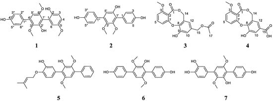
Figure 1.
The structures of compounds 1–7, obtained from Aspergillus candidus HM5-4: asperterphenylcins A–B (1,2), asperdiphenylcins A–B (3,4), 4″-deoxyterprenin (5), terphenyllin (6) and 3″-hydroxyterphenyllin (7).
2. Results
2.1. Structure Elucidation of New Compounds 1–4
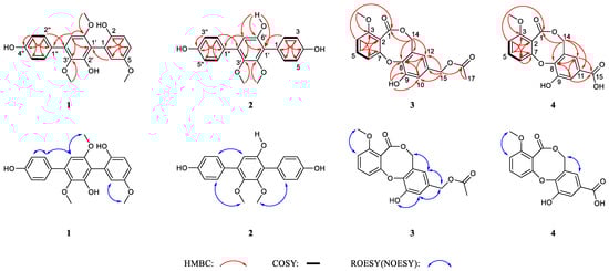
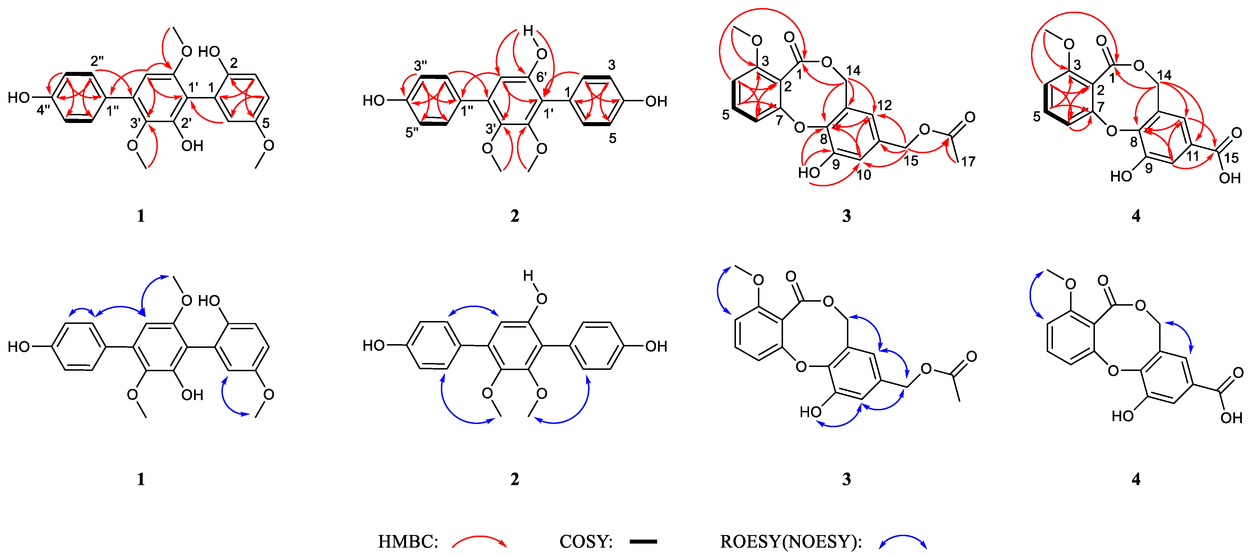
Asperterphenylcin A (1) was isolated as a yellow film. Its molecular formula C21H20O6 was deduced by the HRESIMS ion peak at m/z 391.1139 (calcd. 391.1152 for C21H20O6Na, [M + Na]+; Figure S8), revealing twelve indexes of hydrogen deficiency. The characteristic IR spectrum absorption peak at 3415 cm−1 suggested the existence of a hydroxy group. The 1H NMR spectrum showed eight aromatic protons (from δH 6.38 to δH 7.43) and three methoxy groups (δH 3.29, δH 3.64 and δH 3.73) (Table 1). The 13C NMR spectrum revealed twenty-one carbon signals with the aid of the DEPT and HSQC spectra, which were attributed to eight protonated sp2 carbons (including two overlapping signals at (δC 115.1, δH 6.84) and (δC 129.7, δH 7.43), (δC 103.0, δH 6.38), (δC 114.7, δH 6.76), (δC 115.3, δH 6.83), (δC 123.5, δH 6.69)), ten non-protonated sp2 carbons (δC 117.1, δC 125.0, δC 128.7, δC 132.4, δC 139.3, δC 145.2, δC 146.7, δC 148.1, δC 153.1 and δC 156.7) and three methoxy carbons (δC 55.6, δC 55.7 and δC 60.0) (Table 1). Consideration of the co-isolated compounds 5–7 indicated that compound 1 was most likely a p-terphenyl derivative. The 1H-1H COSY correlations between δH 7.43 (2H, d, J = 8.0 Hz, H-2″/H-6″) and δH 6.84 (2H, d, J = 8.0 Hz, H-3″/H-5″) indicated the presence of a p-substituted benzene ring. The 1,2,5-trisubstituted benzene ring (benzene ring numbering) was established according to the 1H-1H COSY correlation between δH 6.69 (1H, d, J = 8.0 Hz, H-4) and δH 6.76 (1H, d, J = 8.0 Hz, H-3), with the aid of a key HMBC correlation between δH 6.83 (1H, s, H-6) and C-4 (δC 123.5), as shown in Figure 2. Finally, the remaining aromatic proton δH 6.38 (1H, s, H-5′) indicated the presence of a penta-substituted benzene ring. According to the molecular formula of 1, the three methoxy groups contained three oxygen atoms, and the remaining oxygen atoms indicated the substitution of three phenolic hydroxyl groups. Ultimately, the positions of these substituent groups were determined, which were confirmed by the key HMBC correlations between H-5′ (δH 6.38) and C-1′ (δC 117.1), C-3′ (δC 139.3), C-6′ (δC 153.1) and C-1″ (δC 128.7), between H-2″ (δH 7.43) and C-4″ (δC 156.7) and C-4′ (δC 132.4), between H-3 (δH 6.76) and C-1 (δC 125.0) and C-5 (δC 146.7), and between H-4 (δH 6.69) and C-2 (δC 145.2) and C-6 (δC 115.3), along with the ROESY correlations between H-5′ (δH 6.38)/H-2″ (δH 7.43), H-5′ (δH 6.38)/H-6′-OCH3 (δH 3.64) and H-6 (δH 6.83)/H-5-OCH3 (δH 3.73). Thus, the structure of compound 1 was established as depicted in Figure 1.
Table 1.
1H NMR (500 MHz) and 13C NMR (125 MHz) data of compounds 1–2.
Figure 2.
Key HMBC, 1H-1H COSY and ROESY(NOESY) correlations of compounds 1–4.
Asperterphenylcin B (2) was isolated as a yellowish oil with a molecular formula C20H18O5, as determined by HRESIMS data, which displayed the ion peak at m/z 361.1039 (calcd. 361.1046 for C20H18O5Na, [M + Na]+; Figure S18). A comparison of the 1H and 13C NMR spectra for compounds 2 and 1 implied they have similar structure, except that 2 has one less methoxy group than 1 (Table 1). The mass of 2 was 30 units lower than that of 1, which further confirmed the above speculation. The 1H-1H COSY correlations between δH 7.13 (H-2/H-6)/δH 6.77 (H-3/H-5) and δH 7.31 (H-2″/H-6″)/δH 6.81 (H-3″/H-5″) indicated the presence of two p-substituted benzene rings. Thus, the remaining aromatic proton δH 6.56 (1H, s, H-5′) also revealed the presence of penta-substituted benzene rings. The key HMBC correlations between 6′-OH (δH 9.06, s) and C-1′ (δC 121.9), C-5′ (δC 111.2) and C-6′ (δC 150.8) indicated that there is a hydroxyl substitution rather than a methoxy group at the C-6′ (δC 150.8) position. Furthermore, the HMBC correlations between H-2/H-6 (δH 7.13) and C-1′ (δC 121.9) and C-4 (δC 156.0), and between H-2″/H-6″ (δH 7.31) and C-4′ (δC 133.7) and C-4″ (δC 156.6) indicated that there were two hydroxyl groups at the C-4 and C-4″ positions, respectively. Finally, the remaining two methoxy groups were fused at the C-2′ and C-3′ positions which was evidenced by the remaining key HMBC correlations between H-2′-OCH3 (δH 3.49) and C-2′ (δC 151.5) and between H-3′-OCH3 (δH 3.45) and C-3′ (δC 142.9), respectively (Figure 2).
Asperdiphenylcin A (3) was obtained as an orange solid. Its molecular formula was determined as C18H16O7, with eleven degrees of unsaturation based on the HRESIMS data at m/z 367.0790, (calcd. 367.0788 for C18H16O7Na, [M + Na]+; Figure S28). The IR spectrum indicated typical absorption bands for hydroxyl (3445 cm−1) and ester carbonyl (1737 cm−1) groups. The 1H NMR spectrum showed the presence of one methyl (δH 2.04), one methoxy (δH 3.85), two oxymethylenes (δH 5.06 and 4.92) and five aromatic protons (δH 6.63, 6.78, 6.95, 7.09 and 7.56) (Table 2). The 13C NMR spectrum classified by the DEPT and HSQC spectra showed the resonances of one methyl (δC 20.7), one methoxyl (δC 56.3), two oxygenated methylenes (δC 64.7 and 68.2), five protonated sp2 carbons (δC 109.7, 114.2, 117.4, 119.1 and 133.9), seven non-protonated sp2 carbons (δC 115.0, 127.7, 132.7, 143.3, 148.8, 152.0 and 156.7) and two ester-carbonyls (δC 166.2 and 170.1) (Table 2). A detailed analysis of the 1D and 2D NMR data revealed that 3 most resembled barceloneic lactone B [21], which was obtained from the marine fungal strain Aspergillus sp. They shared the same molecular skeleton and had a diphenyl ether skeleton with an eight-membered lactone ring. The aromatic protons at δH 7.09 (1H, d, 8.4, H-4), δH 7.56 (1H, t, 8.3, H-5) and δH 6.78 (1H, d, 8.1, H-6) and the further observed 1H-1H COSY correlation signals between H-4/H-5/H-6 indicated the presence of a 1,2,3-trisubstituted benzene ring in 3. The existence of another 1,2,3,5-tetrasubstituted benzene ring was indicated by the remaining two aromatic protons at δH 6.95 (1H, s, H-10) and δH 6.63 (1H, s, H-12) and the key HMBC correlations between δH 6.95 (1H, s, H-10) and C-8 (δC 143.3) and C-12 (δC 119.1). Based on the downfield shifts of C-7 (δC 152.0) and C-8 (δC 143.3), an oxygen bridge between the C-7 and C-8 positions was deduced. According to the long-range HMBC correlation between δH 7.09 (1H, d, 8.4, H-4) and C-1 (δC 166.2) and the key HMBC correlations between δH 5.06 (2H, s, H-14) and C-1 (δC 166.2), C-8 (δC 143.3), C-13 (δC 127.7) and C-12 (δC 119.1), the presence of a lactone ring between C-2 and C-14 was determined. Furthermore, the remaining methyl (δC 20.7), oxygenated methylene (δC 64.7) and ester-carbonyl (δC 170.1) indicated the presence of a methyl acetate group, which was further confirmed at the C-11 position by key HMBC correlations between δH 4.92 (2H, s, H-15) and C-10 (δC 117.4), C-11 (δC 132.7), C-12 (δC 119.1) and C-16 (δC 170.1), along with the correlation between δH 2.04 (3H, s, H-17) and C-16 (δC 170.1). Ultimately, the planar structure of 3 was elucidated as shown in Figure 1, which was further determined using other key HMBC correlations, shown in Figure 2.
Table 2.
1H NMR and 13C NMR data of compounds 3–4.
Asperdiphenylcin B (4) was obtained as a white crystal. Its molecular formula was determined to be C16H12O7 by the HRESIMS data at m/z 655.1028 (calcd. 655.1058 for C32H24O14Na, [2M + Na]+; Figure S38), indicating the presence of two carbon and two hydrogen atoms less than 3, with eleven degrees of unsaturation. The IR spectrum of 4 also showed the typical absorption bands of hydroxyl (3422 cm−1) and ester carbonyl (1736 cm−1) groups. A comprehensive analysis of the 1H NMR and 13C NMR spectra of 4 revealed that the NMR data of 4 almost resembled those of 3, indicating that they almost shared an identical structure skeleton (Table 2). Specifically, the characteristic peaks in the 13C NMR spectrum of five protonated sp2 carbons (δC 110.8, 115.2, 119.4, 122.8 and 134.5), seven non-protonated sp2 carbons (δC 116.6, 127.9, 128.9, 148.3, 149.8, 152.9 and 158.3), one oxygenated methylene (δC 69.0) and one ester-carbonyl (δC 166.4) in 4 indicated the same diphenyl ether skeleton with an eight-membered lactone ring as in 3, which was further confirmed by the similar key HMBC correlations in Figure 2. The only difference between 4 and 3 was the side chain at C-11, in the light of HMBC correlations between H-10 (δH 7.65) and C-15 (δC 166.6) and between H-12 (δH 7.39) and C-15 (δC 166.6), suggesting that there was a carboxyl group attached to the C-11 in 4 instead of the acetoxymethyl group observed in 3. Thus, the structure of 4 was assigned (Figure 1).
The known compounds 4″-deoxyterprenin (5) [22], terphenyllin (6) [23] and 3″-hydroxyterphenyllin (7) [14] were identified by a comparison of their MS and NMR data with data previously reported in the literature.
2.2. Bioassays of Compounds
2.2.1. Cytotoxic Detection Activity
All the compounds mentioned above were tested for their cytotoxic activities against human myeloid leukemia cells (K562), human liver cancer cells (BEL-7402), human gastric cancer cells (SGC-7901), human non-small cell lung cancer cells (A549) and human HeLa cervical cancer cells. As shown in Table 3, 4″-deoxyterprenin (5) and 3″-hydroxyterphenyllin (7) exhibited certain inhibitory activities against these five types of cancer cells. Compared to the cytotoxic activity of 7, the cytotoxic activity of 5 was much stronger. The IC50 values of 7 were 6.69 ± 0.12 µM against BEL-7402, 3.67 ± 0.17 µM against SGC-7901, and 3.32 ± 0.10 µM against A549, which was close to the IC50 values of positive control cisplatin (4.02 ± 0.06 µM against BEL-7402, 4.11 ± 0.02 µM against SGC-7901, and 1.93 ± 0.02 µM against A549; Table 3). Comparing the structure of compounds 5 and 7 revealed that 5’s cytotoxic activity was greatly enhanced, probably because of the exist of the isopentene group in 5.
Table 3.
Cytotoxic activities of compounds 1–7.
2.2.2. α-Glucosidase Inhibitory Activity
All the above-mentioned compounds were subjected to an α-glucosidase inhibitory activity test by a reported method [24], using genistein as a positive control. The results indicated that compounds 2 and 6 displayed more potent inhibitory activity (IC50 values were 1.26 ± 0.19 and 2.16 ± 0.44 µM) than genistein (IC50 value was 13.04 ± 2.56 µM; Table 4). The inhibitory activity of compound 7 was comparable to that of the positive drug genistein. Compound 1 had the weakest inhibitory activity, but it was still 6.7-times more active than acarbose. As shown in Table 4, all the diphenyl ether derivatives (3 and 4) exhibited no α-glucosidase inhibitory activity and all the p-terphenyl derivatives (1, 2, 6 and 7) except 5 exhibited α-glucosidase inhibitory activity. Comparing the structure of p-terphenyl derivatives 1, 2, 5, 6 and 7 revealed that the α-glucosidase inhibitory activity of 5 completely disappeared, probably because the hydrogen in the hydroxyl group was substituted by an isopentene group in 5.
Table 4.
α-Glucosidase inhibitory activities of compounds 1–7.
2.2.3. Antifungal Activity
The antifungal activities of three known compounds (5–7) were determined by the filter paper disc agar diffusion method. The results revealed that only compound 5 showed inhibitory activity against N. dimidiatum, which can cause a serious disease of stem canker in red-fleshed dragon fruit (Hylocereus polyrhizus); the diameter of its inhibition zone was 31.67 ± 2.36 mm. Carbendazim, as a positive control, displayed inhibitory activity against N. dimidiatum; the diameter of its inhibition zone was 55.00 ± 0.82 mm.
3. Materials and Methods
3.1. General Experimental Procedures
The NMR spectra were recorded on a Bruker AVIII-500 NMR spectrometer (Bruker Corporation, Karlsruhe, Germany) and Bruker DRX-600 spectrometer (Bruker Biospin AG, Fällanden, Germany). The chemical shifts of 1H NMR (500 MHz, 600 MHz) and 13C NMR (125 MHz, 150 MHz,) data were shown in δ (ppm) and referenced against the solvent signal (DMSO-d6, δH 2.50 and δC 39.52; acetone-d6, δH 2.05 and δC 29.84). HRESIMS data were measured on an API QSTAR Pulsar mass spectrometer (Bruker, Bremen, Germany). UV and IR data were tested on a UV-2550 spectrometer (Shimadzu, Kyoto, Japan) and Nicolet 380 Infrared Spectrometer (Thermo Electron Corporation, Madison, WI, USA), respectively. The semi-preparative HPLC was conducted on a Waters 1525 HPLC equipped with an XBridge C18 column (5 μm, 250.0 mm × 10.0 mm; Waters Corporation, Milford, MA, USA). Thin-layer chromatography (TLC) was conducted on pre-coated glass plates (silica gel GF254, Qingdao Marine Chemical Inc., Qingdao, China). Column chromatography (CC) was conducted on silica gel (45–75 µm; Qingdao Marine Chemical Inc., Qingdao, China).
3.2. Fungal Material and Fermentation
The marine fungus A. candidus HM5-4 was isolated from sponges collected from the coast of Lingao County, Hainan Province. The molecular sequence of the strain was cloned using the primers ITS1 (TCCGTAGGTGAACCTGCGG) and ITS4 (TCCTCCGCTTATTGATATGC), and the ITS sequence of the strain was determined. The sequences were submitted to the BLAST database in NCBI (sequence number: OP550289) and were found to be 100% homologous to A. candidus (MH398542.1). Therefore, the fungus was identified as Aspergillus candidus, together with the colony morphology of the fungus. The fungal strain was preserved in the South China Sea Marine Fungus Bank, Institute of Tropical Bioscience and Biotechnology, Chinese Academy of Tropical Agriculture Sciences.
For chemical investigations, the marine fungal strain A. candidus HM5-4 was incubated on PDA medium (consisting of 200.0 g/L potato, 20.0 g/L glucose, 20.0 g/L agar, and 1000.0 mL deionized water) at 28 °C after being obtained from the South China Sea Marine Fungus Bank (maintained at −80 °C in an ultra-low temperature freezer). After three days, three pieces of media with fungi were transferred aseptically to three Erlenmeyer flasks with PDB media (consisting of 200.0 g/L potato, 20.0 g/L glucose, and 1000.0 mL deionized water) and incubated on a rotary shaker (180 rpm) at 28 °C for 72 h. Then, 2.0 mL of seed solution was added to one hundred 1000 mL Erlenmeyer flasks with rice solid media (each flask contained 80.0 g rice and 120.0 mL artificial seawater) for fermentation, respectively. These Erlenmeyer flasks were cultivated under static conditions at 28 °C for 30 days.
3.3. Extraction and Isolation
The whole fermentation cultures of the marine fungal strain A. candidus HM5-4 were ultrasonically extracted with ethyl acetate (EtOAc) at room temperature three times. The filtrates were combined and evaporated in vacuo to yield the crude extract of ethyl acetate (120.0 g). Then, the crude extract was homogeneously dispersed in water and extracted three times with petroleum ether, EtOAc and n-butanol, respectively, and then the extracts were evaporated in vacuo to obtain petroleum ether extract (12.0 g), EtOAc extract (10.0 g) and n-butanol extract (2.0 g). Then, the crude extract of EtOAc (10.0 g) was chromatographed using silica gel column chromatography (CC) via the gradient elution of petroleum ether/EtOAc (1:0, 9:1, 8:2, 7:3, 6:4, 1:1, 3:7, and 0:1, v/v) and CH2Cl2/MeOH (4:1 and 1:1, v/v) to give 21 fractions (Fr.1–Fr.21). Fr.6 (250.6 mg) was fractionated by Sephadex LH-20 CC (eluted with 100% MeOH) to give compound 5 (3.4 mg). Fr.13 (1.3 g) was re-chromatographed on silica gel CC eluted with petroleum ether/EtOAc (5:2, v/v) to give 8 subfractions (Fr.13.1–Fr.13.8). Fr.13.6 (501.7 mg) was fractionated by Sephadex LH-20 CC eluted with 100% MeOH to afford ten subfractions (Fr.13.6.1–Fr.13.6.10). Fr.13.6.4 (108.8 mg) was subjected to silica gel CC eluted with CH2Cl2/MeOH (110:1, v/v) to give 5 subfractions (Fr.13.6.4.1–Fr.13.6.4.5). Fr.13.6.4.2 (9.2 mg) was further purified by semi-preparative reverse-phase HPLC eluted with 55% MeOH to obtain compound 1 (2.2 mg tR = 9.3 min). Fr.13.6.4.5 (4.5 mg) was re-chromatographed on silica gel CC eluted with CH2Cl2/MeOH (85:1; v/v) to yield compound 2 (2.5 mg). Fr.13.8 (572.9 mg) was fractionated by Sephadex LH-20 CC eluted with 100% MeOH to afford six subfractions Fr.13.8.1–Fr.13.8.6. Subfraction Fr.13.8.2 (16.8 mg) was further re-chromatographed on silica gel CC eluted with CH2Cl2/MeOH (130:1; v/v) to obtain compound 3 (2.4 mg). Fr.15 (4.6 g) was chromatographed over silica gel CC via the gradient elution of petroleum ether/EtOAc (1:0, 20:1, 10:1, 3:1, 1:1, and 0:1, v/v) to give 13 subfractions (Fr.15.1–Fr.15.13). Fr.15.9 (135.0 mg) was re-chromatographed on silica gel CC eluted with petroleum ether/CH2Cl2/MeOH (25:10:3; v/v/v) to yield compound 6 (95.1 mg). Fr.15.10 (1.9 g) was fractionated by Sephadex LH-20 CC eluted with 100% MeOH to yield ten subfractions Fr.15.10.1–Fr.15.10.10. Fr.15.10.6 (276.1 mg) was further re-chromatographed on silica gel CC eluted with petroleum ether/EtOAc (12:5; v/v) to afford 3 fractions (Fr.15.10.6.1–Fr.15.10.6.3). Fr.15.10.6.2 (12.5 mg) was purified by semi-preparative reverse-phase HPLC eluted with 45% MeOH to yield compound 7 (5.6 mg tR = 8.6 min). Fr.15.10.6.3 (15.1 mg) was re-chromatographed on silica gel CC eluted with CH2Cl2/MeOH (110:1; v/v) to yield compound 4 (2.1 mg).
Asperterphenylcin A (1): yellow film; UV (MeOH) λmax (log ε): 225 (2.79), 247 (2.55), 276 (2.80) nm; IR(KBr) νmax: 3415, 2929, 1612, 1398, 1236, 1076, 817 cm−1; 1H and 13C NMR data see Table 1; HRESIMS m/z 391.1139 [M + Na]+ (calculated for C21H20O6Na, 391.1152).
Asperterphenylcin B (2): yellowish oil; UV (MeOH) λmax (log ε): 227 (2.81), 247 (2.60), 268 (2.83) nm; IR(KBr) νmax: 3415, 2925, 1608, 1521, 1461, 1398, 1244, 1054, 835 cm−1; 1H and 13C NMR data see Table 1; HRESIMS m/z 361.1039 [M + Na]+ (calculated for C20H18O5Na, 361.1046).
Asperdiphenylcin A (3): orange solid; UV (MeOH) λmax (log ε): 223 (2.77), 257 (2.24), 284 (2.55) nm; IR(KBr) νmax: 3445, 2937, 1737, 1602, 1459, 1280, 1243, 1079 cm−1; 1H and 13C NMR data see Table 2; HRESIMS m/z 367.0790 [M + Na]+ (calculated for C18H16O7Na, 367.0788).
Asperdiphenylcin B (4): white solid; UV (MeOH) λmax (log ε): 222 (2.79), 241 (2.58), 246 (2.59), 268 (2.15), 291 (2.48) nm; IR(KBr) νmax: 3422, 1736, 1602, 1479, 1243, 1078, 763, 414cm−1; 1H and 13C NMR data see Table 2; HRESIMS m/z 655.1028 [2M + Na]+ (calculated for C32H24O14Na, 655.1058).
3.4. Cytotoxic Assay
All above-mentioned compounds were evaluated for their cytotoxicity in five human tumor cell lines: K562, BEL-7402, SGC-7901, A549 and HeLa cells which were bought from the Cell Bank of Type Culture Collection of the Shanghai Institute of Cell Biology, Chinese Academy of Sciences, using modified MTT methods [25]. The details of this are described in our previous paper [19]. The positive control was cisplatin.
3.5. α-Glucosidase Inhibition Assay
All abovementioned compounds were measured for their inhibitory effects against α-glucosidase, using PNPG as the substrate. The enzyme inhibitory assay was carried out by a formerly described method, with some modifications [24]. Compounds were dissolved in DMSO and six concentration gradients were set in turn. All the assays were performed in 0.1 M sodium phosphate buffer (PH = 6.8). The 10 μL sample was mixed with 100 μL α-glucosidase solution (0.2 U/mL, Sigma, Kanagawa, Japan) and shaken well, then added to a 96-well plate and incubated at 37 °C for 15 min. Subsequently, 40 μL of 2.5 mM 4-nitrophenyl-α-D-glucopyranoside was added and further incubated at 37 °C for 15 min. DMSO was used as a control and the blank wells contained buffer in place of substrate. The OD values were tested at 405 nm with a microplate reader. The positive control was genistein. The percentage inhibition was calculated by the following equation:
% inhibition = ((ODcontrol − ODsample)/(ODcontrol − ODblank)) × 100
3.6. Antifungal Activity Test
The inhibitory activities of some compounds against six phytopathogenic fungi (Fusarium oxysporum f. sp. cubense, Colletotrichum gloeosporioides, Thielaviopsis paradoxa, Bipolaris cactivora (Petrak) Alcorn, Neoscytalidium dimidiatum, and Colletotrichum scovillei) were evaluated using the filter paper disc agar diffusion method [26]; 250 μL of the fungal solution was aspirated onto the corresponding solid medium and spread evenly with a cotton swab. Samples were dissolved in MeOH to prepare solutions of 1.0 mg/mL and 3.0 mg/mL concentrations. Carbendazim (10 μL, 1.0 mg/mL) was used as a positive control and MeOH (10 μL) was used as negative control. The diameter of the inhibition zones of the plates cultured at 37 °C for 24 h were measured including the 6 mm disc diameter. Experiments were performed in triplicate. The results are showed as the mean value ± SD.
4. Conclusions
In summary, two undescribed p-terphenyl derivatives, asperterphenylcins A–B (1–2), and two undescribed diphenyl ether derivatives, asperdiphenylcins A–B (3–4), together with three previously described p-terphenyl derivatives—4″-deoxyterprenin (5), terphenyllin (6), and 3″-hydroxyterphenyllin (7)—were obtained from the crude EtOAc extract of a solid-rice culture of a marine sponge-derived fungus A. candidus HM5-4. Bioassays were used to evaluate its cytotoxic activity, α-glucosidase inhibitory activity and antifungal activity. The cytotoxic activity tests exhibited that 4″-deoxyterprenin (5) and 3″-hydroxyterphenyllin (7) showed certain inhibitory activities. Compared to the cytotoxic activity of 7, the cytotoxic activity of 5 was much stronger. The IC50 values of 7 were 6.69 ± 0.12 µM against BEL-7402, 3.67 ± 0.17 µM against SGC-7901, and 3.32 ± 0.10 µM against A549, which was close to the IC50 values of the positive control cisplatin (4.02 ± 0.06 µM against BEL-7402, 4.11 ± 0.02µM against SGC-7901 and 1.93 ± 0.02 µM against A549). Comparing the structures of compounds 5 and 7 revealed that the cytotoxic activity of 5 was greatly enhanced, probably because of the exist of an isopentene group in 5. The results of the antifungal assay showed that compound 5 has an obvious inhibitory effect on N. dimidiatum, which can cause a serious disease of stem canker in red-fleshed dragon fruit (Hylocereus polyrhizus); the diameter of its inhibition zone was 31.67 ± 2.36 mm. The bioassay results for the α-glucosidase inhibitory activity showed that compounds 2, 6 and 7 exhibited potent α-glucosidase inhibitory activity, with IC50 values ranging from 1.26 ± 0.44 to 13.22 ± 0.55 μM. To the best of our knowledge, polyhydroxy phenolic compounds usually possess potent α-glucosidase inhibitory activity. Comparing the structure of p-terphenyl derivatives 1, 2, 5, 6 and 7 revealed that the α-glucosidase inhibitory activity of 5 completely disappeared, probably because the hydrogen in the hydroxyl group was substituted by an isopentene group in 5. In conclusion, p-terphenyl derivatives 2, 6 and 7, with potent α-glucosidase inhibitory activity, possess the potential to be developed as novel α-glucosidase inhibitors and need to be further studied.
Supplementary Materials
The following supporting information can be downloaded at: https://www.mdpi.com/article/10.3390/md22010013/s1, Figures S1–S40: 1D, 2D NMR, MS, UV, and IR spectra of compounds 1–4.
Author Contributions
Conceptualization, Y.Z. and H.D.; methodology, Y.Z. and H.D.; software, Y.Z. and H.D.; validation, S.W., H.P., W.Z. and H.D.; formal analysis, Y.Z. and H.W.; investigation, S.W. and W.C.; resources, Y.Z. and H.D.; data curation, S.W. and W.C.; writing—original draft preparation, S.W., Y.Z. and H.W.; writing—review and editing, S.W., H.P., W.Z. and H.C.; visualization, S.W., H.P. and W.Z.; supervision, Y.Z. and H.D.; project administration, Y.Z. and H.D.; funding acquisition, Y.Z. and H.D. All authors have read and agreed to the published version of the manuscript.
Funding
This work was financially supported by the Natural Science Foundation of Hainan (322MS131), the National Natural Science Foundation of China (41776093), and the Financial Fund of the Ministry of Agriculture and Rural Affairs, China (NFZX2021).
Institutional Review Board Statement
Not applicable.
Informed Consent Statement
Not applicable.
Data Availability Statement
The authors declare that all relevant data supporting the results of this study are available within the article and its Supplementary Materials file, or from the corresponding authors upon request.
Conflicts of Interest
The authors declare no conflicts of interest.
References
- Xu, K.; Gao, Y.; Li, Y.L.; Xie, F.; Zhao, Z.T.; Lou, H.X. Cytotoxic p-terphenyls from the endolichenic fungus Floricola striata. J. Nat. Prod. 2018, 81, 2041–2049. [Google Scholar] [CrossRef] [PubMed]
- Chen, W.H.; Zhang, J.W.; Qi, X.; Zhao, K.; Pang, X.Y.; Lin, X.P.; Liao, S.R.; Yang, B.; Zhou, X.F.; Liu, S.W.; et al. p-Terphenyls as anti-HSV-1/2 agents from a deep-sea-derived Penicillium sp. J. Nat. Prod. 2021, 84, 2822–2831. [Google Scholar] [CrossRef] [PubMed]
- Takahashi, S.; Suda, Y.; Nakamura, T.; Matsuoka, K.; Koshino, H. Total synthesis of kehokorins A–E, cytotoxic p-terphenyls. J. Org. Chem. 2017, 82, 3159–3166. [Google Scholar] [CrossRef] [PubMed]
- Li, W.; Li, X.B.; Lou, H.X. Structural and biological diversity of natural p-terphenyls. J. Asian Nat. Prod. Res. 2018, 20, 1–13. [Google Scholar] [CrossRef] [PubMed]
- Li, W.; Gao, W.; Zhang, M.; Li, Y.L.; Li, L.; Li, X.B.; Chang, W.Q.; Zhao, Z.T.; Lou, H.X. p-terphenyl derivatives from the endolichenic fungus Floricola striata. J. Nat. Prod. 2016, 79, 2188–2194. [Google Scholar] [CrossRef] [PubMed]
- Zhu, J.J.; Li, Z.Y.; Lu, H.H.; Liu, S.Q.; Ding, W.J.; Li, J.Z.; Xiong, Y.H.; Li, C.Y. New diphenyl ethers from a fungus Epicoccum sorghinum L28 and their antifungal activity against phytopathogens. Bioorg. Chem. 2021, 115, 105232. [Google Scholar] [CrossRef]
- Lin, Y.K.; Xie, C.L.; Xing, C.P.; Wang, B.Q.; Tian, X.X.; Xia, J.M.; Jia, L.Y.; Pan, Y.N.; Yang, X.W. Cytotoxic p-terphenyls from the deep-sea-derived Aspergillus candidus. Nat. Prod. Res. 2021, 35, 1627–1631. [Google Scholar] [CrossRef]
- Wang, D.Y.; Wang, Y.; Ouyang, Y.F.; Zhu, W.M. Cytotoxic p-terphenyls from a marine-derived Nocardiopsis species. J. Nat. Prod. 2019, 82, 3504–3508. [Google Scholar] [CrossRef]
- Choi, D.C.; Ki, D.W.; Kim, J.Y.; Lee, I.K.; Yun, B.S. p-Terphenyl glucosides from the culture broth of Phlebiopsis castanea. J. Antibiot. 2023, 76, 52–55. [Google Scholar] [CrossRef]
- Sofian, F.F.; Kikuchi, N.; Koseki, T.; Kanno, Y.; Uesugi, S.; Shiono, Y. Antioxidant p-terphenyl compound, isolated from edible mushroom, Boletopsis leucomelas. Biosci. Biotechnol. Biochem. 2022, 86, 300–304. [Google Scholar] [CrossRef]
- Guo, Z.K.; Abulaizi, A.; Huang, L.; Xiong, Z.J.; Zhang, S.Q.; Liu, T.M.; Wang, R. Discovery of p-terphenyl metabolites as potential phosphodiesterase PDE4D inhibitors from the coral-associated fungus Aspergillus sp. ITBBc1. Mar. Drugs 2022, 20, 679. [Google Scholar] [CrossRef] [PubMed]
- Xu, Y.C.; Wang, Y.; Wu, D.; He, W.W.; Wang, L.P.; Zhu, W.M. p-terphenyls from Aspergillus sp. GZWMJZ-055: Identification, derivation, antioxidant and α-glycosidase inhibitory activities. Front. Microbiol. 2021, 12, 654963. [Google Scholar] [CrossRef] [PubMed]
- Shin, H.J. Natural products from marine fungi. Mar. Drugs. 2020, 18, 230. [Google Scholar] [CrossRef] [PubMed]
- Yurchenko, E.A.; Menchinskaya, E.S.; Pislyagin, E.A.; Chingizova, E.A.; Girich, E.V.; Yurchenko, A.N.; Aminin, D.L.; Mikhailov, V.V. Cytoprotective activity of p-Terphenyl polyketides and flavuside B from marine-derived fungi against oxidative stress in Neuro-2a Cells. Molecules 2021, 26, 3618. [Google Scholar] [CrossRef] [PubMed]
- Peng, G.Y.; Kurtán, T.; Mándi, A.; He, J.; Cao, Z.Y.; Tang, H.; Mao, S.C.; Zhang, W. Neuronal modulators from the coral-associated fungi Aspergillus candidus. Mar. Drugs 2021, 19, 281. [Google Scholar] [CrossRef] [PubMed]
- Zhou, G.L.; Zhang, X.M.; Shah, M.; Che, Q.; Zhang, G.J.; Gu, Q.Q.; Zhu, T.J.; Li, D.H. Polyhydroxy p-terphenyls from a mangrove endophytic fungus Aspergillus candidus LDJ-5. Mar. Drugs 2021, 19, 82. [Google Scholar] [CrossRef]
- Wang, S.; Zeng, Y.B.; Yin, J.J.; Chang, W.J.; Zhao, X.L.; Mao, Y. Two new azaphilones from the marine-derived fungus Penicillium sclerotiorum E23Y−1A. Phytochem. Lett. 2022, 47, 76–80. [Google Scholar] [CrossRef]
- Wang, Z.; Zeng, Y.B.; Zhao, W.B.; Dai, F.H.; Chang, W.J.; Lv, F. Structures and biological activities of brominated azaphilones produced by Penicillium sclerotiorum E23Y−1A. Phytochem. Lett. 2022, 52, 138–142. [Google Scholar] [CrossRef]
- Zeng, Y.B.; Wang, Z.; Chang, W.J.; Zhao, W.B.; Wang, H.; Chen, H.Q.; Dai, F.H.; Lv, F. New azaphilones from the marine-derived fungus Penicillium sclerotiorum E23Y−1A with their anti-inflammatory and antitumor activities. Mar. Drugs 2023, 21, 75. [Google Scholar] [CrossRef]
- Zhao, W.B.; Zeng, Y.B.; Chang, W.J.; Chen, H.Q.; Wang, H.; Dai, F.H.; Lv, F. Potential α-glucosidase inhibitors from the deep-sea sediment-derived fungus Aspergillus insulicola. Mar. Drugs 2023, 21, 157. [Google Scholar] [CrossRef]
- Liu, S.S.; Lu, C.H.; Huang, J.J.; Shen, Y.M. Three new compounds from the marine fungal strain Aspergillus sp. AF119. Rec. Nat. Prod. 2012, 6, 334. [Google Scholar]
- Wei, H.; Inada, H.; Hayashi, A.; Higashimoto, K.; Pruksakorn, P.; Kamada, S.; Arai, M.; Ishida, S.; Kobayashi, P. Prenylterphenyllin and its dehydroxyl analogs, new cytotoxic substances from a marine-derived fungus Aspergillus candidus IF10. J. Antibiot. 2007, 60, 586–590. [Google Scholar] [CrossRef] [PubMed]
- Shan, T.J.; Wang, Y.Y.; Wang, S.; Xie, Y.Y.; Cui, Z.H.; Wu, C.Y.; Sun, J.; Wang, J.; Mao, Z.L. A new p-terphenyl derivative from the insect-derived fungus Aspergillus candidus Bdf-2 and the synergistic effects of terphenyllin. PeerJ 2020, 8, e8221. [Google Scholar] [CrossRef] [PubMed]
- Yang, L.; Yang, Y.L.; Dong, W.H.; Li, W.; Wang, P.; Cao, X.; Yuan, J.Z.; Chen, H.Q.; Mei, W.L.; Dai, H.F. Sesquiterpenoids and 2-(2-phenylethyl) chromones respectively acting as α-glucosidase and tyrosinase inhibitors from agarwood of an Aquilaria plant. J. Enzym. Inhib. Med. Chem. 2019, 34, 853–862. [Google Scholar] [CrossRef]
- Mosmann, T. Rapid colorimetic assay for cellular growth and survival: Application to proliferation and cytotoxicity assays. J. Immunol. Methods 1983, 65, 55–63. [Google Scholar] [CrossRef]
- Li, W.H.; Ding, L.J.; Li, J.; Wen, H.M.; Liu, Y.; Tan, S.L.; Yan, X.J.; Shi, Y.T.; Lin, W.H.; He, S. Novel antimycin analogues with agricultural antifungal activities from the sponge-associated actinomycete Streptomyces sp. NBU3104. J. Agr. Food. Chem. 2022, 70, 8309–8316. [Google Scholar] [CrossRef]


Disclaimer/Publisher’s Note: The statements, opinions and data contained in all publications are solely those of the individual author(s) and contributor(s) and not of MDPI and/or the editor(s). MDPI and/or the editor(s) disclaim responsibility for any injury to people or property resulting from any ideas, methods, instructions or products referred to in the content. |
© 2023 by the authors. Licensee MDPI, Basel, Switzerland. This article is an open access article distributed under the terms and conditions of the Creative Commons Attribution (CC BY) license (https://creativecommons.org/licenses/by/4.0/).