Article

Fucoidan from Fucus vesiculosus Inhibits Inflammatory Response, Both In Vitro and In Vivo

1
School of Biotechnology and Health Sciences, Wuyi University, Jiangmen 529000, China
2
3B’s Research Group, I3Bs—Research Institute on Biomaterials, Biodegradables and Biomimetics, University of Minho, Headquarters of the European Institute of Excellence on Tissue Engineering and Regenerative Medicine, AvePark, Parque de Ciência e Tecnologia, Zona Industrial da Gandra, Barco, 4805-017 Guimarães, Portugal
3
ICVS/3B’s–PT Government Associate Laboratory, Braga, 4710-057 Guimarães, Portugal
4
State Key Laboratory of Quality Research in Chinese Medicine, Institute of Chinese Medical Sciences, University of Macau, Macau, China
5
LAQV-REQUIMTE, Department of Chemistry, University of Aveiro, Campus de Santiago, 3810-193 Aveiro, Portugal
6
CICECO, Department of Chemistry, University of Aveiro, Campus de Santiago, 3810-193 Aveiro, Portugal
*
Authors to whom correspondence should be addressed.
These authors contributed equally to this work.
Mar. Drugs 2023, 21(5), 302; https://doi.org/10.3390/md21050302
Submission received: 21 October 2022 / Revised: 8 May 2023 / Accepted: 10 May 2023 / Published: 17 May 2023

Abstract

:
Fucoidan has been reported to present diverse bioactivities, but each extract has specific features from which a particular biological activity, such as immunomodulation, must be confirmed. In this study a commercially available pharmaceutical-grade fucoidan extracted from Fucus vesiculosus, FE, was characterized and its anti-inflammatory potential was investigated. Fucose was the main monosaccharide (90 mol%) present in the studied FE, followed by uronic acids, galactose, and xylose that were present at similar values (3.8–2.4 mol%). FE showed a molecular weight of 70 kDa and a sulfate content of around 10%. The expression of cytokines by mouse bone-marrow-derived macrophages (BMDMs) revealed that the addition of FE upregulated the expression of CD206 and IL-10 by about 28 and 22 fold, respectively, in respect to control. This was corroborated in a stimulated pro-inflammatory situation, with the higher expression (60 fold) of iNOS being almost completely reversed by the addition of FE. FE was also capable of reverse LPS-caused inflammation in an in vivo mouse model, including by reducing macrophage activation by LPS from 41% of positive CD11C to 9% upon fucoidan injection. Taken together, the potential of FE as an anti-inflammatory agent was validated, both in vitro and in vivo.

1. Introduction

Marine-origin byproducts may present an interesting source of unexplored anti-inflammatory compounds such as polyphenols, proteins, and sulfated polysaccharides (e.g., fucoidan). These marine-derived bioactive compounds may constitute interesting alternatives to currently available synthetic drugs, having potential protective effects over the pathogenesis of inflammatory diseases and lower side effects [1]. Fucoidan is a sulfated polysaccharide mainly composed of fucose units and other carbohydrate monomers as minor components [2]. It can be extracted from different species of brown algae, Fucus vesiculosus being the most reported and well-studied [3]. Fucoidan may present different biological activities such as antitumor, antiviral, anti-angiogenic, and anti-inflammatory [4,5,6,7]. Different structural characteristics have been associated with the biological activity of fucoidans, molecular weight and sulfation degree being the most described ones [8]. More recent studies have also related the bioactivities of fucoidan to sulfates position and branching degree [9,10].
Fucoidan is being investigated for short- and long-term inflammation treatment [11,12]. The anti-inflammatory mechanisms of action of fucoidans comprise antioxidant, transcription factors, adhesion molecules, matrix metalloproteinases, complement cascade properties, and it can also regulate pro-inflammatory enzymes and the expression of related genes [7]. The most discussed possible mechanism of action is the downregulation of MAPK and NF-κB signaling pathways, followed by a decrease in pro-inflammatory cytokines [12]. Different in vitro studies showed promising anti-inflammatory results for fucoidan from Fucus vesiculosus. For example, fucoidan decrease TNF-α and IL-1β levels in mouse macrophages (RAW 264.7 cell line) stimulated by lipopolysaccharide (LPS) [13]. The immunomodulatory properties of photopolymerizable fucoidan were also evaluated in human monocytes (THP-1 cell line) differentiated into macrophages, presenting similar activity as IL-10, by decreasing LPS- and IFN-γ-induced CD86 expression [14]. In LPS-stimulated BV2 microglia cells, fucoidan inhibited the production of nitric oxide (NO) and prostaglandin E2 (PGE2), and reduced the expression of inducible nitric oxide synthase (iNOS), IL-1β, and TNF-α, for example [15]. These anti-inflammatory properties of fucoidan were achieved by the suppression of NF-κB, MAPK, and AKT molecular pathways. A decreased production of pro-inflammatory cytokines (e.g., TNF-α, IL1β, IL-6) was also observed when human keratinocytes and Caco-2 cells were treated with fucoidan [16,17]. Likewise, the inhibition of COX enzymes and hyaluronidase by fucoidan have been observed in a concentration-dependent manner, similarly to the expression of MAPK p38 by LPS-stimulated human mononuclear U937 cells [18]. Recently, it has been proposed by Obluchinskaya et al. that fucoidan inhibited the protein denaturation observed in inflammation, with the effect depending on fucoidan concentration and being mainly associated with fucose contents, although the sulfate content may also play a role [19]. Fucoidan from Fucus vesiculosus has decreased neutrophil infiltration, as well as systemic inflammation, presenting lower levels of TNF-α and IL1β in mice [20]. Similarly, a fucoidan extract from the same species inhibited the recruitment of leucocytes in an inflammation rat model, apparently in a way not dependent on fucoidan’s chemical structure, but without association to P-selectin, in opposition to fucoidans from other brown algae species [21]. Fucoidan is also capable of reducing the inflammation (decreased levels of IL-1α, IL-1β, and IL-10 of bowel disease when orally administered [22]. Moreover, a fucoidan-based cream was capable of inhibiting carrageenan-induced edema in rats upon topical application in a dose-dependent manner, being comparable to a diclofenac gel [23].
All these studies were carried out using purified fucoidans, allowing for the acquisition of structure–function relationships. However, the term “fucoidan” encloses a group of polysaccharides composed of a fucose backbone chain, often sulfated, with a range of chemical characteristics (molecular weight, sugar profile, sulfation degree and pattern, among others), depending on the specific seaweed sample and extraction methodology [24]. Thus, the production of large-scale and commercially available fucoidan extracts, even at a pharmaceutical grade, hardly allows obtaining a unique bioactive structure, and different fucoidan extracts can correspond to different (groups of) chemical entities, which could prevent its anti-inflammatory properties. In this study, we selected a specific commercially available fucoidan extract from Fucus vesiculosus, herein represented as FE, hypothesizing that it contained the active structures able to influence in vitro and in vivo inflammation. Therefore, FE was chemically characterized, and its potential anti-inflammatory activity was assessed both in vitro using the RAW 264.7 cell line and bone-marrow-derived macrophages, and in vivo upon intraperitoneal injection in mice.

2. Results and Discussion

2.1. Structural Characterization of Fucoidan Extract (FE)

Molecular weight and sulfation degree are the most described physicochemical factors influencing fucoidan bioactivity [8]. Moreover, monosaccharide composition and sulfate position may also influence the final biological activity, recently being taken into consideration [9]. The brown seaweed species and specific reproductive phase of the used sample, as well as the extraction method, may be the possible triggers to influence these intrinsic properties [3,25,26]. Given this variability, with the structure–activity relationship not yet fully established, the biological activity of each new extract being produced should not be assumed, but confirmed.
Taking this into consideration, the fucoidan extract (FE), herein studied, was extensively characterized. The total carbohydrate content of FE was 66% (w/w) and had a sulfation degree of around 10% (Table 1). The monosaccharide composition was assessed, confirming that fucose was the main sugar (around 90% mol) (Table 1), with uronic acids (3.8 mol%), galactose (3.3 mol%), and xylose (2.4 mol%) present in smaller amounts. FE had a molecular weight of 70 kDa and a polydispersity (Mw/Mn) of 1.5, confirmed by the gel permeation chromatography (GPC) analysis (Table 1).
Due to the different fucoidan species and extraction methods, it is difficult to define specific and strict ranges for the parameters described above to meet the biological responses. The molecular weight of FE herein studied was within the range that others reported in the literature, from few kDa to over 100 kDa [3,10]. The total of carbohydrate content was slightly increased in this extract when compared to other extracts that have been previously characterized by our group (50–52.5% w/w) and the sulfation degree was lower (around 30% in the other extracts) [9]. Furthermore, this FE presented a higher proportion of Fuc and a lower proportion of other monosaccharides when compared to those same extracts (fucose 71.2–79.1% mol, uronic acids 9.8–15.3% mol, xylose 3.9–8.0% mol, and galactose 3.5–5.5% mol). These differences may be attributed to the purity of this FE, since the polysaccharide extract herein used was a pharmaceutical grade, with a purity of 98%.
To determine the type of glycosidic linkages and the sulfate group position along the polymer chain of FE, a methylation analysis was performed before and after desulfation. If a position is acetylated in the native polysaccharide and becomes methylated after desulfation, it is an indication of a sulfate residue at that position. Thus, FE was mainly sulfated in C-2 and C-4, since the glycosidic linkages 2,3,4-Fuc and 2,4-Fuc decreased after desulfation, and 3,4-Fuc, 2-Fuc, 3-Fuc, and 4-Fuc increased (Table 2). The residue of 2,3-Fuc was not disadvantaged by significant changes and, consequently, FE could be branched at C-2, with the linkage C-3 belonging to the main chain. The residues of the other sugars (t-Xyl, 2-Xyl, and t-Gal) did not show relevant modification in the residue content for native and desulfated FE. This procedure was performed to better understand the level of polymer branching and position of sulfate groups in fucose. The fine characterization of the chemical structure might require the chromatographic purification of FE to obtain fractions composed by (ideally) single chemical components, which is beyond the goal of the present study.

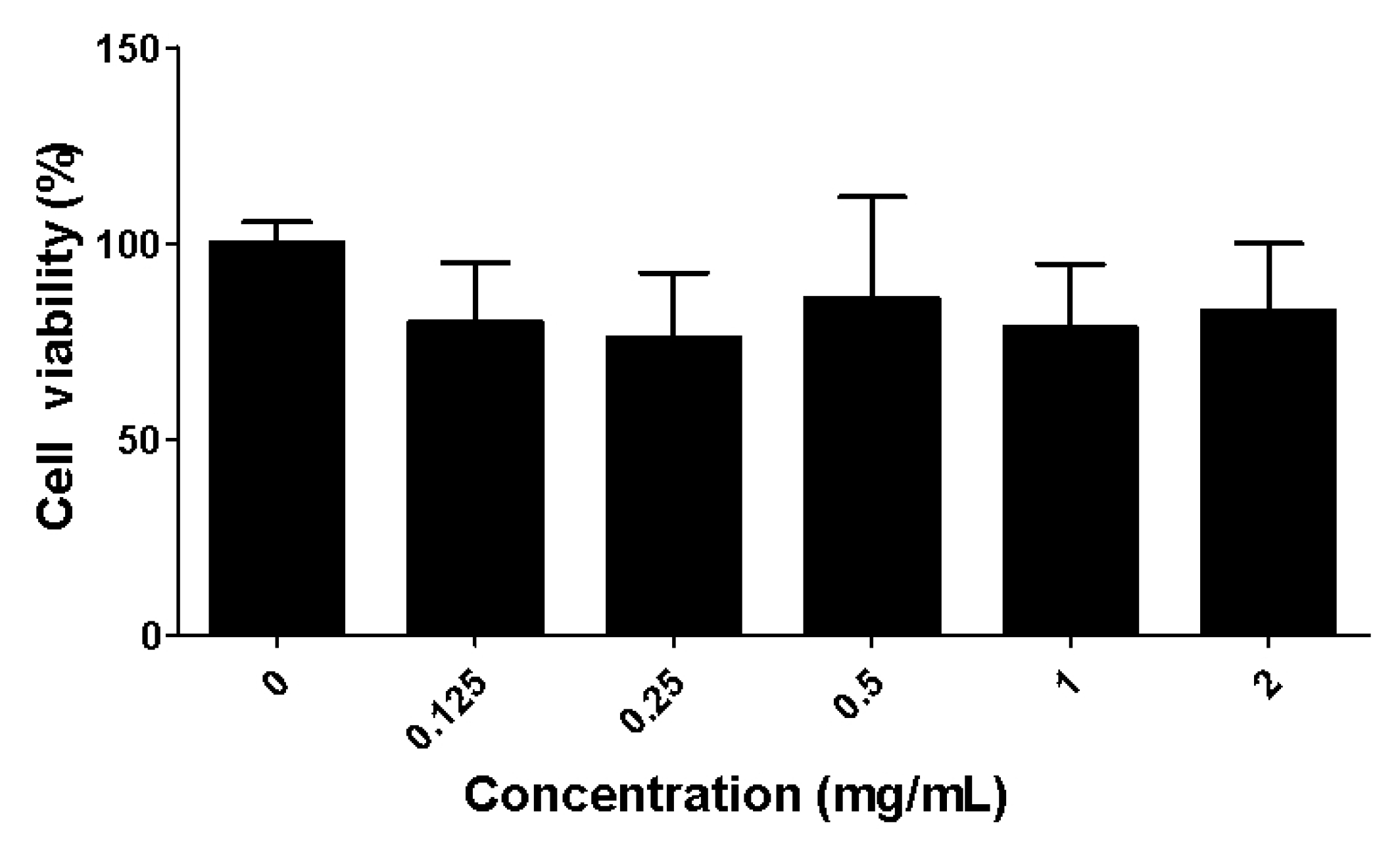
2.2. Toxicity of the Fucoidan Extract (FE) over RAW 264.7 Macrophages

RAW 264.7 murine macrophage cell line is often used as a first screening model of natural products’ bioactivity and to predict their potential effect [22]. The effects of FE on the viability of RAW 264.7 macrophages were assessed for different FE concentrations after 48 h of incubation (Figure 1). There is no positive correlation between cell viability and FE concentrations, and no significant differences were observed for all tested conditions. All further experiments were conducted using 0.1 mg/mL, where negligible cytotoxic effects were observed, since macrophage viability was above 70% (in accordance with ISO 10993-5). This same range of fucoidan concentrations (0.1 mg/mL) did not impair macrophage’ growth, as previously reported by others [13,23].

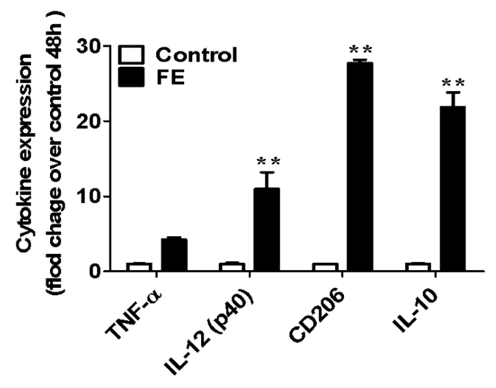
2.3. Expression of Pro- and Anti-Inflammatory Cytokines

Mouse bone-marrow-derived macrophages (BMDMs) are a kind of primary cells, extracted directly from the alive animal. Although RAW 264.7 cells are frequently used as models to assess (anti-)inflammatory response because they are widely available and easy to culture, evidence has revealed that the main biological process involving the cell cycle control, cytoskeleton reorganization, and apoptosis was significantly dissimilar from RAW 264.7 cells to BMDM [27]. Alongside that, BMDMs are proven to be more sensitive and a more realistic response to an inflammatory stimulus [28], thus being selected as the in vitro model in this study. After 48 h incubation with FE, different cytokines (TNF-α and IL-12 as pro-inflammatory, and CD206 and IL-10 as anti-inflammatory) were quantified [29]. TNF-α activates different cell signaling pathways and mediates the production of many other inflammatory mediators, participating in the initiation and progression of the inflammatory response [30]. IL-12 is produced early during infections, and comprises a heavy chain (p. 40, [31]) and a light chain (p. 35, [31]). This cytokine is associated with innate and adaptative immunity through the induction of IFN-γ. IL-10 is produced via a wide variety of activated immune cells, and its main actions are anti-inflammatory, inhibitory, or self-regulating [32]. CD206, a mannose receptor, is a membrane-bound protein, predominantly expressed by macrophages and dendritic cells, acts as a pattern recognition receptor that plays a role in innate and adaptive immunity [33,34].
The effect of FE on the expression of the above-mentioned cytokines by BMDM was studied (Figure 2). The presence of 100 µg/mL FE induced a slight increase in TNF-α expression with no statistically significant differences compared to the control condition. However, the expression of IL-12, CD206 and IL-10 was significantly higher than the control condition. Indeed, the expression of CD206 and IL-10 was much higher than the other two quantified cytokines, demonstrating the anti-inflammatory potential of FE.

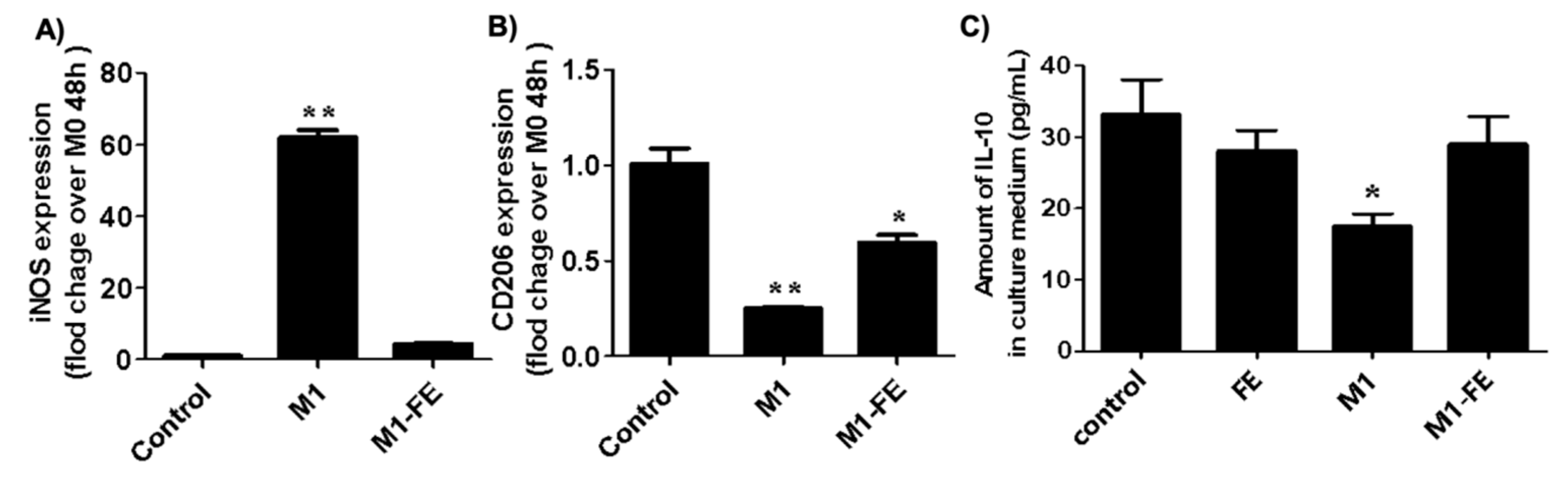
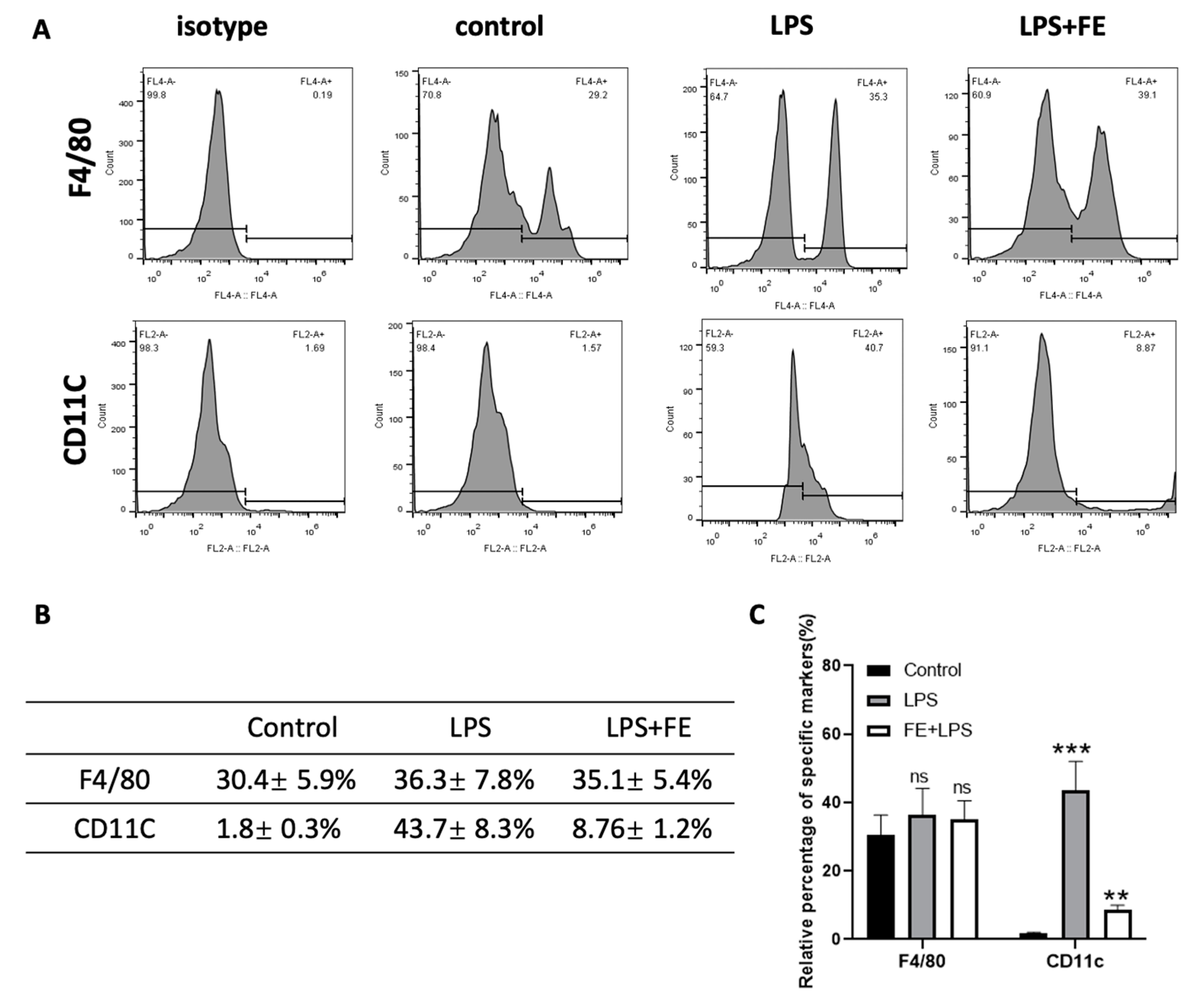
2.4. Reversed Inflammation in M1 Phenotype

Inflammatory stimuli such as LPS (lipopolysaccharides) induces macrophage activation and, consequently, the expression of cytokines that mediates different stages of inflammation [35,36]. Indeed, the inhibition or induction of cytokine production plays a crucial role in the control of inflammation. To assess the effects of FE over the expression of anti- and pro-inflammatory molecules such as inducible nitric oxide synthase (iNOs), CD206, and IL-10 in a simulated inflammatory situation, BMDMs were subject to LPS+IFN-γ stimulation for 12 h. iNOS is one of the direct consequences of an inflammatory process and a major mediator of inflammation in various cell types [37]. As observed in Figure 3A, the expression of iNOS was significantly increased after LPS+IFN-γ stimulation, as expected. This situation was reversed through the addition of FE, with expression levels similar to the basal condition. The addition of FE increased CD206 expression in response to LPS+IFN-γ stimulation. A similar behavior was observed for IL-10 protein, another anti-inflammatory cytokine. Previous studies reported the effect of fucoidan from Fucus vesiculosus at similar concentrations (i.e., 50–100 µg/mL) on different cell types, specifically RAW 264.7, Caco-2, and BV2 microglial cells [13,15,17]. These studies showed that fucoidan decreased the expression of pro-inflammatory cytokines after LPS-stimulation. Fucoidan from species other than Fucus vesiculosus also presented some promising results. Fucoidan extracts from Laminaria japonica reduced the expression of iNOS, TNF-α, and IL-6 [38,39]. In a particular study, Caco-2 cells were used, and both pro- and anti-inflammatory cytokine expressions were assessed [40]. A decreased expression of TNF-α and IL-1β, along with increased levels of IL-10 and IFN-γ, was observed for fucoidan from Sargassum hemiphyllum.
Herein, a decreased expression of pro-inflammatory and an increase in anti-inflammatory mediators validate the anti-inflammatory potential of FE in vitro, after settling a simulated inflammatory situation. These observations state the reversed inflammation capacity of this fucoidan extract (FE).

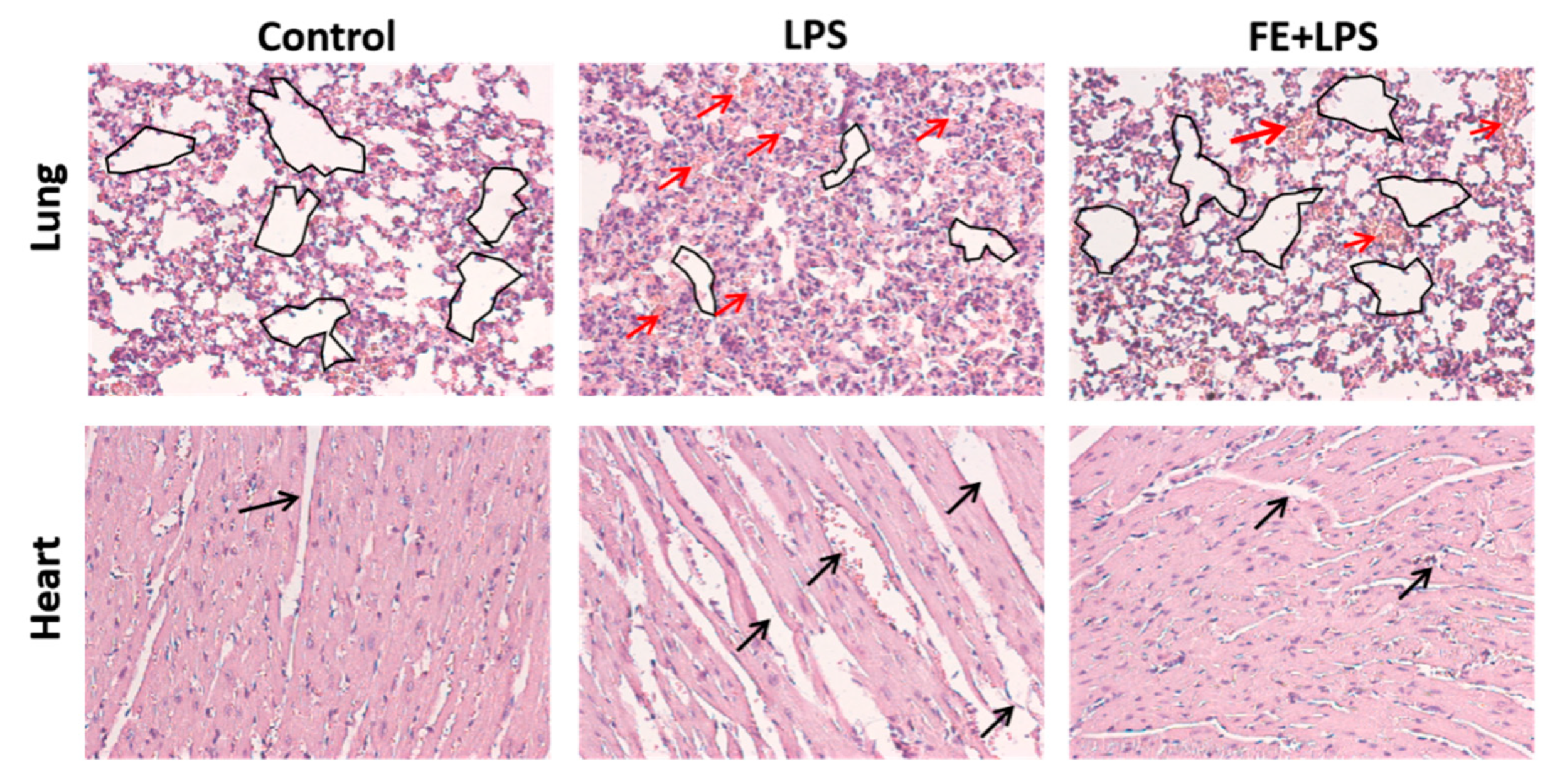
2.5. FE Anti-Inflammatory Response in a Mouse Model

The anti-inflammatory potential of FE was also assessed in vivo through the intraperitoneal injection of LPS in a mouse model. Histologically, the typical features of normal cardiac and lung tissues, or mild morphological changes, were observed in mice injected with FE after LPS stimulation (Figure 4).
These observations are in agreement with the ones of another study, whereby fucoidan administration attenuated myocardial damage [28]. To ascertain the influence of FE over tissue-specific macrophages, intraperitoneal macrophages were marked for the surface markers F4/80 and CD11C (Figure 5). Flow cytometry analysis showed that the F4/80 marker is highly expressed upon FE administration. Indeed, tissue-specific macrophages aid in maintaining homeostasis and triggering the immune system in response to a stimulus [41,42]. Oppositely, the integrin CD11C that is highly expressed in monocytes and macrophages, was downregulated when FE was injected, presenting values similar to the control condition (healthy mice). Therefore, FE did not evoke a CD11C-mediated response, which is an indicator of macrophage activation [43]. These observations indicate that the administration of FE induced a decrease in the inflammatory response upon LPS stimulation.

3. Materials and Methods

3.1. Materials

A pharmaceutical-grade fucoidan extract from Marinova (batch number—DPFVF2015505), herein referred to as FE, with a purity of 98%, was purchased and used as received. Lipopolysaccharides (LPS from E. coli 055:B5) were purchased from InvivoGen Co., Ltd. (San Diego, CA, USA). Enzyme-linked immunosorbent assay (EISA) kits for murine IL-10 were purchased from NeoBioscience Co., Ltd. (Shenzhen, China). FITC anti-mouse F4/80 antibody and APC anti-mouse CD11C antibody for flow cytometric analysis were purchased from Biolegend (San Diego, CA, USA). Recombinant murine IFN-γ was purchased from Peprotech (Cranbury, NJ, USA). The mouse macrophage cell line Raw 264.7 was purchased from American-Type Culture Collection (ATCC, Manassas, VA, USA), and maintained according to the culture guidelines. All reagents used for the cell culture were purchased from Gibco Life Technology (Carlsbad, CA, USA).

3.2. FE Characterization

3.2.1. Molecular Weight

FE molecular weight was determined by GPC using a methodology previously described [9]. Briefly, a Malvern Viscotek TDA 305 (Malvern, UK) system composed of a set of four columns: pre-column Suprema 5 µm 8 × 50 S/N 3111265, Suprema 30 Å 5 µm 8 × 300 S/N 3112751, Suprema 1000 Å 5 µm 8 × 300 S/N 3,112,851 PL, and Aquagel-OH MIXED 8 µm 7.5 × 300 S/N 8M-AOHMIX-46-51, followed by a refractometer (RI-Detector 8110, Bischoff), right-angle light scattering, and viscometer detectors. For the eluent, a solution of 0.1 M NaN3 and 0.01 M NaH2PO4 with a pH = 6.6 was used at a flow rate of 1 mL min−1, and the system kept at 30 °C. To calibrate elution times, a commercial set (Varian ®, Palo Alto, CA, USA) of pullulans with narrow polydispersity and Mp (molecular mass at the maximum chromatographic peak) varying from 0.18 to 708 kDa was used.

3.2.2. Sulfate Content

The sulfate ester content present in FE was determined via elemental analysis using a Truspec 630-200-200 with a TCD detector, with 2 mg of each sample in duplicate. The temperatures were set at 1075 °C (combustion furnace) and 850 °C (after burner temperature). The sulfur value obtained was converted to the sulfate ester content of the FE, using a calculation methodology adapted from [44,45].

3.2.3. Monosaccharide Composition

Neutral monosaccharides were determined as alditol acetates via gas chromatography with a flame ionization detector (GC-FID), using 2-deoxyglucose as the internal standard, as described elsewhere [46]. Briefly, pre-hydrolysis with 72% sulfuric acid was performed on the FE for 3 h at room temperature (RT), followed by a 2.5 h hydrolysis at 100 °C with 1 M sulfuric acid. The obtained monosaccharides were reduced with sodium borohydride and acetylated with acetic anhydride using methylimidazole as the catalyst.
A modification of the colorimetric method 3-phenylphenol was used to quantify the uronic acids [46]. Samples were hydrolyzed with 1 M sulfuric acid at 100 °C for 1 h. Galacturonic acid was used to make an external calibration curve. Hydrolysis was performed in duplicate for all samples. A third analysis was performed concerning the samples with higher variability.
The total content of carbohydrates was determined through the sum of the monosaccharide contents.

3.2.4. Glycosidic Linkage and Substitution Analysis

Methylation analysis was used to determine the glycosidic linkages and the position of sulfate groups [46].
For desulfation, dimethyl sulfoxide (1.8 mL) was used to dissolve the samples (10 mg), followed by the addition of pyridine (0.1 mL), pyromellitic acid (13 mg), NaF (12 mg), and pyridine (0.2 mL), in a sequential order. This solution was stirred at 120 °C for 3 h, cooled, and poured into a NaHCO3 solution (1 mL). The desulfated polysaccharide solution was dialyzed and freeze-dried. Methylation analysis was performed on the desulfated polysaccharides.
Anhydrous dimethylsulfoxide (1 mL) was used to dissolve native and desulfated samples (1–2 mg), and 40 mg NaOH was added in an argon atmosphere. The methylation was performed by stirring the samples for 20 min in 80 μL CH3I, which was added two more times. This was followed by the addition of a mixture of CHCl3/MeOH (1:1, v/v, 3 mL), and dialyzed (12–14 kDa) against 50% EtOH. A 2 M TFA solution at 120 °C for 1 h was used to hydrolyze the methylated sample and, after that, the sample was reduced and acetylated, as previously described for neutral sugar analysis (using NaBD4 instead of NaBH4). Gas chromatography coupled with mass spectrometry (GC-qMS) was used to separate and analyze the partially methylated alditol acetates.

3.3. Biological Assays

3.3.1. Assessment of Cytotoxic Effects of FE on the RAW264.7 Cell Line

RAW 264.7 cells were seeded onto 96-well plates (5000 cells per well). After 12 h, the cells were co-incubated with various concentrations (0.125, 0.25, 0.5, 1, 2 mg/mL) of FE. After another 48 h, the cell viability was tested according to Cell Counting Kit-8 (CCK-8), whereby the reaction reagent, WST-8 (10 μL), was added into each well and incubated for 2 h. The absorbance was measured at 450 nm.

3.3.2. Assessment of the Pro-/Anti-Inflammation Effects of FE on Mouse Bone-Marrow-Derived Macrophages

Mouse bone-marrow-derived macrophages (BMDMs) were extracted from the back limbs of mice and cultured according to previous studies. The primary BMDMs were seeded on a six-well culture plate (2 × 10⁶ cells per well) for direct use, or pre-induced into a M1 phenotype and treated with LPS (100 ng/mL) and IFN-γ (40 ng/mL) for 12 h. After co-incubated with FEs (100 μg/mL) for 24 h, the cells were gently washed with PBS, and their RNA was collected with TRIzol for subsequent RT-qRCR analyses. In parallel, the culture medium was collected and the expression of IL-10 was determined via ELISA. All primers used for RT-qPCR were synthesized by life technologies (China), and their sequences are as follows (F: forward; R: reverse) (Table 3).

3.3.3. Assessment of Anti-Inflammatory Effects of FE on Mice

This study was ethically approved by the Chinese Academy of Sciences, Guangzhou Biomedical and Health Research Institute, attributing the ethical approval code 2020070 on 10 April 2020. Male mice (C57BL/6) were obtained from the Animal Centre of Institute of Chinese Medical Sciences, University of Macau. All animals were raised in specific-pathogen-free animal rooms and treated according to the local policy for animal experiments. Male C57BL/6 mice (18–22 g) were randomly divided into three groups (n = 10 per group) and intraperitoneally injected with saline (Group I), LPS at 10 mg/Kg (Group II), and FE at 10 mg/Kg (30 min before LPS injection (Group III)). Eight hours after the injection, the mice were sacrificed, and the intraperitoneal macrophages were extracted by pump-backed cold PBS from the mouse abdomen, red blood cell lysis buffer was added and the cells were then washed twice with PBS. FITC anti-mouse F4/80 antibody and APC anti-mouse CD11C antibody were used for flow cytometric analysis; meanwhile; both the heart and lung organs were collected, embedded in paraffin, sectioned into 6 μm, and stained with hematoxylin and eosin (H&E).

3.4. Statistical Analysis

The result data were presented as the mean ± standard deviation (SD), from n ≥ 3. Statistical analyses were carried out using a one-way ANOVA (GraphPad Prism, San Diego, CA, USA), with *, ** and *** denoting p < 0.05, p < 0.01 and p < 0.001, respectively.

4. Conclusions

This study elucidated the anti-inflammatory behavior of a commercially available fucoidan extract (FE) at a pharmaceutical grade, obtained from Fucus vesiculosus. The results indicate that FE induced the expression of anti-inflammatory cytokines such as CD206 and IL-10 in BMDMs. The addition of FE reversed the increased expression of iNOs after LPS+IFN-γ stimulation, for values in the range of the control condition. The addition of FE increased CD206 and IL-10 expression in response to LPS+IFN-γ stimulation, proving the anti-inflammatory behavior. Regarding the in vivo studies, FE reversed inflammation after LPS stimulation, both the lung and cardiac tissues being normal when compared with the control group. In conclusion, the anti-inflammatory behavior of the commercial fucoidan extract evaluated in this study was confirmed, revealing that large-scale production could be further explored for application in clinical settings. Nevertheless, further studies are needed to better understand the structure–activity relationship, namely by investigating the chromatographic fractions of FE, with a narrower range of chemical features, and their anti-inflammatory activities.

Author Contributions

Conceptualization, C.O., A.M., C.W., T.H.S. and Y.F.; Methodology, C.O., Q.L., C.N., A.M., C.W. and Y.F.; Validation, M.A.C., A.M., C.W. and T.H.S.; Formal analysis, C.O., Q.L., A.S.F., C.N., A.M. and Y.F.; Investigation, L.W., C.O., Q.L., A.S.F., C.N., M.A.C., C.W. and Y.F.; Data curation, C.O., A.S.F. and Y.F.; Writing—original draft, C.O.; Writing—review and editing, C.N., A.M., C.W., T.H.S. and Y.F.; Supervision, C.N., M.A.C., R.L.R., A.M., C.W. and T.H.S.; Project administration, C.W. and T.H.S.; Funding acquisition, M.A.C., R.L.R., C.W., T.H.S. and Y.F. All authors have read and agreed to the published version of the manuscript.

Funding

This research received funding from project 0474_BLUEBIOLAB_1_E, financed by the European Regional Development Fund through INTERREG España-Portugal 2014–2020, and project ATLANTIDA (ref. NORTE-01-0145-FEDER-000040) supported by the Northern Portugal Regional Programme Norte 2020, under the Portugal 2020 Partnership Agreement. This work was also developed within the scope of the project CICECO-Aveiro Institute of Materials (UIDB/50011/2020, UIDP/50011/2020 and LA/P/0006/2020) and LAQV-REQUIMTE (UIDB/50006/2020), financed by national funds of the Portuguese Foundation for Science and Technology (FCT)/MCTES. A. S. F. thanks FCT for the individual grant (SFRH/BD/102471/2014). This work was also funded by national funds (OE), through FCT, I.P., within the scope of the framework contract seen in numbers 4, 5 and 6 of article 23 of the Decree Law 57/2016, August 29, changed by Law 57/2017, July 19. The authors are also thankful to the financial support from the Natural Science Foundation of China (grant No. 32000936).

Institutional Review Board Statement

The animal experiment was ethically approved by the Chinese Academy of Sciences, Guangzhou Biomedical and Health Research Institute, attributing the ethical approval code 2020070 on 10 April 2020.

Data Availability Statement

The data presented in this study are available in the main text.

Conflicts of Interest

The authors declare no conflict of interest.

References

  1. Fernando, I.P.S.; Nah, J.-W.; Jeon, Y.-J. Potential anti-inflammatory natural products from marine algae. Environ. Toxicol. Pharmacol. 2016, 48, 22–30. [Google Scholar] [CrossRef] [PubMed]
  2. Nunes, C.; Coimbra, M.A. The Potential of Fucose-Containing Sulfated Polysaccharides As Scaffolds for Biomedical Applications. Curr. Med. Chem. 2019, 26, 6399–6411. [Google Scholar] [CrossRef]
  3. Ale, M.; Mikkelsen, J.D.; Meyer, A. Important Determinants for Fucoidan Bioactivity: A Critical Review of Structure-Function Relations and Extraction Methods for Fucose-Containing Sulfated Polysaccharides from Brown Seaweeds. Mar. Drugs 2011, 9, 2106–2130. [Google Scholar] [CrossRef] [PubMed]
  4. Oliveira, C.; Granja, S.; Neves, N.M.; Reis, R.L.; Baltazar, F.; Silva, T.H.; Martins, A. Fucoidan from Fucus vesiculosus inhibits new blood vessel formation and breast tumor growth in vivo. Carbohydr. Polym. 2019, 223, 115034. [Google Scholar] [CrossRef] [PubMed]
  5. Oliveira, C.; Neves, N.M.; Reis, R.L.; Martins, A.; Silva, T.H. A review on fucoidan antitumor strategies: From a biological active agent to a structural component of fucoidan-based systems. Carbohydr. Polym. 2020, 239, 116131. [Google Scholar] [CrossRef]
  6. Wang, W.; Wu, J.; Zhang, X.; Hao, C.; Zhao, X.; Jiao, G.; Shan, X.; Tai, W.; Yu, G. Inhibition of Influenza A Virus Infection by Fucoidan Targeting Viral Neuraminidase and Cellular EGFR Pathway. Sci. Rep. 2017, 7, 40760. [Google Scholar] [CrossRef]
  7. Phull, A.R.; Kim, S.J. Fucoidan as bio-functional molecule: Insights into the anti-inflammatory potential and associated molecular mechanisms. J. Funct. Foods 2017, 38, 415–426. [Google Scholar] [CrossRef]
  8. Cho, M.L.; Lee, B.Y.; You, S.G. Relationship between oversulfation and conformation of low and high molecular weight fucoidans and evaluation of their in vitro anticancer activity. Molecules 2010, 16, 291–297. [Google Scholar] [CrossRef]
  9. Oliveira, C.; Ferreira, A.S.; Novoa-Carballal, R.; Nunes, C.; Pashkuleva, I.; Neves, N.M.; Coimbra, M.A.; Reis, R.L.; Martins, A.; Silva, T.H. The Key Role of Sulfation and Branching on Fucoidan Antitumor Activity. Macromol. Biosci. 2017, 17, 1600340. [Google Scholar] [CrossRef]
  10. Wang, Y.; Xing, M.; Cao, Q.; Ji, A.; Liang, H.; Song, S. Biological Activities of Fucoidan and the Factors Mediating Its Therapeutic Effects: A Review of Recent Studies. Mar. Drugs 2019, 17, 183. [Google Scholar] [CrossRef] [PubMed]
  11. Takahashi, H.; Kawaguchi, M.; Kitamura, K.; Narumiya, S.; Kawamura, M.; Tengan, I.; Nishimoto, S.; Hanamure, Y.; Majima, Y.; Tsubura, S.; et al. An Exploratory Study on the Anti-inflammatory Effects of Fucoidan in Relation to Quality of Life in Advanced Cancer Patients. Integr. Cancer Ther. 2018, 17, 282–291. [Google Scholar] [CrossRef]
  12. Apostolova, E.; Lukova, P.; Baldzhieva, A.; Katsarov, P.; Nikolova, M.; Iliev, I.; Peychev, L.; Trica, B.; Oancea, F.; Delattre, C.; et al. Immunomodulatory and Anti-Inflammatory Effects of Fucoidan: A Review. Polymers 2020, 12, 2338. [Google Scholar] [CrossRef] [PubMed]
  13. Jeong, J.-W.; Hwang, S.J.; Han, M.H.; Lee, D.-S.; Yoo, J.S.; Choi, I.-W.; Cha, H.-J.; Kim, S.; Kim, H.-S.; Kim, G.-Y.; et al. Fucoidan inhibits lipopolysaccharide-induced inflammatory responses in RAW 264.7 macrophages and zebrafish larvae. Mol. Cell. Toxicol. 2017, 13, 405–417. [Google Scholar] [CrossRef]
  14. Amin, M.L.; Mawad, D.; Dokos, S.; Koshy, P.; Martens, P.J.; Sorrell, C.C. Immunomodulatory properties of photopolymerizable fucoidan and carrageenans. Carbohydr. Polym. 2020, 230, 115691. [Google Scholar] [CrossRef] [PubMed]
  15. Park, H.Y.; Han, M.H.; Park, C.; Jin, C.-Y.; Kim, G.-Y.; Choi, I.-W.; Kim, N.D.; Nam, T.-J.; Kwon, T.K.; Choi, Y.H. Anti-inflammatory effects of fucoidan through inhibition of NF-κB, MAPK and Akt activation in lipopolysaccharide-induced BV2 microglia cells. Food Chem. Toxicol. 2011, 49, 1745–1752. [Google Scholar] [CrossRef]
  16. Ryu, M.J.; Chung, H.S. Anti-inflammatory activity of fucoidan with blocking NF-κB and STAT1 in human keratinocytes cells. Nat. Prod. Sci. 2015, 21, 205–209. [Google Scholar]
  17. Bahar, B.; O’Doherty, J.V.; Smyth, T.J.; Ahmed, A.M.; Sweeney, T. A cold water extract of Fucus vesiculosus inhibits lipopolysaccharide (LPS) induced pro-inflammatory responses in the porcine colon ex-vivo model. Innov. Food Sci. Emerg. Technol. 2016, 37, 229–236. [Google Scholar] [CrossRef]
  18. Pozharitskaya, O.; Obluchinskaya, E.; Shikov, A. Mechanisms of Bioactivities of Fucoidan from the Brown Seaweed Fucus vesiculosus L. of the Barents Sea. Mar. Drugs 2020, 18, 275. [Google Scholar] [CrossRef]
  19. Obluchinskaya, E.; Pozharitskaya, O.; Shikov, A. In Vitro Anti-Inflammatory Activities of Fucoidans from Five Species of Brown Seaweeds. Mar. Drugs 2022, 20, 606. [Google Scholar] [CrossRef]
  20. Carvalho, A.C.; Sousa, R.B.; Franco, Á.X.; Costa, J.V.; Neves, L.M.; Ribeiro, R.A.; Sutton, R.; Criddle, D.N.; Soares, P.M.; de Souza, M.H. Protective effects of fucoidan, a P- and L-selectin inhibitor, in murine acute pancreatitis. Pancreas 2014, 43, 82–87. [Google Scholar] [CrossRef]
  21. Cumashi, A.; Ushakova, N.; Preobrazhenskaya, M.; D’Incecco, A.; Piccoli, A.; Totani, L.; Tinari, N.; Morozevich, G.; Berman, A.; Bilan, M.; et al. A comparative study of the anti-inflammatory, anticoagulant, antiangiogenic, and antiadhesive activities of nine different fucoidans from brown seaweeds. Glycobiology 2007, 17, 541–552. [Google Scholar] [CrossRef] [PubMed]
  22. Lean, Q.Y.; Eri, R.; Fitton, J.; Patel, R.; Güven, N. Fucoidan Extracts Ameliorate Acute Colitis. PLoS ONE 2015, 10, e0128453. [Google Scholar] [CrossRef] [PubMed]
  23. Obluchinskaya, E.; Pozharitskaya, O.; Flisyuk, E.; Shikov, A. Formulation, Optimization and In Vivo Evaluation of Fucoidan-Based Cream with Anti-Inflammatory Properties. Mar. Drugs 2021, 19, 643. [Google Scholar] [CrossRef] [PubMed]
  24. Ponce, N.; Stortz, C. A Comprehensive and Comparative Analysis of the Fucoidan Compositional Data Across the Phaeophyceae. Front. Plant Sci. 2020, 11, 556312. [Google Scholar] [CrossRef] [PubMed]
  25. Rodriguez-Jasso, R.M.; Mussatto, S.I.; Pastrana, L.; Aguilar, C.N.; Teixeira, J.A. Microwave-assisted extraction of sulfated polysaccharides (fucoidan) from brown seaweed. Carbohydr. Polym. 2011, 86, 1137–1144. [Google Scholar] [CrossRef]
  26. Obluchinskaya, E.; Pozharitskaya, O.; Zakharov, D.; Flisyuk, E.; Terninko, I.; Generalova, Y.; Smekhova, I.; Shikov, A. The Biochemical Composition and Antioxidant Properties of Fucus vesiculosus from the Arctic Region. Mar. Drugs 2022, 20, 193. [Google Scholar] [CrossRef]
  27. Ng, A.; Tu, C.; Shen, S.; Xu, D.; Oursler, M.; Qu, J.; Yang, S. Comparative Characterization of Osteoclasts Derived From Murine Bone Marrow Macrophages and RAW 264.7 Cells Using Quantitative Proteomics. Jbmr Plus 2018, 2, 328–340. [Google Scholar] [CrossRef]
  28. Li, C.; Gao, Y.; Xing, Y.; Zhu, H.; Shen, J.; Tian, J. Fucoidan, a sulfated polysaccharide from brown algae, against myocardial ischemia–reperfusion injury in rats via regulating the inflammation response. Food Chem. Toxicol. 2011, 49, 2090–2095. [Google Scholar] [CrossRef]
  29. Cavaillon, J.M. Pro- versus anti-inflammatory cytokines: Myth or reality. Cell. Mol. Biol. (Noisy-Le-Grand Fr.) 2001, 47, 695–702. [Google Scholar]
  30. Parameswaran, N.; Patial, S. Tumor necrosis factor-α signaling in macrophages. Crit. Rev. Eukaryot. Gene Expr. 2010, 20, 87–103. [Google Scholar] [CrossRef]
  31. Gee, K.; Guzzo, C.; Che Mat, N.F.; Ma, W.; Kumar, A. The IL-12 family of cytokines in infection, inflammation and autoimmune disorders. Inflamm. Allergy Drug Targets 2009, 8, 40–52. [Google Scholar] [CrossRef] [PubMed]
  32. Steen, E.H.; Wang, X.; Balaji, S.; Butte, M.J.; Bollyky, P.L.; Keswani, S.G. The Role of the Anti-Inflammatory Cytokine Interleukin-10 in Tissue Fibrosis. Adv. Wound Care 2020, 9, 184–198. [Google Scholar] [CrossRef] [PubMed]
  33. Tsuchiya, K.; Suzuki, Y.; Yoshimura, K.; Yasui, H.; Karayama, M.; Hozumi, H.; Furuhashi, K.; Enomoto, N.; Fujisawa, T.; Nakamura, Y.; et al. Macrophage Mannose Receptor CD206 Predicts Prognosis in Community-acquired Pneumonia. Sci. Rep. 2019, 9, 18750. [Google Scholar] [CrossRef] [PubMed]
  34. Nielsen, M.C.; Hvidbjerg Gantzel, R.; Clària, J.; Trebicka, J.; Møller, H.J.; Grønbæk, H. Macrophage Activation Markers, CD163 and CD206, in Acute-on-Chronic Liver Failure. Cells 2020, 9, 1175. [Google Scholar] [CrossRef]
  35. Lee, S.-H.; Ko, C.-I.; Ahn, G.; You, S.; Kim, J.-S.; Heu, M.S.; Kim, J.; Jee, Y.; Jeon, Y.-J. Molecular characteristics and anti-inflammatory activity of the fucoidan extracted from Ecklonia cava. Carbohydr. Polym. 2012, 89, 599–606. [Google Scholar] [CrossRef]
  36. Laskin, D.L.; Pendino, K.J. Macrophages and inflammatory mediators in tissue injury. Annu. Rev. Pharmacol. Toxicol. 1995, 35, 655–677. [Google Scholar] [CrossRef]
  37. Suschek, C.V.; Schnorr, O.; Kolb-Bachofen, V. The role of iNOS in chronic inflammatory processes in vivo: Is it damage-promoting, protective, or active at all? Curr. Mol. Med. 2004, 4, 763–775. [Google Scholar] [CrossRef]
  38. Cui, Y.Q.; Zhang, L.J.; Zhang, T.; Luo, D.Z.; Jia, Y.J.; Guo, Z.X.; Zhang, Q.B.; Wang, X.; Wang, X.M. Inhibitory effect of fucoidan on nitric oxide production in lipopolysaccharide-activated primary microglia. Clin. Exp. Pharmacol. Physiol. 2010, 37, 422–428. [Google Scholar] [CrossRef]
  39. Ni, L.; Wang, L.; Fu, X.; Duan, D.; Jeon, Y.-J.; Xu, J.; Gao, X. In vitro and in vivo anti-inflammatory activities of a fucose-rich fucoidan isolated from Saccharina japonica. Int. J. Biol. Macromol. 2020, 156, 717–729. [Google Scholar] [CrossRef]
  40. Hwang, P.A.; Phan, N.N.; Lu, W.J.; Ngoc Hieu, B.T.; Lin, Y.C. Low-molecular-weight fucoidan and high-stability fucoxanthin from brown seaweed exert prebiotics and anti-inflammatory activities in Caco-2 cells. Food Nutr. Res. 2016, 60, 32033. [Google Scholar] [CrossRef]
  41. Dos Anjos Cassado, A. F4/80 as a Major Macrophage Marker: The Case of the Peritoneum and Spleen. Results Probl. Cell Differ. 2017, 62, 161–179. [Google Scholar] [CrossRef] [PubMed]
  42. Lin, H.H.; Faunce, D.E.; Stacey, M.; Terajewicz, A.; Nakamura, T.; Zhang-Hoover, J.; Kerley, M.; Mucenski, M.L.; Gordon, S.; Stein-Streilein, J. The macrophage F4/80 receptor is required for the induction of antigen-specific efferent regulatory T cells in peripheral tolerance. J. Exp. Med. 2005, 201, 1615–1625. [Google Scholar] [CrossRef] [PubMed]
  43. Arnold, I.C.; Mathisen, S.; Schulthess, J.; Danne, C.; Hegazy, A.N.; Powrie, F. CD11c+ monocyte/macrophages promote chronic Helicobacter hepaticus-induced intestinal inflammation through the production of IL-23. Mucosal Immunol. 2016, 9, 352–363. [Google Scholar] [CrossRef]
  44. Zayed, A.; El-Aasr, M.; Ibrahim, A.; Ulber, R. Fucoidan Characterization: Determination of Purity and Physicochemical and Chemical Properties. Mar. Drugs 2020, 18, 571. [Google Scholar] [CrossRef] [PubMed]
  45. Melo, M.; Feitosa, J.; Freitas, A.; de Paula, R. Isolation and characterization of soluble sulfated polysaccharide from the red seaweed Gracilaria cornea. Carbohydr. Polym. 2002, 49, 491–498. [Google Scholar] [CrossRef]
  46. Pandeirada, C.O.; Maricato, É.; Ferreira, S.S.; Correia, V.G.; Pinheiro, B.A.; Evtuguin, D.V.; Palma, A.S.; Correia, A.; Vilanova, M.; Coimbra, M.A.; et al. Structural analysis and potential immunostimulatory activity of Nannochloropsis oculata polysaccharides. Carbohydr. Polym. 2019, 222, 114962. [Google Scholar] [CrossRef] [PubMed]
Figure 1. Assessment of polysaccharide cytotoxicity in RAW 264.7 murine macrophages. Cell viability was quantified with CCK-8 assay after incubation with FE for 48 h.
Marinedrugs 21 00302 g001
Figure 2. Gene expression of pro- (TNF-α and IL-12) and anti-inflammatory (CD-206 and IL-10) cytokines. Significance was set to a p-value < 0.05. Results are presented as the mean ± standard deviation; ** p < 0.01.
Marinedrugs 21 00302 g002
Figure 3. Expression of iNOS (A) and CD206 (B) determined via RT-qPCR, and the amount of IL-10 quantified by ELISA (C); all inflammatory molecules synthesized by BMDM, stimulated or not stimulated by LPS and IFN-γ for 12 h, and after FE incubation for 48 h. Significance was set to a p-value < 0.05. Results are presented as the mean ± standard deviation; * p < 0.05, ** p < 0.01.
Marinedrugs 21 00302 g003
Figure 4. Morphology of inner organ (lung and heart) stained by H&E. Red (lung) and black (heart) arrows highlight morphological changes in the tissues, more evident in the group treated with LPS but mostly reversed when FE was also injected.
Marinedrugs 21 00302 g004
Figure 5. Flow cytometry analysis of F4/80 and CD11C expressed by peritoneal macrophages. (A) Flow cytometry histograms. (B) Relative percentage of F4/80 and CD11C markers. (C) Statistic analysis of specifically marked cells; ns—difference not statistically significant, ** p < 0.01, *** p < 0.001.
Marinedrugs 21 00302 g005
Table 1. Monosaccharide composition (% mol), total content of sugars, sulfation degree (% w/w), and molecular weight of fucoidan extract (FE).
Monosaccharides (mol%)Total
Sugars
(%, w/w)
Sulfate
(%, w/w)
Mw (kDa)Mw/Mn
FucoseXyloseGalactoseUronic Acids
FE90.4 ± 2.02.4 ± 0.73.3 ± 0.73.8 ± 0.766.1 ± 2.69.9 ± 2.9701.5
Table 2. Methylation analyses before (native) and after the desulfation (desulfated) of FE.
SubstitutionNative FEDesulfated FE
t-Fuc7.2 ± 0.310.8 ± 1.1
2-Fuc6.7 ± 0.69.4 ± 0.3
3-Fuc0.6 ± 0.03.8 ± 0.2
4-Fuc3.4 ± 0.16.5 ± 1.0
2,3-Fuc4.8 ± 0.45.6 ± 0.7
2,4-Fuc9.9 ± 0.47.1 ± 0.5
3,4-Fuc11.6 ± 0.716.8 ± 0.8
2,3,4-Fuc49.9 ± 1.132.7 ± 2.6
Total Fuc93.9 ± 1.292.7 ± 1.1
t-Xyl4.0 ± 0.84.4 ± 0.5
2-Xyl1.5 ± 0.12.0 ± 0.2
Total Xyl5.5 ± 0.96.5 ± 0.7
t-Gal0.6 ± 0.20.8 ± 0.4
Total Gal0.6 ± 0.20.8 ± 0.4
Table 3. Primer sequences used for RT-qPCR procedures.
GeneForward (5′-3′)Reverse (5′-3′)
TNF-αACGGCATGGATCTCAAAGACAGATAGCAAATCGGCTGACG
IL-12 p40AGCAGTAGCAGTTCCCCTGAAGTCCC TTTGGTCCAGTGTG
IL-10GCTCTTACTGACTGGCATGAGCGCAGCTCTAGGAGCATGTG
CD206GCAGGTGGTTTATGGGATGTGGGTTCAGGAGTGTTGTGG
iNOSCCAAGCCCTCACCTACTTCCCTCTGAGGGCTGACACAAGG
GADPHAACGACCCCTTCATTGACTCCACGACATACTCAGCAC-3
Disclaimer/Publisher’s Note: The statements, opinions and data contained in all publications are solely those of the individual author(s) and contributor(s) and not of MDPI and/or the editor(s). MDPI and/or the editor(s) disclaim responsibility for any injury to people or property resulting from any ideas, methods, instructions or products referred to in the content.

