Review

Extraction, Modification and Biomedical Application of Agarose Hydrogels: A Review

1
College of Ocean Food and Biological Engineering, Jimei University, Xiamen 361021, China
2
National R&D Center for Red Alga Processing Technology, Xiamen 361021, China
3
Fujian Provincial Engineering Technology Research Center of Marine Functional Food, Xiamen 361021, China
4
Xiamen Key Laboratory of Marine Functional Food, Xiamen 361021, China
*
Authors to whom correspondence should be addressed.
These authors contributed equally to this paper.
Mar. Drugs 2023, 21(5), 299; https://doi.org/10.3390/md21050299
Submission received: 15 April 2023 / Revised: 4 May 2023 / Accepted: 12 May 2023 / Published: 14 May 2023
(This article belongs to the Section Biomaterials of Marine Origin)

Abstract

:
Numerous compounds present in the ocean are contributing to the development of the biomedical field. Agarose, a polysaccharide derived from marine red algae, plays a vital role in biomedical applications because of its reversible temperature-sensitive gelling behavior, excellent mechanical properties, and high biological activity. Natural agarose hydrogel has a single structural composition that prevents it from adapting to complex biological environments. Therefore, agarose can be developed into different forms through physical, biological, and chemical modifications, enabling it to perform optimally in different environments. Agarose biomaterials are being increasingly used for isolation, purification, drug delivery, and tissue engineering, but most are still far from clinical approval. This review classifies and discusses the preparation, modification, and biomedical applications of agarose, focusing on its applications in isolation and purification, wound dressings, drug delivery, tissue engineering, and 3D printing. In addition, it attempts to address the opportunities and challenges associated with the future development of agarose-based biomaterials in the biomedical field. It should help to rationalize the selection of the most suitable functionalized agarose hydrogels for specific applications in the biomedical industry.

1. Introduction

Seaweed is an enormous marine flora and a major component of marine biological resources. About 90% of aquatic plant species are seaweed [1]. According to the composition of photosynthetic pigments, seaweed is usually divided into three groups: Chlorophyceae (green algae), Phaeophyceae (brown algae), and Rhodophyceae (red algae) [2]. Red algae, in particular, are the most diverse, containing around 6500 species [3]. The components of red algae, such as polysaccharides, proteins, and minerals, have been studied extensively. Polysaccharides have become the most exciting seaweed component for researchers because of their wide range of food and medicinal functions.
Agarose, a natural polysaccharide obtained from red algae, is a linear polysaccharide consisting of d-galactose and 3,6-anhydrous-l-galactose with many hydroxyl groups in its structural unit [4], which readily form hydrogen bonds with hydrogen atoms in the structure or with those of water. Its molecular weight is generally between 80 kDa ~ and 140 kDa. Agarose can form a controlled hydrogel with stability and hysteresis [5]. As shown in Figure 1, at 90–100 °C, the hydrogen bonds between the structural units of agarose break, and agarose are dispersed into the water as random coils to form a clear solution. When the temperature is lowered to 30–40 °C, the molecular chains of agarose are intertwined by hydrogen bonds, forming a double helix structure that is tightly arranged to form a gel. According to recent research, the global agarose market looks promising for the next five years. The global agarose market was estimated to be USD 83.35 million by 2022 and is expected to reach USD 99.35 million by 2028, growing at a compound annual growth rate of 2.97% in the forecast years [6]. The end user segment of agarose is mainly in the biomedical sector, which includes academic institutions, hospitals, diagnostic centers, pharmaceuticals, and biotechnology. The regional features of this market include Asia Pacific, Europe, North America, the Middle East and Africa, and Latin America [6].
As functional biomolecules, bioactive polysaccharides are constantly being developed in the biomedical industry and are attracting considerable interest from scientists [8]. Polysaccharides and their derivatives from natural sources are preferred to synthetic substances in food, household products, and pharmaceuticals because of their biodegradability, non-toxicity, and biocompatibility [9,10,11]. Polysaccharides are now widely used for disease diagnosis, inhibition and treatment, drug delivery, antibacterial and antiviral applications, and tissue engineering [9,12,13,14]. Agarose is a natural source of neutral gel polysaccharide, which plays a vital role in the biomedical industry. It can inhibit the growth of bacteria on the surfaces of medical devices because of its low viscosity as an antimicrobial material [15,16]. The excellent biocompatibility of agarose makes it suitable as a hydrogel for the controlled release of drugs [17,18] and materials for tissue engineering [19,20]. Agarose gel is electrically neutral and rigid; therefore, it is widely used as a filling material for gel electrophoresis and separation media. Agarose can be modified by compounding or grafting with other materials to provide specific adhesion and a pH response [17], making agarose more promising for tissue regeneration and other applications.
In the present work, the main emphasis has been placed on applying agarose and its derivatives in the biomedical field. Firstly, an overview of agarose extraction is presented. Next, the preparation of agarose derivatives is summarized, and the principles, advantages, and disadvantages of the modification methods are briefly discussed. A comprehensive review of agarose applications in the biomedical field is then presented. This review considers four categories: agarose gel electrophoresis, agarose separation media, agarose coatings, agarose as a drug delivery vehicle, and agarose-based tissue engineering materials.

2. Extraction and Modification of Agarose

2.1. Agarose Extraction

In 1937, Araki first isolated agarose from agar by acetylation, and then scientists began to study the extraction of agarose [21]. Agar is generally extracted from red algae, and electrically neutral agarose can be obtained by removing the agar proteins [22]. At present, many studies on agarose extraction techniques have been reported [22,23]. According to the extraction principle, it can be classified into agaropectin precipitation, agarose precipitation, ion exchange, ionic liquid, and complex extraction. Table 1 shows the extraction methods for agarose.

2.1.1. Agaropectin Precipitation Method

Agar generally consists of agarose and agaropectin, and agarose and agaropectin dissolve at different rates in different solvents. Therefore, the researchers used a specific solvent to dissolve the agar and separate the insoluble portion; the part dissolved in the solvent was agarose. Jeon et al. [23] isolated agarose with a low sulfate content and high strength from agar by precipitating agaropectin-containing sulfate using DMSO. Santos et al. mixed quaternary ammonium compounds with an agar solution to form a precipitate and dried the filtrate to obtain agarose. To solve the problem of the difficult separation of agarose during the extraction of agarose by the quaternary method, Blethen et al. [24] used λ-carrageenan together with agarose to form an easily separable precipitate, which could be quickly separated by quaternary ammonium salts.

2.1.2. Agarose Precipitation Method

The agarose precipitation method is also based on the difference in solubility of agarose and agaropectin in the solvent. The difference is that the precipitated part is agarose. The most commonly used method is the polyethylene glycol method [22]. Duckworth et al. [25] added polyethylene glycol to the agar solution, dissolved in 0.05 M NaCl at 50–60 °C, stirred continuously to produce a precipitate, collected the precipitate after centrifugation, washed the precipitate with polyethylene glycol and NaCl solution, and dried to obtain agarose. Chew et al. [22] dissolved polyethylene glycol in ethanol at 70–75 °C, mixed the same amount of agar solution with a polyethylene glycol-ethanol solution, stirred, produced a white flocculent precipitate, and centrifuged. The precipitate was washed with distilled water and acetone, freeze-dried, and ground to obtain agarose.

2.1.3. Ion Exchange Method

Ion exchange is the ion exchange process between ions in a solution and ions in an ion exchange resin to remove specific ions from the solution [26]. The anion exchange resin method is commonly used to extract agarose [27]. Anion exchange resins adsorb negatively charged anions from the solution and can be used to separate agarose from agar. Duckworth et al. [25] added an agar solution treated with polyethylene glycol to DEAE-Sephadex A-50, eluted with distilled water, and then added the eluate to ethanol and precipitated to obtain agarose. Zhang et al. [28] added an agar solution to DEAE-cellulose suspension and stirred at 70 °C for two hours; agarose was prepared by freezing, dehydration, and drying.

2.1.4. Ionic Liquid Method

Ionic liquids (ILs) are widely used to extract various chemicals or biopolymers from biomass because of their high ability to dissolve polar and non-polar compounds [29]. In addition, ILs can be effectively recovered and recycled. In recent years, ILs have also been used as a novel solvent for agarose extraction. With constant stirring, Sharma et al. [30] added the ILs solution to the alkali-treated seaweed extract, and the resulting precipitate was washed with gradient IPA and vacuum-dried agarose products. Trivedi et al. [29] mixed seaweed powder with a preheated ILs solution, heated and filtered after 2–3 s of microwave treatment, and the filtrate was precipitated with methanol. The precipitate was collected and removed with a methanol solution; purified agarose was obtained by vacuum drying after removing residual ILs with a methanol solution.
Table 1. Extraction methods for agarose.
MethodReagentsSeaweed TypeHighlightsCite
Agaropectin precipitation DMSOGelidium amansii1 wt% concentration gel strength was 1190 g/cm2;
sulfate content was 0.28 wt%
Jeon et al. [23]
Quaternary ammonium compounds Gracilaria cylindrica1 wt% concentration gel strength was 935 g/cm2;
sulfate content was 0.17 wt%
Santos et al. [24]
Gelidium amansii1 wt% concentration gel strength was 742 g/cm2;
sulfate content was 0.63 wt%
Chew et al. [22]
Ion exchange DEAE-Sephadex A-50Purified agar solutionsulfate content was 0.05 wt%;
pyruvic acid content was below 0.01 wt%
Duckworth et al. [25]
DEAE-Cellulose suspensionAhnfeltia plicata1 wt% concentration gel strength was 1417 g/cm2;
sulfate content was 0.15 wt%
Zhang et al. [28]
Ionic liquid Choline-based bio-ionic liquidsGracilaria dura1 wt% concentration gel strength was 1250 g/cm2;
sulfate content was 0.21 wt%
Sharma et al. [30]
1-ethyl-3-methylimidazolium acetate,
choline acetate,
1-ethyl-3-methylimidazolium diethyl phosphate
Gracilaria dura1 wt% concentration gel strength was 600 g/cm2;
sulfate content was 1.95 wt%;
yield was 39 wt%
Trivedi et al. [29]
Heat-compatible; strong-anion exchange; isopropanol Gracilaria amansii1 wt% concentration gel strength was 853 g/cm2;
sulfate content was 0.14 wt%
Wang et al. [31]

2.1.5. Agarose Extraction by a Complex Method

To improve the purity of agarose, researchers have attempted to combine different techniques to extract agarose. Zhang et al. [28] used a mixture of polyethylene glycol and anion exchange resin to remove agaropectin. Agarose was dissolved after extraction with polyethylene glycol and further purified by anion exchange resin. Wang et al. [31] preliminarily separated agarose and impurities by heat-compatible anion exchange resin after treating the agar with EDTA and then dissolved agarose before adding isopropanol for alcohol precipitation, collecting the precipitate and freeze-drying and purifying to obtain biotechnological grade agarose. This method effectively reduced the sulfate content of agarose.
Nowadays, with the increasing awareness of environmental protection, researchers are constantly improving the extraction of agarose to develop a simple, fast, efficient, economical, and environmentally friendly method. In recent studies, researchers have found that enzymatic desulphurization efficiently obtains agarose [32]. The DNA electrophoresis profiles of the agarose gel obtained were indistinguishable from those of commercially available agarose [33,34]. In addition, researchers have found that H2O2 can effectively remove the C-4 sulfate group of d-galactose from the agar and that the H2O2 decomposition products are non-polluting; therefore, the H2O2 method is expected to be a new agarose production method [35,36].

2.2. Modification of Agarose

Agarose gel exhibits brittleness and contractility, which makes it problematic in applications such as tissue engineering and wound dressings. To improve these shortcomings, biological, physical, and chemical modification methods were used to obtain a good performance by changing the molecular structure of agarose [37]. Table 2 shows the modification methods for agarose.
Biological modification, typically using enzymatic modifications, such as reducing the sulfate content by sulfatase [38], is a green, gentle, and efficient method of modifying polysaccharides. A physical modification is the alteration of the original morphology and structure of polysaccharides with physical techniques such as micronization, ultrasound, high-pressure homogenization, moist heat treatment, and pulsed electric fields [39]. It alters the original properties of the polysaccharide by affecting physical interactions, including hydrogen bonding, electrostatic interactions, hydrophobic interactions, or intermolecular interactions [40], Krömmelbein et al. [41] treated agarose hydrogel with electron irradiation. They found that the glycosidic bonds of agarose hydrogel were weakened, and carbonyl-containing substances were formed during electron irradiation treatment. Chemical modification aims to add new groups to a polysaccharide and provide it with unique properties [39]. Sulphation, carboxymethylation, acetylation, and phosphorylation are the main methods for the chemical modification of polysaccharides. The chemical modification of polysaccharides leads to structural diversity and helps to create new gel properties. In general, biological modifications are costly and require strict reaction conditions, while physical modifications do not increase polysaccharides’ biological activity and usually result in reduced quality [40]. By contrast, chemical modifications are preferred by researchers due to their simplicity, high efficiency, and reproducibility.
At present, low-melting agarose is usually prepared by chemical modification. Zhang et al. [42] showed that the low-melting agarose designed by oxy alkylation has good thermal reversibility, mechanical properties, and separation efficiency and is also a promising and economical low-melting agarose; it is the most common low-melting agarose on the market. Xiao et al. [43] successfully modified agarose with octenyl succinic anhydride (OSA), which showed new gel properties such as low gel strength, low melting temperature, and high transparency compared to the native agarose. Furthermore, Xiao et al. [44] prepared agarose-fatty acid esters using fatty acid derivatives. They found that the preferred substitution site was the hydroxyl group at the C-2 position of D-galactose. They also found that long-chain fatty acids and highly substituted agarose had a higher emulsifying capacity than short-chain fatty acids and low-substituted derivatives. Oza et al. [45] prepared a new fluorescent polymeric material by grafting the nucleobase guanine onto agarose using a water-based method and potassium persulfate as an initiator. These studies suggest that the properties of introduced side chains can predict new properties of modified products, which also provides ideas for the development of other agarose derivatives.
Table 2. Modification of agarose.
MethodTypeApplicationModification
Reagents
ResultsCite
BiomodificationEnzymatic modificationEdible packaging filmGalactose oxidase GAO-5FAgarose is oxidized to polyaldehydes and can be cross-linked with gelatine for application in food packaging films.Cao et al. [46]
Enzymatic modification GH50 Agarosease Aga3420Efficient production of high purity neoagarobiose (NA2) in a low temperature environment.Zhang et al. [47]
Physical
modification
Electronic irradiation Material properties of agarose hydrogels can be adjusted at a low doses of high energy electron irradiation.Krömmelbein et al. [41]
UltrasoundDrug delivery1-MHz UltrasoundHigh frequency ultrasound sonication enhances the internal diffusivity of agarose gels and aid drug delivery.Tsukamoto et al. [48]
Compounding MucinCrosslinking between mucin and agarose results in increased swelling, adhesion, hygroscopicity, and thermal properties.Builders et al. [49]
CompoundingDrug deliveryChitosan/γ-aluminaHydrogel nanocomposites are efficient drug delivery systems for the chemotherapeutic agent 5-FU and simultaneously reduce its adverse effects.Bayat et al. [50]
CompoundingDrug delivery Fe3O4/CSAbility to release curcumin in response to pHPourmadadi et al. [51]
Chemical
modification
Esterification Octenylsuccinic anhydrideReduced gel strength, lower melting temperature and increased transparencyXiao et al. [43]
EsterificationEmulsifiersFatty acid derivativesImproved emulsification propertiesXiao et al. [44]
SensorNucleobase guanineGood fluorescent activityOza et al. [45]
EsterificationDrug delivery Carbonyl diimidazoleAdsorbs more hydrophobic dyes and controls the release of hydrophobic dyesEvans et al. [52]
EsterificationMicrocapsulesDodecenylsuccinic anhydridePrepared agarose microcapsules were used for the encapsulation of DHA and showed good oxidative stability and release properties.Xiao et al. [53]
ConjugatedImmunoaffinity chromatography columnsAvermectin polyclonal antibodiesEnables the rapid and sensitive simultaneous determination of avermectin, ivermectin, doramectin, and eprinomectin residues in the bovine liver and muscle in combination with LC-MA-MAHou et al. [54]
CouplingHydrophobic interaction chromatography supportPhenyl ligandMicrospheres can be used to isolate lysozyme and bovine serum proteins and can tolerate higher flow rates.Gustavsson et al. [55]

3. Biomedical Applications of Agarose and Its Derivatives

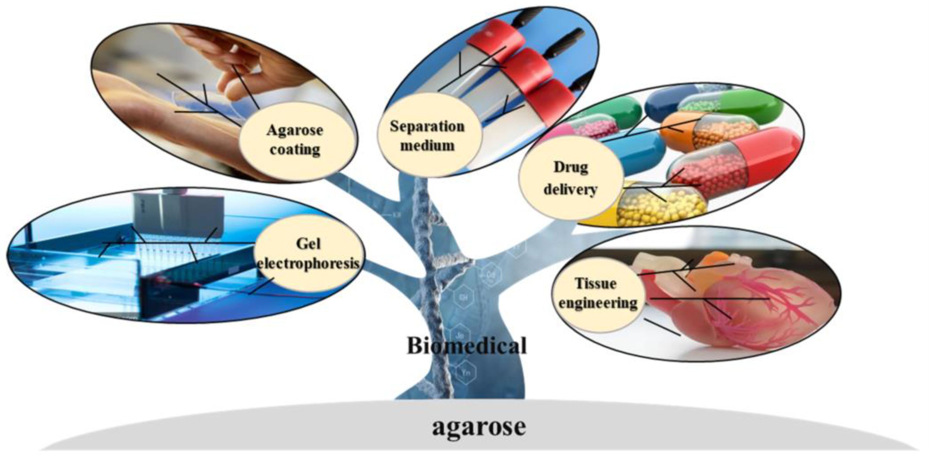
As the sol—agarose chains that form a porous network through hydrogen bonding [43]—are gelled, it allows the diffusion of high molecular weight substances. Meanwhile, many biological processes, such as the separation and purification of biological macromolecules, the complexation of proteins, and cell growth and multiplication, depend on buffers. For this reason, the hydrophilicity of the medium is essential for use in biological processes. Agarose is a polyhydroxy hydrocolloid, and the diffusion of biomolecules in the gel is not clearly different from that in an aqueous solution [56]. Synthetic and inorganic biomaterials, such as polyacrylamide gel and porous glass, are limited in separation and purification because of their nonspecific adsorption with some biomacromolecules. In theory, biologically active substances interact weakly with agarose and can remain biologically active in agarose gel [57]. It is possible to functionalize agarose to obtain specific properties that can lead to the derivation of different types of chromatographic separation media, extending the range of separation applications [58]. Figure 2 shows the application of agarose and its derivatives in the biomedical field.

3.1. Agarose Gel Electrophoresis

Electrophoresis is increasingly used in several fields, including clinical chemistry, toxicology, pharmacology, and immunology. Electrically charged molecules are driven through the agarose matrix by an electric field. They are separated in the agarose gel matrix according to their size, conformation, shape, and amount of charge they carry [59]. For this reason, agarose gel electrophoresis is widely used to separate and identify biological macromolecules such as nucleic acids, polysaccharides, proteins, and viruses [60,61].
The concentration of agarose gel electrophoresis is usually in the range of 0.5 to 2% (w/w) [62,63]. Agarose gel electrophoresis has the advantage of easy operation, simple equipment, a low sample volume, and high resolution. It serves the dual purpose of a “molecular sieve” and “electrophoresis” and is widely used in the study of nucleic acids [59]. In particular, chemically modified agarose with a low melting point has a higher sieving capacity and is ideal for the electrophoresis of DNA and RNA, allowing the recovery of the natural forms of DNA from the gel [64]. Green et al. [65] separated DNA fragments of different sizes using low-melting agarose and then recovered DNA by organic extraction from molten agarose using phenol-chloroform.
Zhang et al. prepared low melting point agarose through the oxy alkylation of agarose using ethylene oxide, 1,2-epoxypropane, and 1,2-epoxy butane [42]. They found that oxy-alkylated agarose underwent a superior separation performance in DNA gel electrophoresis. Recently, cyclic RNA migration under various conditions, various monoclonal and polyclonal antibodies, and the molecular weight of serum albumin have also been shown to be analyzed by agarose gel electrophoresis. Li et al. [62] characterized monoclonal antibodies developed against short peptide phosphotyrosines for reactive kinase phosphorylation by agarose gel electrophoresis. They optimized electrophoretic conditions by adjusting critical parameters of the electrophoretic process. Tomioka et al. [66] performed the agarose gel electrophoresis of commercially available bovine serum proteins using UltraPure and MetaPhor agarose. They found that the agarose gel electrophoresis obtained with MetaPhor agarose had a more significant molecular sieving effect, with high-resolution size differences and the ability to show more than 4%, oligomeric bands. In addition, agarose gel electrophoresis has been used to separate proteins and their complexes. Sakuma and colleagues [67] effectively extended the scope of agarose gel electrophoresis for biomedical applications by using agarose gel electrophoresis to analyze physically modified, aggregated, or post-translationally modified proteins. Agarose can also be mixed with polyacrylamide and dextran to support gel electrophoresis.

3.2. Agarose Separation Medium

Agarose gel is a hydrophilic medium that is highly compatible with biological macromolecules. Its porous properties make it ideal for separating proteins and nucleic acids [68]. Additionally, agarose can be customized for various applications by incorporating other functional groups as needed [69]. Agarose is a popular chromatographic separation media due to its availability in various forms, such as affinity, ion exchange, and hydrophobic chromatography media [70]. These different forms are based on distinct separation principles, making agarose one of the most widely used polysaccharide-based separation media.

3.2.1. Affinity Chromatography

Affinity chromatography is a technique used to separate and purify biological macromolecules by taking advantage of their specific recognition or reversible binding to corresponding molecules [71]. Agarose is a preferred matrix for affinity chromatography due to its low cost, large pore size, minimal non-specific binding to biological reagents, and stability across a wide range of pH values [72,73]. Affinity chromatography can be classified into four main types based on the interaction systems between biological macromolecules and ligands. These types are affinity chromatography, biomimetic affinity chromatography, immunoaffinity chromatography, and metal ion affinity chromatography.
Biological affinity chromatography is a kind of affinity chromatography that uses highly selective biological specificity to select interacting substances [74]. The enzyme-substrate, enzyme-inhibitor, and hormone-receptor pairs are commonly used. Yi et al. [75] exploited the biospecific choice between enzymes and enzyme inhibitors by using ST/SC (SpyTag/SpyCatcher) chemistry and targeted the immobilization of the green fluorescent protein as a model protein on agarose microspheres. Subsequently, ST-GFP (green fluorescent protein) was replaced by ST-PqsA (a key enzyme in the Pseudomonas aeruginosa quorum sensing pathway), and PqsA inhibitors were isolated by a biospecific selection between PqsA and PqsA inhibitors. Biomimetic affinity chromatography uses molecular interactions to synthesize ligands that mimic specific structures and sites on biological molecules as stationary phases for protein adsorption. Bai et al. [76] isolated and purified formate dehydrogenase (FDH) from bovine cell extracts by immobilizing four triazine reactive dyes on agarose and found that Procion Blue HERB was a suitable dye ligand for FDH, enabling simple, inexpensive, and efficient agarose affinity chromatography. Immunoaffinity chromatography is a separation system in which one of the antigens and antibodies is used as a ligand to affinity adsorb the other side. Hou et al. [54] prepared immunoaffinity chromatography columns to separate avermectin, ivermectin, doramectin, and ivermectin from samples by coupling avermectin polyclonal antibodies to CNBr-activated agarose, followed by an analysis of the residues eluting from the columns by high-performance liquid chromatography-tandem mass spectrometry. Metal ion affinity chromatography uses metal ion complexes to bind to proteins for separation and purification. The polymer, supported by super-porous agarose particles, has a high osmotic pressure, a high tolerance to interference mechanisms, and low back pressure. Zheng et al. [77] constructed complex immobilized metal ion affinity agarose particles to selectively isolate and purify histidine-tagged proteins using super porous agarose particles as supports, flexible copolymer brushes as backbones, and Ni2+-chelated iminodiacetic acid as ligands.

3.2.2. Size Exclusion Chromatography

Size exclusion chromatography (SEC) is a chromatographic technique for the separation of polymer samples based on the correspondence between the pore size of the gel pores and the molecular size of the polymer, with the advantages of the simplicity of the operation and high sample recovery [78]. Because of the 3D network structure of the gel filtration media, biomacromolecules with high molecular weight are blocked on the outside as they pass through the pores of gel particles and elute directly downwards at a rapid rate. By contrast, biomacromolecules with a low molecular weight can enter the interior of the gel particles as they pass through the column and are retained at a slow elution rate. The molecular sieving effect of the gels allows the molecular size of the samples to be screened for different elution times for biomolecules of different molecular weights [79]. Dextran, polyacrylamide, and agarose gel are the most commonly used gel filtration media. In particular, agarose gels have a higher mechanical strength and sieve stability, allowing for higher flow rates, a fuller range of pH conditions, and molecular weights. Size exclusion chromatography is often divided into gel permeation chromatography and gel filtration chromatography, depending on the mobile phase.
Zhao et al. [80] prepared homogeneous agarose microspheres of controlled particle size by an emulsion membrane method, followed by multi-step cross-linking and dextran grafting to obtain high-resolution chromatography media, finely controlling the molecular range with good pressure resistance. Site-specific glycosylation and its associated heterogeneity affect the functional activity of glycoproteins, and studies have shown that aberrant glycosylation can be closely associated with many diseases. Yet, the analysis of intact glycopeptides remains a significant challenge. For this purpose, Zhao et al. [81] developed a method to enrich intact tryptic N-glycopeptides using the excellent properties of the acrylamide–agarose composite gel and the hydrophilic properties of volume exclusion chromatography.

3.2.3. Ion Exchange Chromatography

Ion exchange chromatography (IEC) is a separation method based on the difference in electrostatic interactions between the sample and the stationary phase (ion exchange chromatography medium [82,83]). It has the advantages of wide applicability, high resolution, no effect on the biological activity of the sample, and high resolution, and it is widely used for the separation and purification of biological macromolecules [84] such as proteins [85]. The derivatizability of the hydroxyl groups of agarose allows ligands of different charge types and densities to be introduced into the gel [82]. Depending on the properties of ion-exchange ligands coupled to the agarose surface, IEC can be divided into anion-exchange chromatography media and cation-exchange chromatography. Currently, the most frequently used functional ligands are diethyl aminoethyl [86], carboxymethyl [87], and sulfopropyl [88], which make an agarose-based ion-exchange medium for weak anions, strong anions, weak cations, and strong cations, respectively. Separation can be achieved according to the difference in electrostatic interactions between the separation medium and the molecules because of the difference in charge type, charge number, and charge distribution on the surface of different molecules. In specialty biochemical separation media, the Agarose range of ion exchange media is currently a mainstream product. Anion-exchange chromatography separates and purifies positively charged proteins (lysozyme and cytochrome C) using an agarose-based anion-exchange medium. Silva-Santos et al. [89] used anion-exchange chromatography with HiTrap Q-agarose as the anion-exchange medium to purify ssDNA generated by aPCR from the reaction mixture. Barroca-Ferreira et al. [90] used Q-Sepharose anion-exchange chromatography to efficiently and finely purify prostaglandin 1.0 from K. pastoris small bioreactor lysates in a series of separation and purification steps; this is involved in cellular communication, stimulating cell proliferation and is specific to the cancer microenvironment. Cation exchange chromatography combines an ion exchange stationary phase with a negatively charged moiety coupled to a positively charged cation for separation and purification. Li et al. [91] investigated the effect of chain length and ionic strength on lysozyme adsorption and chromatographic interactions with γ-globulin by developing a cation exchange medium and co-grafting sodium methacrylate onto the commercial agarose gel Sepharose FF. Zhao et al. [70] developed a method by combining pre-crosslinking and surfactant micelle swelling to produce highly crosslinked macroporous agarose microspheres with a homogeneous network structure, low backpressure, and high flow rate, and, based on this, they created a carboxymethyl-coupled cation exchange chromatography media that could effectively separate proteins at high flow rates. Using sulphopropyl-Sepharose cation exchange chromatography, Li et al. [92] isolated and purified a 17.5 kDa bean protein inhibitor with anti-proliferative activity against leukemia and lymphoma cells from canola.

3.2.4. Hydrophobic Interaction Chromatography

Hydrophobic interaction chromatography (HIC) is a method that uses the reversible binding of a hydrophobic target in the mobile phase to the separation media together with a hydrophobic binding partner to achieve separation and purification [93]. HIC is widely used for the isolation and purification of biomolecules, such as proteins and peptides [55,93,94], for the advantages of mild reaction conditions, the high recovery of biomolecules, a low environmental impact, and cost savings [95,96]. They are typically composed of a hydrophobic matrix with a hydrophobic ligand. Currently, HIC media are commonly used with natural polysaccharides such as agarose and cellulose and synthetic polymers such as polystyrene and polyacrylates [97].
Agarose hydrophobic chromatography media consisting of agarose and hydrophobic ligands were introduced by chemical modification. Here, there are two main types of hydrophobic ligands: a hydrocarbon group with different carbon chain lengths, CH3(CH2)n-X, where X can be NH2, COOH, or OH, and hydrophobicity increases with the value of n; the second is a butyl or phenyl group with general hydrophobicity [98,99]. Its separation is based solely on the strength of the hydrophobic interaction, as the absence of groups such as NH2, COOH, or OH eliminates the interference of hydrogen bonding or charge interactions.
Superporous agarose beads are frequently employed as a foundation for hydrophobic chromatographic media. Gustavsson et al. [55] conducted a study in which they separated ribonuclease A, lysozyme, and bovine serum albumin using hydrophobic agarose chromatographic media prepared from phenyl-derived super porous agarose beads. The results showed that these proteins could be effectively separated and purified. The use of the chromatographic method resulted in higher flow rates compared to the use of homogeneous agarose beads as support. Hydrophobic chromatography was found to provide even higher flow rates. Lipases play a crucial role in various fields, such as oleochemistry, organic chemistry, biofuels, and pharmaceuticals, due to their ability to catalyze the hydrolysis of triacylglycerols into glycerol and free fatty acids. Mehta et al. [100] purified lipases from Aspergillus fumigatus through the use of octyl-Sepharose column chromatography for this purpose. The activation of angiogenesis by an essential fibroblast growth factor (bFGF) and vascular endothelial growth factor A (VEGFA) could promote tumorigenesis. However, the use of peptidomes containing bFGF/VEGFA can effectively block the activation of these growth factors, making them a promising option for the development of therapeutic tumor vaccines. Holková et al. [101] employed phenyl agarose CL-4B hydrophobic chromatography to isolate and purify lipoxygenases from poppy cultures. These enzymes play a crucial role in regulating growth, development, and resistance to biotic and abiotic stresses.

3.3. Agarose Coating

Infections caused by various bacterial biofilms have become one of the most common public health problems in clinical settings [102]. Bacterial proliferation and perithecia formation on medical devices pose a serious health challenge to patients [15]. As a result, the provision of anti-fouling and anti-bacterial capabilities for medical devices has become a hot research topic [103]. One of the most effective ways of doing this is to form coatings of a certain thickness and adhesion on the surface of the equipment. Agarose and its derivatives have been widely used as substrates for functional hydrogel coatings due to their excellent lubricity, biocompatibility, electrical conductivity, and mechanical properties [102,104].
Agarose shows no excellent anti-fouling or anti-bacterial properties. Early research has shown that agarose gel could resist the attachment of A. aeruginosa at a pH below 8.0, but when the pH increased beyond 8.2, the bacteria aggregated and was deposited on the agarose surface. Therefore, the majority of research has been carried out to produce novel coatings by combining agarose with other substances, commonly combining materials with antimicrobial properties and agarose [105]. Eric et al. [106] developed a novel coating by incorporating Cu/bioactive glass into an agarose matrix and showed that released Cu improved anti-adhesive properties and slowed biofilm formation on the biomaterial surface. Li et al. [107] combined ZnO nanoparticles and Ag nanoparticles on silk glue–agarose composites, which showed excellent antibacterial activity against Gram-positive and Gram-negative bacteria and great potential as a novel antibacterial biomaterial. Rather than simply compounding the agarose, chemical reactions such as crosslinking and grafting bound the agarose to different molecules of different sizes. This method improved the antibacterial and contamination resistance of agarose while maintaining superior gelling properties. Li and colleagues [103] introduced acrylate groups onto agarose molecules to form a covalent, crosslinked agarose coating on the surface of a medical silicone diaphragm that reduced the formation of bacteria by more than two orders of magnitude. He grafted and cross-linked agarose and quaternary ammonium chitosan using thiol-alcohol chemistry as an antimicrobial coating. This crosslinked coating effectively inhibited the biofilm formation of Gram-negative and Gram-positive bacteria. Interestingly, after 30 days of repeated wiping with ethanol, autoclave, or lysozyme solutions, the layer retained its antimicrobial activity [108].

3.4. Drug Delivery

A wide variety of drug carriers have been developed and are in use. Still, drug delivery has been on a path of innovation due to multidimensional challenges from materials to cost [109]. Carbohydrate polymers are one of the most important materials for the design of efficient drug carriers and have great potential to meet a wide range of needs [110,111]. Common carbohydrate polymers such as chitosan, starch, and cellulose have been widely used in drug delivery systems but still have considerable drawbacks. For example, natural chitosan is only soluble in dilute acidic aqueous media and loses its mechanical properties [112]; starch has poor mechanical properties and high-water solubility [113]; cellulose is insoluble in organic and aqueous solvents and has poor antibacterial properties [114]. All of these are unavoidable shortcomings. Agarose, with its non-toxicity, gelatinization, and gel structure with suitable viscoelasticity and thermal reversibility, is a common substrate for constructing particles, microcapsules [115], and microspheres and is excellent for controlled drug release. Drug release systems can be built using the biocompatibility and haemocompatibility of agarose [116]. For example, active ingredients can be encapsulated in agarose hydrogel nanoparticles and prepared by the emulsion template method [117]. Agarose can also be mixed with various polysaccharides (e.g., chitosan, cyclodextrin, and konjac glucomannan), proteins, and magnetic nanoparticles to form complexes with enhanced physicochemical properties for efficient drug delivery [116]. In general, agarose hydrogel drug delivery systems are dominated by mass diffusion release (Fickian diffusion) in agarose hydrogels, where the diffusion time depends on the network structure and the size of the particles [118]. In targeted drug delivery systems, agarose gels can be adapted in terms of their pore size, structure, and function by adjusting the agarose concentration [119], mixing with other compounds, and making chemical modifications to create an interesting and versatile drug delivery system [118].
In 1994, Haglund et al. [120] loaded Ibuprofen and Indomethacin onto agarose beads and studied their release mechanisms. To reduce the severe side effects associated with high doses of cyclophosphamide as a premedication in oncological treatment, Sakai et al. [121] encapsulated cells that were genetically modified to express cytochrome P450 2B1 enzymes in sub-sieve-sized agarose capsules before placing the cell-encapsulated microcapsules in arteries near the tumor via a microcatheter and activating them locally. The side effects were effectively reduced without reducing the dose. The subcapsules of agarose were smaller in diameter than standard agarose, reducing surgical trauma while also reducing the occlusion of the dripping vessel.
Apart from agarose pellets for drug delivery, agarose hydrogels are widely used in drug delivery systems due to their biocompatibility and solute permeability [122]. However, natural agarose has a high mechanical strength and solidification temperature [42], making it difficult to load thermosensitive drugs due to their inactivation during natural agarose gelation. As mentioned above, Kim et al. [42] prepared low gel temperature agarose by introducing β-cyclodextrin into ethylenediamine-modified agarose and found that this gel could be used for the sustained release of the thermosensitive drug doxorubicin because of its low mechanical strength and solidification temperature. In addition, hydrogel complexes of agarose with polysaccharides and proteins are often used as carriers for drug delivery systems to compensate for the shortcomings of natural agarose hydrogel in drug delivery. Rossi et al. [123] used agarose in a synthetic hydrogel with a Carbomer 974P macromolecule monomer as a spatially blocked and molecularly structured drug and mimetic molecule sodium fluorescein as a drug delivery vehicle.
To overcome the limitations of conventional therapeutics and increase the relevance and sensitivity of the drug delivery process, materials that respond to a range of endogenous (pH, enzyme expression, and redox potential) or exogenous (temperature, ultrasound, magnetic fields, and light) stimuli can be used as substrates for drug delivery systems [124]. Agarose hydrogel is electrically conductive, pH-responsive, and thermally reversible, making it an ideal tunable drug delivery system for loading and controlling the release of drugs by stimulating a sensitive response under different environmental conditions, increasing the retention time of the drug, and thus, reducing the dose frequency and toxic effects of the drug [125]. Endogenous drug delivery directly transfers a carrier-bound drug to a target site. Rajabzadeh-Khosroshahi et al. [126] prepared a degradable and biocompatible chitosan/agarose/graphitic carbon nitride nanocomposite for the loading and delivery of anti-cancer curcumin. It was found that the nanocomposite particles were effective at enhancing the bioavailability of curcumin in pH-sensitive drug release studies. Pourmadadi et al. [127] developed an agarose/chitosan double nanoemulsion as a drug delivery vehicle for curcumin/5-fluorouracil using a green synthesis approach. This nanocomposite was highly efficient for delivering curcumin/5-fluorouracil and has excellent potential for the targeted treatment of cancer cells. Quercetin, a drug with anticancer properties, is limited in cancer treatment due to its low solubility, low permeability, and short half-life. To this end, Samadi et al. [128] loaded quercetin into a pH-responsive agarose-polyvinylpyrrolidone-hydroxyapatite hydrogel nanocomposite and encapsulated it in the internal aqueous phase of a w/o/w emulsion and found that it was effective at enhancing the loading capacity, sustained release, and apoptosis-inducing effects of quercetin. Exogenous drug delivery is the release of a drug from a carrier to a target site that is triggered by various stimuli. Dong et al. [129] developed a drug-loaded MXene/agarose hydrogel drug delivery system with high photothermal conversion efficiency and photothermal stability by loading MXene nanosheets together with the anticancer drug adriamycin into a low-melting point agarose hydrogel, allowing for controlled drug release by near-infrared light irradiation. Hu et al. [130] prepared a pH- and magnetic field-responsive drug delivery system for the administration of adriamycin hydrochloride by grafting and copolymerizing 2-hydroxyethyl acrylate and vinyl acetate Fe3O4@agarose nanoparticles for the efficient and sustained release of adriamycin hydrochloride in the presence of weak acid or external magnetic fields.

3.5. Tissue Engineering

Tissue engineering combines tissue cells with specific biological activity and biomaterials to construct tissues and organs in vitro or in vivo to maintain, repair, regenerate or improve the function of damaged tissues and organs. The scaffold material serves as an artificial extracellular matrix for tissue engineering [131]. It supports cell attachment, growth, reproduction, metabolism, and the formation of new tissues. Because of the unique properties of human tissues, determining the best type of scaffold remains challenging. The most researched materials are biodegradable polymers, ceramic-like materials, composite materials, and materials derived from biological sources [132]. Natural carbohydrate polymers are crucial in tissue engineering scaffolds because they are non-toxic and biocompatible [133]. Agarose has unique electrical neutrality and gelling properties compared to other common carbohydrates such as chitosan and alginate [134]. There are a wide variety of agarose-based tissue scaffolds, including hydrogel, 3D/4D printed scaffolds, etc.

3.5.1. Agarose Hydrogel

Hydrogel, with its ability to provide a microenvironment for cell adhesion, proliferation, and migration, and facilitate biomolecular exchange, is essential to tissue engineering. Agarose has excellent potential for various tissue engineering applications because of its controlled self-assembly properties and tunable water adsorption capacity [133,135]. The structure, composition, and mechanical properties of single agarose are not conducive to customization and are usually complex with other polysaccharides to extend its range of applications [134]. Su et al. [136] prepared the agarose polydopamine (APG) hydrogel as scaffolds for skin wound healing. APG has good cell adhesion and high cell migration on the surface of the hydrogel, allowing cells to penetrate the hydrogel network. This study also showed that a composite hydrogel accelerated the healing process of full-thickness skin defects. In addition, the hydrogel can be used for cartilage tissue engineering. Singh et al. [134] prepared mulberry and non-mulberry silk hydrogel mixed with agarose and evaluated cartilage tissue formation in vitro. The results showed that mixed hydrogel exhibited enhanced cell proliferation and collagen deposition from sulfated glycosaminoglycans, clearly indicating that the composite hydrogel is a potential alternative for cartilage repair.

3.5.2. 3D/4D Printed Brackets

Three-dimensional printing uses computer-aided design models to produce solid objects in a precise layer-by-layer deposition [137], enabling biological structures that mimic the structure and functional properties of in vivo tissues and are of great importance in the field of tissue engineering [109,138]. An integral part of 3D printing is bio-ink; therefore, the selection of the right bio-ink is crucial and must meet basic requirements such as gelation, shear thinning, printability, and viscoelasticity [139,140]. Agarose gel naturally possesses these properties and can meet printing requirements under certain conditions. Fan et al. [139] demonstrated that a hybrid matrigel-agarose hydrogel could be used as bio-ink for 3D printing, showing that the agarose component of the composite system could hold the 3D printed structure while the matrigel provided the necessary microenvironment for cell growth. Four-dimensional (4D) printing has recently attracted increasing research interest due to its unique properties. Four-dimensional printing uses special smart materials to print objects with more complex structures that can change shape over time in response to specific external stimuli [141]. Jinhua Guo prepared a 4D-printed agarose/laponite/polymerized acrylamide hydrogel using agarose as a substrate. The experimental results showed that the 4D gel could further deform and that the hydrogel changed shape over time through external treatment followed by cooling [142].

4. Materials and Methods

For a comprehensive description of the latest research on agarose extraction, agarose modification, and its applications in biomedical science, Science Direct (Elsevier B. V., Amsterdam, The Netherlands), Pubmed (US National Library of Medicine. Bethesda, MD, USA) and Web of Science (Clarivate PLC, London, UK) databases are all available for documentation searching. In the section on agarose extraction, the keywords “agarose, extraction” were used to search, and the results were filtered by title, abstract, and full text to select the best literature on agarose extraction from 1937 to 2023 for analysis. In the agarose modification and applications section, a search was conducted using the keywords “agarose, modification, biomedical applications”, and the results were filtered for title, abstract, full text, and articles published in top journals of the field within the last five years.

5. Conclusions

Agarose is a carbohydrate polymer with modifiable properties that are becoming increasingly attractive in biomedicine. The rigidity, porosity, electrical neutrality and hydrophilic properties of agarose hydrogel provide it with unique advantages for separation, and purification. Additionally, agarose contains numerous hydroxyl groups, which can be modified through chemical methods to obtain specific functional groups and the further derivation of different types of chromatographic separation media, which broadens its range of separation applications. In addition, its excellent biocompatibility makes agarose one of the most promising carriers for drug delivery systems. In particular, in targeted drug applications, agarose can be loaded into lesions for targeted drug delivery. Because of its inert structure, agarose has excellent biocompatibility. It is possible to modulate the structure of agarose to provide controlled permeability to air and nutrients and regulate cell adhesion. Therefore, agarose-based biomaterials could provide a living space environment for cells in a biomimetic environment.

6. Prospects and Challenges

Focusing on the biomedical field, agarose, and modified agarose show great potential for application because of their unique physicochemical properties. A great deal of research on agarose and its derivatives is still needed, however, to improve green extraction and modification. Firstly, the source of agarose needs to be considered, including differences in the composition or molecular weight of agarose when extracted from seaweeds grown in different environments; it is also necessary to select more appropriate extraction and modification techniques for agarose and choose eco-friendly solvents to improve yield and quality. Furthermore, predicting the function of agarose gels by modulating chemical modifications is a challenge because the relationship between the chemical structure and mechanical properties of agarose still needs to be discovered.
Chromatography is one of the most efficient and gentle methods in separation and purification techniques. To achieve high-resolution purification, the particle size of microspheres and their homogeneity, as well as their fine pore structure, are very important factors. Agarose microspheres, commonly prepared by mechanical stirring and homogenous emulsification methods, are not uniform in size and require secondary sieving. Recently, membrane emulsification technology has been developed to prepare microspheres, which are characterized by uniform and controllable particle size, mild reaction conditions, simple operation, and green process [80]. However, several factors must be considered to obtain the desired microspheres, including the type of membrane, pH, solution viscosity, temperature, transmembrane pressure, continuous phase flow rate, and preparation efficiency. Therefore, this technique needs to be further explored.
Natural agarose is enriched with hydroxyl groups and is capable of forming hydrogen bonds with drugs and bioactive molecules for delivery. However, a limitation exists in employing raw agarose-based materials as drug delivery systems: the most cited regulations being a relatively high dissolution temperature, low degradation rate, low potential for the adsorption of hydrophobic drug molecules, and the slow adsorption/desorption of certain drugs [118]. Many studies have aimed at solving these problems through chemical modifications, yet the toxicity and biocompatibility of modified agarose still need to be addressed. Several studies have demonstrated the low cell adhesion, low in vitro cytotoxicity, and good biocompatibility of modified agarose [52,143,144]. However, most studies have remained in vitro, and the study of modified agarose-loaded drugs in animals still needs to be improved, which indicates that modified agarose-based drug delivery systems still have a long way to progress.
In the field of tissue engineering, agarose, and its derivatives are similarly lacking in animal testing. The exploration of biocompatibility, hemocompatibility, regenerative capacity, and toxicity in animals has, therefore, become a focus of research. At the same time, the natural brittleness of agarose has been a neglected aspect of research. Flexible agar hydrogels with self-healing capabilities have been developed [145]. With similar studies that can be drawn upon to address the in vivo compatibility of hydrogels, innovative agarose-based biomaterials are bound to have an increasing impact on biomedical applications.

Author Contributions

Conceptualization, Y.R., Q.X. and A.-F.X.; investigation, F.J. and X.-W.X.; writing—original draft preparation, F.J. and X.-W.X.; writing—review and editing, Q.X. and A.-F.X.; visualization, H.-F.W. and J.C.; funding acquisition, F.-Q.C., Q.X. and A.-F.X. All authors have read and agreed to the published version of the manuscript.

Funding

This research was funded by the Fujian Province Natural Science Foundation of China (2022J01805) and the National Natural Science Foundation of China (22278173, 32201941).

Institutional Review Board Statement

Not applicable.

Data Availability Statement

Not applicable.

Conflicts of Interest

The authors declare no conflict of interest.

References

  1. Ramkumar, V.S.; Prakash, S.; Ramasubburayan, R.; Pugazhendhi, A.; Gopalakrishnan, K.; Kannapiran, E.; Rajendran, R.B. Seaweeds: A resource for marine bionanotechnology. Enzyme Microb. Technol. 2016, 95, 45–57. [Google Scholar]
  2. El-Beltagi, H.S.; Mohamed, A.A.; Mohamed, H.I.; Ramadan, K.M.A.; Barqawi, A.A.; Mansour, A.T. Phytochemical and Potential Properties of Seaweeds and Their Recent Applications: A Review. Mar. Drugs 2022, 20, 342. [Google Scholar] [CrossRef] [PubMed]
  3. Nan, F.R.; Feng, J.; Lv, J.P.; Liu, Q.; Fang, K.P.; Gong, C.Y.; Xie, S.L. Origin and evolutionary history of freshwater Rhodophyta: Further insights based on phylogenomic evidence. Sci. Rep. 2017, 7, 2934. [Google Scholar] [CrossRef] [PubMed]
  4. Graham, S.; Marina, P.F.; Blencowe, A. Thermoresponsive polysaccharides and their thermoreversible physical hydrogel networks. Carbohydr. Polym. 2019, 207, 143–159. [Google Scholar] [CrossRef]
  5. Lahaye, M. Developments on gelling algal galactans, their structure and physico-chemistry. J. Appl. Phycol. 2001, 13, 173–184. [Google Scholar] [CrossRef]
  6. MarketWatch Global Agarose Market Analysis and Business Growth Outlook [2023–2030]. Available online: https://www.marketwatch.com/ (accessed on 12 April 2023).
  7. Alba, K.; Kontogiorgos, V. Seaweed Polysaccharides (Agar, Alginate Carrageenan). In Encyclopedia of Food Chemistry; Elsevier: Amsterdam, The Netherlands, 2018. [Google Scholar]
  8. Lee, Y.E.; Kim, H.; Seo, C.; Park, T.; Lee, K.B.; Yoo, S.Y.; Hong, S.C.; Kim, J.T.; Lee, J. Marine polysaccharides: Therapeutic efficacy and biomedical applications. Arch Pharm. Res. 2017, 40, 1006–1020. [Google Scholar] [CrossRef]
  9. Sharma, A.; Kaur, I.; Dheer, D.; Nagpal, M.; Kumar, P.; Venkatesh, D.N.; Puri, V.; Singh, I. A propitious role of marine sourced polysaccharides: Drug delivery and biomedical applications. Carbohydr. Polym. 2023, 308, 120448. [Google Scholar] [CrossRef]
  10. Xiao, Q.; Ma, M.Z.; Chen, J.; Zhang, Y.H.; Chen, F.Q.; Weng, H.F.; Xiao, A.F. Preparation of macroporous rigid agarose microspheres by pre-crosslinking with cyclic anhydride. Int. J. Biol. Macromol. 2022, 222, 41–54. [Google Scholar] [CrossRef]
  11. Song, R.; Murphy, M.; Li, C.S.; Ting, K.; Soo, C.; Zheng, Z. Current development of biodegradable polymeric materials for biomedical applications. Drug Des. Dev. Ther. 2018, 12, 3117–3145. [Google Scholar] [CrossRef]
  12. Gaharwar, A.K. Engineered Biomaterials for in Situ Tissue Regeneration. Tissue Eng. Part A 2022, 28, S590. [Google Scholar] [CrossRef]
  13. Ciancia, M.; Fernandez, P.V.; Leliaert, F. Diversity of Sulfated Polysaccharides From Cell Walls of Coenocytic Green Algae and Their Structural Relationships in View of Green Algal Evolution. Front. Plant Sci. 2020, 11, 554585. [Google Scholar] [CrossRef] [PubMed]
  14. Lopez-Heredia, M.A.; Lapa, A.; Mendes, A.C.; Balcaen, L.; Samal, S.K.; Chai, F.; Van der Voort, P.; Stevens, C.V.; Parakhonskiy, B.V.; Chronakis, I.S.; et al. Bioinspired, biomimetic, double-enzymatic mineralization of hydrogels for bone regeneration with calcium carbonate. Mater. Lett. 2017, 190, 13–16. [Google Scholar] [CrossRef]
  15. Wu, F.; Xu, T.T.; Zhao, G.Y.; Meng, S.S.; Wan, M.M.; Chi, B.; Mao, C.; Shen, J. Mesoporous Silica Nanoparticles-Encapsulated Agarose and Heparin as Anticoagulant and Resisting Bacterial Adhesion Coating for Biomedical Silicone. Langmuir 2017, 33, 5245–5252. [Google Scholar] [CrossRef] [PubMed]
  16. Chu, W.T.; Ma, Y.H.; Zhang, Y.N.; Cao, X.J.; Shi, Z.Y.; Liu, Y.; Ding, X.J. Significantly improved antifouling capability of silicone rubber surfaces by covalently bonded acrylated agarose towards biomedical applications. Colloids Surf. B-Biointerfaces 2023, 222, 112979. [Google Scholar] [CrossRef] [PubMed]
  17. Hu, Y.; Kim, Y.; Hong, I.; Kim, M.; Jung, S. Fabrication of Flexible pH-Responsive Agarose/Succinoglycan Hydrogels for Controlled Drug Release. Polymers 2021, 13, 2049. [Google Scholar] [CrossRef]
  18. Apte, G.; Lindenbauer, A.; Schemberg, J.; Rothe, H.; Nguyen, T.H. Controlling Surface-Induced Platelet Activation by Agarose and Gelatin-Based Hydrogel Films. ACS Omega 2021, 6, 10963–10974. [Google Scholar] [CrossRef]
  19. Krommelbein, C.; Xie, X.F.; Seifert, J.; Konieczny, R.; Friebe, S.; Kas, J.; Riedel, S.; Mayr, S.G. Electron beam treated injectable agarose/alginate beads prepared by electrospraying. Carbohydr. Polym. 2022, 298, 120024. [Google Scholar] [CrossRef]
  20. Kinoshita, K.; Iwase, M.; Yamada, M.; Yajima, Y.; Seki, M. Fabrication of multilayered vascular tissues using microfluidic agarose hydrogel platforms. Biotechnol. J. 2016, 11, 1415–1423. [Google Scholar] [CrossRef]
  21. Araki, C.; Arai, K. Studies on the chemical constitution of agar-agar. XXIV. Isolation of a new disaccharide as a reversion product from acidic hydrolysate. Bull. Chem. Soc. Jpn 1967, 40, 1452–1456. [Google Scholar] [CrossRef]
  22. Chew, K.W.; Show, P.L.; Yap, Y.J.; Juan, J.C.; Phang, S.M.; Ling, T.C.; Chang, J.S. Sonication and grinding pre-treatments on Gelidium amansii seaweed for the extraction and characterization of Agarose. Front. Environ. Sci. Eng. 2018, 12, 2. [Google Scholar] [CrossRef]
  23. Jeon, Y.-J.; Athukorala, Y.; Lee, J.-H. Characterization of Agarose Product from Agar Using DMSO. ALGAE 2005, 20, 61–67. [Google Scholar] [CrossRef]
  24. Santos, G.A.; Doty, M.S. Agarose from Gracilaria-Cylindrica. Bot. Mar. 1983, 26, 31–34. [Google Scholar] [CrossRef]
  25. Duckworth, M.; Yaphe, W. Preparation of agarose by fractionation from the spectrum of polysaccharides in agar. Anal. Biochem. 1971, 44, 636–641. [Google Scholar] [CrossRef]
  26. Ozturk, Y.; Ekmekci, Z. Removal of sulfate ions from process water by ion exchange resins kk. Miner. Eng. 2020, 159, 106613. [Google Scholar] [CrossRef]
  27. Cook, R.B.; Witt, H.J. Agarose composition, AQUEOUS Gel and Method of Making Same. U.S. Patent 4,290,911, 22 September 1981. [Google Scholar]
  28. Zhang, Y.; Fu, X.T.; Duan, D.L.; Xu, J.C.; Gao, X. Preparation and characterization of agar, agarose, agaropectin from the red alga Ahnfeltia plicata. J. Oceanol. Limnol. 2019, 37, 815–824. [Google Scholar] [CrossRef]
  29. Trivedi, T.; Kumar, A. Efficient Extraction of Agarose from Red Algae Using Ionic Liquids. Green Sustain. Chem. 2014, 4, 190–201. [Google Scholar] [CrossRef]
  30. Sharma, M.; Chaudhary, J.P.; Mondal, D.; Meena, R.; Prasad, K. A green and sustainable approach to utilize bio-ionic liquids for the selective precipitation of high purity agarose from an agarophyte extract. Green Chem. 2015, 17, 2867–2873. [Google Scholar] [CrossRef]
  31. Wang, T.P.; Chang, L.L.; Chang, S.N.; Wang, E.C.; Hwang, L.C.; Chen, Y.H.; Wang, Y.M. Successful preparation and characterization of biotechnological grade agarose from indigenous Gelidium amansii of Taiwan. Process Biochem. 2012, 47, 550–554. [Google Scholar] [CrossRef]
  32. Xiao, Q.; Yin, Q.; Ni, H.; Cai, H.N.; Wu, C.Z.; Xiao, A.F. Characterization and immobilization of arylsulfatase on modified magnetic nanoparticles for desulfation of agar. Int. J. Biol. Macromol. 2017, 94, 576–584. [Google Scholar] [CrossRef]
  33. Shukla, M.K.; Kumar, M.; Prasad, K.; Reddy, C.R.K.; Jha, B. Partial characterization of sulfohydrolase from Gracilaria dura and evaluation of its potential application in improvement of the agar quality. Carbohydr. Polym. 2011, 85, 157–163. [Google Scholar] [CrossRef]
  34. Wang, X.Y.; Duan, D.L.; Fu, X.T. Enzymatic desulfation of the red seaweeds agar by Marinomonas arylsulfatase. Int. J. Biol. Macromol. 2016, 93, 600–608. [Google Scholar] [CrossRef]
  35. Zhang, C.; An, D.; Xiao, Q.; Chen, F.Q.; Zhang, Y.H.; Weng, H.F.; Xiao, A.F. Convenient Agarose Preparation with Hydrogen Peroxide and Desulfation Process Analysis. Mar. Drugs 2021, 19, 297. [Google Scholar] [CrossRef]
  36. Zhang, C.; An, D.; Xiao, Q.; Weng, H.F.; Zhang, Y.H.; Yang, Q.M.; Xiao, A.F. Preparation, characterization, and modification mechanism of agar treated with hydrogen peroxide at different temperatures. Food Hydrocoll. 2020, 101, 105527. [Google Scholar] [CrossRef]
  37. Wu, Z.; Li, H.; Zhao, X.W.; Ye, F.Y.; Zhao, G.H. Hydrophobically modified polysaccharides and their self-assembled systems: A review on structures and food applications. Carbohydr. Polym. 2022, 284, 119182. [Google Scholar] [CrossRef]
  38. Zhang, Y.H.; Song, X.N.; Lin, Y.; Xiao, Q.; Du, X.P.; Chen, Y.H.; Xiao, A.F. Antioxidant capacity and prebiotic effects of Gracilaria neoagaro oligosaccharides prepared by agarase hydrolysis. Int. J. Biol. Macromol. 2019, 137, 177–186. [Google Scholar] [CrossRef]
  39. Punia, S. Barley starch modifications: Physical, chemical and enzymatic—A review. Int. J. Biol. Macromol. 2020, 144, 578–585. [Google Scholar] [CrossRef]
  40. Zhang, B.; Lan, W.; Xie, J. Chemical modifications in the structure of marine polysaccharide as serviceable food processing and preservation assistant: A review. Int. J. Biol. Macromol. 2022, 223, 1539–1555. [Google Scholar] [CrossRef]
  41. Krommelbein, C.; Mutze, M.; Konieczny, R.; Schonherr, N.; Griebel, J.; Gerdes, W.; Mayr, S.G.; Riedel, S. Impact of high-energy electron irradiation on mechanical, structural and chemical properties of agarose hydrogels. Carbohydr. Polym. 2021, 263, 117970. [Google Scholar] [CrossRef]
  42. Zhang, N.; Wang, J.L.; Ye, J.; Zhao, P.; Xiao, M.T. Oxyalkylation modification as a promising method for preparing low-melting-point agarose. Int. J. Biol. Macromol. 2018, 117, 696–703. [Google Scholar] [CrossRef]
  43. Xiao, Q.; Weng, H.F.; Chen, G.; Xiao, A.F. Preparation and characterization of octenyl succinic anhydride modified agarose derivative. Food Chem. 2019, 279, 30–39. [Google Scholar] [CrossRef]
  44. Xiao, Q.; Chen, G.; Xiao, A.F. Preparation, characterization, and emulsification properties of agarose fatty acid derivatives with different hydrophobic chains. Int. J. Biol. Macromol. 2019, 141, 906–918. [Google Scholar] [CrossRef]
  45. Oza, M.D.; Meena, R.; Prasad, K.; Paul, P.; Siddhanta, A.K. Functional modification of agarose: A facile synthesis of a fluorescent agarose-guanine derivative. Carbohydr. Polym. 2010, 81, 878–884. [Google Scholar] [CrossRef]
  46. Cao, N.; Xia, G.; Sun, H.; Zhao, L.; Cao, R.; Jiang, H.; Mao, X.; Liu, Q. Characterization of a Galactose Oxidase from Fusarium odoratissimum and Its Application in the Modification of Agarose. Foods 2023, 12, 603. [Google Scholar] [CrossRef]
  47. Zhang, M.Y.; Wang, J.X.; Zeng, R.Y.; Wang, D.Q.; Wang, W.X.; Tong, X.F.; Qu, W. Agarose-Degrading Characteristics of a Deep-Sea Bacterium Vibrio Natriegens WPAGA4 and Its Cold-Adapted GH50 Agarase Aga3420. Mar. Drugs 2022, 20, 692. [Google Scholar] [CrossRef]
  48. Tsukamoto, A.; Tanaka, K.; Kumata, T.; Yoshida, K.; Watanabe, Y.; Miyata, S.; Furukawa, K.S.; Ushida, T. 1-MHz ultrasound enhances internal diffusivity in agarose gels. Appl. Acoust. 2013, 74, 1117–1121. [Google Scholar] [CrossRef]
  49. Builders, P.F.; Kunle, O.O.; Adikwu, M.U. Preparation and characterization of mucinated agarose: A mucin-agarose physical crosslink. Int. J. Pharm. 2008, 356, 174–180. [Google Scholar] [CrossRef]
  50. Bayat, F.; Pourmadadi, M.; Eshaghi, M.M.; Yazdian, F.; Rashedi, H. Improving Release Profile and Anticancer Activity of 5-Fluorouracil for Breast Cancer Therapy Using a Double Drug Delivery System: Chitosan/Agarose/γ-Alumina Nanocomposite@Double Emulsion. J. Cluster Sci. 2023, 1–13. [Google Scholar] [CrossRef]
  51. Pourmadadi, M.; Ahmadi, M.; Yazdian, F. Synthesis of a novel pH-responsive Fe3O4/chitosan/agarose double nanoemulsion as a promising Nanocarrier with sustained release of curcumin to treat MCF-7 cell line. Int. J. Biol. Macromol. 2023, 235, 123786. [Google Scholar] [CrossRef]
  52. Evans, C.; Morimitsu, Y.; Nishi, R.; Yoshida, M.; Takei, T. Novel hydrophobically modified agarose cryogels fabricated using dimethyl sulfoxide. J. Biosci. Bioeng. 2022, 133, 390–395. [Google Scholar] [CrossRef]
  53. Xiao, Q.; Chen, G.; Zhang, Y.H.; Weng, H.F.; Cai, M.H.; Xiao, A.F. Evaluation of a novel self-emulsifiable dodecenyl succinylated agarose in microencapsulation of docosahexaenoic acid (DHA) through spray-chilling process. Int. J. Biol. Macromol. 2020, 163, 2314–2324. [Google Scholar] [CrossRef]
  54. Hou, X.L.; Li, X.W.; Ding, S.Y.; He, J.H.; Jiang, H.Y.; Shen, J.Z. Simultaneous analysis of avermectins in bovine tissues by LC-MS-MS with immunoaffinity chromatography cleanup. Chromatographia 2006, 63, 543–550. [Google Scholar] [CrossRef]
  55. Gustavsson, P.E.; Axelsson, A.; Larsson, P.O. Superporous agarose beads as a hydrophobic interaction chromatography support. J. Chromatogr. A 1999, 830, 275–284. [Google Scholar] [CrossRef] [PubMed]
  56. Zhang, L.Y.; Xiao, Q.; Xiao, Z.C.; Zhang, Y.H.; Weng, H.F.; Chen, F.Q.; Xiao, A.F. Hydrophobic modified agar: Structural characterization and application in encapsulation and release of curcumin. Carbohydr. Polym. 2023, 308, 120644. [Google Scholar] [CrossRef]
  57. Zhao, L.S.; Li, S.S.; Liang, C.; Qiao, L.Z.; Du, K.F. High-strength and low-crystallinity cellulose/agarose composite microspheres: Fabrication, characterization and protein adsorption. Biochem. Eng. J. 2021, 166, 107826. [Google Scholar] [CrossRef]
  58. Zhao, L.; Huang, Y.D.; Zhu, K.; Miao, Z.; Zhao, J.Z.; Che, X.J.; Hao, D.X.; Zhang, R.Y.; Ma, G.H. Manipulation of pore structure during manufacture of agarose microspheres for bioseparation. Eng. Life Sci. 2020, 20, 504–513. [Google Scholar] [CrossRef] [PubMed]
  59. Tantray, J.A.; Mansoor, S.; Wani, R.F.C.; Nissa, N.U. (Eds.) Chapter 24—Agarose gel electrophoresis. In Basic Life Science Methods; Academic Press: Cambridge, MA, USA, 2023; pp. 103–106. [Google Scholar]
  60. Li, C.; Arakawa, T. Agarose native gel electrophoresis of proteins. Int. J. Biol. Macromol. 2019, 140, 668–671. [Google Scholar] [CrossRef]
  61. Tomioka, Y.; Arakawa, T.; Akuta, T.; Nakagawa, M.; Ishibashi, M. Analysis of proteins by agarose native gel electrophoresis in the presence of solvent additives. Int. J. Biol. Macromol. 2022, 198, 26–36. [Google Scholar] [CrossRef] [PubMed]
  62. Li, C.; Akuta, T.; Nakagawa, M.; Sato, T.; Shibata, T.; Maruyama, T.; Okumura, C.J.; Kurosawa, Y.; Arakawa, T. Agarose native gel electrophoresis for characterization of antibodies. Int. J. Biol. Macromol. 2020, 151, 885–890. [Google Scholar] [CrossRef] [PubMed]
  63. Abe, B.T.; Wesselhoeft, R.A.; Chen, R.; Anderson, D.G.; Chang, H.Y. Circular RNA migration in agarose gel electrophoresis. Mol. Cell 2022, 82, 1768–1777.e3. [Google Scholar] [CrossRef]
  64. Song, N.L.; He, X.; Zhao, Q.R.; Yan, T.D.; Wen, L. Cloning and expression of the tumstatin active peptides-T-7 and its derivant-T-7-NGR. Clin. Exp. Med. 2009, 9, 165–171. [Google Scholar]
  65. Green, M.R.; Sambrook, J. Recovery of DNA from Low-Melting-Temperature Agarose Gels: Organic Extraction. Cold Spring Harb. Protoc. 2020, 2020, 100461. [Google Scholar] [CrossRef] [PubMed]
  66. Tomioka, Y.; Nakagawa, M.; Sakuma, C.; Nagatoishi, S.; Tsumoto, K.; Arakawa, T.; Akuta, T. Ladder observation of bovine serum albumin by high resolution agarose native gel electrophoresis. Int. J. Biol. Macromol. 2022, 215, 512–520. [Google Scholar] [CrossRef] [PubMed]
  67. Sakuma, C.; Tomioka, Y.; Li, C.; Shibata, T.; Nakagawa, M.; Kurosawa, Y.; Arakawa, T.; Akuta, T. Analysis of protein denaturation, aggregation and post-translational modification by agarose native gel electrophoresis. Int. J. Biol. Macromol. 2021, 172, 589–596. [Google Scholar] [CrossRef] [PubMed]
  68. Lira, R.B.; Steinkuhler, J.; Knorr, R.L.; Dimova, R.; Riske, K.A. Posing for a picture: Vesicle immobilization in agarose gel. Sci. Rep. 2016, 6, 25254. [Google Scholar] [CrossRef]
  69. Pourmadadi, M.; Yazdian, F.; Koulivand, A.; Rahmani, E. Green synthesized polyvinylpyrrolidone/titanium dioxide hydrogel nanocomposite modified with agarose macromolecules for sustained and pH-responsive release of anticancer drug. Int. J. Biol. Macromol. 2023, 240, 124345. [Google Scholar] [CrossRef]
  70. Zhao, X.; Huang, L.; Wu, J.; Huang, Y.D.; Zhao, L.; Wu, N.; Zhou, W.Q.; Hao, D.X.; Ma, G.H.; Su, Z.G. Fabrication of rigid and macroporous agarose microspheres by pre-cross-linking and surfactant micelles swelling method. Colloids Surf. B-Biointerfaces 2019, 182, 110377. [Google Scholar] [CrossRef]
  71. Iftekhar, S.; Ovbude, S.T.; Hage, D.S. Kinetic Analysis by Affinity Chromatography. Front. Chem. 2019, 7, 673. [Google Scholar] [CrossRef]
  72. Behar, G.; Renodon-Corniere, A.; Mouratou, B.; Pecorari, F. Affitins as robust tailored reagents for affinity chromatography purification of antibodies and non-immunoglobulin proteins. J. Chromatogr. A 2016, 1441, 44–51. [Google Scholar] [CrossRef]
  73. Yin, J.L.; Zheng, H.W.; Lin, H.; Sui, J.X.; Wang, B.C.; Pavase, T.R.; Cao, L.M. Boronic acid-functionalized agarose affinity chromatography for isolation of tropomyosin in fishes. J. Sci. Food Agric. 2019, 99, 6490–6499. [Google Scholar] [CrossRef]
  74. Fang, Y.M.; Lin, D.Q.; Yao, S.J. Review on biomimetic affinity chromatography with short peptide ligands and its application to protein purification. J. Chromatogr. A 2018, 1571, 1–15. [Google Scholar] [CrossRef]
  75. Yi, Y.; Shi, K.F.; Ding, S.W.; Hu, J.M.; Zhang, C.; Mei, J.F.; Ying, G.Q. A general strategy for protein affinity-ligand oriented-immobilization and screening for bioactive compounds. J. Chromatogr. B-Anal. Technol. Biomed. Life Sci. 2023, 1218, 123591. [Google Scholar] [CrossRef]
  76. Bai, Y.L.; Yang, S.T. Production and separation of formate dehydrogenase from Candida boidinii. Enzyme Microb. Technol. 2007, 40, 940–946. [Google Scholar] [CrossRef]
  77. Zheng, H.W.; Wang, C.Y.; Pavase, T.R.; Xue, C.H. Fabrication of copolymer brushes grafted superporous agarose gels: Towards the ultimate ideal particles for efficient affinity chromatography. Colloids Surf. B-Biointerfaces 2022, 217, 112705. [Google Scholar] [CrossRef] [PubMed]
  78. Barth, H.G. Size Exclusion Chromatography: A Teaching Aid for Physical Chemistry. J. Chem. Educ. 2018, 95, 1125–1131. [Google Scholar] [CrossRef]
  79. Zhao, L.; Huang, L.; Huang, Y.D.; Zhu, K.; Che, X.J.; Du, Y.X.; Gao, J.W.; Hao, D.X.; Zhang, R.Y.; Wang, Q.B.; et al. Preparation and structural regulation of macroporous agarose microspheres for highly efficient adsorption of giant biomolecules. Colloid. Polym. Sci. 2022, 300, 691–705. [Google Scholar] [CrossRef]
  80. Zhao, L.; Che, X.J.; Huang, Y.D.; Zhu, K.; Du, Y.X.; Gao, J.W.; Zhang, R.Y.; Zhang, Y.Q.; Ma, G.H. Regulation on both pore structure and pressure-resistant property of uniform agarose microspheres for high-resolution chromatography. J. Chromatogr. A 2022, 1681, 463461. [Google Scholar] [CrossRef]
  81. Zhao, T.; Zhang, C.; Ma, W.D.; Xiong, Y.; Yao, J.; Yan, G.Q.; Chen, G.; Lu, H.J. A practical approach to enrich intact tryptic N-glycopeptides through size exclusion chromatography and hydrophilicity (SELIC) using an acrylamide-agarose composite gel system. Anal. Chim. Acta 2019, 1058, 107–116. [Google Scholar] [CrossRef]
  82. Stone, M.C.; Tao, Y.Y.; Carta, G. Protein adsorption and transport in agarose and dextran-grafted agarose media for ion exchange chromatography: Effect of ionic strength and protein characteristics. J. Chromatogr. A 2009, 1216, 4465–4474. [Google Scholar] [CrossRef]
  83. Zhang, Z.R.; Zhou, S.Y.; Han, L.J.; Zhang, Q.Y.; Pritts, W.A. Impact of linker-drug on ion exchange chromatography separation of antibody-drug conjugates. Mabs 2019, 11, 1113–1121. [Google Scholar] [CrossRef]
  84. Apolinar-Valiente, R.; Williams, P.; Nigen, M.; Tamayo, V.M.; Doco, T.; Sanchez, C. Fractionation of Acacia seyal gum by ion exchange chromatography. Food Hydrocoll. 2020, 98, 105283. [Google Scholar] [CrossRef]
  85. Kristl, A.; Luksic, M.; Pompe, M.; Podgornik, A. Effect of Pressure Increase on Macromolecules’ Adsorption in Ion Exchange Chromatography. Anal. Chem. 2020, 92, 4527–4534. [Google Scholar] [CrossRef] [PubMed]
  86. Oksanen, H.M.; Domanska, A.; Bamford, D.H. Monolithic ion exchange chromatographic methods for virus purification. Virology 2012, 434, 271–277. [Google Scholar] [CrossRef] [PubMed]
  87. Cao, Y.L.; Ding, Y.Y.; Zhang, L.P.; Shi, G.; Sang, X.X.; Ni, C.H. Preparation of surface-modified, micrometer-sized carboxymethyl chitosan drug-loaded microspheres. J. Appl. Polym. Sci. 2018, 135, 45731. [Google Scholar] [CrossRef]
  88. Ljunglof, A.; Lacki, K.M.; Mueller, J.; Harinarayan, C.; van Reis, R.; Fahrner, R.; Van Alstine, J.M. Ion exchange chromatography of antibody fragments. Biotechnol. Bioeng. 2007, 96, 515–524. [Google Scholar] [CrossRef] [PubMed]
  89. Silva-Santos, A.R.; Paulo, P.M.R.; Prazeres, D.M.F. Scalable purification of single stranded DNA scaffolds for biomanufacturing DNA-origami nanostructures: Exploring anion-exchange and multimodal chromatography. Sep. Purif. Technol. 2022, 298, 121623. [Google Scholar] [CrossRef]
  90. Barroca-Ferreira, J.; Goncalves, A.M.; Santos, M.F.A.; Santos-Silva, T.; Maia, C.J.; Passarinha, L.A. A chromatographic network for the purification of detergent-solubilized six-transmembrane epithelial antigen of the prostate 1 from Komagataella pastoris mini-bioreactor lysates. J. Chromatogr. A 2022, 1685, 463576. [Google Scholar] [CrossRef]
  91. Li, X.; Liu, Y.; Sun, Y. Development of poly(methacrylate)-grafted Sepharose FF for cation-exchange chromatography of proteins. J. Chromatogr. A 2020, 1634, 461669. [Google Scholar] [CrossRef]
  92. Li, M.; Liu, Q.; Cui, Y.; Li, D.; Wang, H.; Ng, T.B. Isolation and Characterization of a Phaseolus vulgaris Trypsin Inhibitor with Antiproliferative Activity on Leukemia and Lymphoma Cells. Molecules 2017, 22, 187. [Google Scholar] [CrossRef]
  93. Lienqueo, M.E.; Salazar, O.; Henriquez, K.; Calado, C.R.C.; Fonseca, L.P.; Cabral, J.M.S. Prediction of retention time of cutinases tagged with hydrophobic peptides in hydrophobic interaction chromatography. J. Chromatogr. A 2007, 1154, 460–463. [Google Scholar] [CrossRef]
  94. Hall, T.; Kelly, G.M.; Emery, W.R. Use of mobile phase additives for the elution of bispecific and monoclonal antibodies from phenyl based hydrophobic interaction chromatography resins. J. Chromatogr. B-Anal. Technol. Biomed. Life Sci. 2018, 1096, 20–30. [Google Scholar] [CrossRef]
  95. Rodler, A.; Ueberbacher, R.; Beyer, B.; Jungbauer, A. Calorimetry for studying the adsorption of proteins in hydrophobic interaction chromatography. Prep. Biochem. Biotechnol. 2019, 49, 1–20. [Google Scholar] [CrossRef] [PubMed]
  96. Fekete, S.; Murisier, A.; Verscheure, L.; Sandra, K.; Guillarme, D. Hydrophobic Interaction Chromatography (HIC) for the Characterization of Therapeutic Monoclonal Antibodies and Related Products, Part 2: Practical Considerations. LC GC Eur. 2021, 34, 139–148. [Google Scholar]
  97. Wang, L.H.; Fu, Q.X.; Yu, J.Y.; Liu, L.F.; Ding, B. Nanoparticle-doped polystyrene/polyacrylonitrile nanofiber membrane with hierarchical structure as promising protein hydrophobic interaction chromatography media. Compos. Commun. 2019, 16, 33–40. [Google Scholar] [CrossRef]
  98. Ren, K.; Li, Y.; Shi, F.; Wang, X.Y. Separation of lipopolysaccharides containing different fatty acid chains using hydrophobic interaction chromatography. Anal. Methods 2012, 4, 838–843. [Google Scholar] [CrossRef]
  99. Brandts, P.M.; Middelkoop, C.M.; Gelsema, W.J.; Deligny, C.L. Hydrophobic Interaction Chromatography of Simple Compounds on Alkyl-Agaroses with Different Alkyl Chain Lengths and Chain Densities—Mechanism and Thermodynamics. J. Chromatogr. 1986, 356, 247–259. [Google Scholar] [CrossRef]
  100. Mehta, A.; Grover, C.; Gupta, R. Purification of lipase from Aspergillus fumigatus using Octyl Sepharose column chromatography and its characterization. J. Basic Microbiol. 2018, 58, 857–866. [Google Scholar] [CrossRef]
  101. Holkova, I.; Rauova, D.; Mergova, M.; Bezakova, L.; Mikus, P. Purification and Product Characterization of Lipoxygenase from Opium Poppy Cultures (Papaver somniferum L.). Molecules 2019, 24, 4268. [Google Scholar] [CrossRef]
  102. Ghosh, S.; Saraswathi, A.; Indi, S.S.; Hoti, S.L.; Vasan, H.N. Ag@AgI, Core@Shell Structure in Agarose Matrix as Hybrid: Synthesis, Characterization, and Antimicrobial Activity. Langmuir 2012, 28, 8550–8561. [Google Scholar] [CrossRef]
  103. Li, M.; Neoh, K.G.; Kang, E.T.; Lau, T.; Chiong, E. Surface Modifi cation of Silicone with Covalently Immobilized and Crosslinked Agarose for Potential Application in the Inhibition of Infection and Omental Wrapping. Adv. Funct. Mater. 2014, 24, 1631–1643. [Google Scholar] [CrossRef]
  104. Chen, X.Y.; Li, H.J.; Qiao, X.N.; Jiang, T.Z.; Fu, X.; He, Y.; Zhao, X. Agarose oligosaccharide- silver nanoparticle- antimicrobial peptide-composite for wound dressing. Carbohydr. Polym. 2021, 269, 118258. [Google Scholar] [CrossRef]
  105. Stickler, D.J.; Lear, J.C.; Morris, N.S.; Macleod, S.M.; Downer, A.; Cadd, D.H.; Feast, W.J. Observations on the adherence of Proteus mirabilis onto polymer surfaces. J. Appl. Microbiol. 2006, 100, 1028–1033. [Google Scholar] [CrossRef] [PubMed]
  106. Wers, E.; Lefeuvre, B. New hybrid agarose/Cu-Bioglass® biomaterials for antibacterial coatings. Korean J. Chem. Eng. 2017, 34, 2241–2247. [Google Scholar] [CrossRef]
  107. Li, W.T.; Huang, Z.X.; Cai, R.; Yang, W.; He, H.W.; Wang, Y.J. Rational Design of Ag/ZnO Hybrid Nanoparticles on Sericin/Agarose Composite Film for Enhanced Antimicrobial Applications. Int. J. Mol. Sci. 2021, 22, 105. [Google Scholar] [CrossRef]
  108. Li, M.; Mitra, D.; Kang, E.T.; Lau, T.; Chiong, E.; Neoh, K.G. Thiol-ol Chemistry for Grafting of Natural Polymers to Form Highly Stable and Efficacious Antibacterial Coatings. ACS Appl. Mater. Interfaces 2017, 9, 1847–1857. [Google Scholar] [CrossRef]
  109. Matai, I.; Kaur, G.; Seyedsalehi, A.; McClinton, A.; Laurencin, C.T. Progress in 3D bioprinting technology for tissue/organ regenerative engineering. Biomaterials 2020, 226, 119536. [Google Scholar] [CrossRef] [PubMed]
  110. McClements, D.J. Encapsulation, protection, and release of hydrophilic active components: Potential and limitations of colloidal delivery systems. Adv. Colloid Interface Sci. 2015, 219, 27–53. [Google Scholar] [CrossRef] [PubMed]
  111. Demchenko, D.V.; Pozharitskaya, O.N.; Shikov, A.N.; Flisyuk, E.V.; Rusak, A.V.; Makarov, V.G. Rheological Study of Agar Hydrogels for Soft Capsule Shells. Pharm. Chem. J. 2014, 47, 556–558. [Google Scholar] [CrossRef]
  112. Felfel, R.M.; Gideon-Adeniyi, M.J.; Hossain, K.M.Z.; Roberts, G.A.F.; Grant, D.M. Structural, mechanical and swelling characteristics of 3D scaffolds from chitosan-agarose blends. Carbohyd. Polym. 2019, 204, 59–67. [Google Scholar] [CrossRef]
  113. Ribba, L.; Garcia, N.L.; D’Accorso, N.; Goyanes, S. Chapter 3—Disadvantages of Starch-Based Materials, Feasible Alternatives in Order to Overcome These Limitations. In Starch-Based Materials in Food Packaging; Villar, M.A., Barbosa, S.E., García, M.A., Castillo, L.A., López, O.V., Eds.; Academic Press: Cambridge, MA, USA, 2017; pp. 37–76. [Google Scholar]
  114. Devi, L.S.; Das, A.J.; Das, A.B. Characterization of high amylose starch-microcrystalline cellulose based floatable gel for enhanced gastrointestinal retention and drug delivery. Carbohydr. Polym. Technol. Appl. 2022, 3, 100185. [Google Scholar] [CrossRef]
  115. Shikov, A.; Pozharitskaya, O.; Makarov, V.; Makarova, M. New Technology for Preparation of Herbal Extracts and Soft Halal Capsules on its Base. Am. -Eurasian J. Sustain. Agric. 2009, 3, 130–134. [Google Scholar]
  116. Awadhiya, A.; Tyeb, S.; Rathore, K.; Verma, V. Agarose bioplastic-based drug delivery system for surgical and wound dressings. Eng. Life Sci. 2017, 17, 204–214. [Google Scholar] [CrossRef] [PubMed]
  117. Zhang, B.; Yang, T.Y.; Wang, Q.B.; Zhang, G.F.; Huo, J.S.; Huang, J.; Wang, L.Y. Fabrication of uniform alginate-agarose microcapsules loading FeSO4 using water-oil-water-oil multiple emulsions system combined with premix membrane emulsification technique. Colloids Surf. A-Physicochem. Eng. Asp. 2016, 498, 128–138. [Google Scholar] [CrossRef]
  118. Yazdi, M.K.; Taghizadeh, A.; Taghizadeh, M.; Stadler, F.J.; Farokhi, M.; Mottaghitalab, F.; Zarrintaj, P.; Ramsey, J.D.; Seidi, F.; Saeb, M.R.; et al. Agarose-based biomaterials for advanced drug delivery. J. Control Release 2020, 326, 523–543. [Google Scholar] [CrossRef]
  119. Pozharitskaya, O.; Shikov, A.; Demchenko, D.; Flisyuk, E.; Makarov, V. Effect of Plasticizers on Moisture Absorption and Mechanical Properties of Agar Films. Farmatsiya 2017, 66, 18–23. [Google Scholar]
  120. Haglund, B.O.; Upadrashta, S.M.; Neau, S.H.; Cutrera, M.A. Dissolution Controlled Drug-Release from Agarose Beads. Drug Dev. Ind. Pharm. 1994, 20, 947–959. [Google Scholar] [CrossRef]
  121. Sakai, S.; Kawabata, K.; Tanaka, S.; Harimoto, N.; Hashimoto, I.; Mu, C.J.; Salmons, B.; Ijima, H.; Kawakami, K. Subsieve-size agarose capsules enclosing ifosfamide-activating cells: A strategy toward chemotherapeutic targeting to tumors. Mol. Cancer Ther. 2005, 4, 1786–1790. [Google Scholar] [CrossRef] [PubMed]
  122. Kim, C.; Jeong, D.; Kim, S.; Kim, Y.; Jung, S. Cyclodextrin functionalized agarose gel with low gelling temperature for controlled drug delivery systems. Carbohydr. Polym. 2019, 222, 115011. [Google Scholar] [CrossRef]
  123. Rossi, F.; Santoro, M.; Casalini, T.; Veglianese, P.; Masi, M.; Perale, G. Characterization and Degradation Behavior of Agar-Carbomer Based Hydrogels for Drug Delivery Applications: Solute Effect. Int. J. Mol. Sci. 2011, 12, 3394–3408. [Google Scholar] [CrossRef]
  124. Armenia, I.; Ayllon, C.C.; Herrero, B.T.; Bussolari, F.; Alfranca, G.; Grazu, V.; de la Fuente, J.M. Photonic and magnetic materials for on-demand local drug delivery. Adv. Drug Delivery Rev. 2022, 191, 114584. [Google Scholar] [CrossRef]
  125. Guo, Y.; Wang, Y.; Chen, H.; Jiang, W.; Zhu, C.; Toufouki, S.; Yao, S. A new deep eutectic solvent-agarose gel with hydroxylated fullerene as electrical “switch” system for drug release. Carbohydr. Polym. 2022, 296, 119939. [Google Scholar] [CrossRef]
  126. Rajabzadeh-Khosroshahi, M.; Pourmadadi, M.; Yazdian, F.; Rashedi, H.; Navaei-Nigjeh, M.; Rasekh, B. Chitosan/agarose/graphitic carbon nitride nanocomposite as an efficient pH-sensitive drug delivery system for anticancer curcumin releasing. J. Drug Delivery Sci. Technol. 2022, 74, 103443. [Google Scholar] [CrossRef]
  127. Pourmadadi, M.; Ahmadi, M.; Abdouss, M.; Yazdian, F.; Rashedi, H.; Navaei-Nigjeh, M.; Hesari, Y. The synthesis and characterization of double nanoemulsion for targeted Co-Delivery of 5-fluorouracil and curcumin using pH-sensitive agarose/chitosan nanocarrier. J. Drug Delivery Sci. Technol. 2022, 70, 102849. [Google Scholar] [CrossRef]
  128. Samadi, A.; Pourmadadi, M.; Yazdian, F.; Rashedi, H.; Navaei-Nigjeh, M.; Eufrasio-da-silva, T. Ameliorating quercetin constraints in cancer therapy with pH-responsive agarose-polyvinylpyrrolidone -hydroxyapatite nanocomposite encapsulated in double nanoemulsion. Int. J. Biol. Macromol. 2021, 182, 11–25. [Google Scholar] [CrossRef] [PubMed]
  129. Dong, Y.J.; Li, S.S.; Li, X.Y.; Wang, X.Y. Smart MXene/agarose hydrogel with photothermal property for controlled drug release. Int. J. Biol. Macromol. 2021, 190, 693–699. [Google Scholar] [CrossRef] [PubMed]
  130. Hu, X.Y.; Wang, Y.M.; Zhang, L.L.; Xu, M.; Zhang, J.F.; Dong, W. Magnetic field-driven drug release from modified iron oxide-integrated polysaccharide hydrogel. Int. J. Biol. Macromol. 2018, 108, 558–567. [Google Scholar] [CrossRef]
  131. Alizadeh, R.; Zarrintaj, P.; Kamrava, S.K.; Bagher, Z.; Farhadi, M.; Heidari, F.; Komeili, A.; Gutierrez, T.J.; Saeb, M.R. Conductive hydrogels based on agarose/alginate/chitosan for neural disorder therapy. Carbohydr. Polym. 2019, 224, 115161. [Google Scholar] [CrossRef]
  132. Hasan, M.L.; Padalhin, A.R.; Kim, B.; Lee, B.T. Preparation and evaluation of BCP-CSD-agarose composite microsphere for bone tissue engineering. J. Biomed. Mater. Res. Part B-Appl. Biomater. 2019, 107, 2263–2272. [Google Scholar] [CrossRef]
  133. Zarrintaj, P.; Manouchehri, S.; Ahmadi, Z.; Saeb, M.R.; Urbanska, A.M.; Kaplan, D.L.; Mozafari, M. Agarose-based biomaterials for tissue engineering. Carbohydr. Polym. 2018, 187, 66–84. [Google Scholar] [CrossRef]
  134. Singh, Y.P.; Bhardwaj, N.; Mandal, B.B. Potential of Agarose/Silk Fibroin Blended Hydrogel for in Vitro Cartilage Tissue Engineering. ACS Appl. Mater. Interfaces 2016, 8, 21236–21249. [Google Scholar] [CrossRef]
  135. Garakani, S.S.; Khanmohammadi, M.; Atoufi, Z.; Kamrava, S.K.; Setayeshmehr, M.; Alizadeh, R.; Faghihi, F.; Bagher, Z.; Davachi, S.M.; Abbaspourrad, A. Fabrication of chitosan/agarose scaffolds containing extracellular matrix for tissue engineering applications. Int. J. Biol. Macromol. 2020, 143, 533–545. [Google Scholar] [CrossRef]
  136. Su, T.; Zhang, M.Y.; Zeng, Q.K.; Pan, W.H.; Huang, Y.J.; Qian, Y.N.; Dong, W.; Qi, X.L.; Shen, J.L. Mussel-inspired agarose hydrogel scaffolds for skin tissue engineering. Bioact. Mater. 2021, 6, 579–588. [Google Scholar] [CrossRef] [PubMed]
  137. Mandal, S.; Nagi, G.K.; Corcoran, A.A.; Agrawal, R.; Dubey, M.; Hunt, R.W. Algal polysaccharides for 3D printing: A review. Carbohydr. Polym. 2023, 300, 120267. [Google Scholar] [CrossRef] [PubMed]
  138. Teixeira, M.C.; Lameirinhas, N.S.; Carvalho, J.P.F.; Silvestre, A.J.D.; Vilela, C.; Freire, C.S.R. A Guide to Polysaccharide-Based Hydrogel Bioinks for 3D Bioprinting Applications. Int. J. Mol. Sci. 2022, 23, 6564. [Google Scholar] [CrossRef] [PubMed]
  139. Fan, R.; Piou, M.; Darling, E.; Cormier, D.; Sun, J.; Wan, J.D. Bio-printing cell-laden Matrigel-agarose constructs. J. Biomater. Appl. 2016, 31, 684–692. [Google Scholar] [CrossRef] [PubMed]
  140. Zamboulis, A.; Michailidou, G.; Koumentakou, I.; Bikiaris, D.N. Polysaccharide 3D Printing for Drug Delivery Applications. Pharmaceutics 2022, 14, 145. [Google Scholar] [CrossRef]
  141. Ding, A.; Lee, S.J.; Tang, R.; Gasvoda, K.L.; He, F.; Alsberg, E. 4D Cell-Condensate Bioprinting. Small 2022, 18, e2202196. [Google Scholar] [CrossRef]
  142. Guo, J.; Zhang, R.; Zhang, L.; Cao, X. 4D Printing of Robust Hydrogels Consisted of Agarose Nanofibers and Polyacrylamide. ACS Macro Lett. 2018, 7, 442–446. [Google Scholar] [CrossRef]
  143. El-Kady, A.M.; Ali, A.A.; El-Fiqi, A. Controlled delivery of therapeutic ions and antibiotic drug of novel alginate-agarose matrix incorporating selenium-modified borosilicate glass designed for chronic wound healing. J. Non-Cryst. Solids 2020, 534, 119889. [Google Scholar] [CrossRef]
  144. Veisi, H.; Varshosaz, J.; Rostami, M.; Mirian, M. Thermosensitive TMPO-oxidized lignocellulose/cationic agarose hydrogel loaded with deferasirox nanoparticles for photothermal therapy in melanoma. Int. J. Biol. Macromol. 2023, 238, 124126. [Google Scholar] [CrossRef]
  145. Nie, Z.; Peng, K.L.; Lin, L.Z.; Yang, J.Y.; Cheng, Z.K.; Gan, Q.; Chen, Y.; Feng, C.G. A conductive hydrogel based on nature polymer agar with self-healing ability and stretchability for flexible sensors. Chem. Eng. J. 2023, 454, 139843. [Google Scholar] [CrossRef]
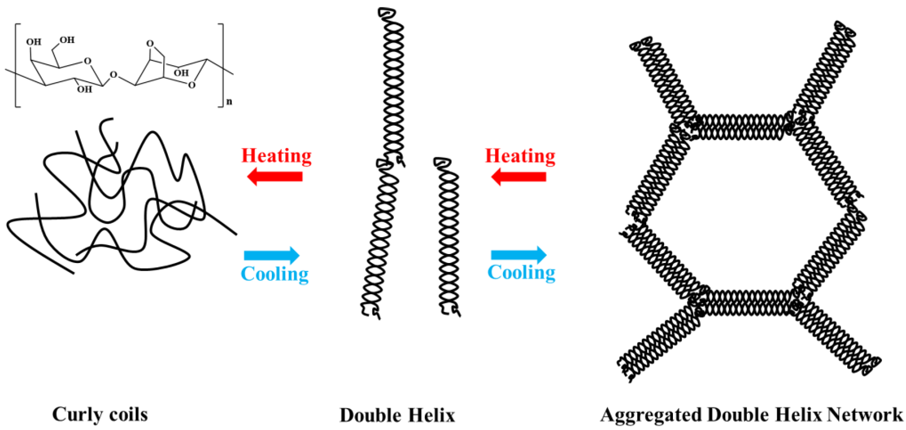
Figure 1. Structure and gelling mechanism of agarose [7].
Marinedrugs 21 00299 g001
Figure 2. Application of agarose.
Marinedrugs 21 00299 g002
Disclaimer/Publisher’s Note: The statements, opinions and data contained in all publications are solely those of the individual author(s) and contributor(s) and not of MDPI and/or the editor(s). MDPI and/or the editor(s) disclaim responsibility for any injury to people or property resulting from any ideas, methods, instructions or products referred to in the content.