Article

Ferroptosis Inhibitory Compounds from the Deep-Sea-Derived Fungus Penicillium sp. MCCC 3A00126

1
College of Marine Sciences, Shanghai Ocean University, 999 Hucheng Ring Road, Shanghai 201306, China
2
Key Laboratory of Marine Genetic Resources, Third Institute of Oceanography, Ministry of Natural Resources, 184 Daxue Road, Xiamen 361005, China
3
Institute of Drug Discovery Technology, Ningbo University, Ningbo 315211, China
*
Author to whom correspondence should be addressed.
Mar. Drugs 2023, 21(4), 234; https://doi.org/10.3390/md21040234
Submission received: 6 December 2022 / Revised: 27 March 2023 / Accepted: 7 April 2023 / Published: 10 April 2023
(This article belongs to the Special Issue Bioactive Compounds from the Deep-Sea-Derived Microorganisms)

Abstract

:
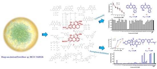
Two new xanthones (1 and 2) were isolated from the deep-sea-derived fungus Penicillium sp. MCCC 3A00126 along with 34 known compounds (336). The structures of the new compounds were established by spectroscopic data. The absolute configuration of 1 was validated by comparison of experimental and calculated ECD spectra. All isolated compounds were evaluated for cytotoxicity and ferroptosis inhibitory activities. Compounds 14 and 15 exerted potent cytotoxicity against CCRF-CEM cells, with IC50 values of 5.5 and 3.5 μM, respectively, whereas 26, 28, 33, and 34 significantly inhibited RSL3-induced ferroptosis, with EC50 values of 11.6, 7.2, 11.8, and 2.2 μM, respectively.

Graphical Abstract

1. Introduction

The oceans cover over 70% of the world’s surface, with 95% of them being deeper than 1000 m. In recent years, nearly half of the new marine natural products (MNPs) have been isolated from marine microorganisms [1,2,3], especially fungi, the most diverse and abundant eukaryotes on Earth, which can be distributed in any currently known extreme environment [4]. As a region rarely explored, the deep sea is characterized by a high pressure, a low/high (such as hydrothermal mouth) temperature, a high salt concentration, the absence of light, oligotrophic conditions, a high halogen content, and so on. To adapt to such extreme environments, deep-sea-derived microorganisms must develop special metabolic mechanisms, giving rise to tremendous secondary metabolites with unique structures and potent bioactivities [5]. For more than half a century, MNPs have been continuously discovered, but those from the deep sea are rare [6,7]. In recent years, with the development of deep-sea sample collection technology, reports of deep-sea MNPs have increased significantly. As an important group of deep-sea microorganisms, fungi can produce a large number of structurally novel and biologically active secondary metabolites, which have attracted extensive attention from researchers. For example, vercytochalasins A and B are two novel, biosynthetically related cytochalasins isolated from Curvularia verruculosa, the endophytic fungus of the deep-sea lobster Shinkaia crosnieri. Vercytochalasin A is the most potent natural product against angiotensin-I-converting enzyme (ACE), with an IC50 value of 505 nM [8]. Chevalinulins A and B are two indole alkaloids with a rare spiro-[bicyclo[2.2.2]octane-diketopiperazine] skeleton. They both exhibit significant in vivo proangiogenic activity in transgenic zebrafish [9].
Xanthones, also known as 9H-xanthen-9-ones, are a class of yellow compounds bearing a dibenzo-γ-pyrone scaffold. They are widely distributed in plants, lichens, and microorganisms of terrestrial and marine origin, and exhibit diverse biological activities such as antiviral [10], cytotoxic [11], antibacterial [12], antifungal [13], and hypoglycemic [14] activities. The molecular skeleton of xanthones can bind with a variety of targets, so this family of compounds is often called “privileged structures” [15]. They are regarded as typical aromatic polyketone and appear in the form of fully aromatic or hydrogenated derivatives [16]. In general, xanthones can be classified into monomers, dimers, and heterodimers. According to the degree of hydrogenation of the skeleton aromatic ring, they can be further split into four subclasses: fully aromatic xanthones, dihydro-, tetrahydro-, and hexahydro-xanthones [16]. From 2010 to 2021, 100 marine xanthones were reported, among which 51 were new compounds. Most of the new xanthones were derived from marine fungi, especially deep-sea fungi isolated from deep-sea sediments or organisms [17]. Therefore, deep-sea-derived fungi are undoubtedly an important resource for the discovery of xanthones with novel structures and significant bioactivities.
During our ongoing search for bioactive secondary metabolites from deep-sea-derived fungi [18,19,20,21], the crude extract of Penicillium sp. MCCC 3A00126 isolated from the Eastern Pacific Ocean at a depth of 5246 m showed significant cytotoxicity against acute lymphoblastic leukemia CCRF-CEM with the cell survival rate of 29.8 % under the concentration of 10 μg/mL. Therefore, it was subjected to a systematic chemical investigation. As a result, two novel (1 and 2) and 13 known (315) xanthones were isolated and purified, along with 21 known miscellaneous compounds (1636) (Figure 1). Herein, the details of isolation, structure, and bioactivity are reported.

2. Results and Discussion

Compound 1 was obtained as a colorless gum. The molecular formula C18H18O9 was determined by the positive HR-ESI-MS (high resolution electrospray mass spectrometry spectrum) at m/z 401.0839 [M + Na]+, suggesting ten degrees of unsaturation. The 1H (Figure S1 in the Supplementary Materials) and 13C (Figure S2 in the Supplementary Materials) NMR (nuclear magnetic resonance) spectroscopic data (Table 1) exhibited the presence of one methoxyl [δH 3.84 (3H, s); δC 53.4 q], one methyl singlet [δH 2.16 (3H, s); δC 20.8 q], one oxygenated [δH 5.13 (2H, s); δC 65.0 t], and two aliphatic [δH 2.25 (2H, m), 2.87 (2H, m); δC 24.2 t, 26.1 t] methylene groups, one oxygenated aliphatic [δH 4.09 (1H, dd, J = 10.3, 3.6 Hz); δC 72.6 d] and two olefinic [δH 6.75 (1H, s), 6.86 (1H, s); δC 109.9 d, 105.5 d] methines, as well as ten quaternary carbons including one oxygenated aliphatic (δC 76.2 s), six olefinic (δC 109.6 s, 117.0 s, 145.0 s, 156.1 s, 160.7 s, 167.4 s), and three carbonyl (δC 170.5 s, 172.7 s, 182.0 s] carbons. These signals were very similar to aspergillusone B (4) [22], except for an additional acetyl group [δH 2.16 (3H, s); δC 20.8 q, 170.5 s] at the C-11 position. This was supported by the downfield shifts of H-11 from δH 4.76 to δH 5.13 and C-11 from δC 64.4 to δC 65.0. Further confirmation was obtained by the HMBC (heteronuclear multiple-bond correlation) correlations of H2-11 to the carbonyl group of the acetyl moiety; and the 1H-1H COSY (correlation spectroscopy) cross peaks of H2-6 to H2-5/H-7 (Figure 2).
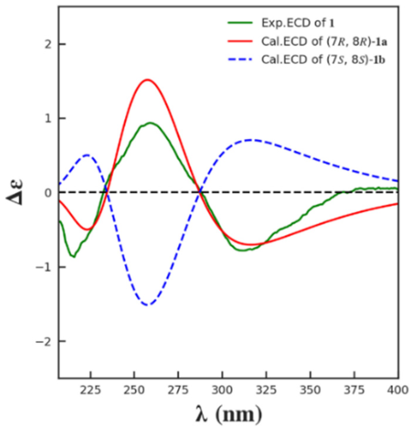
The coupling constants between H-7 and H2-6 of 1 (J = 10.3 Hz, 3.6 Hz) indicated the same pseudoaxial position of H-7 as that of 4 [22], as it was found in a simple MM2 conformational study of both possible 7,8-anti and 7,8-syn diols (Figure 2). The observed optical rotation value of 1 ([α ] D 25 −82.5) was close to that of 4 ([α ] D 25 −46.3) in the same concentration (c 0.2) and the same solvent (CHCl3), (c 0.2, CHCl3), suggesting they have the same absolute configuration at C-7 and C-8. For the further confirmation, the ECD (electron circular dichroism) spectra were calculated for (7R,8R)-1 (1a) and its enantiomer (7S,8S)-1 (1b) using Yinfo Cloud Computing Platform (https://cloud.yinfotek.com, accessed on 13 June 2022). Thirty states of each seven conformers were calculated to generate the ECD curves. As shown in Figure 3, the calculated ECD spectrum of 1a was consistent with that of the experimental one. On the basis of the above evidence, compound 1 was then elucidated as 11-O-acetylaspergillusone B.
Compound 2 was obtained as a amorphous yellow solid. The molecular formula C17H12O7 was established by its positive HR-ESI-MS spectrum at m/z 351.0482 [M + Na]+. The 1H and 13C data of 2 showed the presence of two methoxyls, five methines and ten quaternary carbons, which were closely related to those of huperxanthone A (13) [14], except that the sp2 quaternary carbon at C-7 (δC 151.1 s) in 13 was changed as an sp2 methine (δC 123.1 d) in 2. By detailed analysis of its HSQC (heteronuclear single quantum correlation), 1H–1H COSY, HMBC, and NOESY (nuclear Overhauser effect) spectroscopic data, compound 2 was then established as 7-dehydroxyhuperxanthone A.
By comparison of the NMR and MS data with those published in the literature, 34 known compounds were identified as 13 xanthones: (7R,8R)-α-diversonolic ester (3) [23,24], aspergillusone B (4) [22], 8-hydroxy-6-methyl-9-oxo-9H-xanthene-1-carboxylate (5) [25], yicathin B (6) [26], pinselin (7) [27], sydowinins A (8) and B (9) [28], huperxanthone C (10) [14], 13-O-acetylsydowinin B (11) [29], 8-(methoxycarbonyl)-1-hydroxy-9-oxo-9H-xanthene-3-carboxylic acid (12) [25], huperxanthone A (13) [14], sterigmatocystin (14) [30], 5-methoxysterigmatocystin (15) [31]; six anthraquinones: versicolorin B (16) [32], 8-O-methylversicolorin B (17) [33], anthraquinone aversin (18) [34], averufin (19) [35], 6-O-methylaverufin (20) [36], questin (21) [37]; five sesquiteroenoids: (S)-(+)-sydonic acid (22) [38], (S)-(+)-11-dehydrosydonic acid (23) [38], (−)-12-acetoxy-1-deoxysydonic acid (24) [39], (7S,11S)-(+)-12-acetoxysydonic acid (25) [38], (−)-(7R,10R)-iso-10-hydroxysydowic acid (26) [39]; four diphenyl ethers: diorcinol (27) [40], verticilatin (28) [41], (R)-3-((2-(2-hydroxypropan-2-yl)-6-methyl-2,3-dihydrobenzofuran-4-yl)oxy)-5-methylphenol (29) [42], (3S)-3,4-dihydro-5-(3-hydroxy-5-methylphenoxy)-2,2,7-trimethyl-2H-chromen-3-ol (30) [43]; one polyketone: 3-hydroxy-5-methylphenyl-2,4-dihydroxy-6-methylbenzoate (31) [44]; four indole alkaloids: brevianamide F (32) [45], notoamide I (33) [46], notoamide C (34) [47], psychrophilin D (35) [48], and one steroid: 5a,8a-epidioxy-22E-ergosta-6,9(11)-trien-3β-ol (36) [49].
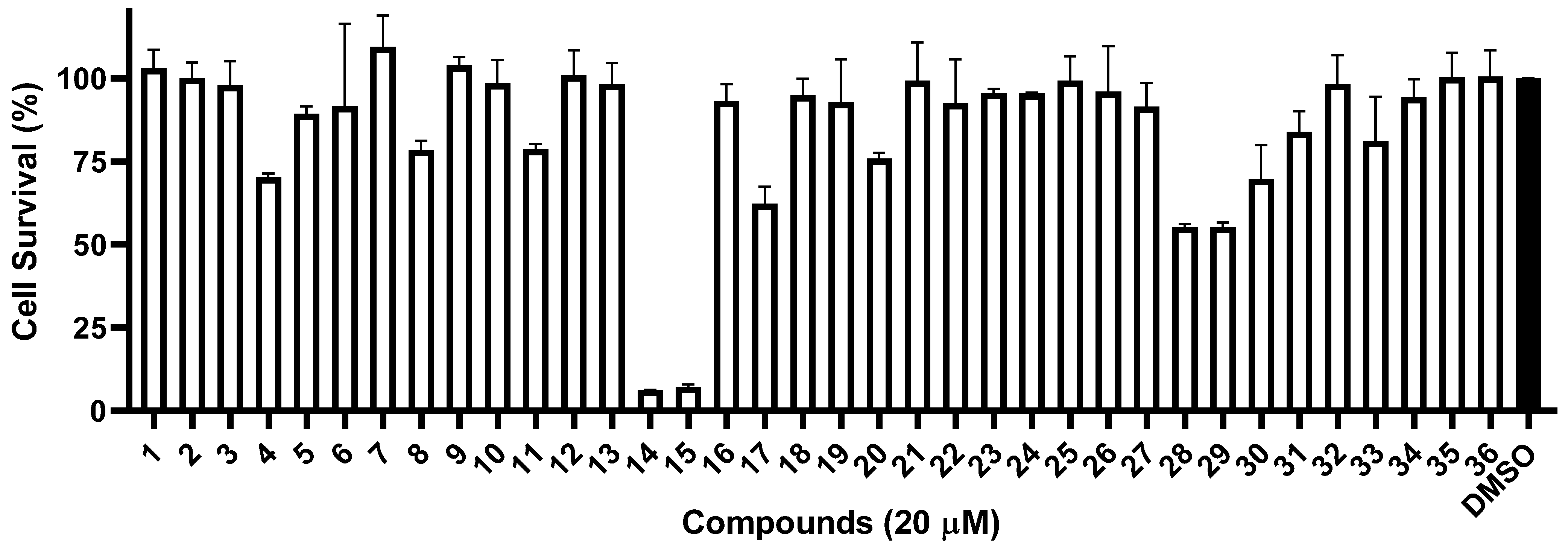
Since the crude extract of Penicillium sp. MCCC 3A00126 showed a potent anti-proliferative effect on CCRF-CEM, all 36 isolates were subjected to cytotoxicity tests on the same acute lymphoblastic leukemia using the CCK-8 assay. As shown in Figure 4, under a concentration of 20 μM, two compounds, 14 and 15, exerted potent activity, with cell survival rates of 6.2% and 7.3%, respectively, while seven compounds, 4, 8, 11, 17, 20, 28, and 29, showed weak effects, with cell survival rates of 70.2%, 78.5%, 78.8%, 62.3%, 75.8%, 55.3%, and 55.3%, respectively. Interestingly, compounds 14 and 15 possess a difuran ring at C-5 and C-6, which might be the key to the bioactivity.
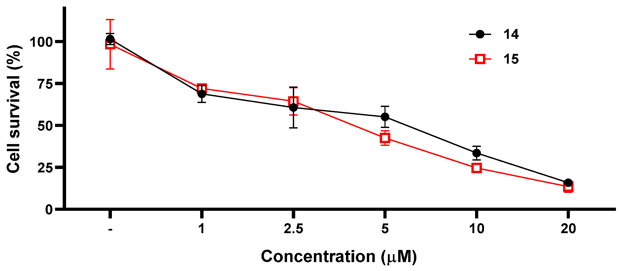
Compounds 14 and 15 were further evaluated to determine their 50% inhibiting concentration (IC50) against CCRF-CEM using five different concentrations: 1 μM, 2.5 μM, 5.0 μM, 10.0 μM, and 20.0 μM. The IC50 values of 14 and 15 were found to be 5.5 μM and 3.5 μM, respectively (Figure 5).
Ferroptosis is an iron-dependent mode of necroptosis induced by certain small molecules, such as RSL3 (the glutathione peroxidase 4 inhibitor), which is different from apoptosis, necrolysis, and autophagy [50]. Its main characteristics are the generation of ROS (reactive oxygen species), LPO (lipid peroxidation), and iron accumulation. RSL3 acts on specific targets in cells and causes a reduction in antioxidants GSH (glutathione) and GPX4 (glutathione peroxidase 4), resulting in the accumulation of ROS in cells, LPO in cells, and ferroptosis in cells under the synergistic effect of iron [51]. Many tumor cells that are easy to metastasize are prone to ferroptosis, so inducing and inhibiting ferroptosis for pharmacological regulation has great potential in the treatment of certain cancers.
To further investigate whether these isolates could inhibit ferroptosis, RSL3, the GPX4 inhibitor, was used to induce ferroptosis in CCRF-CEM cells. As a result, compounds 26, 28, 33, and 34 exerted strong inhibition, with cell survival rates of 83.9%, 110.0%, 99.0%, and 105.2%, respectively, under a concentration of 20 μM. Additionally, compounds 3, 27, 29, and 30 showed weak activity, with cell survival rates of 36.0%, 16.6%, 19.5%, and 28.8%, respectively (Figure 6).
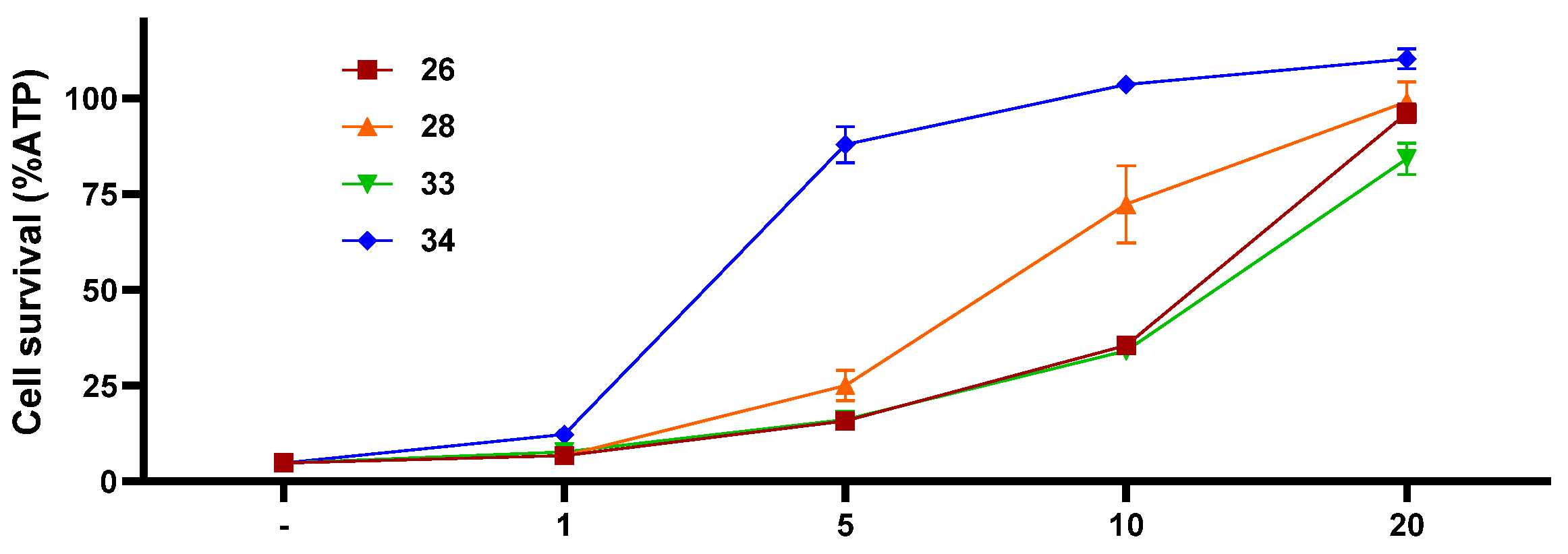
To determine the 50% effective concentration (EC50) of compounds 26, 28, 33, and 34, four different concentrations (1 μM, 5.0 μM, 10.0 μM, and 20.0 μM) were adopted on RSL3-induced ferroptosis in CCRF-CEM cells, providing corresponding IC50 values of 11.6 μM, 7.2 μM, 11.8 μM, and 2.2 μM, respectively (Figure 7).
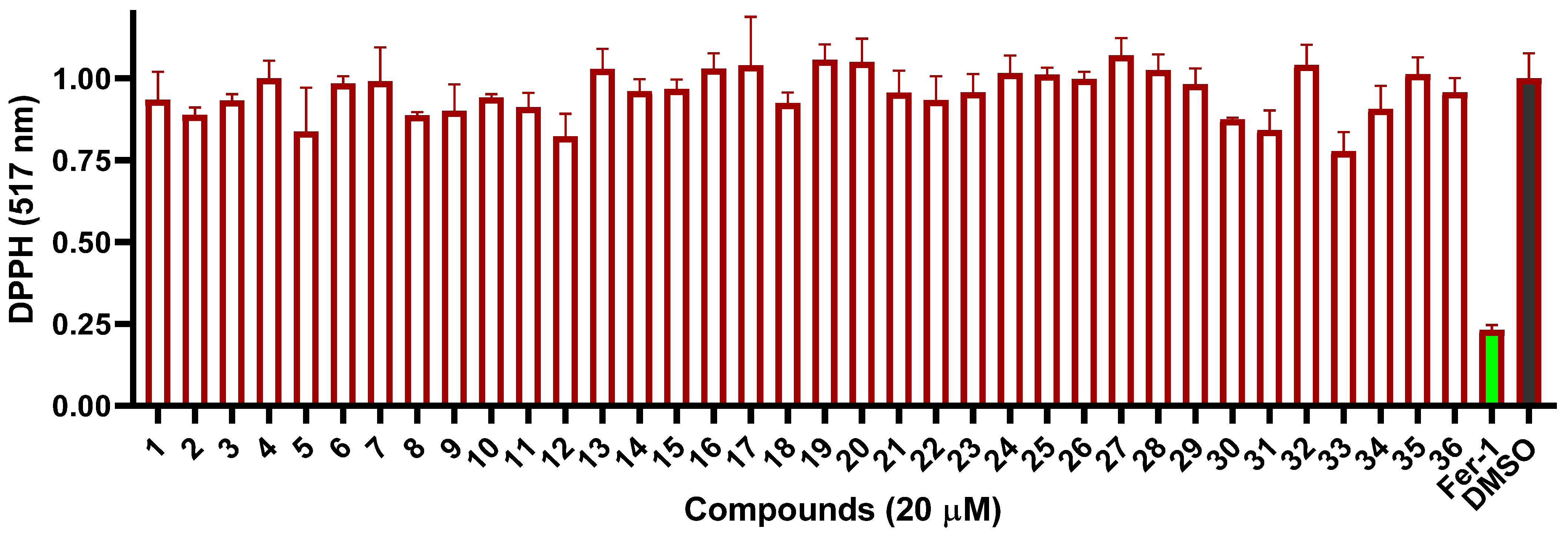
As ferroptosis was triggered by lipid peroxidation, many ferroptosis inhibitors exhibited antioxidant activity, such as ferrostain-1 (Fer-1) [50,52,53]. Therefore, the 2,2-diphenyl-1-picrylhydrazyl (DPPH) assay was performed on these compounds to evaluate their intrinsic antioxidant potential. However, none showed positive effects under a concentration of 20 μM (Figure 8), indicating that compounds 26, 28, 33, and 34 might exert ferroptosis inhibition by other mechanisms instead of DPPH.

3. Materials and Methods

3.1. General Experimental Procedures

The HR-ESI-MS spectra were obtained on a Waters Xevo G2 Q-TOF mass spectrometer equipped with a Spray™ ESI source in both the positive and negative ion mode. NMR spectra were recorded in CDCl3, CD3OD, or DMSO-d6 on a Bruker Avance III 400 Mz spectrometer at room temperature. Optical rotation was measured by an Anton Paar MCP 100 polarimeter. UV and ECD spectra were acquired on a JASCO J-810 spectropolarimeter. Preparative HPLC (high-performance liquid chromatography) separations for purification were carried out on an Agilent 1260 system with a semi-preparative chromatographic column (COSMOSIL 5C18-MS-II, 5PFP, SL-II, 250 mm × 10 mm). Materials for column chromatography (CC) included silica gel, ODS (octadecylsilyl), and Sephadex LH-20.

3.2. Biological Material

The deep-sea-derived fungus Penicillium sp. MCCC 3A00126 was isolated from a sediment sample collected from the Eastern Pacific Ocean at a depth of 5246 m by Professor De-Zan Ye of the Third Institute of Oceanography, Ministry of Natural Resources, in September 2003. The 18S rRNA gene sequence alignment (AM18217) showed great similarity (99.88%) to Penicillium sp. PB g (GenBank accession number MK372218.1); therefore, it was identified to be Penicillium sp. It was deposited at the Marine Culture Collection of China (Xiamen, China) with accession number MCCC 3A00126.

3.3. Fermentation, Isolation, and Purification

The strains stored at −80 °C were inoculated into a 250 mL conical flask containing 100 mL PDB culture medium to conduct initial activation for three days in a shaking table culture at 28 °C. Then, under the same culture conditions, 1 mL of the fungal liquid was placed in another 1 L conical flask containing 250 mL of PDB medium to conduct secondary activation.
The secondary activated fungal strain was inoculated into 60 Erlenmeyer flasks containing 80 g rice and 120 mL distilled water. The fermentation was kept under static conditions at 25 °C. After 40 days, 400 mL of EtOAc was added to each flask over 12 h. The organic solvent was filtered. The procedure was repeated four times. The organic solvents were combined and concentrated to a small volume. The latter was dissolved in MeOH and extracted by PE (petroleum ether) three times. The MeOH layer was concentrated to provide a crude extract (26.3 g), which was subjected to MPLC (medium-pressure liquid chromatography) over silica gel using the CH2Cl2/MeOH gradient (100% → 70%) to obtain four fractions (Fr.1–Fr. 4). Fraction Fr.1 (0.46 g) was separated by repeated column chromatography (CC) over ODS (MeOH/H2O) and Sephadex LH-20 (CH2Cl2/MeOH, 1:1 and 0:1), then purified by semi-prep. HPLC (C18, 10 × 250 mm, MeOH/H2O, 80% → 100%), providing 2 (15.0 mg) and 5 (14.0 mg). Fraction Fr.2 (1.1 g) was subjected to CC over ODS and Sephadex LH-20, then purified by semi-prep. HPLC (C18, 10 × 250 mm, MeOH/H2O, 70% → 90%), yielding 14 (2.0 mg), 15 (3.0 mg), and 18 (5.4 mg). Fraction Fr.3 (3.7 g) was separated by ODS CC with MeOH/H2O (30% → 80%, 3 h, 80% → 90%, 3 h) to obtain thirteen subfractions (Fr.3.1–Fr.3.13). Subfractions (Fr.3.1–Fr.3.9) were subjected to CC over Sephadex LX-20 using MeOH to yield compounds 3 (2.0 mg), 10 (9.0 mg), 11 (4.0 mg), 13 (8.0 mg), 16 (2.0 mg), 19 (2.5 mg), 20 (6.0 mg), 28 (9.5 mg), and 36 (28.0 mg), respectively. Sub-fraction Fr.3.10 was subjected to CC over Sephadex LH-20 (CH2Cl2/MeOH, 1:1) and silica gel (CH2Cl2/MeOH, 45:1), followed by separation using HPLC (C18, 10 mm × 250 mm, MeOH/H2O, 80% → 90%) to give 8 (15.0 mg) and 9 (2.0 mg). Fr.3.11 was separated by CC over LH-20 (MeOH) and silica gel (CH2Cl2/MeOH, 55:1) to obtain 12 (1.0 mg). Fr.3.12 was purified by semi-prep. HPLC (PFP-pentafluorophenyl group, 10 mm × 250 mm, MeOH/H2O, 60% → 90%), yielding 21 (3.0 mg), 29 (6.6 mg), and 30 (1.3 mg). Fr.3.13 was further separated by HPLC (C18, 10 mm × 250 mm, MeOH/H2O, 55% → 85%) to yield 6 (3.0 mg).
Using similar procedures, fraction Fr.4 (3.3 g) was separated into thirteen subfractions (Fr.4.1–Fr. 4.13) by CC over ODS (MeOH/H2O, 5 → 60%, 4 h, 60% → 100%, 2 h). Compounds 1 (8.0 mg), 4 (23.3 mg), 17 (23.0 mg), 22 (39.0 mg), 27 (7.0 mg), and 31 (19.8 mg) were obtained from Fr.4.1–Fr.4.6 by CC over Sephadex LH-20 (MeOH). Fr.4.7 and Fr.4.8 were separated by HPLC (C18, 10 mm × 250 mm, MeOH/H2O, 35% → 65%) to obtain 26 (1.0 mg) and 32 (2.6 mg), respectively. Fr.4.9 was subjected to Sephadex LH-20 CC, eluting with MeOH, and then purification by HPLC (C18, 10 mm × 250 mm, MeOH/H2O, 55% → 85%) afforded 24 (3.0 mg) and 25 (9.5 mg). Fr.4.10 was separated by HPLC on silica gel (10 mm × 250 mm, CH2Cl2/MeOH, 100% → 90%) to yield 35 (12.0 mg). Compounds 23 (2.5 mg) and 33 (1.7 mg) were also separated from Fr.4.11 using HPLC (C18, 10 mm × 250 mm, MeOH/H2O, 60% → 80%). Fr.4.12 was chromatographed by CC over silica gel (PE/EtOAc, 3:1) to yield 7 (3.2 mg) and Fr.4.13 was subjected HPLC (C18, 10 mm × 250 mm, MeOH/H2O, 55% → 85%) to yield 34 (0.7 mg).
11-O-Acetylaspergillusone B (1). Colorless gum; [α ] D 25 −82.5 (c 0.2, CHCl3), −19.0 (c 0.1, MeOH); UV (2.6 mM, MeOH) λmax (log ε) 212 (2.32), 240 (2.42), 289 (1.69), 331 (2.04) nm; ECD (2.6 mM, MeOH) λmaxε) 216 (1.99), 263 (2.06), 313 (1.79); 1H and 13C NMR data, Table 1; HRESIMS m/z 401.0839 [M + Na]+ (calcd for C18H18O9Na, 401.0849).
7-Dehydroxyhuperxanthone A (2). Yellow amorphous solid; UV (3.0 mM, MeOH) λmax (log ε) 266 (3.23); 1H and 13C NMR data, Table 1; HRESIMS m/z 351.0482 [M + Na]+ (calcd for C17H12O7Na, 351.0481).

3.4. ECD Calculation

ECD calculations were performed using Yinfo Cloud Computing Platform, a user-friendly and versatile web server for biomedicinal, material, and statistical research (https://cloud.yinfotek.com, accessed on 13 June 2022). The conformational analysis of compound 1 was carried out using the MMFF94 force field at an energy cutoff of 7.0 kcal/mol and an RSMD threshold of 0.5 Å. A total of thirteen conformations were obtained from the conformational analysis, and seven of which, accounting for more than 1%, were selected for further screening. The seven conformers were relocated and confirmed at the PM6, HF/6-31G(d), and B3LYP/6-1G(d) level to obtain three dominant conformers. Further, the calculation of the theoretical ECD spectra at the B3LYP/6-311G(d, p) level was conducted in MeOH. The final spectrum was obtained by averaging each conformer using the Boltzmann distribution.

3.5. Cytotoxic Experiment

CCRF-CEM cells (CL-0058), kindly provided by Procell Life Science & Technology Co., Ltd. (Wuhan, China), were cultured in RIPM-1640 at 37 °C in a humidified atmosphere containing 5% CO2 with 10% inactive fetal bovine serum, 2 mM l-glutamine, 100 IU penicillin, and 100 mg/mL streptomycin. The cytotoxicity experiment was conducted using the CCK-8 (Cell Counting Kit-8) assay. Briefly, 4000 cells were seeded on a 96-well plate. After 24 h, different concentrations of the tested compounds were added, and the incubation continued for 48 h. The CCK-8 assay was performed with MD Spectra Max Paradigm.

3.6. Ferroptosis Inhibitory Assay

As previously reported [54], 10,000 CCRF-CEM cells were seeded on a 96-well plate for 24 h. Then, ferrostatin-1 (1 μM, as the positive control) and different concentrations of the tested compounds, ranging from 1 μM to 20 μM, were added for first-round screening. After 0.5 h, RSL3 (2 μM) was added to trigger ferroptosis. Four hours later, cellular ATP was detected using the Cell Titer Glo Luminescent Cell Viability assay kit (G7570, Promega, Madison, WI, USA) according to the manufacturer’s instructions. Then, the EC50 values were determined as the indicated concentration.

3.7. DPPH Assay

According to a reported procedure [55], the stable radical DPPH (2,2-diphenyl-1-picrylhydrazyl) was dissolved in MeOH to a final working concentration of 100 μM. Then, 1 μL of the indicated compounds dissolved in DMSO (10 mM) was added to a final concentration of 100 μM, inverted several times, and allowed to incubate at room temperature for 30 minutes. Samples were then aliquoted to a 96-well microplate and absorbance at 517 nm was recorded using Spectra Max Paradigm (Molecular Devices, San Jose, CA, USA). The relative DPPH normalized to the background (MeOH only) showed the mean ± SD, and the experiments were triplicated.

4. Conclusions

Systematic chemical investigation of the deep-sea fungus Penicillium sp. 3A00126 yielded 36 compounds, comprising fifteen xanthones (315), six anthraquinones (1621), five sesquiterpenoids (2226), four diphenyl ethers (2730), one polyketone (31), four indole alkaloids (3235), and one steroid (36). Compound 1, named 11-O-acetylaspergillusone B, is a new tetrahydroxanthone, and compound 2, 7-dehydroxyhuperxanthone A, is a new, fully aromatic xanthone. All 36 isolates were tested for cytotoxicity and ferroptosis inhibitory effects. Sterigmatocystin (14) and 5-methoxysterigmatocystin (15) showed potent cytotoxicity against CCRF-CEM cells, with IC50 values of 5.5 μM and 3.5 μM, respectively, whereas (−)-(7R,10R)-iso-10-hydroxysydowic acid (26), verticilatin (28), notoamide I (33), and notoamide C (34) significantly inhibited RSL3-induced ferroptosis, with EC50 values of 11.6 μM, 7.2 μM, 11.8 μM, and 2.2 μM, respectively.

Supplementary Materials

The following are available online at https://www.mdpi.com/article/10.3390/md21040234/s1, Figures S1–S13: One-dimensional and two-dimensional NMR and HR-ESI-MS spectra of compound 1 and 2.

Author Contributions

X.-W.Y. designed the project; Y.-J.H. and Z.-B.Z. isolated and purified compounds. L.X., M.-M.X. and Y.Z. performed the fermentation. H.-B.M. conducted the biological experiments. Y.-J.H., H.-Y.Y. and X.-W.Y. analyzed the data and wrote the paper, while critical revision of the publication was performed by all authors. All authors have read and agreed to the published version of the manuscript.

Funding

The work was financially supported by Xiamen Southern Oceanographic Center Project (22GYY007HJ07) and the National Natural Science Foundation of China (22177143).

Institutional Review Board Statement

Not applicable.

Informed Consent Statement

Not applicable.

Data Availability Statement

Not applicable.

Conflicts of Interest

The authors declare no conflict of interest.

References

  1. Carroll, A.R.; Copp, B.R.; Davis, R.A.; Keyzers, R.A.; Prinsep, M.R. Marine natural products. Nat. Prod. Rep. 2023, 40, 275–325. [Google Scholar] [CrossRef] [PubMed]
  2. Carroll, A.R.; Copp, B.R.; Davis, R.A.; Keyzers, R.A.; Prinsep, M.R. Marine natural products. Nat. Prod. Rep. 2021, 38, 362–413. [Google Scholar] [CrossRef] [PubMed]
  3. Carroll, A.R.; Copp, B.R.; Davis, R.A.; Keyzers, R.A.; Prinsep, M.R. Marine natural products. Nat. Prod. Rep. 2022, 39, 1122–1171. [Google Scholar] [CrossRef] [PubMed]
  4. Zain Ul Arifeen, M.; Ma, Y.N.; Xue, Y.R.; Liu, C.H. Deep-sea fungi could be the new arsenal for bioactive molecules. Mar. Drugs 2019, 18, 9. [Google Scholar] [CrossRef] [PubMed] [Green Version]
  5. Sun, C.; Mudassir, S.; Zhang, Z.; Feng, Y.; Chang, Y.; Che, Q.; Gu, Q.; Zhu, T.; Zhang, G.; Li, D. Secondary metabolites from deep-sea derived microorganisms. Curr. Med. Chem. 2020, 27, 6244–6273. [Google Scholar] [CrossRef] [PubMed]
  6. Skropeta, D.; Wei, L. Recent advances in deep-sea natural products. Nat. Prod. Rep. 2014, 31, 999–1025. [Google Scholar] [CrossRef] [PubMed]
  7. Skropeta, D. Deep-sea natural products. Nat. Prod. Rep. 2008, 25, 1131–1166. [Google Scholar] [CrossRef] [Green Version]
  8. Hu, X.; Li, X.; Yang, S.; Li, X.; Wang, B.; Meng, L. Vercytochalasins A and B: Two unprecedented biosynthetically related cytochalasins from the deep-sea-sourced endozoic fungus Curvularia verruculosa. Chin. Chem. Lett. 2023, 34, 107516. [Google Scholar] [CrossRef]
  9. Yan, L.H.; Li, P.H.; Li, X.M.; Yang, S.Q.; Liu, K.C.; Wang, B.G.; Li, X. Chevalinulins A and B, proangiogenic alkaloids with a spiro[bicyclo[2.2.2]octane-diketopiperazine] skeleton from deep-sea cold-seep-derived fungus Aspergillus chevalieri CS-122. Org. Lett. 2022, 24, 2684–2688. [Google Scholar] [CrossRef]
  10. Liu, F.A.; Lin, X.; Zhou, X.; Chen, M.; Huang, X.; Yang, B.; Tao, H. Xanthones and quinolones derivatives produced by the deep-sea-derived fungus Penicillium sp. SCSIO Ind16F01. Molecules 2017, 22, 1999. [Google Scholar] [CrossRef] [Green Version]
  11. Isaka, M.; Palasarn, S.; Kocharin, K.; Saenboonrueng, J. A cytotoxic xanthone dimer from the entomopathogenic fungus Aschersonia sp. BCC 8401. J. Nat. Prod. 2005, 68, 945–946. [Google Scholar] [CrossRef] [PubMed]
  12. Lee, Y.M.; Li, H.; Hong, J.; Cho, H.Y.; Bae, K.S.; Kim, M.A.; Kim, D.K.; Jung, J.H. Bioactive metabolites from the sponge-derived fungus Aspergillus versicolor. Arch. Pharm. Res. 2010, 33, 231–235. [Google Scholar] [CrossRef] [PubMed]
  13. Fredimoses, M.; Zhou, X.; Ai, W.; Tian, X.; Yang, B.; Lin, X.; Liu, J.; Liu, Y. Emerixanthone E, a new xanthone derivative from deep sea fungus Emericella sp. SCSIO 05240. Nat. Prod. Res. 2019, 33, 2088–2094. [Google Scholar] [CrossRef] [PubMed]
  14. Ma, T.T.; Shan, W.G.; Ying, Y.M.; Ma, L.F.; Liu, W.H.; Zhan, Z.J. Xanthones with α-glucosidase inhibitory activities from Aspergillus versicolor, a fungal endophyte of Huperzia serrata. Helv. Chim. Acta 2015, 98, 148–152. [Google Scholar] [CrossRef]
  15. Lesch, B.; Brase, S. A short, atom-economical entry to tetrahydroxanthenones. Angew. Chem. Int. Ed. Engl. 2004, 43, 115–118. [Google Scholar] [CrossRef]
  16. Masters, K.S.; Brase, S. Xanthones from fungi, lichens, and bacteria: The natural products and their synthesis. Chem. Rev. 2012, 112, 3717–3776. [Google Scholar] [CrossRef]
  17. Verissimo, A.C.S.; Pinto, D.; Silva, A.M.S. Marine-derived xanthone from 2010 to 2021: Isolation, bioactivities and total synthesis. Mar. Drugs 2022, 20, 347. [Google Scholar] [CrossRef]
  18. Niu, S.; Xie, C.L.; Xia, J.M.; Liu, Q.M.; Peng, G.; Liu, G.M.; Yang, X.W. Botryotins A–H, tetracyclic diterpenoids representing three carbon skeletons from a deep-sea-derived Botryotinia fuckeliana. Org. Lett. 2020, 22, 580–583. [Google Scholar] [CrossRef]
  19. Xie, C.L.; Liu, Q.; He, Z.H.; Gai, Y.B.; Zou, Z.B.; Shao, Z.Z.; Liu, G.M.; Chen, H.F.; Yang, X.W. Discovery of andrastones from the deep-sea-derived Penicillium allii-sativi MCCC 3A00580 by OSMAC strategy. Bioorg. Chem. 2021, 108, 104671. [Google Scholar] [CrossRef]
  20. Xie, C.L.; Zhang, D.; Guo, K.Q.; Yan, Q.X.; Zou, Z.B.; He, Z.H.; Wu, Z.; Zhang, X.K.; Chen, H.F.; Yang, X.W. Meroterpenthiazole A, a unique meroterpenoid from the deep-sea-derived Penicillium allii-sativi, significantly inhibited retinoid X receptor (RXR)-α transcriptional effect. Chin. Chem. Lett. 2022, 33, 2057–2059. [Google Scholar] [CrossRef]
  21. He, Z.H.; Xie, C.L.; Wu, T.; Yue, Y.T.; Wang, C.F.; Xu, L.; Xie, M.M.; Zhang, Y.; Hao, Y.J.; Xu, R.; et al. Tetracyclic steroids bearing a bicyclo[4.4.1] ring system as potent antiosteoporosis agents from the deep-sea-derived fungus Rhizopus sp. W23. J. Nat. Prod. 2023, 86, 157–165. [Google Scholar] [CrossRef] [PubMed]
  22. Trisuwan, K.; Rukachaisirikul, V.; Kaewpet, M.; Phongpaichit, S.; Hutadilok-Towatana, N.; Preedanon, S.; Sakayaroj, J. Sesquiterpene and xanthone derivatives from the sea fan-derived fungus Aspergillus sydowii PSU-F154. J. Nat. Prod. 2011, 74, 1663–1667. [Google Scholar] [CrossRef] [PubMed]
  23. Holker, J.S.E.; O’Brien, E.; Simpson, T.J. The structures of some metabolites of Penicillium diversum: α- and β-diversonolic esters. J. Chem. Soc. Perkin Trans. 1 1983, 1365–1368. [Google Scholar] [CrossRef]
  24. Nicolaou, K.C.; Li, A. Total syntheses and structural revision of α- and β-diversonolic esters and total syntheses of diversonol and blennolide C. Angew. Chem. Int. Ed. Engl. 2008, 47, 6579–6582. [Google Scholar] [CrossRef] [PubMed]
  25. Shao, C.; Wang, C.; Wei, M.; Gu, Y.; Xia, X.; She, Z.; Lin, Y. Structure elucidation of two new xanthone derivatives from the marine fungus Penicillium sp. (ZZF 32#) from the south china sea. Magn. Reson. Chem. 2008, 46, 1066–1069. [Google Scholar] [PubMed]
  26. Sun, R.R.; Miao, F.P.; Zhang, J.; Wang, G.; Yin, X.L.; Ji, N.Y. Three new xanthone derivatives from an algicolous isolate of Aspergillus wentii. Magn. Reson. Chem. 2013, 51, 65–68. [Google Scholar] [CrossRef]
  27. Cui, X.; Zhu, G.; Liu, H.; Jiang, G.; Wang, Y.; Zhu, W. Diversity and function of the antarctic krill microorganisms from Euphausia superba. Sci. Rep. 2016, 6, 36496. [Google Scholar] [CrossRef] [Green Version]
  28. Hamasaki, T.; Sato, Y.; Hatsuda, Y. Structure of sydowinin A, sydowinin B, and sydowinol, metabolites from Aspergillus sydowi. Agric. Biol. Chem. 2014, 39, 2341–2345. [Google Scholar] [CrossRef]
  29. Song, X.Q.; Zhang, X.; Han, Q.J.; Li, X.B.; Li, G.; Li, R.J.; Jiao, Y.; Zhou, J.C.; Lou, H.X. Xanthone derivatives from Aspergillus sydowii, an endophytic fungus from the liverwort Scapania ciliata S. Lac and their immunosuppressive activities. Phytochem. Lett. 2013, 6, 318–321. [Google Scholar] [CrossRef]
  30. Zhu, F.; Lin, Y.C. Three xanthones from a marine-derived mangrove endophytic fungus. Chem. Nat. Compd. 2007, 43, 132–135. [Google Scholar] [CrossRef]
  31. Shao, C.; She, Z.; Guo, Z.; Peng, H.; Cai, X.; Zhou, S.; Gu, Y.; Lin, Y. 1H and 13C NMR assignments for two anthraquinones and two xanthones from the mangrove fungus (ZSUH-36). Magn. Reson. Chem. 2007, 45, 434–438. [Google Scholar] [CrossRef] [PubMed]
  32. Graybill, T.L.; Casillas, E.G.; Pal, K.; Townsend, C.A. Silyl triflate-mediated ring-closure and rearrangement in the synthesis of potential bisfuran-containing intermediates of aflatoxin biosynthesis. J. Am. Chem. Soc. 1999, 121, 7729–7746. [Google Scholar] [CrossRef]
  33. Dou, Y.; Wang, X.; Jiang, D.; Wang, H.; Jiao, Y.; Lou, H.; Wang, X. Metabolites from Aspergillus versicolor, an endolichenic fungus from the lichen Lobaria retigera. Drug Discov. Ther. 2014, 8, 84–88. [Google Scholar] [CrossRef] [PubMed] [Green Version]
  34. Liu, X.B.; Zheng, N.; Liang, L.Q.; Zhao, D.M.; Qin, Y.Y.; Li, J.; Yang, R.Y. Secondary metabolites from the endophytic fungus Fusarium equiseti and their antibacterial activities. Chem. Nat. Compd. 2019, 55, 1141–1144. [Google Scholar] [CrossRef]
  35. Yamazaki, M.; Satoh, Y.; Maebayashi, Y.; Horie, Y. Monoamine oxidase inhibitors from a fungus, Emericella navahoensis. Chem. Pharm. Bull. 1988, 36, 670–675. [Google Scholar] [CrossRef] [Green Version]
  36. Steyn, P.S.; Vleggaar, R.; Wessels, P.L.; Cole, R.J.; Scott, D.B. Structure and carbon-13 nuclear magnetic resonance assignments of versiconal acetate, versiconol acetate, and versiconol, metabolites from cultures of Aspergillus parasiticus treated with dichlorvos. J. Chem. Soc. Perkin Trans. 1 1979, 451–459. [Google Scholar] [CrossRef]
  37. Liu, D.; Yan, L.; Ma, L.; Huang, Y.; Pan, X.; Liu, W.; Lv, Z. Diphenyl derivatives from coastal saline soil fungus Aspergillus iizukae. Arch. Pharm. Res. 2015, 38, 1038–1043. [Google Scholar] [CrossRef]
  38. Lu, Z.; Zhu, H.; Fu, P.; Wang, Y.; Zhang, Z.; Lin, H.; Liu, P.; Zhuang, Y.; Hong, K.; Zhu, W. Cytotoxic polyphenols from the marine-derived fungus Penicillium expansum. J. Nat. Prod. 2010, 73, 911–914. [Google Scholar] [CrossRef]
  39. Li, X.B.; Zhou, Y.H.; Zhu, R.X.; Chang, W.Q.; Yuan, H.Q.; Gao, W.; Zhang, L.L.; Zhao, Z.T.; Lou, H.X. Identification and biological evaluation of secondary metabolites from the endolichenic fungus Aspergillus versicolor. Chem. Biodivers. 2015, 12, 575–592. [Google Scholar] [CrossRef]
  40. Sanchez, J.F.; Chiang, Y.M.; Szewczyk, E.; Davidson, A.D.; Ahuja, M.; Elizabeth Oakley, C.; Woo Bok, J.; Keller, N.; Oakley, B.R.; Wang, C.C. Molecular genetic analysis of the orsellinic acid/F9775 gene cluster of Aspergillus nidulans. Mol. Biosyst. 2010, 6, 587–593. [Google Scholar] [CrossRef] [PubMed]
  41. Wei, P.Y.; Li, L.; Yang, C.G.; Luo, D.Q.; Zheng, Z.H.; Lu, X.H.; Shi, B.Z. A novel oxybis cresol verticilatin with highly varying degrees of biological activities from the insect pathogenic fungus Paecilomyces verticillatus. J. Asian Nat. Prod. Res. 2014, 16, 1153–1157. [Google Scholar] [CrossRef]
  42. Hu, S.S.; Jiang, N.; Wang, X.L.; Chen, C.J.; Fan, J.Y.; Wurin, G.G.; Ge, H.M.; Tan, R.X.; Jiao, R.H. Prenylated diphenyl ethers from the mantis-associated fungus Aspergillus versicolor GH-2. Tetrahedron Lett. 2015, 56, 3894–3897. [Google Scholar] [CrossRef]
  43. Wang, X.; Mou, Y.; Hu, J.; Wang, N.; Zhao, L.; Liu, L.; Wang, S.; Meng, D. Cytotoxic polyphenols from a sponge-associated fungus Aspergillus versicolor Hmp-48. Chem. Biodivers. 2014, 11, 133–139. [Google Scholar] [CrossRef]
  44. Eamvijarn, A.; Kijjoa, A.; Bruyere, C.; Mathieu, V.; Manoch, L.; Lefranc, F.; Silva, A.; Kiss, R.; Herz, W. Secondary metabolites from a culture of the fungus Neosartorya pseudofischeri and their in vitro cytostatic activity in human cancer cells. Planta Med. 2012, 78, 1767–1776. [Google Scholar] [CrossRef] [PubMed] [Green Version]
  45. Grundmann, A.; Li, S.M. Overproduction, purification and characterization of FtmPT1, a brevianamide F prenyltransferase from Aspergillus fumigatus. Microbiology 2005, 151, 2199–2207. [Google Scholar] [CrossRef]
  46. Tsukamoto, S.; Kato, H.; Samizo, M.; Nojiri, Y.; Onuki, H.; Hirota, H.; Ohta, T. Notoamides F-K, prenylated indole alkaloids isolated from a marine-derived Aspergillus sp. J. Nat. Prod. 2008, 71, 2064–2407. [Google Scholar] [CrossRef] [PubMed]
  47. Kato, H.; Yoshida, T.; Tokue, T.; Nojiri, Y.; Hirota, H.; Ohta, T.; Williams, R.M.; Tsukamoto, S. Notoamides A–D: Prenylated indole alkaloids isolated from a marine-derived fungus, Aspergillus sp. Angew. Chem. Int. Ed. Engl. 2007, 46, 2254–2256. [Google Scholar] [CrossRef] [PubMed]
  48. Peng, J.; Gao, H.; Zhang, X.; Wang, S.; Wu, C.; Gu, Q.; Guo, P.; Zhu, T.; Li, D. Psychrophilins E-H and versicotide C, cyclic peptides from the marine-derived fungus Aspergillus versicolor ZLN-60. J. Nat. Prod. 2014, 77, 2218–2223. [Google Scholar] [CrossRef]
  49. Kobori, M.; Yoshida, M.; Ohnishi-Kameyama, M.; Takei, T.; Shinmoto, H. 5α,8α-Epidioxy-22E-ergosta-6,9(11),22-trien-3β-ol from an edible mushroom suppresses growth of HL60 leukemia and HT29 colon adenocarcinoma cells. Biol. Pharm. Bull. 2006, 29, 755–759. [Google Scholar] [CrossRef] [Green Version]
  50. Dixon, S.J.; Lemberg, K.M.; Lamprecht, M.R.; Skouta, R.; Zaitsev, E.M.; Gleason, C.E.; Patel, D.N.; Bauer, A.J.; Cantley, A.M.; Yang, W.S.; et al. Ferroptosis: An iron-dependent form of nonapoptotic cell death. Cell 2012, 149, 1060–1072. [Google Scholar] [CrossRef] [Green Version]
  51. Gao, M.; Monian, P.; Pan, Q.; Zhang, W.; Xiang, J.; Jiang, X. Ferroptosis is an autophagic cell death process. Cell Res. 2016, 26, 1021–1032. [Google Scholar] [CrossRef] [PubMed] [Green Version]
  52. Hofmans, S.; Vanden Berghe, T.; Devisscher, L.; Hassannia, B.; Lyssens, S.; Joossens, J.; Van Der Veken, P.; Vandenabeele, P.; Augustyns, K. Novel ferroptosis inhibitors with improved potency and ADME properties. J. Med. Chem. 2016, 59, 2041–2053. [Google Scholar] [CrossRef] [PubMed]
  53. Stockwell, B.R. Ferroptosis turns 10: Emerging mechanisms, physiological functions, and therapeutic applications. Cell 2022, 185, 2401–2421. [Google Scholar] [CrossRef]
  54. Feng, H.; Li, F.; Tang, P. Circ_0000745 regulates NOTCH1-mediated cell proliferation and apoptosis in pediatric T-cell acute lymphoblastic leukemia through adsorbing miR-193b-3p. Hematology 2021, 26, 885–895. [Google Scholar] [CrossRef] [PubMed]
  55. Peng, X.; Tan, Q.; Wu, L.; Wu, D.; Xu, J.; Zhou, H.; Gu, Q. Ferroptosis Inhibitory aromatic abietane diterpenoids from Ajuga decumbens and structural revision of two 3,4-epoxy group-containing abietanes. J. Nat. Prod. 2022, 85, 1808–1815. [Google Scholar] [CrossRef] [PubMed]
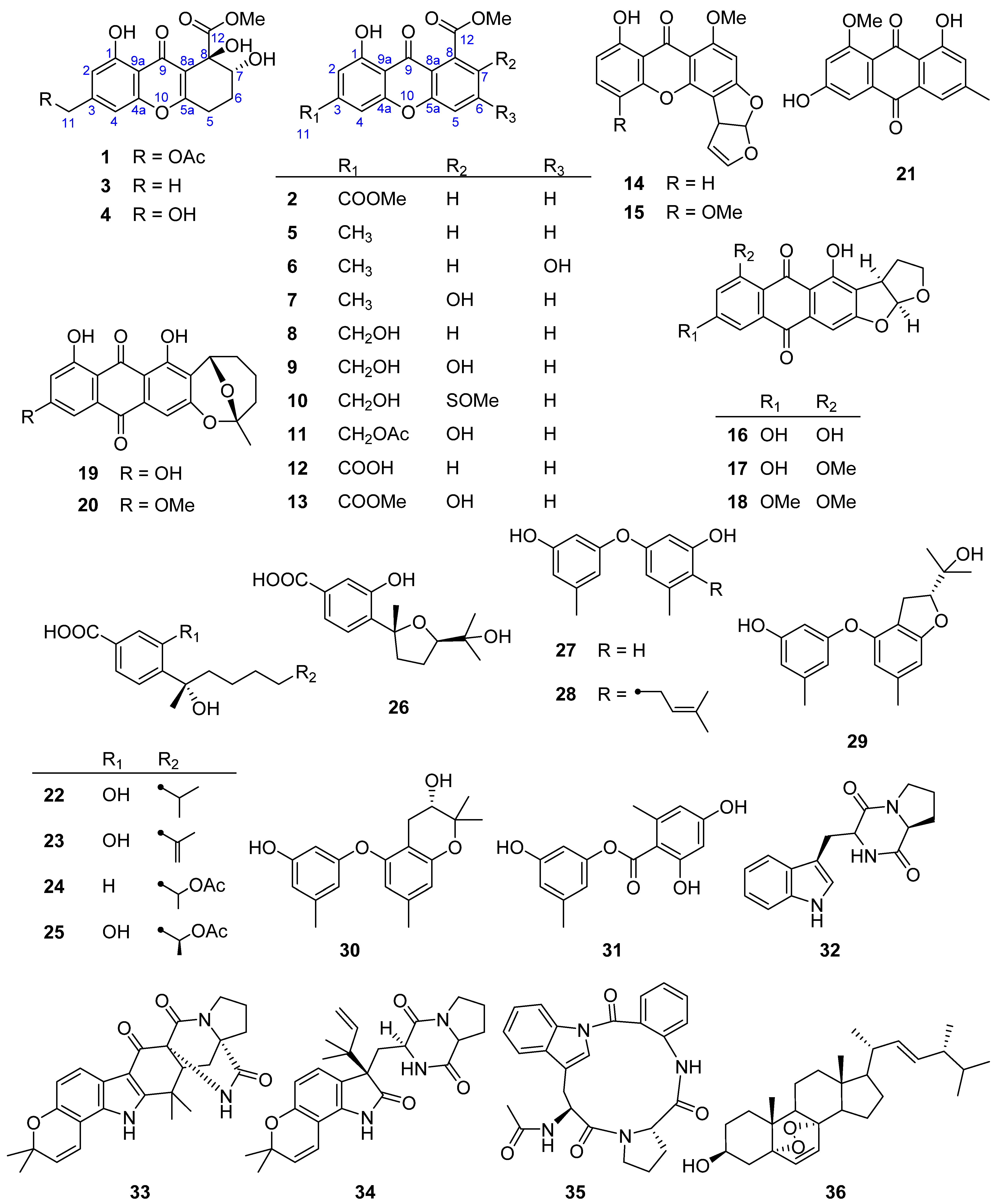
Figure 1. Chemical structures of compounds 136 isolated from Penicillium sp. MCCC 3A00126.
Marinedrugs 21 00234 g001
Figure 2. Key 1H–1H COSY and HMBC correlations and MM2 model of the most stable conformer in the 7,8-anti diol found in 1.
Marinedrugs 21 00234 g002
Figure 3. Calculated and experimental ECD spectra of 1.
Marinedrugs 21 00234 g003
Figure 4. Cytotoxicity of compounds 136 against CCRF-CEM cells.
Marinedrugs 21 00234 g004
Figure 5. IC50 values of compounds 14 and 15 against CCRF-CEM cells.
Marinedrugs 21 00234 g005
Figure 6. Inhibitory effects of compounds 136 on RSL3-induced ferroptosis in CCRF-CEM cells.
Marinedrugs 21 00234 g006
Figure 7. EC50 values of compounds 26, 28, 33, and 34 on RSL3-induced ferroptosis in CCRF-CEM cells.
Marinedrugs 21 00234 g007
Figure 8. DPPH radical scavenging activities of compounds 136.
Marinedrugs 21 00234 g008
Table 1. 1H (400 MHz) and 13C (100 MHz) NMR data of 1 and 2 in CDCl3 (δ in ppm, J in Hz within parenthesis).
No.12
δCδHδCδH
1160.7 C 161.7 C
2109.9 CH6.75 s111.6 CH7.44 s
3145.0 C 137.6 C
4105.5 CH6.86 s108.1 CH7.60 s
4a156.1 C 155.4 C
526.1 CH22.87 m119.6 CH7.59 (d, 8.0)
624.2 CH22.25 m135.5 CH7.81 (dd, 8.0, 7.2)
772.6 CH4.09 (dd, 10.3, 3.6)123.1 CH7.36 (d, 7.2)
876.2 C 133.7 C
8a117.0 C 117.4 C
9182.0 C 180.9 C
9a109.6 C 111.0 C
10167.4 C 156.2 C
1165.0 CH25.13 s165.3 C
12172.7 C 169.2 C
11-OMe 52.8 CH33.98 s
11-OAc170.5 C
20.8 CH32.16 s
12-OMe53.4 CH33.84 s53.2 CH34.04 s
1-OH 11.99 s 12.20 s
Disclaimer/Publisher’s Note: The statements, opinions and data contained in all publications are solely those of the individual author(s) and contributor(s) and not of MDPI and/or the editor(s). MDPI and/or the editor(s) disclaim responsibility for any injury to people or property resulting from any ideas, methods, instructions or products referred to in the content.