1. Introduction
Literature survey focusing on the secondary metabolites of soft corals of the genus
Sarcophyton revealed that they are a reservoir of diverse natural products, particularly diterpenes. Till now, more than 300 diterpenes have been reported from ca. 18 species of this genus, except undefined species. Moreover, many diterpenes exhibited a broad range of notable biological activities, such as anti-inflammatory effects [
1,
2]. Their intriguing scaffolds and excellent bioactivities have gained great attention from synthetic chemists as challenging targets for total synthesis [
3,
4].
Of them, sarsolenane diterpenes are extremely rare in nature, represented so far by just five compounds from the above-mentioned genus. They are sarsolenone and 7-deacetyl-sarsolenone from the soft coral
Sarcophyton solidum [
5,
6], and dihydrosarsolenone, methyl dihydrosarsolenoneate, and secodihydrosarsolenone from the soft coral
Sarcophyton trocheliophorum [
7,
8]. The absolute configuration of dihydrosarsolenone was established by TDDFT ECD calculations, while the absolute configurations of methyl dihydrosarsolenoneate and 7-deacetyl-sarsolenone were deduced from the comparison of their ECD spectra with those of the corresponding reference compounds, respectively [
6,
7]. To date, dihydrosarsolenone, methyl dihydrosarsolenoneate, and secodihydrosarsolenone have merely been subjected to the bioassay, and the results showed that only secodihydrosarsolenone exhibited moderate PTP1B inhibitory activity [
7,
8].
Capnosane diterpenes are also in the minority of diterpenes from the genus
Sarcophyton, which include sarsolilides A–C from the soft corals
S. solidum [
6,
9] and
S. trocheliophorum [
7], sarcophyolides B–D from the soft coral
Sarcophyton elegans [
10], and trocheliophols A–S and sarcophytrols A–C from the soft coral
S. trocheliophorum [
11,
12]. To establish the absolute configurations of capnosane diterpenes, different techniques, such as TDDFT ECD calculations, X-ray diffraction, the modified Mosher’s method, and CD were applied [
7,
10,
11,
12]. Only the inhibitory effects of capnosane diterpenes against PTP1B, inflammation-related NF-κB, bacterial pathogens, and tumor cell lines were evaluated. The results showed that sarsolilides A and B were potential PTP1B inhibitors [
7] and sarcophyolide B was cytotoxic against A2780 human ovarian tumor cells [
10], while the trocheliophols H, I, and S showed inhibitory potential against phytopathogens and human disease-related Gram-positive and Gram-negative bacteria [
11]. Interestingly, two cembrane–capnosane heterodimers, bissubvilides A and B, and sarsolilide B were discovered in the soft coral
Sarcophyton subviride recently [
13].
In the course of our ongoing research aiming for structurally novel and biologically active secondary metabolites from South China Sea soft corals [
14,
15,
16,
17,
18,
19], the title animal
Sarcophyton mililatensis was collected from the Xigu Island, Hainan Province, China. Among all of the
Sarcophyton species,
S. mililatensis has rarely been investigated. There are only four reports of the chemical constituents and bioactivities of
S. mililatensis [
18,
20,
21,
22]. Our previous chemical investigation on the South China Sea soft coral
S. mililatensis led to the discovery of sarcomililate A, an unprecedented diterpenoid with a tricyclo [11.3.0.0
2,16]hexadecane carbon framework [
18]. Inspired by this research, and in order to disclose more chemically intriguing marine natural products, especially trace constituents, we recently conducted a continuing chemical investigation of the Et
2O-soluble extract of the title soft coral. This study resulted in the isolation and characterization of three unusual diterpenes mililatensols A–C (
1–
3) bearing the rare sarsolenane and capnosane carbon frameworks (
Figure 1). Hereto, the isolation, structure elucidation, anti-inflammatory, and SARS-CoV-2 M
pro-inhibitory activities of these new isolates are described.
2. Results and Discussion
The acetone extract of the soft coral
S. mililatensis was partitioned between Et
2O and H
2O. The Et
2O-soluble portion was repeatedly chromatographed over silica gel, Sephadex LH-20, and RP-HPLC to yield the new records of sarsolenane and capnosane diterpenes, namely mililatensols A–C (
1–
3) (
Figure 1).
Compound
1, a white powder, possessed the molecular formula C
20H
30O, which was deduced from the molecular ion peak at
m/
z 286.2293 ([M]
+, calcd. for C
20H
30O, 286.2291) (
Figure S1), indicating six degrees of unsaturation. The IR absorption at 3282 cm
−1 (
Figure S8) revealed the existence of the hydroxyl group, which was in agreement with the presence of a secondary alcohol functionality [
δH 4.37 (1H, dt,
J = 2.8, 9.4 Hz, H-10),
δC 68.4 (CH, C-10)] as indicated by the
1H and
13C NMR data (
Table 1,
Figures S2 and S3). Its
13C NMR spectrum exhibited 20 carbon resonances assigned to four methyls, six methylenes, five methines (including three olefinic,
δC 126.8, 127.6 and 128.1, and an oxygenated,
δC 68.4), and five quaternary carbons (all olefinic,
δC 122.6, 130.0, 131.3, 132.5, and 139.4) (
Table 1), which were deduced from DEPT and HSQC experiments (
Figures S3 and S4). In addition, its
1H NMR spectrum (
Figure S2) displayed signals of four vinyl methyls at
δH 1.64 (3H, s, H
3-18), 1.64 (3H, s, H
3-19), 1.67 (3H, s, H
3-16) and 1.69 (3H, s, H
3-17), and three olefinic protons at
δH 4.95 (1H, t,
J = 7.4 Hz, H-7), 5.03 (1H, d,
J = 10.2 Hz, H-3) and 5.20 (1H, d,
J = 9.4 Hz, H-11), which were attributable to three trisubstituted double bonds (
Table 1). As revealed by the
1H and
13C NMR data, there were four double bonds, accounting for four degrees of unsaturation. The remaining two degrees of unsaturation indicated the presence of two rings in the molecule. Analysis of the
1H–
1H COSY correlations (
Figure 2 and
Figure S5) readily disclosed four spin-coupling segments from H
2-5 via H
2-6 to H-7, from H
2-9 via H-10 to H-11, from H
2-13 to H
2-14, and from H
2-20 via H-2 to H-3. On the basis of an HMBC experiment (
Figure 2 and
Figure S6), these four fragments could be fully connected by inserting the “loose ends” of the quaternary carbon atoms of C-1, C-4, C-8, and C-12. The cyclododecane ring was constructed by the characteristic HMBC cross-peaks of H
3-18/C-3, C-4 and C-5, H
3-19/C-7, C-8 and C-9, H-11/C-12 and C-13. Moreover, the key HMBC correlations of H-2/C-1 and C-14, H
2-13/C-12, and C-20 disclosed a typical cyclohexane ring. The joints of the two-carbon framework were C-2 and C-12, also indicated by the above-mentioned HMBC correlations. Furthermore, the HMBC correlations from H
3-16 to C-15 and from H
3-17 to C-1 and C-15 revealed a tetrasubstituted double bond Δ
1(15) at C-1 in the molecule. Thus, the planar structure of
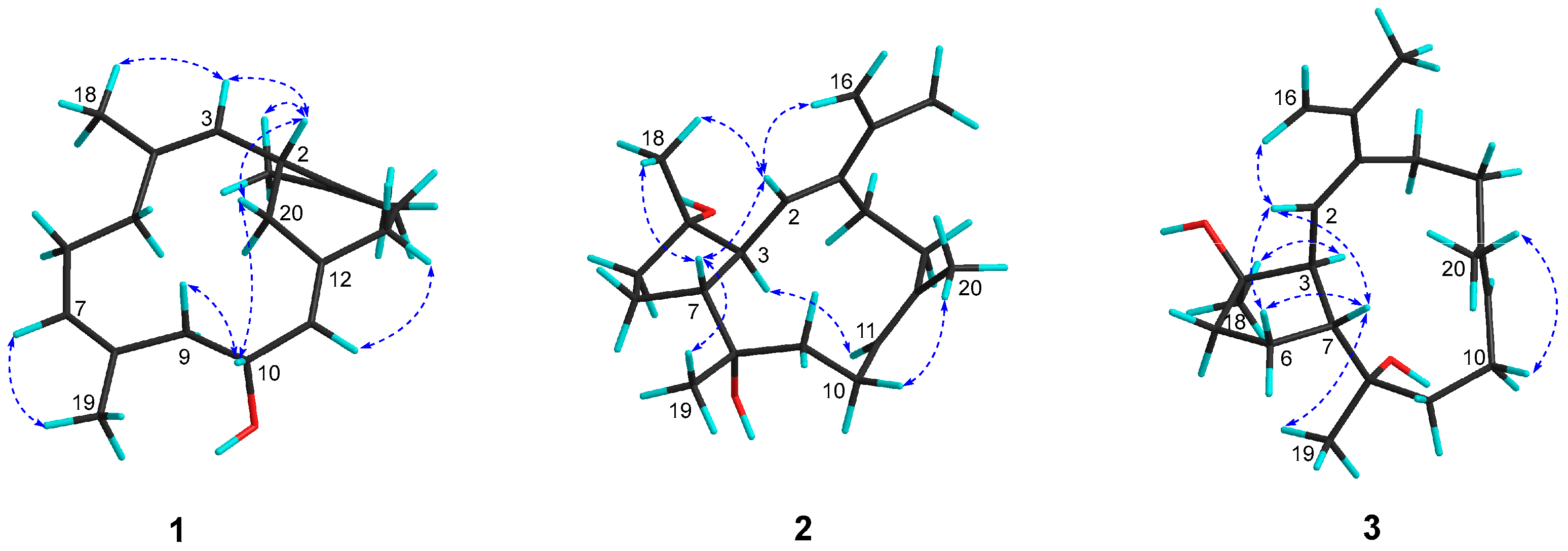
1 was delineated as a new record of sarsolenane diterpenes. The NOESY (
Figure 3 and
Figure S7) cross-peaks of H-3/H
3-18, H-7/H
3-19, and H-11/H-13a (
δH 2.08) indicated that the double bonds Δ
3, Δ
7, and Δ
11 in
1 took
Z-geometry, respectively. The relative configurations of the two chiral carbons (C-2 and C-10) could be deduced from the NOESY cross-peaks of H-10 (
δH 4.37)/H-20a (
δH 2.30) and H-2 (
δH 3.73)/H-20a. Hereto, the structure of
1 was temporally depicted as shown in
Figure 1.
Compound
2 was obtained as colorless oil. Its molecular formula was ascertained as C
20H
32O
2 by the pseudo-molecular ion peak at
m/
z 327.2297 ([M + Na]
+, calcd. for C
20H
32O
2Na, 327.2295) (
Figure S9), requiring five degrees of unsaturation. The IR spectrum of compound
2 (
Figure S16) revealed characteristic absorption for hydroxyl at 3445 cm
−1. Its
1H and
13C NMR data (
Table 1,
Figures S10–S14) disclosed the presence of two trisubstituted double bonds (
δH 5.54 (1H, d,
J = 9.6 Hz, H-2),
δC 130.0 (C-2), 142.8 (C-1) and
δH 5.29 (1H, dd,
J = 5.4, 10.0 Hz, H-11),
δC 127.9 (C-11), 135.4 (C-12)), one terminal double bond (
δH 4.99 (1H, s, H-16a), 5.09 (1H, s, H-16b),
δC 112.8 (C-16), 141.3 (C-15)), two unprotonated oxygenated carbons (
δC 74.8 (C-8), 82.2 (C-4)), two vinyl methyls (
δH 1.63 (3H, s, H
3-20),
δC 18.8 (C-20), and
δH 1.91 (3H, s, H
3-17),
δC 21.9 (C-17)), and two methyls bonded to tertiary carbons (
δH 1.12 (3H, s, H
3-18),
δC 24.2 (C-18), and 1.16 (3H, d, H
3-19),
δC 31.9 (C-19)), accounting for three degrees of unsaturation. The remaining two degrees of unsaturation suggested that
2 was a bicyclic diterpene. Comparison of these spectroscopic data with those of the known compound pavidolide D (
4) previously isolated from the soft coral
Sinularia pavida [
23] suggested a structural resemblance between them. In fact, a similar substructure of 5/9-fused bicyclo rings was present in both two compounds, which is a typical feature of the capnosane skeleton. The major difference between compounds
2 and
4 centered on the C-1 isopropyl substituent, where the two doublet methyl resonances in
4 were replaced by a pair of singlets of an exo-methylene group (
δH 4.99, 5.09;
δC 112.8, 141.3) and an allylic singlet methyl (
δH 1.91,
δC 21.9) in
2 (
Figure 1). Interpretation of the diagnostic HMBC correlations from H
3-17 to C-1, C-15, and C-16 (
Figure 2 and
Figure S14) associated with the extra one degree of unsaturation in
2 were all consistent with an isopropenyl substituent at C-1. Therefore, compound
2 was concluded to be the 16,17-dehydro derivative of
4. The (
E,
E) geometry of the double bonds Δ
1 and Δ
11 in
2 was confirmed upon the observation of the NOESY cross-peaks of H-16b (
δH 5.09)/H-2 (
δH 5.54) and H-10a (
δH 2.34)/H
3-20 (
δH 1.63) (
Figure 3 and
Figure S15). Furthermore, the NOE interactions of H-7 (
δH 2.05)/H
3-18 (
δH 1.12) and H-7/H
3-19 (
δH 1.16), and the lack of H-7/H-3 (
δH 2.73) revealed H-7, H
3-18, and H
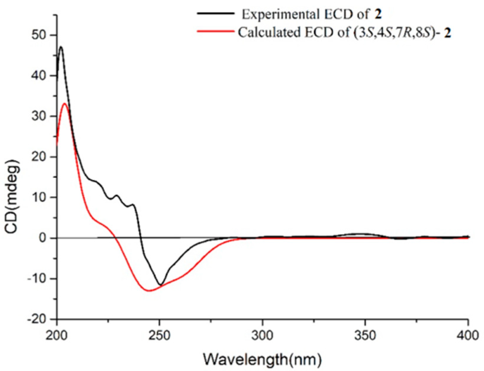
3-19 were co-facial, while H-3 was the opposite. To establish the absolute configuration of
2, ECD spectra of both enantiomeric forms (3
S,4
S,7
R,8
S and 3
R,4
R,7
S,8
R) were calculated by employing time-dependent density functional theory (TDDFT), using NMR-demonstrated conformation (
Figure S25) as the initial structure input. The ECD calculations were conducted with the B3LYP/6-311G(d) basis set using the IEFPCM solvent continuum model with CH
3OH as the solvent. As shown in
Figure 4, the experimental ECD spectrum of
2 in CH
3OH displayed one negative π−π* Cotton effect at 250 nm (Δ
ε −1.11). Perfect agreement between the experimental ECD spectrum and the calculated one allowed the assignment of the absolute configuration as (3
S,4
S,7
R,8
S).
The colorless oil
3 had the same molecular formula C
20H
32O
2 as
2, which was disclosed by the pseudo-molecular ion peak in the HRESIMS experiment (
m/
z 327.2298 [M + Na]
+, calcd. for C
20H
32O
2Na, 327.2295) (
Figure S17). The
1H and
13C NMR (
Table 1,
Figures S18 and S19) as well as IR data (
Figure S24) of
3 closely resembled those of
2, while 2D NMR (
1H–
1H COSY, HSQC, HMBC,
Figures S20–S22) analysis revealed both
3 and
2 having the same gross structure (
Figure 2). The difference was due to the apparent upfield-shifted H-3 (
δH 2.37) in
3 comparison with that (
δH 2.73) of
2 accompanied by the downfield-shifted C-18 (
δC 26.8), suggesting
3 to be a C-4 epimer of
2 [
23]. The presence of NOESY correlation between H-3 and H
3-18 (
δH 1.22) and the lack of NOE interaction between H-7 (
δH 2.50) and H
3-18, as revealed in the ROESY spectrum (
Figure 3 and
Figure S23) of
3, further supported the structural assignment. Thus, the absolute configuration of
3 could be assigned as (3
S,4
R,7
R,8
S).
The discovery of mililatensols A–C (
1–
3) with two different carbobicyclic skeletons represents an example of the productivity of the soft coral
S. mililatensis. It is worth pointing out that this is the first report of sarsolenane and capnosane diterpenes from the title animal. This study, as well as our previous research on the South China Sea soft coral
S. mililatensis, permitted an upgrade of our knowledge on the structurally diverse marine diterpenes, especially those produced by soft corals of the genus
Sarcophyton. Moreover, it is intriguing to note that till now, the sarsolenane and capnosane diterpenes have only been co-isolated from two species
S. solidum [
5,
6,
9] and
S. trocheliophorum [
7] besides
S. mililatensis. These new findings, as well as the limited previous investigations on the title animal, revealed the rarely studied soft coral
S. mililatensis is a biochemical warehouse for terpenes.
All the isolates were subjected to the bioassay for anti-inflammatory effects on LPS-induced TNF-α release in RAW264.7 macrophages. The results showed that compounds 1–3 exhibited the inhibition ratios of 26.8%, 11.4%, and 20.1% at a concentration of 20 μmol/L, indicating none of them possessed obvious activities.
The preliminary virtual screening for inhibitory potential against SARS-CoV-2 was performed by molecular docking experiments, using the highly resolved SARS-CoV-2 M
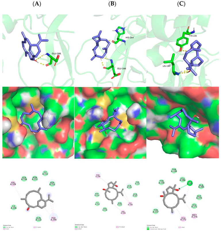
pro crystal structure (PDB: 6LU7 with a resolution of 2.16 Å). As shown, the hydrogen bond was formed between the C-10 hydroxyl of sarsolenane diterpene
1 and Glu166 (
Figure 5A, upper row), which were lying in the active site. Moreover,
1 occupied the hydrophobic pocket, which promoted Van der Waals interactions with Val104 and Phe294 (
Figure 5A, mid and lower row). As for capnosane diterpene
2, both C-4 and C-8 hydroxyls participated in hydrogen bonds with His164 and Glu166 (
Figure 5B, upper row), respectively, while for compound
3, its C-4 and C-8 hydroxyls participated in hydrogen bonds with LEU-287 and TYR-239 (
Figure 5C, upper row), respectively. In addition, both capnosane diterpenes
2 and
3 laid in the hydrophobic pocket through Van der Waals interactions with a number of key amino acids (
Figure 5B,C, mid and lower rows). The low binding affinities of compounds
1–
3 (
Table 2) revealed these three diterpenes were potential SARS-CoV-2 M
pro inhibitors.
3. Materials and Methods
3.1. General Experimental Procedures
Optical rotations were recorded on a Perkin-Elmer 241MC polarimeter. IR spectra were obtained on a Nicolet 6700 spectrometer (Thermo Scientific, Waltham, MA, USA). CD spectra were measured on a JASCO J-810 instrument. NMR spectra were measured on a Bruker DRX-500 or Bruker DRX-600 spectrometer (Bruker Biospin AG, Fällanden, Germany). Chemical shifts (δ) were reported in ppm with reference to the solvent signals, and coupling constants (J) were in Hz. ESIMS spectra were obtained on a Finngan-MAT-95 mass spectrometer. HRESIMS spectra were measured on an Agilent 1290-6545 UHPLC-QTOF mass spectrometer. Commercial silica gel (Qingdao Haiyang Chemical Group Co., Ltd., Qingdao, China, 200–300 and 400–600 mesh), Sephadex LH-20 gel (Amersham Biosciences, Piscataway, NJ, USA) were used for column chromatography, and precoated silica gel plates (Yan Tai Zi Fu Chemical Group Co., Yantai, China, G60 F-254) were used for analytical TLC. Reversed-phase (RP) HPLC was performed on an Agilent 1260 series liquid chromatography equipped with a DAD G1315D detector at 210 and 254 nm. A semi-preparative ODS-HG-5 column [5 µm, 250 × 9.4 mm] was employed for the purifications. All solvents used for column chromatography and HPLC were of analytical grade (Shanghai Chemical Reagents Co., Ltd., Shanghai, China) and chromatographic grade (Dikma Technologies Inc., Foothill Ranch, CA, USA), respectively.
3.2. Biological Material
The soft corals of Sarcophyton mililatensis were collected at a depth of −20 m by SCUBA diving from the coast of Xigu Island, Hainan Province, China, in May 2014. They were frozen immediately after collection, and identified by Prof. X.-B. Li from Hainan University. A voucher specimen (No. 14S-80) is available for inspection at Shanghai Institute of Materia Medica, Chinese Academy of Sciences.
3.3. Extraction and Isolation
The frozen animals (400 g, dry weight) were cut into pieces and extracted exhaustively with acetone at room temperature (3 × 1.5 L). The organic extract was evaporated to give a dark brown residue that was partitioned between Et2O and H2O. The upper layer was concentrated under reduced pressure to give a Et2O portion (13.5 g). The Et2O extract was separated into twenty-one fractions (A–U) by gradient silica gel column chromatography [0→100% Et2O (EE) in petroleum ether (PE)]. Fraction J was further purified by Sephadex LH-20 [PE/CH2Cl2/MeOH (2:1:1)], followed by silica gel column chromatography [PE/EE (2:1)] to give three subfractions. Subfraction J2D was further purified by RP-HPLC [MeCN/H2O (90:10), 3.0 mL/min] to give compound 2 (2.5 mg, tR = 6.2 min). Similarly, subfraction J2F was subjected to RP-HPLC [MeCN/H2O (82:18), 3.0 mL/min] to yield compound 3 (3.1 mg, tR = 8.6 min). Fraction P was further purified by Sephadex LH-20 [PE/CH2Cl2/MeOH (2:1:1)], followed by silica gel column chromatography [PE/acetone (3:1)] to afford three subfractions. Purification of subfraction P2C by RP-HPLC [MeOH/H2O (90:10), 3.0 mL/min] to give compound 2 (3.2 mg, tR = 12.4 min).
3.4. Spectroscopic Data of Compounds
Mililatensol A (
1): white powder;
−51.8 (
c 0.25, CHCl
3); IR (KBr):
νmax 3282, 2952, 2932, 2852, 1436, 1385, 1196, 1180, 1131, 1076, 1042, 990, 635 cm
−1; For
1H NMR (CDCl
3, 600 MHz) and
13C NMR (CDCl
3, 125 MHz) spectral data, see
Table 1; HREIMS
m/z 286.2293 (M
+; calcd. for C
20H
30O, 286.2291).
Mililatensol B (
2): colorless oil;
−50.8 (
c 0.2, CHCl
3); IR (KBr):
νmax 3445, 2917, 2849, 1383, 1196, 1180, 1132, 1076 cm
−1; For
1H NMR (CDCl
3, 600 MHz) and
13C NMR (CDCl
3, 125 MHz) spectral data, see
Table 1; HRESIMS
m/z 327.2297 ([M + Na]
+; calcd. for C
20H
32NaO
2, 327.2295).
Mililatensol C (
3): colorless oil;
−32.0 (
c 0.25, CHCl
3); IR (KBr):
νmax 3447, 2921, 2851, 1494, 1383, 1196, 1180, 1132, 1076 cm
−1; For
1H NMR (CDCl
3, 600 MHz) and
13C NMR (CDCl
3, 125 MHz) spectral data, see
Table 1; HRESIMS
m/z 327.2298 ([M + Na]
+; calcd. for C
20H
32NaO
2, 327.2295).
3.5. Anti-Inflammatory Activity Assay
The murine macrophage cell line RAW264.7 was obtained from American Type Culture Collection (ATCC, Manassas, VA, USA). In the bioassay for anti-inflammation, cells were cultured in DMEM containing 10% FBS, 2 mmol/L L-glutamine, 100 μg/mL streptomycin, and 100 U/mL penicillin in a humidified incubator of 5% CO2 at 37 °C. For the cytotoxicity part, RAW264.7 cells were incubated with compounds or the media (0.125% DMSO in DMEM containing 10% FBS) for 24 h, respectively. CCK-8 reagents (20 μL per well) were added, and the OD values were collected after 1 h incubation at 450 nm (650 nm calibration) by a microplate reader (Molecular Devices, Sunnyvale, CA, USA). For the anti-inflammatory activity assay, RAW264.7 cells were incubated with compounds or the media (0.125% DMSO in DMEM containing 10% FBS), and then cells were primed with LPS (1 μg/mL) for 24 h. The supernatants were centrifuged and then measured using the mouse TNF-α ELISA kit. The CC50 and IC50 were estimated using the log (inhibitor) vs. normalized response non-linear fit (Graph Pad Prism 6.0, GraphPad Software, San Diego, CA, USA). Dexamethasone was used as a positive control.
3.6. Molecular Docking
AutoDock 4.2 and AutoDock Tools 1.5.7 software were downloaded from the official website (
https://autodock.scripps.edu/ (accessed on 20 August 2022)), compounds
1–
3 by Chem3D was optimized to export the mol2 format files, and the common crystal structure was obtained from the RCSB protein database (PDB ID: 6LU7). The 6LU7 receptor was imported into the software Pymol, and the 02J, 010, AVL, PJE groups contained in the 6LU7 receptor file could be deleted, where the water molecules could also be deleted in AutoDock, and the pdb file was finally exported. The resulting files were imported into AutoDock, hydrated, merged with non-polar hydrogen atoms, and saved as 6LU7.pdbqt. AutoDock 4.2 can choose flexible or rigid docking; this experiment adopted flexible docking. The AutoDock reads into the ligand and also hydrogenates the ligand to set it to a ligand. The Ligand subroutine in the AutoDock Tools 1.5.7 software can automatically detect the number of rotatable bonds that can rotate to dock with the receptor molecule during docking. The active site of 6LU7 was not detected, so the coordinate value was set to (−26.427, 12.578, 58.908) directly, and the lattice spacing of the docking parameter was set to 0.603Å.


