Article

Jellyfish as an Alternative Source of Bioactive Antiproliferative Compounds

1
Department of Ecosustainable Marine Biotechnology, Stazione Zoologica Anton Dohrn, Villa Comunale, 80121 Napoli, Italy
2
Fundación MEDINA, Centro de Excelencia en Investigación de Medicamentos Innovadores en Andalucía, Avda. del Conocimiento 34, 18016 Granada, Spain
3
Department of Integrative Marine Ecology, Stazione Zoologica Anton Dohrn, Villa Comunale, 80121 Napoli, Italy
*
Author to whom correspondence should be addressed.
These authors contributed equally to the work.
Mar. Drugs 2022, 20(6), 350; https://doi.org/10.3390/md20060350
Submission received: 5 May 2022 / Revised: 20 May 2022 / Accepted: 23 May 2022 / Published: 25 May 2022
(This article belongs to the Section Marine Chemoecology for Drug Discovery)

Abstract

:
Jellyfish are commonly considered a nuisance for their negative effects on human activities (e.g., fisheries, power plants and tourism) and human health. However, jellyfish provide several benefits to humans and are commonly eaten in eastern countries. Additionally, recent studies have suggested that jellyfish may become a source of high-value molecules. In this study, we tested the effects of the methanolic extracts and enriched fractions, obtained by solid-phase extraction fractionation, from the scyphomedusae Pelagia noctiluca, Rhizostoma pulmo, Cotylorhiza tuberculata and the cubomedusa Caryddea marsupialis on different human cancer cell lines in order to evaluate a potential antiproliferative activity. Our results indicated that fraction C from Caryddea marsupialis-(CM) and C. tuberculata oral arms (CTOA) were the most active to reduce cell viability in a dose-dependent manner. LC/MS based dereplication analyses highlighted that both bioactive fractions contained mainly fatty acids and derivatives, with CM additionally containing small peptides (0.7–0.8 kDa), which might contribute to its higher biological activity. The mechanism of action behind the most active fraction was investigated using PCR arrays. Results showed that the fraction C of CM can reduce the expression of genes involved in apoptosis inhibition in melanoma-treated cells, which makes jellyfish a potential new source of antiproliferative drugs to be exploited in the future.

1. Introduction

The marine environment and the organisms living within it are being increasingly exploited as a source of natural defense metabolites and bioactive compounds [1,2,3]. Within marine organisms, jellyfish, particularly scyphomedusae, are raising increasing interest as a source of compounds for biotechnological applications [4,5,6]. Generally considered a nuisance, particularly during their sudden and massive appearances (blooms), because they interfere with human activities at sea along the coasts (e.g., tourism, fisheries and industries) by stinging swimmers, damaging fishing gears and caught fish as well as clogging power plant water inflows [7], jellyfish have been an important part of the diet of eastern populations, particularly the Chinese [8]. Despite the lack of specific studies, scyphomedusae appear to be a healthy food for their low carbohydrate and lipid content and, conversely, their high protein content [4]. In addition to the biochemical composition, recent studies have highlighted the antioxidant activity of several scyphomedusae, including Cotylorhiza tuberculata, Rhizostoma pulmo, R. luteum, Catostylus tagi and Rhopilema esculentum [9,10,11,12,13], which makes them a healthy food [14] and a source of antioxidant compounds [13].
In addition to the exploitation for antioxidant compounds, jellyfish possess collagen, which is the polymer most abundant in this group. Collagen extracted from jellyfish has shown a high biocompatibility with human collagen [15,16], which prompted biomedical applications, including a substrate for microglia culture [15] and scaffolds for tissue growth and regeneration [17,18]). Although the venom extracted from several jellyfish causes painful stings to humans, it has shown effects to be considered a promising tool for pharmacological products [5]. For example, the venom extracted from Nemopilema nomurai was tested on heart and muscle myoblasts in mice and blood cells from different organisms, including humans [19] and a model animal [20], as well as human hepatocellular carcinoma (HepG2) cells [21]. However, the information necessary to develop the application of compounds extracted from jellyfish for biotechnology, particularly biomedicals, is still very limited.
The scyphomedusae Pelagia noctiluca (Forsskål, 1775), Rhizostoma pulmo (Macri, 1778) and Cotylorhiza tuberculata (Macri, 1778) are endemic and often blooming in the Mediterranean Sea [22,23,24,25,26]. P. noctiluca is well known not only for its blooms, but also for stinging swimmers in coastal areas along the whole Mediterranean since ancient times [22,23,24,25]. The venom extracted from the scyphomedusa has shown antiproliferative as well as cytolytic and cytotoxic activities [27,28], and induces oxidative stress [29]. Additionally, analgesic [30] as well as anti-inflammatory activities have been detected in the crude venom extracted from P. noctiluca.
R. pulmo and C. tuberculata occasionally bloom in the Mediterranean Sea [22,24,25,26], but they are being studied for their antioxidant activity and their biochemical composition, for which they have been suggested to become a novel food for Mediterranean populations [14]. Because these scyphomedusae induce mild stings (R. pulmo) or are completely harmless to humans (C. tuberculata), their venom has been less studied than the crude venom of other scyphomedusae [31]. Nevertheless, in vitro bioassays have indicated that the tissue of R. pulmo have cytolytic and hemolytic activities [32]. A metalloproteinase, named rhizoproteinase, was isolated from the tentacles of this species and showed anticoagulant activity [33], while Cariello and co-workers isolated rhizolysin, a high-molecular-weight protein with hemolytic activity [34]. The intermediate phase (IP) of the hydro alcoholic extract of C. tuberculata has shown antiproliferative activity on MCF-7 breast cancer cell viability [35]. In particular, IP showed a concentration dependent activity, with no effects on non-malignant human epidermal keratinocytes (HEKa).
The cubomedusa Carybdaea marsupialis is found typically in the Mediterranean Sea [36]. Recorded in the Adriatic Sea for the first time in 1985 [37] and in Tunisia in 2015 [38], it appears in aggregations of solitary individuals [39]. The apparent increase in the individuals belonging to this species has been attributed to a greater availability of substrate for polyps [22]. Like most cubomedusae, C. marsupialis induces painful stings to humans, which become lethal only in rare cases. A pore-forming toxin was extracted from C. marsupialis collected in the Adriatic Sea [40], Sanchez-Rodrigues and collaborators isolated one neurotoxin and three cytolysins from specimens identified as C. marsupialis in the Caribbean Sea and tested their effect in vivo on sea crabs [41]. The presence of venom-derived neurotoxins that act on membrane proteins of the vertebrate nervous system was corroborated by bioassays on oocytes expressing membrane proteins from rat brain cells [42].
In this study, we tested the effect of total extracts and fractions from the scyphomedusae P. noctiluca, R. pulmo, C. tuberculata and the cubomedusa C. marsupialis on different human tumor cell lines (i.e., human hepatocellular liver carcinoma, melanoma and alveolar basal epithelial adenocarcinoma) in order to evaluate their possible antiproliferative activities. Our results indicated that raw extracts and the fraction enriched in glycolipids and phospholipids of C. tuberculata (particularly the oral arms) and C. marsupialis were the most active and their mechanism of action was studied using cell-death PCR array.

2. Results

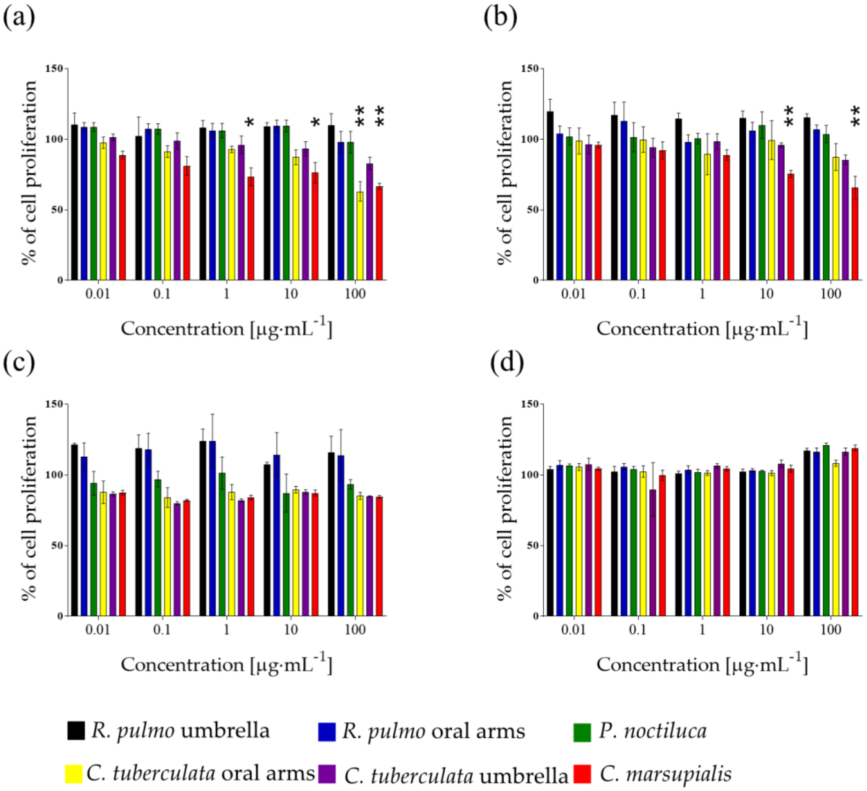
2.1. Raw Extracts Screening

In order to evaluate the antiproliferative activity of the collected jellyfish, raw methanolic extracts of C. tuberculata umbrella (CTU), C. tuberculata oral arms (CTOA), C. marsupialis (CM), R. pulmo umbrella (RPU), R. pulmo oral arms (RPOA) and P. noctiluca (PN) were tested on different human tumor cell lines: hepatocellular liver carcinoma (HepG2), human melanoma cells (A2058), adenocarcinomic human alveolar basal epithelial cells (A549) and human normal lung fibroblasts cell lines (MRC5). Total extract from CM was active on both A2058 and A549 cell lines (Figure 1a,b). In particular, CM was active on A2058 when tested at 1, 10 and 100 µg·mL1 (p < 0.05, 0.05 and 0.01, respectively), and on A549 at 10 and 100 µg·mL1 (Figure 1a,b). CTOA was active only on A2058 at 100 µg·mL1 (p < 0.01; Figure 1a). Cell proliferation of HepG2 cells and normal cells (MRC5) was not affected by jellyfish total extracts (Figure 1c,d). CTU, RPU, RPOA and PN raw extract did not significantly affect cell proliferation of tested cell lines (Figure 1a–d).

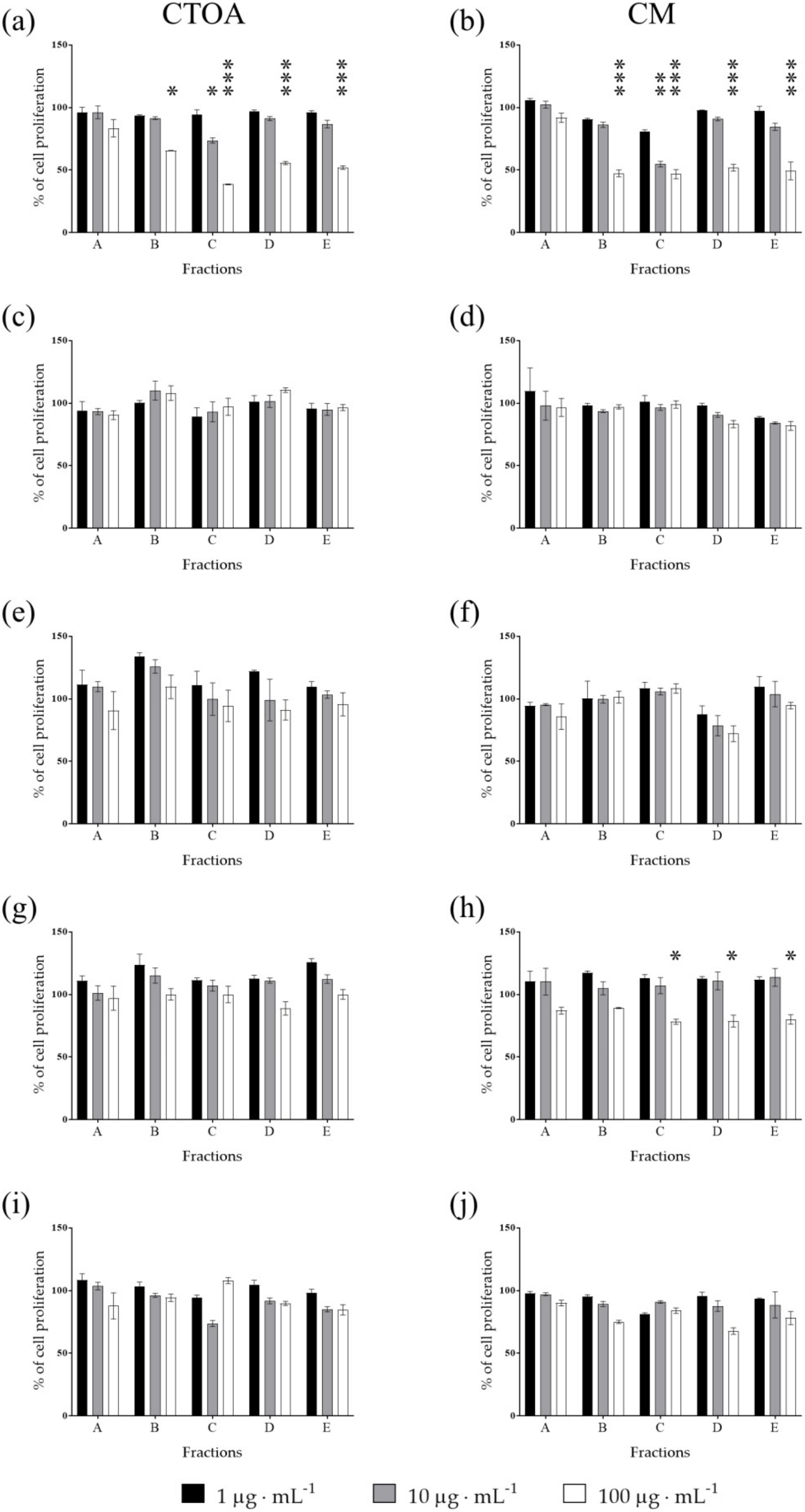
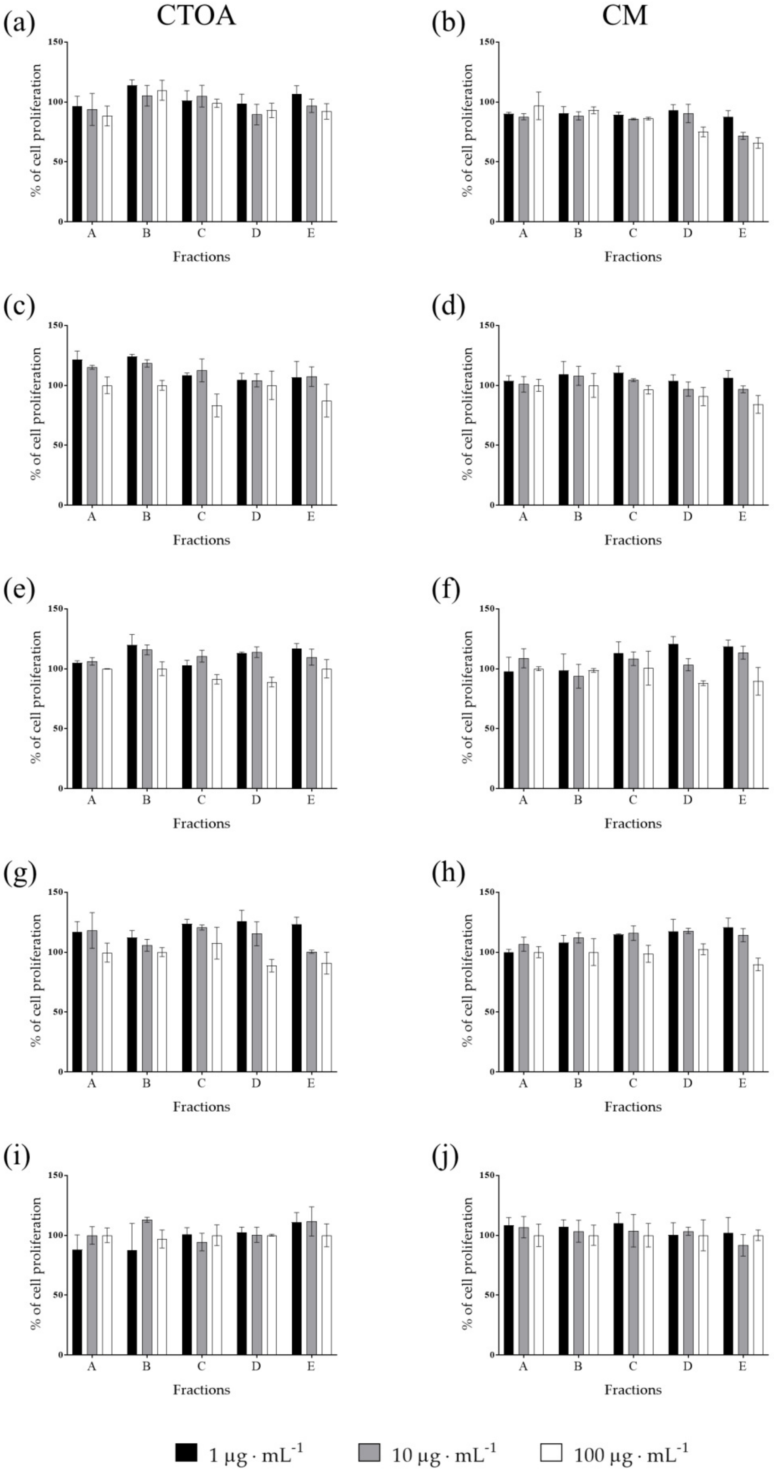
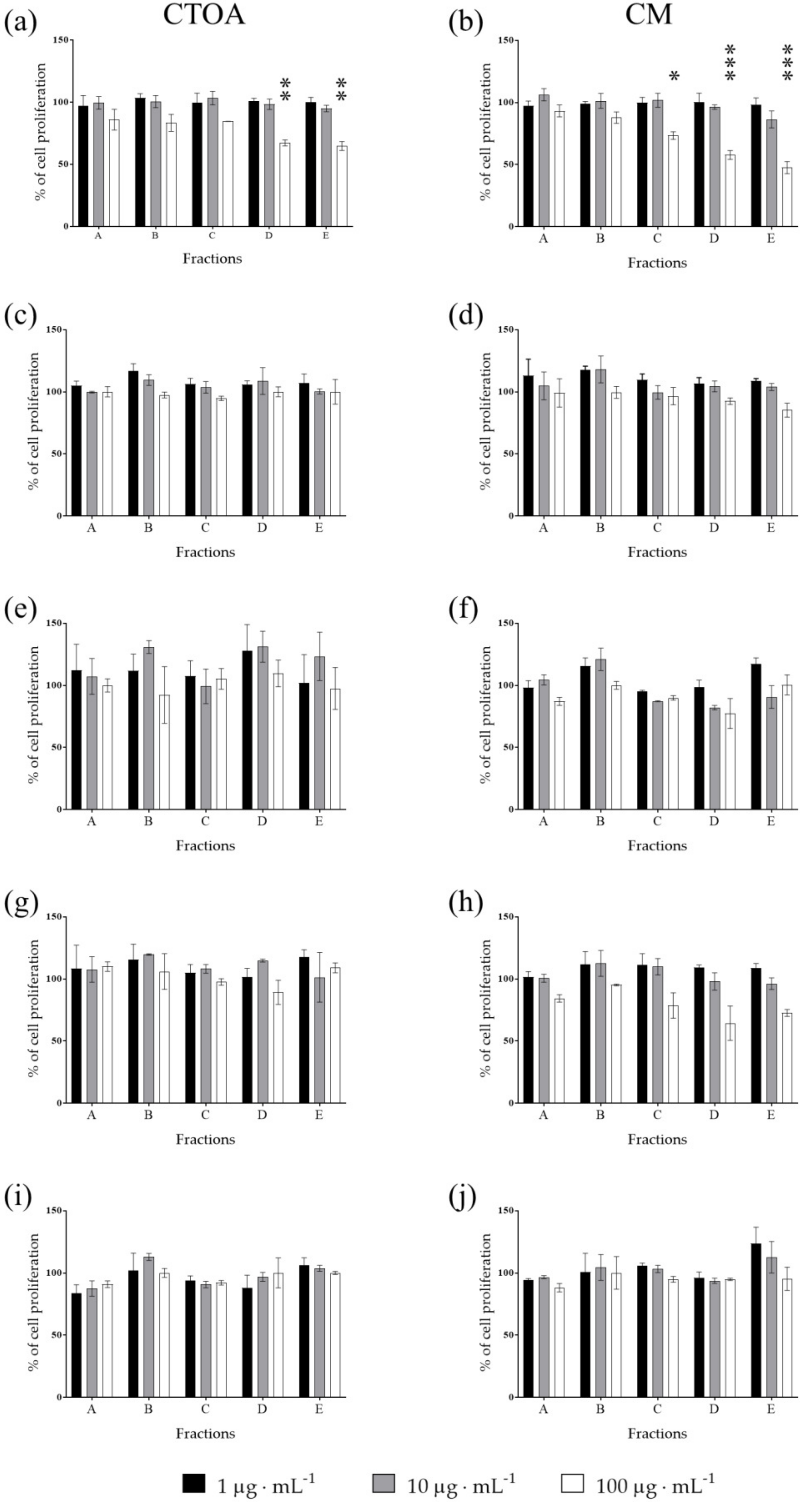
2.2. Bioactivity Testing of CTOA and CM Fractions

Raw extracts of the most active jellyfish (CTOA and CM) were fractioned using a Chromabond SPE column (see methods) and fractions (A-E) were then tested in order to identify the most active ones at 24, 48 and 72 h (Figure 2, Figure 3 and Figure 4). After 72 h of treatment, fraction C of both CTOA and CM showed the highest activity, both at 10 µg·mL1 and 100 µg·mL1 against A2058 cells (p < 0.05 and 0.001 for CTOA 10 µg·mL1 and 100 µg·mL1, and p < 0.01 and 0.001 for CM 10 µg·mL1 and 100 µg·mL1). Fractions B, D and E of both CTOA and CM were active at 100 µg·mL1 on A2058 cells (p < 0.05 for CTOA fraction B and p < 0.001 for CTOA fraction D and E, while p < 0.01 for CM fractions B, D and E; Figure 4a,b). Conversely, a significant antiproliferative activity was not found against the other cancer cell lines (Figure 4c–j). In addition, after 24 h of treatment (Figure 2), fractions from both CTOA and CM did not show any antiproliferative activity. On the contrary, after 48 h of treatment, fraction D and E at 100 µg·mL1 from CTOA and fractions C, D, and E at 100 µg·mL1 from CM exerted antiproliferative activity on A2058 cell line (Figure 3), and cell proliferation after 48 h of treatment was higher than the one after 72 h of treatment.
In addition, the half-maximal inhibitory concentration (IC50) of fraction C was evaluated for both CTOA and CM on A2058 cells (Figure 5). In particular, the corresponding C fractions were tested at increasing concentrations, from 0.1 to 100 µg·mL1. Fraction C obtained from CTOA had an IC50 = 8.10 µg·mL1, while fraction C of CM was the most active, with an IC50 = 0.34 µg·mL1 (Figure 5).

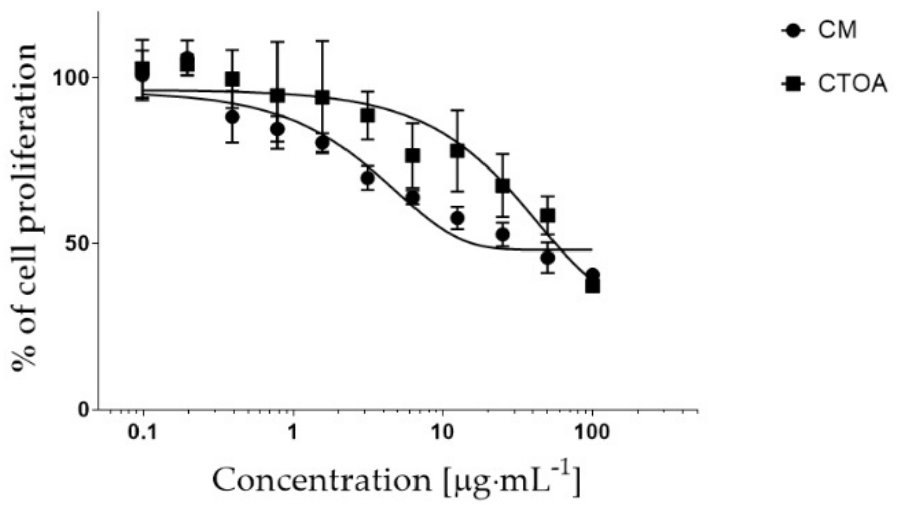
2.3. Mechanism of Action of Fraction C of CTOA and CM

In order to elucidate the cell death metabolic pathway induced by fraction C of the most active jellyfish, expression levels of selected genes involved in various cell death pathways were evaluated using a PCR array in A2058 cells treated in the presence of enriched fraction C of both CTOA and CM (respectively at 20 μg·mL1 and 1 μg·mL1). Gene transcription was considered to be affected by compounds if expression values were greater than a two-fold difference with respect to the control (DMSO alone; Student’s t-test p value < 0.05). Both differentially up-regulated and down-regulated genes are reported in Table 1.
Gene expression analyses indicated that apoptotic peptidase activating factor 1 (APAF1) was up-regulated in both CTOA- and CM-treated cells. APAF1 plays an important role in apoptosis, since it is essential for apoptosome formation. The in vivo loss of APAF-1 expression is generally associated with tumor progression [43] and its up-regulation is associated with the antitumor activity of taxane [44]. S100 calcium binding protein A7A (S100A7), also known as psoriasin, has been found to be up-regulated in CM-treated cells. S110A7 has been found to be up-regulated in psoriatic lesions [45] and when secreted it can be a chemoattractant which regulates the migration of neutrophils or T helper cells [46]. Beclin 1 (BECN1), an autophagy-related gene [47], is over-expressed in CM-treated cells. The DENN/MADD domain containing 4A (DENND4) was down-regulated in both CTOA- and CM-treated cells. DENND4 is a regulator of Rab-GTPase (a class of protein involved in membrane trafficking [48]) [49]. The X-linked inhibitor of apoptosis (XIAP), nucleolar protein 3 (apoptosis repressor with CARD domain, NOL3) and Forkhead box I1 (FOXI1) were down-regulated in CM-treated cells. XIAP, strongly inhibited by CM treatment (−149.92 ± 93.7 fold), is an apoptosis inhibitor; it suppresses apoptosis by binding caspase and inhibiting caspase activation [50]. XIAP modulation is becoming an important target for cancer therapy, in fact, it may confer therapeutic resistance and may modulate signaling factors involved in other processes, such as necroptosis, autophagy and immunosuppression [51]. NOL3 is an apoptosis suppressor and is related to chemoresistance and radiotolerance in cancer cell [52]. FOXI1 is a transcription factor related to cell growth and differentiation and it has been associated with metastasis in breast cancer [53]. Altogether, gene expression analyses suggested an apoptotic signal by APAF1 increase in both CTOA- and CM-treated cells, and also decrease in the apoptosis repressors XIAP and NOL3 in CM-treated cells.

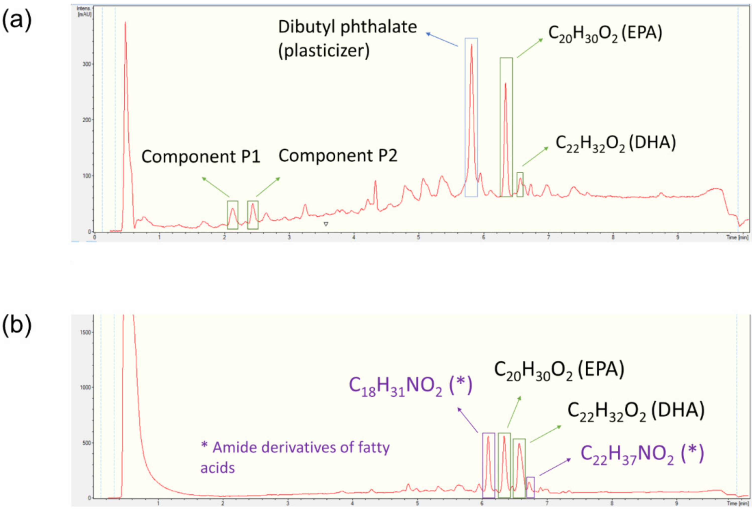
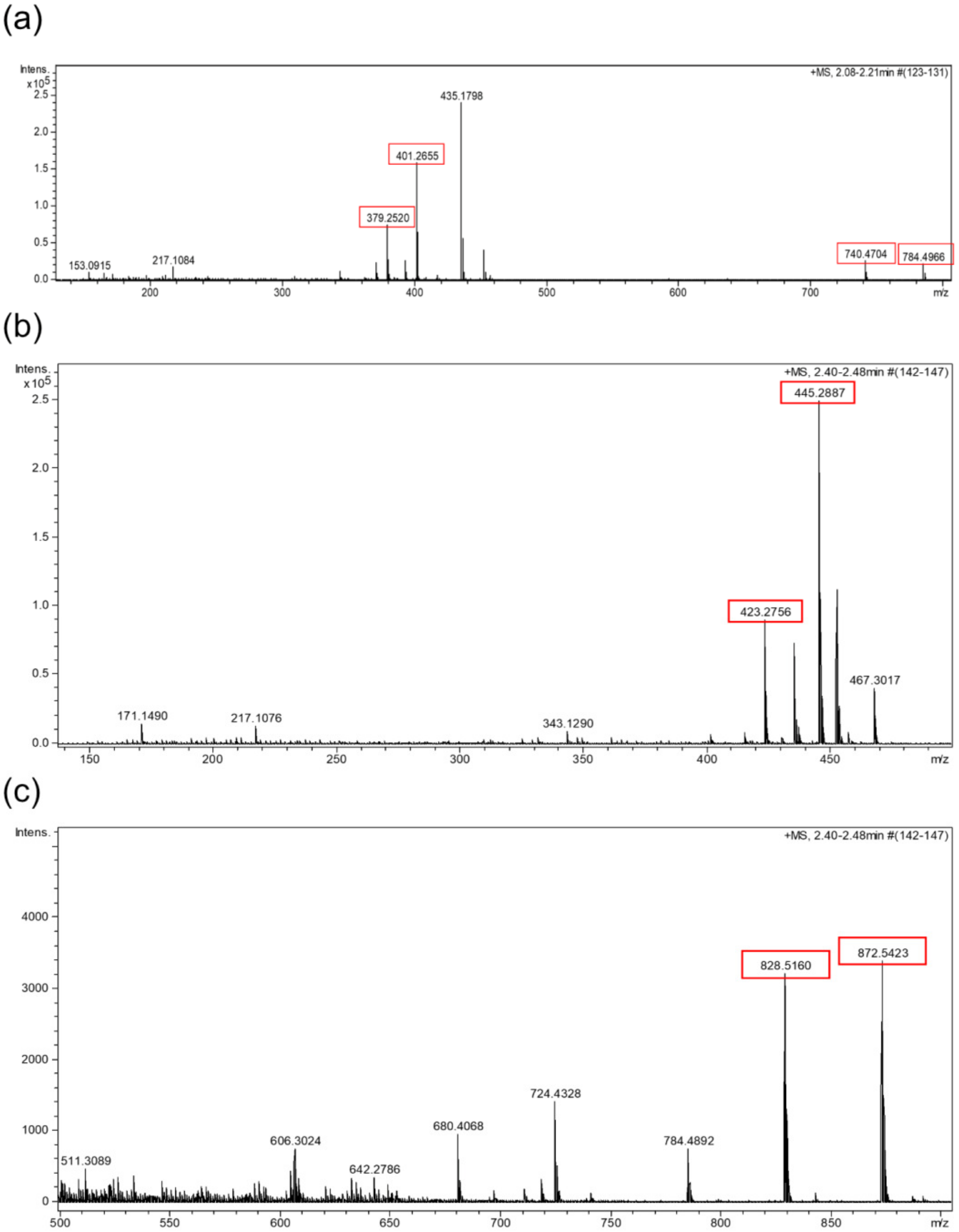
2.4. HPLC-UV-HRMS Dereplication of the Fractions

The bioactive fractions from the raw extracts CM and CTOA were dereplicated by HPLC-UV-HRESIMS according to the procedures reported in the Materials and Methods section. LC-UV chromatograms of fractions C from CM and CTOA are reported in the figures below together with the molecular formula and the putative identity of the most representative components detected by HRESIMS (Figure 6a,b).
Four different unknown compounds were detected in fraction C of the CM extract. Component P1 consisted of a mixture of two compounds (P1A and P1B) which displayed the HRMS adducts [M + H]+ and [M + H + NH4]2+ (Figure 7a). Component P2 corresponded to another mixture of two compounds (P2A and P2B), which also displayed HRMS adducts [M + H]+ and [M + H + NH4]2+ (Figure 7b,c). Considering the observed adducts, compounds P1A, P1B, P2A and P2B were assigned the molecular formulae C39H61N7O7, C41H65N7O8, C43H69N7O9 and C45H73N7O10, respectively. The best coincidences with these formulae in the Dictionary of Natural Products database (DNP) corresponded to cyclic peptides, but only P1A and P2A had hits in the database. Box jellyfishes are known to be outstanding producers of toxic proteins and peptides which are present in their venoms [54,55], and small cyclic peptides have also been isolated from jellyfish-derived fungi [56]. C39H61N7O7 retrieved as a hit the peptide pseudacyclin A in the DNP, while C43H69N7O9 retrieved the peptide taxlllaid E [57,58]. Compounds present in fraction C are most likely to be peptides, but further jellyfish sampling and studies are necessary to confirm their identity.
Other compounds present within the fractions of both species were the well-known polyunsaturated fatty acids, eicosapentaenoic acid (EPA) and dodecosahexaenoic acid (DHA). Both compounds were reported to be present in jellyfish extracts according to the literature [58,59,60]. EPA is already known to possess antiproliferative properties. In particular, EPA reduces the cell viability of lung carcinoma cells A549 by 50% after 72 h of treatment at a concentration of 6.05 μM [61]. DHA has also been reported to possess antiproliferative properties. It reduced the viability of breast (MDA-MB-231 and MCF-7), pancreatic (MiaPaca-2) and colorectal (CaCo-2, SW-620) cancer cell lines at concentrations between 10 and 100 µM [62]. Both compounds can therefore be contributing to the biological activity observed when testing the fractions.
Finally, CTOA contained two components with molecular formulae C18H31NO2 and C22H37NO2. These compounds are most likely amide derivatives of polyunsaturated fatty acids. They are reported as natural self-defense agents in plants with several known biological activities [63], but a few of them have been reported in marine natural sources alike [64,65]. This would be the first time this class of compounds is reported in jellyfish. However, further isolation and characterization procedures should be performed in order to confirm their identity and their potential contribution to the bioactivity observed.

3. Discussion

This study shows that C. tuberculata oral arms and whole C. marsupialis extracts have antiproliferative properties against human cell lines and, in particular, melanoma A2058 and lung A549 cells. Results showed that, after fractionation, fraction C was specific against melanoma cells. It has been previously reported that bioactivity is not identical between total extracts and fractions, because of less salt concentration in fractions [66], as well as synergistic effects of components in the total extracts or loss of unstable compounds in fractions. Fraction C of both species was active against melanoma human cells at 10 and 100 µg/mL in both species. Fraction C was enriched in glycolipids and phospholipids, according to the solid phase extraction fractionation procedure by Cutignano et al. [66]. This class of compounds has been previously shown to exert various biological activities, such as anticancer [67] and immunomodulatory [68,69]. These new findings open exciting potential scenarios on the use of jellyfish extracts for cancer treatment or prevention, as possible innovative drugs or food additives.
The biological activity against cancer cells by C. tuberculata was unexpected, considering that this scyphomedusa is harmless to humans [31] and there are no toxic or antiproliferative compounds known from this species to date [5]. Conversely, C. marsupialis is known to induce painful stings [31] and produces compounds with negative effects on human cells, such as hemolysin, a compound with hemolytic activity, and four cytolysins (i.e., CmHl1, CmHl5, CmH17 and CmNt) [40,41]. However, these biological activities found in compounds extracted from C. marsupialis have found little application until now.
In line with its harmlessness, C. tuberculata appears to be a promising source of compounds which benefit human health, such as collagen and antioxidant peptides [9]. This Mediterranean scyphomedusa contains the highest amount of collagen of all other scyphomedusae where its content was determined, together with the other Mediterranean rhizostome species, R. pulmo, and more than the quantity contained in other scyphomedusae consumed as food in the eastern seas [4]. Additionally, C. tuberculata hosts the dinoflagellate endosymbionts Philozoon medusarum [70], which produces several compounds through photosynthesis which support the metabolism of the scyphomedusa, although these pathways are not yet well defined [71].
In addition to the compounds mentioned above, fraction C of both species, which was the most active, was enriched in fatty acids and fatty acid amide derivatives [66]. Considering the high content in fatty acids (omega 3 and 6), antioxidant, phenolic compounds and proteins, jellyfish have been proposed as a new biomass potentially useful as human food worldwide [9,14]. Overall, our findings suggest that both C. marsupialis and C. tuberculata may have important applications in the biomedical field, in addition to those described above.
Our results are in line with recent reviews [4,5], which highlighted that jellyfish are an unexploited source of high-value compounds, which are already used or have promising applications in several biotechnological fields, including nutraceutical, cosmeceutical and biomedical sectors. The advantage of using jellyfish as a source of high-value compounds is two-fold. Firstly, jellyfish are abundant and often give rise to blooms which interfere with fisheries and power plant activities along the coasts [7]. Secondly, they are cheap to obtain as a by-catch of the above-mentioned activities and using them for their compounds may actually become a way to recycle their biomass within the context of a circular economy and a sustainable use of resources [72,73,74]. In particular, the extraction of high-value compounds may be an effective way to exploit the resource in western countries, where the consumption of jellyfish as food is extremely limited due to the different dietary tradition compared to eastern countries.

4. Materials and Methods

4.1. Sample Collection and Processing

Two scyphomedusae (P. noctiluca and R. pulmo) and the cubomedusa C. marsupialis were collected in the Gulf of Naples between 2017 and 2019, while the scyphomedusa C. tuberculata was caught in Palinuro during August 2019 (Table 2). All scyphomedusae were collected using a dipnet from the boat, while the cubomedusa was collected directly by SCUBA divers. All specimens were transported to the SZN in plastic buckets filled with seawater from the sampling site. In laboratory, medusae were dissected and frozen at −30 °C. Samples were then freeze-dried to remove the water and concentrate the organic matter.

4.2. Extract Preparation

Lyophilized jellyfish were resuspended in methanol (proportion 1:5, w/v). Samples were vortexed to ensure that methanol completely soaked the freeze-dried jellyfish that were then placed on ice for 30 min, then the obtained suspension was sonicated with 2 short bursts of 30 s each, followed by intervals of 30 s for cooling. Samples were then centrifuged at 3000 rpm for 20 min, and the supernatant was transferred into a rotary evaporator system (Rotavapor). Once a consistent reduction of total volume was reached, samples were evaporated under a nitrogen stream.

4.3. Fractionation of the Raw Extract

Fractionation of each extract (an aliquot with a maximum weight of 20 mg) was performed by solid phase extraction (SPE) using Chromabond® HR-X cartridges (6 mL/500 mg) as reported by Cutignano et al. [66]. Briefly, the samples were loaded into the cartridges and then eluted using different mixtures of solvents to get five different fractions: 100% H2O (2 mL, discarded); 100% H2O (6 mL, fraction A); CH3OH/H2O (50:50, 9 mL, fraction B); CH3CN/H2O (70:30, 9 mL, fraction C); 100% CH3CN (9 mL, fraction D); CH2Cl2/CH3OH (90:10, 9 mL, fraction E). Each fraction was evaporated under reduced pressure, weighted, and preserved at −20 °C.

4.4. Cell Lines

Human hepatocellular liver carcinoma (HepG2; ATCC® HB-8065™) were cultured in EMEM medium, human melanoma cells (A2058; ATCC®CRL-11147TM) were cultured in DMEM, human keratynocytes (HaCaT, CLS n. 300493) were cultured in DMEM medium, adenocarcinomic human alveolar basal epithelial cells (A549; ATCC®CL-185TM) were cultured in F-12K medium and human normal lung fibroblasts (MRC-5; ATCC® CCL-171™) were cultured in EMEM medium. The media were supplemented with 10% fetal bovine serum, 50 U·mL−1 penicillin and 50 μg·mL−1 streptomycin. Human cell lines A2058, A549, MRC-5 and HepG2 were bought at ATCC (https://www.lgcstandards-atcc.org/ accessed on 1 June 2019). The HACAT cells were bought at the CEINGE facility cell bank (https://www.ceinge.unina.it/en/cell-cultures accessed on 1 June 2019).

4.5. In Vitro Antiproliferative Assays

Human tumor cell lines (HepG2, A2058 and A549) and the normal cell lines (MRC5 and HaCaT) were seeded in 96-well microtiter plates at a density of 1 × 10⁴ cells/well and incubated at 37 °C to allow cell adhesion to the plates. After 16 h, the medium was replaced with fresh medium containing increasing concentrations of extracts or fractions (100, 10, 1, 0.1 and 0.01 μg·mL−1) for 24, 48 or 72 h. Extracts and fractions were dissolved in dimethyl sulfoxide (DMSO) with the maximum concentration for DMSO used of 1% (v/v). Each concentration was tested at least in triplicates. After 72 h, 3-(4,5-dimethyl-2-thizolyl)-2,5-diphenyl-2H-tetrazolium bromide (MTT; A2231,0001, AppliChem Panreac Tischkalender, Darmstadt, GmbH) was added. Briefly, the medium was replaced with a medium containing MTT at 0.5 mg·mL−1 and plates were incubated for 3 h at 37 °C. After incubation, cells were treated with isopropyl alcohol for 30 min at room temperature to dissolve MTT. Absorbance was measured at OD = 570 nm using a microplate reader (Multiskan™ FC Microplate Photometer, Thermo Fisher Scientific, Waltham, MA, USA). Cell proliferation was expressed as a percentage of cell proliferation in the presence of the tested samples, with respect to untreated control cultures (with only DMSO).

4.6. RNA Extraction and Reverse Transcription-Quantitative Polymerase Chain Reaction (RT-qPCR)

A2058 cells were seeded in 6-well plates (400,000 per well) and left 16 h for attachment. The seeded cells were then treated in the presence of fraction C of both CM and CTOA samples at 1 μg·mL−1 for 48 h at 37 °C. Cells were washed by adding phosphate buffered saline (PBS 1X). Cells were lysed by adding 1 mL of Trisure Reagent (Meridian bioscience, Memphis, TN, USA). RNA was isolated as previously described [75]. RNA concentration, quality, and purity were assessed using an ND-1000 UV-Vis spectrophotometer (NanoDrop Technologies, Thermo Fisher Scientific, Waltham, MA, USA), monitoring the absorbance at 260 nm, and the 260/280 nm and 260/230 nm ratios (both ratios were about 2.0). RNA quality was evaluated by gel electrophoresis that showed intact RNA. About 500 ng of RNA underwent a reverse transcription reaction using the RT2 first strand kit (cat. 330401, Qiagen, Hilden, Germany) according to the manufacturer’s instructions. The RT-qPCR analysis was performed in duplicate using the RT2 Profiler PCR Array kit (cat. PAHS-212ZE-4, Qiagen, Hilden, Germany) to analyze the expression of 84 genes involved in cell death signaling pathways. Plates were run on a ViiA7 (Applied Biosystems, Foster City, CA, USA, 384-well blocks). The PCR program consisted of a denaturation step at 95 °C for 20 s followed by 40 cycles at 95 °C for 15 s and 60 °C for 1 min. The cycle threshold (Ct)-values were analyzed with PCR array data analysis online software (GeneGlobe Data Analysis Center at https://geneglobe.qiagen.com/us/analyze, accessed on 10 September 2021, Qiagen, Hilden, Germany). Real-time data were expressed as the fold of expression, describing the changes in gene expression between cells treated in the presence fraction C of CM and cells treated in the presence of DMSO alone (control). The PCR array data analysis software uses Student’s t-test for statistical analysis. Only expression values greater than a two-fold difference with respect to the controls were considered significant.

4.7. Dereplication

Fractions were analyzed by HPLC-UV-HRESIMS on an Agilent 1200 RR coupled to a Bruker maXis QToF spectrometer with electrospray ionization, as reported by [76]. Data were analyzed following the guidelines developed by Fundación MEDINA [77] and compared with the data available on the Fundación MEDINA internal database and the Dictionary of Natural Products and Reaxys databases. The identity of the components was putatively assigned by comparison of their HRMS spectra, UV maxima, retention time, natural source and biological activity with the data available in DNP (Dictionary of natural products) and Reaxys (“Others; Isolated from natural source” filter).

4.8. Statistics

Arithmetic means ± the standard deviations (SD) were calculated and compared by a two-tailed Student t-test. Differences at p < 0.05 were regarded as significant.

Author Contributions

Conceptualization, I.D. and C.L.; formal analysis, G.R., K.A.M., J.M. and C.L.; writing—original draft preparation, G.R., K.A.M., I.D. and C.L.; writing—review and editing, G.R., K.A.M., I.D., F.R. and C.L.; supervision, C.L. All authors have read and agreed to the published version of the manuscript.

Funding

This research received no external funding.

Institutional Review Board Statement

Not applicable.

Informed Consent Statement

Not applicable.

Data Availability Statement

Not applicable.

Acknowledgments

Gennaro Riccio was supported by a research grant within the project “Antitumor Drugs and Vaccines from the Sea (ADViSE)” (CUP B43D18000240007–SURF 17061BP000000011; PG/2018/0494374) funded by POR Campania FESR 2014–2020 “Technology Platform for Therapeutic Strategies against Cancer”-Action 1.1.2 and 1.2.2. We are grateful to Marco Cannavacciuolo, Augusto Passarelli, Gianluca Zazo, Roberto Gallia, Francesco Terlizzi, Enzo Rando, and Ferdinando Tramontano for collecting jellyfish in the Gulf of Naples and Raffaella Casotti, Leo Brunet, Lorenzo Auciello, and Leonardo Auciello for C. tuberculata sampling from Palinuro.

Conflicts of Interest

The authors declare no conflict of interest.

References

  1. Jaspars, M.; De Pascale, D.; Andersen, J.H.; Reyes, F.; Crawford, A.D.; Ianora, A. The marine biodiscovery pipeline and ocean medicines of tomorrow. J. Mar. Biol. Assoc. U. K. 2016, 96, 151–158. [Google Scholar] [CrossRef] [Green Version]
  2. Romano, G.; Costantini, M.; Sansone, C.; Lauritano, C.; Ruocco, N.; Ianora, A. Marine microorganisms as a promising and sustainable source of bioactive molecules. Mar. Environ. Res. 2017, 128, 58–69. [Google Scholar] [CrossRef] [PubMed]
  3. Saide, A.; Martínez, K.A.; Ianora, A.; Lauritano, C. Unlocking the health potential of microalgae as sustainable sources of bioactive compounds. Int. J. Mol. Sci. 2021, 22, 4383. [Google Scholar] [CrossRef] [PubMed]
  4. Merquiol, L.; Romano, G.; Ianora, A.; D’Ambra, I. Biotechnological applications of scyphomedusae. Mar. Drugs 2019, 17, 604. [Google Scholar] [CrossRef] [PubMed] [Green Version]
  5. D’Ambra, I.; Lauritano, C. A Review of toxins from cnidaria. Mar. Drugs 2020, 18, 507. [Google Scholar] [CrossRef]
  6. Stabili, L.; Rizzo, L.; Caprioli, R.; Leone, A.; Piraino, S. Jellyfish bioprospecting in the mediterranean sea: Antioxidant and lysozyme-like activities from Aurelia coerulea (cnidaria, scyphozoa) extracts. Mar. Drugs 2021, 19, 619. [Google Scholar] [CrossRef]
  7. Richardson, A.J.; Bakun, A.; Hays, G.C.; Gibbons, M.J. The jellyfish joyride: Causes, consequences and management responses to a more gelatinous future. Trends Ecol. Evol. 2009, 24, 312–322. [Google Scholar] [CrossRef]
  8. Hsieh, Y.H.P.; Leong, F.M.; Rudloe, J. Jellyfish as food. Hydrobiologia 2001, 451, 11–17. [Google Scholar] [CrossRef]
  9. Leone, A.; Lecci, R.M.; Durante, M.; Meli, F.; Piraino, S. The bright side of gelatinous blooms: Nutraceutical value and antioxidant properties of three Mediterranean jellyfish (Scyphozoa). Mar. Drugs 2015, 13, 4654–4681. [Google Scholar] [CrossRef] [Green Version]
  10. Prieto, L.; Enrique-Navarro, A.; Volsi, R.L.; Ortega, M.J. The large jellyfish Rhizostoma luteum as sustainable a resource for antioxidant properties, nutraceutical value and biomedical applications. Mar. Drugs 2018, 16, 396. [Google Scholar] [CrossRef] [Green Version]
  11. Morais, Z.B.; Pintao, A.M.; Costa, I.M.; Calejo, M.T.; Bandarra, N.M.; Abreu, P. Composition and in vitro antioxidant effects of jellyfish Catostylus tagi from sado estuary (SW Portugal). J. Aquat. Food Prod. Technol. 2009, 18, 90–107. [Google Scholar] [CrossRef]
  12. Zhang, M.; Liu, W.; Li, G. Isolation and characterisation of collagens from the skin of largefin longbarbel catfish (Mystus macropterus). Food Chem. 2009, 115, 826–831. [Google Scholar] [CrossRef]
  13. De Domenico, S.; De Rinaldis, G.; Paulmery, M.; Piraino, S.; Leone, A. Barrel Jellyfish (Rhizostoma pulmo) as Source of Antioxidant Peptides. Mar. Drugs 2019, 17, 134. [Google Scholar] [CrossRef] [PubMed] [Green Version]
  14. Leone, A.; Lecci, R.M.; Milisenda, G.; Piraino, S. Mediterranean jellyfish as novel food: Effects of thermal processing on antioxidant, phenolic, and protein contents. Eur. Food Res. Technol. 2019, 245, 1611–1627. [Google Scholar] [CrossRef] [Green Version]
  15. Mearns-Spragg, A.; Tilman, J.; Tams, D.; Barnes, A. The Biological Evaluation of Jellyfish Collagen as a New Research Tool for the Growth and Culture of iPSC Derived Microglia. Front. Mar. Sci. 2020, 7, 689. [Google Scholar] [CrossRef]
  16. Addad, S.; Exposito, J.Y.; Faye, C.; Ricard-Blum, S.; Lethias, C. Isolation, characterization and biological evaluation of jellyfish collagen for use in biomedical applications. Mar. Drugs 2011, 9, 967–983. [Google Scholar] [CrossRef] [Green Version]
  17. Bernhardt, A.; Paul, B.; Gelinsky, M. Biphasic scaffolds from marine collagens for regeneration of osteochondral defects. Mar. Drugs 2018, 16, 91. [Google Scholar] [CrossRef] [Green Version]
  18. Hoyer, B.; Bernhardt, A.; Lode, A.; Heinemann, S.; Sewing, J.; Klinger, M.; Notbohm, H.; Gelinsky, M. Jellyfish collagen scaffolds for cartilage tissue engineering. Acta Biomater. 2014, 10, 883–892. [Google Scholar] [CrossRef]
  19. Kang, C.; Munawir, A.; Cha, M.; Sohn, E.T.; Lee, H.; Kim, J.S.; Yoon, W.D.; Lim, D.; Kim, E. Cytotoxicity and hemolytic activity of jellyfish Nemopilema nomurai (Scyphozoa: Rhizostomeae) venom. Comp. Biochem. Physiol. C Toxicol. Pharmacol. 2009, 150, 85–90. [Google Scholar] [CrossRef]
  20. Lee, H.; Bae, S.K.; Kim, M.; Pyo, M.J.; Kim, M.; Yang, S.; Won, C.K.; Yoon, W.D.; Han, C.H.; Kang, C.; et al. Anticancer Effect of Nemopilema nomurai Jellyfish Venom on HepG2 Cells and a Tumor Xenograft Animal Model. Evidence-based Complement. Altern. Med. 2017, 2017, 2752716. [Google Scholar] [CrossRef] [Green Version]
  21. Choudhary, I.; Lee, H.; Pyo, M.J.; Heo, Y.; Chae, J.; Yum, S.S.; Kang, C.; Kim, E. Proteomic investigation to identify anticancer targets of Nemopilema nomurai jellyfish venom in human hepatocarcinoma HepG2 cells. Toxins 2018, 10, 194. [Google Scholar] [CrossRef] [PubMed] [Green Version]
  22. Boero, F. General Fiaheries Commission for the Mediterranen Review of Jellyfish Blooms in the Mediterranean and Black Sea. In Studies and Reviews. General Fisheries Commission for the Mediterranean; FAO: Rome, Italy, 2013; Volume 92, p. 53. [Google Scholar]
  23. Goy, J.; Morand, P.; Etienne, M. Long-term fluctuations of Pelagia noctiluca (Cnidaria, Scyphomedusa) in the western Mediterranean Sea. Prediction by climatic variables. Deep Sea Res. Part A Oceanogr. Res. Pap. 1989, 36, 269–279. [Google Scholar] [CrossRef]
  24. United Nations Environment Programme. Mediterranean Action Plan. Jellyfish blooms in the Mediterranean. In Proceedings of the II Workshop on Jellyfish in the Mediterranean Sea, Trieste, Italy, 2–5 September 1987. [Google Scholar]
  25. United Nations Environment Programme. Mediterranean Action Plan. In Proceedings of the Workshop on Jellyfish Blooms in the Mediterranean, Athens, Greece, 31 October–4 November 1983. [Google Scholar]
  26. Kikinger, R. Cotylorhiza tuberculata (Cnidaria: Scyphozoa)—Life History of a Stationary Population. Mar. Ecol. 1992, 13, 333–362. [Google Scholar] [CrossRef]
  27. Mariottini, G.L.; Sottofattori, E.; Mazzei, M.; Robbiano, L.; Carli, A. Cytotoxicity of the venom of Pelagia noctiluca forskål (Cnidaria: Scyphozoa). Toxicon 2002, 40, 695–698. [Google Scholar] [CrossRef]
  28. Ayed, Y.; Boussabbeh, M.; Zakhama, W.; Bouaziz, C.; Abid, S.; Bacha, H. Induction of cytotoxicity of Pelagia noctiluca venom causes reactive oxygen species generation, lipid peroxydation induction and DNA damage in human colon cancer cells. Lipids Health Dis. 2011, 10, 232. [Google Scholar] [CrossRef] [Green Version]
  29. Morabito, R.; Costa, R.; Rizzo, V.; Remigante, A.; Nofziger, C.; La Spada, G.; Marino, A.; Paulmichl, M.; Dossena, S. Crude venom from nematocysts of Pelagia noctiluca (Cnidaria: Scyphozoa) elicits a sodium conductance in the plasma membrane of mammalian cells. Sci. Rep. 2017, 7, 41065. [Google Scholar] [CrossRef] [PubMed] [Green Version]
  30. Ayed, Y.; Dellai, A.; Mansour, H.B.; Bacha, H.; Abid, S. Analgesic and antibutyrylcholinestrasic activities of the venom prepared from the Mediterranean jellyfish Pelagia noctiluca (Forsskal, 1775). Ann. Clin. Microbiol. Antimicrob. 2012, 11, 15. [Google Scholar] [CrossRef] [Green Version]
  31. Mariottini, G.L.; Pane, L. Mediterranean jellyfish venoms: A review on scyphomedusae. Mar. Drugs 2010, 8, 1122–1152. [Google Scholar] [CrossRef] [Green Version]
  32. Allavena, A.; Mariottini, G.L.; Carli, A.M.; Contini, S.; Martelli, A. In vitro evaluation of the cytotoxic, hemolytic and clastogenic activities of Rhizostoma pulmo toxin(s). Toxicon 1998, 36, 933–936. [Google Scholar] [CrossRef]
  33. Rastogi, A.; Sarkar, A.; Chakrabarty, D. Partial purification and identification of a metalloproteinase with anticoagulant activity from Rhizostoma pulmo (Barrel Jellyfish). Toxicon 2017, 132, 29–39. [Google Scholar] [CrossRef]
  34. Cariello, L.; Romano, G.; Spagnuolo, A.; Zanetti, L. Isolation and partial characterization of Rhizolysin, a high molecular weight protein with hemolytic activity, from the jellyfish Rhizostoma pulmo. Toxicon 1988, 26, 1057–1065. [Google Scholar] [CrossRef]
  35. Leone, A.; Lecci, R.M.; Durante, M.; Piraino, S. Extract from the zooxanthellate jellyfish Cotylorhiza tuberculata modulates gap junction intercellular communication in human cell cultures. Mar. Drugs 2013, 11, 1728–1762. [Google Scholar] [CrossRef] [PubMed] [Green Version]
  36. Acevedo, M.J.; Straehler-Pohl, I.; Morandini, A.C.; Stampar, S.N.; Bentlage, B.; Matsumoto, G.I.; Yanagihara, A.; Toshino, S.; Bordehore, C.; Fuentes, V.L. Revision of the genus Carybdea (Cnidaria: Cubozoa: Carybdeidae): Clarifying the identity of its type species Carybdea marsupialis. Zootaxa 2019, 4543, 515–548. [Google Scholar] [CrossRef] [PubMed]
  37. Boero, F.; Minelli, A. First record of Carybdea marsupialis (L., 1758) (Cnidaria, Cubozoa) from the Adriatic Sea. Boll. Mus. Civ. Stor. Nat. Venezia 1986, 35, 179–180. [Google Scholar]
  38. Gueroun, S.K.M.; Acevedo, M.J.; Kéfi-Daly Yahia, O.; Deidun, A.; Fuentes, V.L.; Piraino, S.; Daly Yahia, M.N. First records of Carybdea marsupialis proliferation (Cnidaria: Cubozoa) along the eastern Tunisian coast (Central Mediterranean). Ital. J. Zool. 2015, 82, 430–435. [Google Scholar] [CrossRef]
  39. Bordehore, C.; Fuentes, V.L.; Atienza, D.; Barberá, C.; Fernandez-Jover, D.; Roig, M.; Acevedo-Dudley, M.J.; Canepa, A.J.; Gili, J.M. Detection of an unusual presence of the cubozoan Carybdea marsupialis at shallow beaches located near Denia, Spain (south-western Mediterranean). Mar. Biodivers. Rec. 2011, 4, e69. [Google Scholar] [CrossRef] [Green Version]
  40. Rottini, G.; Gusmani, L.; Parovel, E.; Avian, M.; Patriarca, P. Purification and properties of a cytolytic toxin in venom of the jellyfish Carybdea marsupialis. Toxicon 1995, 33, 315–326. [Google Scholar] [CrossRef]
  41. Sánchez-Rodríguez, J.; Torrens, E.; Segura-Puertas, L. Partial purification and characterization of a novel neurotoxin and three cytolysins from box jellyfish (Carybdea marsupialis) nematocyst venom. Arch. Toxicol. 2006, 80, 163–168. [Google Scholar] [CrossRef]
  42. Lazcano-Pérez, F.; Arellano, R.O.; Garay, E.; Arreguín-Espinosa, R.; Sánchez-Rodríguez, J. Electrophysiological activity of a neurotoxic fraction from the venom of box jellyfish Carybdea marsupialis. Comp. Biochem. Physiol. Part C Toxicol. Pharmacol. 2017, 191, 177–182. [Google Scholar] [CrossRef]
  43. Anichini, A.; Mortarini, R.; Sensi, M.; Zanon, M. APAF-1 signaling in human melanoma. Cancer Lett. 2006, 238, 168–179. [Google Scholar] [CrossRef]
  44. Bakhshoudeh, M.; Mehdizadeh, K.; Hosseinkhani, S.; Ataei, F. Upregulation of apoptotic protease activating factor-1 expression correlates with anti-tumor effect of taxane drug. Med. Oncol. 2021, 38, 88. [Google Scholar] [CrossRef] [PubMed]
  45. Watson, P.H.; Leygue, E.R.; Murphy, L.C. Psoriasin (S100A7). Int. J. Biochem. Cell Biol. 1998, 30, 567–571. [Google Scholar] [CrossRef]
  46. Jinquan, T.; Vorum, H.; Larsen, C.G.; Madsen, P.; Rasmussen, H.H.; Gesser, B.; Etzerodt, M.; Honoré, B.; Celis, J.E.; Thestrup-Pedersen, K. Pscoriasin: A novel chemotactic protein. J. Investig. Dermatol. 1996, 107, 5–10. [Google Scholar] [CrossRef] [PubMed] [Green Version]
  47. Hill, S.M.; Wrobel, L.; Rubinsztein, D.C. Post-translational modifications of Beclin 1 provide multiple strategies for autophagy regulation. Cell Death Differ. 2019, 26, 617–629. [Google Scholar] [CrossRef] [PubMed]
  48. Homma, Y.; Hiragi, S.; Fukuda, M. Rab family of small GTPases: An updated view on their regulation and functions. FEBS J. 2021, 288, 36–55. [Google Scholar] [CrossRef] [PubMed]
  49. Marat, A.L.; Dokainish, H.; McPherson, P.S. DENN domain proteins: Regulators of Rab GTPases. J. Biol. Chem. 2011, 286, 13791–13800. [Google Scholar] [CrossRef] [Green Version]
  50. Abbas, R.; Larisch, S. Targeting XIAP for Promoting Cancer Cell Death-The Story of ARTS and SMAC. Cells 2020, 9, 663. [Google Scholar] [CrossRef] [Green Version]
  51. Devi, G.R.; Finetti, P.; Morse, M.A.; Lee, S.; de Nonneville, A.; Van Laere, S.; Troy, J.; Geradts, J.; McCall, S.; Bertucci, F. Expression of X-Linked Inhibitor of Apoptosis Protein (XIAP) in Breast Cancer Is Associated with Shorter Survival and Resistance to Chemotherapy. Cancers 2021, 13, 2807. [Google Scholar] [CrossRef]
  52. Mercier, I.; Vuolo, M.; Madan, R.; Xue, X.; Levalley, A.J.; Ashton, A.W.; Jasmin, J.F.; Czaja, M.T.; Lin, E.Y.; Armstrong, R.C.; et al. ARC, an apoptosis suppressor limited to terminally differentiated cells, is induced in human breast cancer and confers chemo- and radiation-resistance. Cell Death Differ. 2005, 12, 682–686. [Google Scholar] [CrossRef] [Green Version]
  53. Onodera, Y.; Takagi, K.; Neoi, Y.; Sato, A.; Yamaguchi, M.; Miki, Y.; Ebata, A.; Miyashita, M.; Sasano, H.; Suzuki, T. Forkhead box i1 in breast carcinoma as a potent prognostic factor. Acta Histochem. Cytochem. 2021, 54, 123–130. [Google Scholar] [CrossRef]
  54. Brinkman, D.L.; Konstantakopoulos, N.; McInerney, B.V.; Mulvenna, J.; Seymour, J.E.; Isbister, G.K.; Hodgson, W.C. Chironex fleckeri (box jellyfish) venom proteins: Expansion of a cnidarian toxin family that elicits variable cytolytic and cardiovascular effects. J. Biol. Chem. 2014, 289, 4798–4812. [Google Scholar] [CrossRef] [Green Version]
  55. Jouiaei, M.; Casewell, N.R.; Yanagihara, A.A.; Nouwens, A.; Cribb, B.W.; Whitehead, D.; Jackson, T.N.W.; Ali, S.A.; Wagstaff, S.C.; Koludarov, I.; et al. Firing the sting: Chemically induced discharge of cnidae reveals novel proteins and peptides from box jellyfish (Chironex fleckeri) Venom. Toxins 2015, 7, 936–950. [Google Scholar] [CrossRef] [PubMed]
  56. La Kim, E.; Li, J.L.; Xiao, B.; Hong, J.; Yoo, E.S.; Yoon, W.D.; Jung, J.H. A new cyclic tetrapeptide from the jellyfish-derived fungus Phoma sp. Chem. Pharm. Bull. 2012, 60, 1590–1593. [Google Scholar] [CrossRef] [Green Version]
  57. Pavlaskova, K.; Nedved, J.; Kuzma, M.; Zabka, M.; Sulc, M.; Sklenar, J.; Novak, P.; Benada, O.; Kofronova, O.; Hajduch, M.; et al. Characterization of pseudacyclins A-E, a suite of cyclic peptides produced by Pseudallescheria boydii. J. Nat. Prod. 2010, 73, 1027–1032. [Google Scholar] [CrossRef]
  58. Sipos, J.C.; Ackman, R.G. Jellyfish (Cyanea capillata) Lipids: Fatty Acid Composition. J. Fish. Res. Board Canada 1968, 25, 1561–1569. [Google Scholar] [CrossRef]
  59. Zhu, S.; Ye, M.; Xu, J.; Guo, C.; Zheng, H.; Hu, J.; Chen, J.; Wang, Y.; Xu, S.; Yan, X. Lipid Profile in Different Parts of Edible Jellyfish Rhopilema esculentum. J. Agric. Food Chem. 2015, 63, 8283–8291. [Google Scholar] [CrossRef]
  60. Stenvers, V.; Chi, X.; Javidpour, J. Seasonal variability of the fatty acid composition in Aurelia aurita (Cnidaria: Scyphozoa): Implications for gelativore food web studies. J. Plankton Res. 2020, 42, 440–452. [Google Scholar] [CrossRef]
  61. Yang, P.; Cartwright, C.; Chan, D.; Ding, J.; Felix, E.; Pan, Y.; Pang, J.; Rhea, P.; Block, K.; Fischer, S.M.; et al. Anticancer activity of fish oils against human lung cancer is associated with changes in formation of PGE2 and PGE3 and alteration of Akt phosphorylation. Mol. Carcinog. 2014, 53, 566–577. [Google Scholar] [CrossRef] [PubMed] [Green Version]
  62. Newell, M.; Baker, K.; Postovit, L.M.; Field, C.J. A critical review on the effect of docosahexaenoic acid (DHA) on cancer cell cycle progression. Int. J. Mol. Sci. 2017, 18, 1784. [Google Scholar] [CrossRef] [PubMed] [Green Version]
  63. Tanvir, R.; Javeed, A.; Rehman, Y. Fatty acids and their amide derivatives from endophytes: New therapeutic possibilities from a hidden source. FEMS Microbiol. Lett. 2018, 365, fny114. [Google Scholar] [CrossRef] [PubMed]
  64. Han, B.; McPhail, K.L.; Ligresti, A.; Di Marzo, V.; Gerwick, W.H. Semiplenamides A-G, Fatty Acid Amides from a Papua New Guinea Collection of the Marine Cyanobacterium Lyngbya semiplena. J. Nat. Prod. 2003, 66, 1364–1368. [Google Scholar] [CrossRef] [PubMed]
  65. Selvin, J.; Shanmughapriya, S.; Gandhimathi, R.; Seghal Kiran, G.; Rajeetha Ravji, T.; Natarajaseenivasan, K.; Hema, T.A. Optimization and production of novel antimicrobial agents from sponge associated marine actinomycetes Nocardiopsis dassonvillei MAD08. Appl. Microbiol. Biotechnol. 2009, 83, 435–445. [Google Scholar] [CrossRef] [PubMed]
  66. Cutignano, A.; Nuzzo, G.; Ianora, A.; Luongo, E.; Romano, G.; Gallo, C.; Sansone, C.; Aprea, S.; Mancini, F.; D’Oro, U.; et al. Development and application of a novel SPE-method for bioassay-guided fractionation of marine extracts. Mar. Drugs 2015, 13, 5736–5749. [Google Scholar] [CrossRef] [PubMed] [Green Version]
  67. Haque, F.; Khan, M.S.A.; AlQurashi, N. ROS-Mediated Necrosis by Glycolipid Biosurfactants on Lung, Breast, and Skin Melanoma Cells. Front. Oncol. 2021, 11, 253. [Google Scholar] [CrossRef] [PubMed]
  68. Manzo, E.; Cutignano, A.; Pagano, D.; Gallo, C.; Barra, G.; Nuzzo, G.; Sansone, C.; Ianora, A.; Urbanek, K.; Fenoglio, D.; et al. A new marine-derived sulfoglycolipid triggers dendritic cell activation and immune adjuvant response. Sci. Rep. 2017, 7, 6286. [Google Scholar] [CrossRef]
  69. Banchet-Cadeddu, A.; Hénon, E.; Dauchez, M.; Renault, J.H.; Monneaux, F.; Haudrechy, A. The stimulating adventure of KRN 7000. Org. Biomol. Chem. 2011, 9, 3080–3104. [Google Scholar] [CrossRef]
  70. LaJeunesse, T.C.; Wiedenmann, J.; Casado-Amezúa, P.; D’Ambra, I.; Turnham, K.E.; Nitschke, M.R.; Oakley, C.A.; Goffredo, S.; Spano, C.A.; Cubillos, V.M.; et al. Revival of Philozoon Geddes for host-specialized dinoflagellates, ‘zooxanthellae’, in animals from coastal temperate zones of northern and southern hemispheres. Eur. J. Phycol. 2022, 57, 166–180. [Google Scholar] [CrossRef]
  71. Djeghri, N.; Pondaven, P.; Stibor, H.; Dawson, M.N. Review of the diversity, traits, and ecology of zooxanthellate jellyfishes. Mar. Biol. 2019, 166, 147. [Google Scholar] [CrossRef]
  72. D’Ambra, I.; Merquiol, L. Jellyfish from Fisheries By-Catches as a Sustainable Source of High-Value Compounds with Biotechnological Applications. Mar. Drugs 2022, 20, 266. [Google Scholar] [CrossRef]
  73. Coppola, D.; Lauritano, C.; Palma Esposito, F.; Riccio, G.; Rizzo, C.; De Pascale, D. Fish Waste: From Problem to Valuable Resource. Mar. Drugs 2022, 19, 116. [Google Scholar] [CrossRef]
  74. Coppola, D.; Oliviero, M.; Vitale, G.A.; Lauritano, C.; D’Ambra, I.; Iannace, S.; de Pascale, D. Marine collagen from alternative and sustainable sources: Extraction, processing and applications. Mar. Drugs 2020, 18, 214. [Google Scholar] [CrossRef] [PubMed] [Green Version]
  75. Riccio, G.; Bottone, S.; La Regina, G.; Badolati, N.; Passacantilli, S.; Rossi, G.B.; Accardo, A.; Dentice, M.; Silvestri, R.; Novellino, E.; et al. A Negative Allosteric Modulator of WNT Receptor Frizzled 4 Switches into an Allosteric Agonist. Biochemistry 2018, 57, 839–851. [Google Scholar] [CrossRef] [PubMed]
  76. Martín, J.; Crespo, G.; González-Menéndez, V.; Pérez-Moreno, G.; Sánchez-Carrasco, P.; Pérez-Victoria, I.; Ruiz-Pérez, L.M.; González-Pacanowska, D.; Vicente, F.; Genilloud, O.; et al. MDN-0104, an antiplasmodial betaine lipid from Heterospora chenopodii. J. Nat. Prod. 2014, 77, 2118–2123. [Google Scholar] [CrossRef] [PubMed]
  77. Pérez-Victoria, I.; Martín, J.; Reyes, F. Combined LC/UV/MS and NMR Strategies for the Dereplication of Marine Natural Products. Planta Med. 2016, 82, 857–871. [Google Scholar] [CrossRef] [PubMed] [Green Version]
Figure 1. Antiproliferative assay. Raw methanolic extracts were tested at increasing concentration (10 and 100 ng·mL−1, 1, 10 and 100 μg·mL−1) on A2058 (a), A549 (b), HepG2 (c) and MRC5 (d) cell lines. Cell proliferation was normalized using a control sample, containing DMSO only. Results are expressed as a percentage survival after 72 h exposure (n = 3; * for p < 0.05; ** for p < 0.01).
Marinedrugs 20 00350 g001
Figure 2. Antiproliferative assay. Fractions obtained from raw extracts of CTOA and CM were tested at increasing concentration (1, 10 and 100 μg·mL−1) on A2058 (a,b), A549 (c,d), HepG2 (e,f), HaCaT (g,h) and MRC5 (i,j) cell lines. Cell proliferation was normalized using a control sample, containing only DMSO. Results are expressed as percent of cell proliferation after 24 h exposure.
Marinedrugs 20 00350 g002
Figure 3. Antiproliferative assay. Fractions obtained from raw extracts of CTOA and CM were tested at increasing concentration (1, 10 and 100 μg·mL−1) on A2058 (a,b), A549 (c,d), HepG2 (e,f), HaCaT (g,h) and MRC5 (i,j) cell lines. Cell proliferation was normalized using a control sample, containing only DMSO. Results are expressed as percent of cell proliferation after 48 h exposure (n = 3; * for p < 0.05; ** for p < 0.01; ***p < 0.001).
Marinedrugs 20 00350 g003
Figure 4. Antiproliferative assay. Fractions obtained from raw extracts of CTOA and CM were tested at increasing concentration (1, 10 and 100 μg·mL1) on A2058 (a,b), A549 (c,d), HepG2 (e,f), HaCaT (g,h) and MRC5 (i,j) cell lines. Cell proliferation was normalized using a control sample, containing only DMSO. Results are expressed as percent of cell proliferation after 72 h exposure (n = 3; * for p < 0.05; ** for p < 0.01; ***p < 0.001).
Marinedrugs 20 00350 g004
Figure 5. Antiproliferative curve of fraction C of both CM and CTOA. Fraction C was tested at increasing concentrations, from 0.1 to 100 µg·mL−1, in A2058 cells. Results are expressed as a percentage of cell proliferation after 72 h exposure (n = 3).
Marinedrugs 20 00350 g005
Figure 6. LC-UV chromatogram of fraction C from CM (a) and CTOA (b).
Marinedrugs 20 00350 g006
Figure 7. HRMS spectrum expansion of component P1 (a). [M + H]+ and [M + H + NH4]2+ adducts of the compounds P1A and P1B are highlighted. P1A m/z values for [M + H]+ and [M + H + NH4]2+ are 740.4704 and 379.2520, respectively. P1B m/z values for [M + H]+ and [M + H + NH4]2+ are 784.4966 and 401.2655, respectively. HRMS spectrum expansions of component P2 (b,c). [M + H]+ and [M + H + NH4]2+ adducts of the compounds P2A and P2B are highlighted. P2A m/z values for [M + H]+ and [M + H + NH4]2+ are 828.5160 and 423.2756, respectively. P2B m/z values for [M + H]+ and [M + H + NH4]2+ are 872.5423 and 445.2887, respectively.
Marinedrugs 20 00350 g007
Table 1. Transcriptional modulation of genes involved in human cell death signaling pathways in treated A2058 cells. Gene transcription is not considered affected by compound treatment if fold regulation is in the range ± 2.
UniGEneRefSeqSymbolDescriptionFoldSD
Genes up-regulated by CTOA fraction C
Hs.552567NM_001160APAF1Apoptotic peptidase activating factor 12.770.245
Genes down-regulated by CTOA fraction C
Hs.592068NM_020655JPH3Junctophilin 3−2.540.792
Hs.241570NM_000594TNFTumor necrosis factor−2.220.227
Hs.654567NM_005848DENND4ADENN/MADD domain containing 4A−2.160.019
Genes up-regulated by CM fraction C
Hs.442337NM_176823S100A7AS100 calcium binding protein A7A4.280.917
Hs.552567NM_001160APAF1Apoptotic peptidase activating factor 14.210.079
Hs.716464NM_003766BECN1Beclin 12.650.056
Hs.578973NM_015247CYLDCylindromatosis (turban tumor syndrome)2.200.428
Hs.181301NM_004079CTSSCathepsin S2.040.141
Genes down-regulated by CM fraction C
Hs.356076NM_001167XIAPX-linked inhibitor of apoptosis−149.929.751
Hs.513667NM_003946NOL3Nucleolar protein 3 (apoptosis repressor with CARD domain)−19.254.862
Hs.87236NM_012188FOXI1Forkhead box I1−6.871.369
Hs.654567NM_005848DENND4ADENN/MADD domain containing 4A−6.140.416
Hs.592068NM_020655JPH3Junctophilin 3−2.790.292
Hs.696238NM_001166BIRC2Baculoviral IAP repeat containing 2−2.260.553
Hs.592244NM_000074CD40LGCD40 ligand−2.120.137
Table 2. The species names, taxonomy, collection site and date of the specimens used in this study.
TaxonSpeciesCollection SiteCollection Date
CubomedusaeCarybdea marsupialisGulf of Naples23/10/2018
ScyphomedusaePelagia noctilucaGulf of Naples15/05/2019
Rhizostoma pulmoGulf of Naples27/10/2017
CotylorhizatuberculataPalinuro26/08/2019
Publisher’s Note: MDPI stays neutral with regard to jurisdictional claims in published maps and institutional affiliations.

