Article

Paspalines C–D and Paxillines B–D: New Indole Diterpenoids from Penicillium brefeldianum WZW-F-69

1
State Key Laboratory of Cellular Stress Biology, School of Life Sciences, Xiamen University, Xiamen 361102, China
2
State-Province Joint Engineering Laboratory of Targeted Drugs from Natural Products, Xiamen University, Xiamen 361102, China
*
Author to whom correspondence should be addressed.
Mar. Drugs 2022, 20(11), 684; https://doi.org/10.3390/md20110684
Submission received: 30 September 2022 / Revised: 25 October 2022 / Accepted: 27 October 2022 / Published: 29 October 2022

Abstract

:
Five new indole diterpenoids named paspaline C–D (12) and paxilline B–D (35), as well as eleven known analogues (616), were identified from fungus Penicillium brefeldianum strain WZW-F-69, which was isolated from an abalone aquaculture base in Fujian province, China. Their structures were elucidated mainly through 1D- and 2D-NMR spectra analysis and ECD comparison. Compound 1 has a 6/5/5/6/6/8 hexacyclic ring system bearing 2,2-dimethyl-1,3-dioxocane, which is rare in natural products. Compound 2 has an unusual open F-ring structure. The cytotoxic activities against 10 cancer cell lines and antimicrobial activities against model bacteria and fungi of all compounds were assayed. No compound showed antimicrobial activity, but at a concentration of 1 μM, compounds 1 and 6 exhibited the highest inhibition rates of 71.2% and 83.4% against JeKo-1 cells and U2OS cells, respectively.

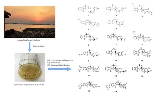
Graphical Abstract

1. Introduction

Indole diterpenoids are a family of secondary metabolites featuring a common core structure comprising an indole derived from indole-3-glycerol phosphate, and a cyclic diterpene skeleton derived from geranylgeranyl diphosphate. A select group of ascomycetous fungi, including aspergillus, penicillium, and zygomycetous fungi, is the main producer [1,2]. They were known initially for tremorgenic and neurotoxic activities partly due to their inhibition of potassium ion channels in the nervous systems [3,4], but in recent years, more diverse bioactivities, such as acyl-CoA:cholesterol acyltransferase (ACAT) inhibitors [5], M phase cell cycle inhibitors [6], and in vitro anti-proliferative, anti-insetan activities [7], etc., were discovered, which indicated their potency as drug lead compounds. Among the known indole diterpenoids, paspaline-type and paxiline-type congeners are the largest classes with the most multitudinous structures [8]. Paspline (6) is the simplest member of complex indole diterpenoids, and paxilline is the proposed precursor of terpendoles and other indole ditrpenoids, such as lotirems and janthitrems [9].
During our searching for bioactive natural compounds from marine fungi, one fungus, Penicillium brefeldianum, strain WZW-F-69, was isolated from an aquaculture base of abalone at Fujian province, China. From the culture, 16 indole diterpenoids, most of which belonged to the paspaline and paxiline types, were identified; thereinto, 5 were elucidated for the first time (Figure 1). Their cytotoxic and antimicrobial activities were tested, some of them showed to be biologically active. Herein, the structural elucidation and bioassay assay are described.

2. Results and Discussion

2.1. Structure Elucidation of the New Compounds

The ethyl acetate extract of the fermentation broth of WZW-F-69 was chromatographed on preparative HPLC columns, Sephadex LH-20 to give compounds 116.
Paspaline C (1) was obtained as a colorless solid. The HR-ESI-MS analysis exhibited [M + H]+ ion at m/z 480.3474 (calculated 480.3472, C31H46NO3+, error: 0.2 ppm), which was appropriate for the molecular formula C31H45NO3, indicating 10 degrees of unsaturation. The 1H-NMR spectrum (Table 1) revealed the presence of one exchangeable proton (1-NH), four aromatic protons (H-20–H-23), and seven single methyl groups. 13C-NMR spectrum showed 31 signals, classified by DEPT and HSQC analysis as nine none-protonated carbons including four olefinic carbons (C-2, C-18, C-19 and C-24), one acetal carbon (C-31), one oxygenated sp3 carbon (C-27), three non-oxygenated sp3 carbons (C-3, C-4 and C-12), and eight methine groups, including four olefinic methines (C-20–C-23) and four sp3 methine carbons (C-7, C-9, C-13 and C-16).
The H-H COSY from H-20 to H-23, as well as the HMBC correlations (Figure 2) from H-1 to C-2/C-18/C-19/C-24, hinted at the presence of a 2,3-disubstituted indole residue (ring A and B). The indole molecule had six degrees of unsaturation; the remaining four degrees of unsaturation indicated that compound 1 has a hexacyclic skeleton. Further analysis of the HMBC correlation from H3-25 to C-2/C-3/C-4/C-16 and from H-17 to C-2/C-3/C-16/C-18 revealed that ring C was ortho-fused with ring B at C-2 and C-18. Ring D and ring E were established mainly by the COSY correlations from H-13 to H-15 through H-14, and HMBC correlations from H3-26 to C-3/C-4/ C-5/C-13, as well as H3-30 to C-7/C-11/C-12/C-13. A sub-structure featuring a 6/5/5/6/6-fused system (ring A–E) search resulted in many hits, most of which were indole diterpenoids, mainly the derivatives of paspaline (6), which was co-isolated with 1. Detailed comparison of the NMR data between 1 and 6 showed that compound 1 had an additional 2-oxypropyl unit. The observed HMBC correlations from H-9 to C-31 and from H-32 to C-7 established a 2,2-dimethyl-1,3-dioxocane ring (ring F), so the planar structure of compound 1 was solved. It had a 6/5/5/6/6/8 hexacyclic system.
The relative configuration of 1 was determined mainly by analysis of the NOESY spectrum (Figure 3). The NOE correlations of H-16 and H3-26 with H3-30 established the β-orientations of H-16/H3-26/H3-30. The correlations of H-7 and H3-25 with H-13 indicated the α-orientations of H-7/H-13/H3-25, which was same as those of known compound 6. However, the configuration of C-9 in compound 1 needs to be reconfirmed, as the cyclization of ring F might transform it. Careful analysis of the NOE spectrum revealed the correlation of H-13/H3-33, H3-32/H3-28, and H-26/H3-29; the three correlations established the α-orientation of H-9, then the relative configuration (RC) of compound 1 was 3S*, 4S*, 7S*, 9S*, 12S*, 13R*, and 16S*.
Paspaline D (2) had the molecular formula of C30H43NO4 established by the HR-ESI-MS analysis which exhibited the [M + H]+ ion at m/z 504.3081 (calculated 504.3084, C30H43NNaO4+, error: 0.2 ppm). Comparison of the 1H- and 13C- NMR spectra of 1 and 2 showed two similar sets of data, the main differences being C-7 and C-31. For compound 1, C-31 (δ106.5 ppm) was an aldehyde acetal carbon, and H-7 was a typical oxygenous chemical shift (δ3.59 ppm), while in compound 2, C-31 (δ170.7 ppm) was a carbonyl carbon, and the chemical shift of H-7 (δ4.80 ppm) shifted downfield obviously. Detailed analysis of 2D-NMR spectra (Figure 2) revealed that compound 2 had an identical 6/5/5/6/6 system, but had no ring F; an acetoxyl group and a 3, 4-dihydroxy-4-methylpentyl group were attached at C-7 and C-12 with ring E, respectively, so its planar structure was established.
The relative configuration of 2 was established by analysis of the NOE spectrum (Figure 3). The α orientations of H-7/H-13/H3-25 and β orientations of H-16/H3-26/H3-30 were the same as those in compound 1. The H-7/H-9 correlation helped in confirming the configuration of C-9 as S*.
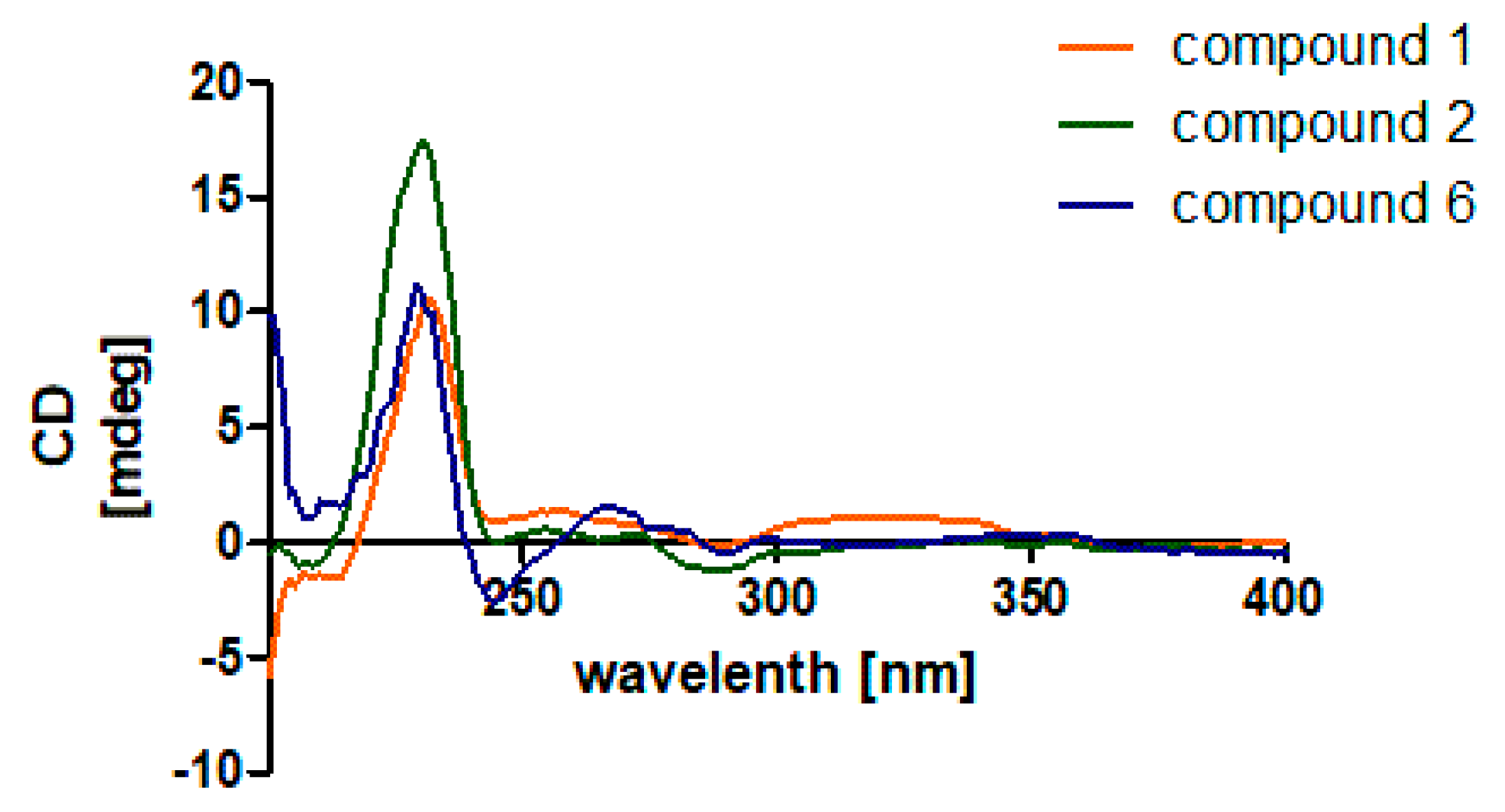
The ECD experiment was used to determine the absolute configuration (AC) of compounds 1 and 2 (Figure 4). A negative cotton curve at 230–250 nm corresponds to the AC of 3S, 4S, 13R and 16S of indole diterpenoids with the 6/5/5/6/6 skeleton of ring A-E as the π→π* transition of the indole ring [10]. Compounds 1, 2 and 6 had the same negative at about 230 nm (Figure 4) and positive at about 260 nm. Based on the result and biosynthetic considerations, the AC of compounds 1 and 2 was established as 3S, 4S, 7S, 9S, 12S, 13R, and 16S. This was confirmed by single crystal X-ray crystallographic analysis of compound 6 (Figure S69).
Paxilline B (3) had a molecular formula of C34H43NO5 based on the HR-ESI-MS analysis which exhibited an [M + H]+ ion at m/z 546.3217 (calculated 546.3214, C34H44NO5+, error: 0.3 ppm). The 1H- and 13C-NMR spectra of 3 (Table 2) are very similar to those of compound 7, O-acetylpaxilline, which revealed that compound 5 is an indole diterpene compound. The 1H-NMR spectrum at δ 6.88 (dd, J = 7.8, 0.8) /7.03 (dd, J = 8.1, 7.3)/7.01 (dd, J = 8.1, 1.0) suggested a 1, 2, 3-trisubstitued phenyl nucleus, indicating an isopentenyl group located at potion 20 or 23 of indole deterpenoid skeleton. The HMBC correlations of H-21 to C-32, CH3-35 and CH3-36 to C-33 and C-34 confirmed a 3-methyl-2-butenyl unit at position 20, so the planar structure of 3 was 20-prenylated -27-O-acetyl paxilline.
Paxilline C (4) had the same molecular formula C34H43NO5 as compound 3 based on (+)-HR-ESI-MS [M + H] + value 546.3220. The two compounds had highly similar 1H-NMR and 13C-NMR spectra (Table 2), except for the position of the isopentenyl group. Detailed analysis of 2D-NMR spectra (Table S4) revealed that for compound 4, the isopentenyl is at C-21, so the planar structure of 4 is 21-prenylated -27-O-acetyl paxilline.
The relative configurations of 3 and 4 can be inferred through NOE spectra analysis. Two compounds had the same NOE correlations from H-16 to H3-26, from H-7 to H-9, and H3-25. Their ECD spectra are highly consistent with those of compound 7 (Figure S63), indicating that compounds 3 and 4 have the absolute configuration of 3S, 4R, 7S, 9R, 13S, and 16S.
Paxilline D (5) was obtained as a colorless solid, and its molecular formula was determined as C28H35NO4 based on (+)-HR-ESI-MS data [M + H]+ 450.2638 (calculated 450.2639 for C28H36NO4+, error: 0.1 ppm). It had highly similar 1H-NMR and 13C-NMR spectra with 13-desoxypaxilline (10) (molecular formula: C27H33NO3, Figures S39 and S40). Detailed analysis revealed that compound 5 had an additional methoxy group (δH 3.40, δC 49.1), which was consistent with their molecular formulas. HMBC correlations from H3-30 to C-7, as well as the typical aldehyde acetal chemical shift of C-7 (δC 96.5) established the planar structure of 5 as 7-methoxy-13-desoxypaxilline.
The relative structure of compound 5 was determined by NOE through connection from H-9 to H-13, H3-30 and H3-25, which indicated the same orientation of these atoms (Table S5). Compound 5 showed a negative cotton effect at 200–250 nm in the ECD spectrum, that corresponded to that of paxilline. Therefore, the absolute configuration of compound 5 was assigned as 3S, 4S, 7R, 9R, 12R, 16S.
In addition to compounds 15, 11 congeners—paspaline (6) (Figure 5) [11], 1’-O-acetyl paxilline (7) [12], 20-prenylated paxilline (8) [13], 22-prenylated paxilline (9) [14], 13-deoxy-paxilline (10) [15], paspalicine (11) [16], sherinine K (12) [17], 3-deoxo- 4b-deoxy paxilline (13) [18], 10-hydroxy-13-desoxy paxilline(14) [19], pyrapaxilline (15) [20], and shearinine F(16) [21]—were also isolated and identified. Their structures were determined mainly by analysis of their NMR spectra and comparison with the literature. Based on the substitution around the phenyl ring of the indole moiety and the olefinic bond of ring F, 16 compounds can be divided into 4 groups: compounds 1, 2 and 6 belong to the paspaline group, which lack substitution on the indole ring and have no olefinic bond on ring F; compounds 5, 7, 10, 11, 13 and 14 belong to the paxilline group which have an olefinic bond on ring F; compounds 3, 4, 8 and 9 are the 3rd group which have one isopentene group on the indole ring; and compounds 12, 15 and 16 have two isopentyl substituent groups on the indole ring. The biogenetic pathway of the five new compounds is proposed in Figure 6. Emindole SB is likely a biosynthetic precursor of 6 and other advanced indole diterpenoids.

2.2. Bioactivities of Compounds

Compounds 116 were evaluated for their antimicrobial and cytotoxic activities with the cell lines and microbial strains in our lab. All tested compounds showed no activities against Escherichia coli CMCC44103, Staphlococcus aures CMCC26003, Aspergillus niger ACCC3005, and Candida albicans AS2.538 by the disk diffusion method at 10 mg/mL, nor cytotoxic activity at 1 μM against MDA-MB-231 (human breast cancer cells), A375 (human melanoma cells), BGC-823 (human pancreatic cancer cells), SKGT4 and Kyle 450 (two human esophagus cancer cell) lines. However, compounds 1 and 6 exhibited moderate inhibitory activity against HepG-2 (human hepatoma carcinoma cells), U2OS (human osteosarcoma cells), MCF-7 (human breast cancer cells), Jeko-1 (human mantle cell lymphoma cells) and HL-60 (human acute promyelocytic leukemia cells). Their average inhibition rates were in the range of 50% to 80% at a concentration of 1 μM (Table 3). Our result is in line with the literature [3,22] that paspline- and paxilline-type indole diterpenoids seldom have anti-microbial activity, while some of them have cytotoxic activity, which differs from different cancer cell lines.

3. Materials and Methods

3.1. General Apparatus

The NMR data were recorded on Bruker Advance-III spectrometer (Bruker, Karlsruhe, Germany). Mass spectra were performed on the Waters 2767 LC-MS system using the Nova-pak silica column (Waters, Milford, CT, USA). HR-ESI-MS was recorded on Thermo Scientific Q Exactive HF Orbitrap-FTMS (Thermo Fisher, Waltham, MA, USA). Analytic HPLC was operated on Agilent 1200 series liquid chromatography with an Agilent XDB-C18 column (9.4 × 250 mm, 5 μm) and UV detector (Agilent, Santa Clara, CA, USA). Semi-preparative HPLC was operated on Welch Sail 1000 liquid chromatography with Prep RP-18 column (28 × 4 cm) and UV detector (Welch, Baltimore, MD, USA). Mid-press reverse phase chromatography was operated on Buchi Switzerland system with BUCHI RP-18 column (20 × 4 cm, 170 g) (Büchi, Uster, Switzerland). Column chromatography (CC) was performed with silica gel (300–400 mesh, Yantai Jiangyou silicon development Co., Ltd., Yantai, China) and Sephadex LH-20 (GE Healthcare, Chicago, IL, USA).

3.2. Fungal Material

The fungal strain WZW-F-69 was isolated from soil near an abalone aquaculture base of Fujian province, China (N 23°47′727″, E 117°29′079″), in August 2015. After being purified, the strain was identified with the ITS gene sequence (Sequence S1) to be Penicillium brefeldianum (100% consistent with Genebank KM243921.1). The strain was preserved in our lab at −80 °C.

3.3. Fermentation and Extraction

For chemical investigation, the fungal strain WZW-F-69 was cultivated in 100 mL liquid seawater potato-dextrose medium (including extract of potato 200 g, glucose 20 g, sea salt 30 g, water 1 L), 220 rpm, at 28 °C for 5–7 days to prepare the seed culture. Then, 5 mL of the seed solution was inoculated on solid rice culture media (150 g rice in 150 mL water) in a 2.5 L stainless steel box. A total of 3 kg of the rice media was incubated at 28 °C for 20 days. The rice culture media were collected and soaked in ethyl acetate (EA) overnight. After the solvent was removed in vacuum, it yielded a crude extract (20 g). The crude extract was redissolved in MeOH: petroleum ether (PE, 1:1, v/v) three times. The combined MeOH extract (10 g) was grouped by the RP-18 silica gel column mid pressure chromatography (28 × 4 cm, 170 g), and the elute solution changed from 100% H2O to 100% MeOH in 2 h, 30 mL/min.

3.4. Isolation and Purification

The 70% MeOH elution, component D (802.8 mg), was purified by Sephadex LH-20 (2.5 × 140 cm, 140g in MeOH, 15 s/drip, 1 tube/h) and gave 7 fractions (D-1 to D-7). Compound 2 (3.2 mg) was purified from D-2 with Agilent HPLC (MeOH 80%, 5 µm, 4.6 × 250 mm, 3 mL/min). D-3 afforded 11 fractions (D-3-1 to D-3-11) with Sephadex LH-20 (2 × 80 cm, 80 g, in acetone, 15 s/drip, 1 tube/h). Compounds 1 (1.0 mg), 6 (9.6 mg) and 14 (3.2 mg) were purified from D-3-4, D-3-9 and D-3-6, respectively. D-4 afforded 7 fractions (D-4-1 to D-4-7) by Sephadex LH-20 in acetone (2.5 × 15 cm, 140 g, 15 s/drip, 1 tube/h). D-4-1 yielded 7 (58.0 mg), 10 (4.6 mg), 11 (6.3 mg) after being subjected to a silica gel column chromatography (2 × 8 cm, 10 g) with elution of PE:EtoAc (from 35:1 to 0:1 in five hours). Compound 5 (7.3 mg) was yielded from D-4-2 through Agilent HPLC (MeCN/H2O, 80%).
The 80% MeOH elution, component E (3.280 g), was purified by Sephadex LH-20 (2.5 × 140 cm, 140 g, in CHCl2/MeOH, 1:2, v:v) then by Sephadex LH-20 (140 g, acetone) and silica gel column chromatography (24 g, PE: EtoAc, 100:0 to 0:100 in 3 h), afforded 3 (3.2 mg) and 4 (1.0 mg).
The 90% MeOH eluted part, component F (1.075 g), yielded 11 fractions (F-1 to F-11) after Sephadex LH-20 chromatography (140 g, in MeOH). F-8 was subjected to silica gel CC (4 g, PE:EtoAc, 100:0 to 0:100 in two hours) to gain 3 sub-fractions (F-8-1 to F-8-3). F-8-2 afforded 12 (2.1 mg) by Agilent HPLC (MeCN-H2O, 90%, 5 µm, 4.6 × 250 mm, 3 mL/min).
The potion of 90–100% MeOH, component G (1.662 g), gave 9 (5.3 mg), 13 (1.8 mg), 15 (4.1 mg) and 16 (5.0 mg) after Sephadex LH-20 (2.5 × 140 cm, 140 g, in MeOH and 2.5 × 140 cm, 140 g, in acetone).
Another patch of the MeOH extract named component H (1.5 g) followed Sephadex LH-20 (2.5 × 140 cm, 140g, in MeOH), silica gel CC (30 g, PE: EtoAc, 100:0 to 0:100 in 3 h) and Agilent HPLC (MeCN/H2O, 80%) and yielded 8 (1.0 mg).
Paspaline C (1): colorless solid, [α]25D: 0 (c 1.0, MeOH), HR-ESI-MS at m/z 480.3474 [M + H]+, calculated for C31H45NO3, 480.3472. UV (MeOH), λmax (log ε): 284 (1.4), 225 (5.1). IR (KBr) νmax 3430, 2939, 2253, 1651, 1455, 1086, 1049, 1025, 1004, 824, 762, 615. For 1H-NMR(CDCl3, 600 MHz) and 13C-NMR(CDCl3, 150 MHz) data, see Table 1; for UV spectrum, see Figure S64.
Paspaline D (2): colorless solid, [α]25D: +34 (c 1.0, MeOH), HR-ESI-MS at m/z 504.3081 [M + Na]+, calculated for C35H45NO4Na, 504.3084. UV (MeOH), λmax (log ε): 280 (1.2). IR (KBr) νmax 3440, 2974, 2114, 1634, 1456, 1364, 1276, 1160, 1048, 1032, 911, 471. For 1H-NMR (CDCl3, 600 MHz) and 13C-NMR (CDCl3, 150 MHz) data, see Table 1; for UV spectrum, see Figure S65.
Paxilline B (3): colorless solid, [α]25D: +24 (c 1.0, MeOH), HR-ESI-MS at m/z 546.3217 [M + H]+, calculated for C34H44NO5, 546.3214. UV. UV (MeOH), λmax (log ε): 285 (1.0), 225 (3.8). IR (KBr) νmax 3430, 2948, 2843, 1658, 1453, 1377, 1179, 1113, 1020, 635. 1H-NMR (CDCl3, 600 MHz) and 13C-NMR (CDCl3, 150 MHz) data, see Table 2; for UV spectrum, see Figure S66.
Paxilline C (4): colorless solid, [α]25D: +60 (c 1.0, MeOH), HR-ESI-MS at m/z 546.3220 [M + H]+, calculated for C34H44NO5, 546.3214. UV (MeOH), λmax (log ε): 290 (1.1), 240 (4.3). IR (KBr) νmax 3403, 2982, 2937, 2849, 1709, 1676, 1624, 1449, 1368, 1259, 1124, 1025, 960, 928, 742, 709, 606, 583. For 1H-NMR (CDCl3, 600 MHz) and 13C-NMR (CDCl3, 150 MHz) data, see Table 2; for UV spectrum, see Figure S67.
Paxilline D (5): colorless solid, [α]25D: 0 (c 1.0, MeOH), HR-ESI-MS at m/z 450.2638 [M + H]+, calculated for C28H36NO4, 450.2639. UV (MeOH), λmax (log ε): 230 (1.5). IR (KBr) νmax 3400, 2928, 1658, 1448, 1373, 1303, 1258, 1127, 1032, 742. For 1H-NMR (CDCl3, 600 MHz) and 13C-NMR (CDCL3, 150 MHz) data, see Table 2; for UV spectrum, see Figure S68.

3.5. Antimicrobial Activity

The antimicrobial activity was evaluated with the disk diffusion methodology [23]. Four indicator organisms (Staphococcus aureus CMCC26003, Escherichia coli CMCC44103, Aspergillus niger ACCC3005, Canidida albicans AS2.538) were used. The spores at 1×10⁶/mL were poured onto culture media on a Petri dish (φ = 9 cm): PDA medium for fungal indicators, and Luria–Bertain (LB) medium for bacterial indicators. The sterilized filter paper (φ = 5 cm) with 5 µL of compound solution at 10 mg/mL was put on the surface of the culture medium; 24–48 h later, the inhibition diameters were measured. Amphotricin B and gentamicin at 1 mg/mL were used as positive controls for the fungal and bacterial indicators, respectively.

3.6. Cytotoxic Activity

A375, MDA-MB-231, HepG2, BGC823, U2OS and MCF-7 cell lines were grown in DMEM medium. SKGT4, Kyse450, JeKo-1 and HL60 cells were grown in RPMI1640 medium. Two media were supplemented with 10% FBS and 1% penicillin or streptomycin. Then, 198 µL of the medium with about 5000 cancer cells was plated in each well of a 96-well plate. Then, 2 µL of the test compound at 10 mM in DMSO was added in triplicate. Forty-eight hours later, the cell viability was determined using CellTiter 96® AQueous Assay Reagents according to the instruction manual at 490 nm. Data were normalized to the control group (DMSO).

4. Conclusions

In summary, 5 new indole diterpenoids (15) and 11 known congeners (6–16) were identified from sea-mud-derived fungal Penicillium Brefeldianum WZW-F-69. Even though the main structural elements resemble the reported indole diterpenes, paspaline C (1) has a rare 2,2-dimethyl-1,3-dioxocane ring F, while paspaline D (2) has an unusual open ring F structure. Paxilline B (3) and paxilline C (4) are isopentenyl ring A derivatives of paxilline, while paxilline D (5) has an aldehyde acetyl carbon (C-7), which makes them chemically unique. Although the AC of new compounds was established mainly by the comparison of ECD spectra with known compounds 6 and 7, the consistence with known compounds, such as 6-hydroxylpaspslinine [3], 4a-demethylpaspaline-4a-carboxylic acid [7], whose AC was confirmed with experimental and calculated ECD, supported our assignment. Compounds 1 and 6 exhibited cytotoxic activity against several cancer cell lines. In summary, the identification of diverse structural compounds from WZW-F-69 illustrates the huge potential of finding more new bioactive lead compounds from marine fungi for future research.

Supplementary Materials

The following are available online at https://www.mdpi.com/article/10.3390/md20110684/s1, Sequence S1: Taxonomy of Penicillium Brefeldianum WZW-F-69, Tables S1–S5: NMR (600 MHz, CDCl3) data for Compounds 15, Figures S1–S30: 1D and 2D NMR spectra of compounds 15, Figures S31–S52: 1H-NMR, 13C-NMR of compounds 616, Figures S53–S57: HR-ESI-MS reports of 15, Figures S58–S62: ECD spectra of compounds 15, Figure S63: ECD spectrum of compounds 3, 4 and 7, Figures S64–S68: UV spectra of compounds 15, Figure S69: x-ray single crystal diffraction structure of compound 6.

Author Contributions

Methodology and writing—original draft preparation, W.L.; methodology, H.L.; resource, Z.W.; validation, J.S.; methodology, Z.Z. and L.Y.; project administration and funding acquisition, X.D.; supervision and writing—review and editing, Q.X. All authors have read and agreed to the published version of the manuscript.

Funding

This research was funded by the National Natural Science Foundation of China (Nos. 22025702 and 91853203 to X.D.), and the Fundamental Research Funds for the central Universities of China (No. 2011121037 to Q.X.).

Conflicts of Interest

The authors declare no conflict of interest.

References

  1. Ariantari, N.P.; Ancheeva, E.; Wang, C.; Mandi, A.; Knedel, T.-O.; Kurtan, T.; Chaidir, C.; Mueller, W.E.G.; Kassack, M.U.; Janiak, C.; et al. Indole diterpenoids from an endophytic Penicillium sp. J. Nat. Prod. 2019, 82, 1412–1423. [Google Scholar] [CrossRef] [PubMed]
  2. Breteche, A.; Duflos, M.; Dassonville, A.; Nourrisson, M.R.; Brelet, J.; Le Baut, G.; Grimaud, N.; Petit, J.Y. New N-pyridinyl(methyl)-indole-2- and 3-(alkyl) carboxamides and derivatives acting as systemic and topical inflammation inhibitors. J. Enzym. Inhib. Med. Chem. 2002, 17, 415–424. [Google Scholar] [CrossRef] [PubMed]
  3. Hu, X.Y.; Meng, L.H.; Li, X.; Yang, S.Q.; Li, X.M.; Wang, B.G. Three new indole diterpenoids from the sea-anemone-derived fungus Penicillium sp. AS-79. Mar. Drugs 2017, 15, 137. [Google Scholar] [CrossRef]
  4. Munday-Finch, S.C.; Wilkins, A.L.; Miles, C.O. Isolation of paspaline B, an indole-diterpenoid from Penicillium paxilli. Phytochemistry 1996, 41, 327–332. [Google Scholar] [CrossRef]
  5. Huang, X.H.; Tomoda, H.; Nishida, H.; Masuma, R.; Omura, S. Terpendoles, novel ACAT inhibitors produced by Albophoma yamanashiensis. I. Production, isolation and biological properties. J. Antibiot. 1995, 48, 1–4. [Google Scholar] [CrossRef]
  6. Sallam, A.A.; Houssen, W.E.; Gissendanner, C.R.; Orabi, K.Y.; Foudah, A.I.; El Sayed, K.A. Bioguided discovery and pharmacophore modeling of the mycotoxic indole diterpene alkaloids penitrems as breast cancer proliferation, migration, and invasion inhibitors. MedChemComm 2013, 4, 1360–1369. [Google Scholar] [CrossRef]
  7. Fan, Y.; Wang, Y.; Liu, P.; Fu, P.; Zhu, T.; Wang, W.; Zhu, W. Indole diterpenoids with anti-H1N1 activity from the aciduric fungus Penicillium camemberti-1492. J. Nat. Prod. 2013, 76, 1328–1336. [Google Scholar] [CrossRef] [PubMed]
  8. Liu, C.; Noike, M.; Minami, A.; Oikawa, H.; Dairi, T. A fungal prenyltransferase catalyzes the regular di-prenylation at positions 20 and 21 of paxilline. Biosci. Biotechnol. Biochem. 2014, 78, 448–454. [Google Scholar] [CrossRef]
  9. Reddy, P.; Guthridge, K.; Vassiliadis, S.; Hemsworth, J.; Hettiarachchige, I.; Spangenberg, G.; Rochfort, S. Tremorgenic mycotoxins: Structure diversity and biological activity. Toxins 2019, 11, 302. [Google Scholar] [CrossRef]
  10. Zhou, L.M.; Kong, F.D.; Fan, P.; Ma, Q.Y.; Xie, Q.Y.; Li, J.H.; Zheng, H.Z.; Zheng, Z.H.; Yuan, J.Z.; Dai, H.F.; et al. Indole-diterpenoids with protein tyrosine phosphatase inhibitory activities from the marine-derived fungus Penicillium sp. KFD28. J. Nat. Prod. 2019, 82, 2638–2644. [Google Scholar] [CrossRef]
  11. Kim, D.E.; Zweig, J.E.; Newhouse, T.R. Total synthesis of paspaline A and emindole PB enabled by computational augmentation of a transform-guided retrosynthetic strategy. J. Am. Chem. Soc. 2019, 141, 1479–1483. [Google Scholar] [CrossRef]
  12. Liu, C.; Tagami, K.; Minami, A.; Matsumoto, T.; Frisvad, J.C.; Suzuki, H.; Ishikawa, J.; Gomi, K.; Oikawa, H. Reconstitution of biosynthetic machinery for the synthesis of the highly elaborated indole diterpene penitrem. Angew. Chem. Int. Ed. 2015, 54, 5748–5752. [Google Scholar] [CrossRef] [PubMed]
  13. Bauer, J.I.; Gross, M.; Cramer, B.; Wegner, S.; Hausmann, H.; Hamscher, G.; Usleber, E. Detection of the tremorgenic mycotoxin paxilline and its desoxy analog in ergot of rye and barley: A new class of mycotoxins added to an old problem. Anal. Bioanal. Chem. 2017, 409, 5101–5112. [Google Scholar] [CrossRef] [PubMed]
  14. Kozak, L.; Pocsi, I.; Molnar, I.; Kozak, L.; Szilagyi, Z.; Toth, L.; Molnar, I. Tremorgenic and neurotoxic paspaline-derived indole-diterpenes: Biosynthetic diversity, threats and applications. Appl. Microbiol. Biotechnol. 2019, 103, 1599–1616. [Google Scholar] [CrossRef] [PubMed]
  15. Saikia, S.; Parker, E.J.; Koulman, A.; Scott, B. Defining paxilline biosynthesis in Penicillium paxilli: Functional characterization of two cytochrome P450 monooxygenases. J. Biol. Chem. 2007, 282, 16829–16837. [Google Scholar] [CrossRef] [PubMed]
  16. Fehr, T.; Acklin, W. Isolation of 2 new indole derivatives from the mycelia of Claviceps paspali. Helv. Chim. Acta 1966, 49, 1907–1910. [Google Scholar] [CrossRef]
  17. Springer, J.P.; Clardy, J. Paspaline and paspalicine, two indole-mevalonate metabolites from Claviceps paspali. Tetrahedron Lett. 1980, 21, 231–234. [Google Scholar] [CrossRef]
  18. Nozawa, K.; Horie, Y.; Udagawa, S.; Kawai, K.; Yamazaki, M. Studies on fungal products. Part XXIV. Isolation of a new tremorgenic indoloditerpene, 1′-O-acetylpaxilline, from Emericella striata and distribution of paxilline in Emericella spp. Chem. Pharm. Bull. 1989, 37, 1387–1389. [Google Scholar] [CrossRef]
  19. Mantle, P.G.; Weedon, C.M. Biosynthesis and transformation of tremorgenic indole-diterpenoids by Penicillium paxilli and Acremonium lolii. Phytochemistry 1994, 36, 1209–1217. [Google Scholar] [CrossRef]
  20. Belofsky, G.N.; Gloer, J.B.; Wicklow, D.T.; Dowd, P.F. Antiinsectan alkaloids: Shearinines A–C and a new paxilline derivative from the ascostromata of Eupenicillium shearii. Tetrahedron 1995, 51, 3959–3968. [Google Scholar] [CrossRef]
  21. Springer, J.P.; Clardy, J.; Wells, J.M.; Cole, R.J.; Kirksey, J.W. Structure of paxilline, a tremorgenic metabolite of Penicillium paxilli. Tetrahedron Lett. 1975, 16, 2531–2534. [Google Scholar] [CrossRef]
  22. Gao, S.S.; Li, X.M.; Williams, K.; Proksch, P.; Ji, N.Y.; Wang, B.G. Rhizovarins A-F, indole-diterpenes from the mangrove-derived endophytic fungus Mucor Irregularis QEN-189. J. Nat. Prod. 2016, 79, 2066–2074. [Google Scholar] [CrossRef] [PubMed]
  23. Mc, G.J. The filter paper disc method of assaying antibiotics. J. Bacteriol. 1945, 50, 717. [Google Scholar]
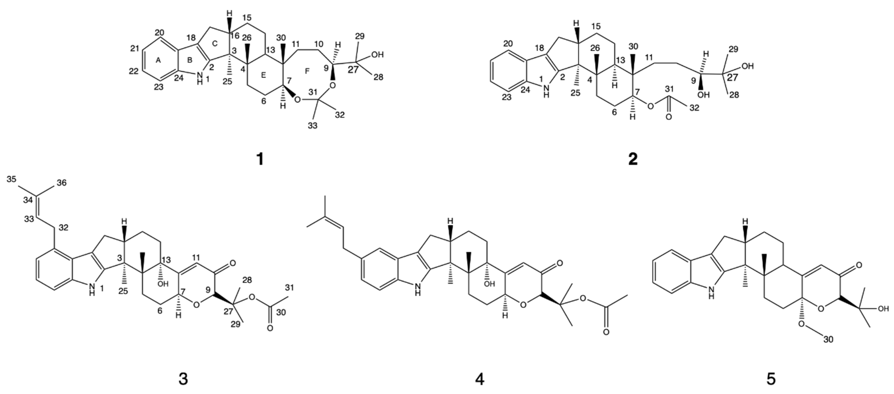
Figure 1. Structures of compounds 15.
Marinedrugs 20 00684 g001
Figure 2. Key HMBC and key COSY correlations of 1 and 2.
Marinedrugs 20 00684 g002
Figure 3. Key NOESY correlations of 1 and 2.
Marinedrugs 20 00684 g003
Figure 4. ECD spectra of compound 1, 2 and 6.
Marinedrugs 20 00684 g004
Figure 5. Structures of compounds 616.
Marinedrugs 20 00684 g005
Figure 6. Proposed biosynthesis pathway of compounds 15.
Marinedrugs 20 00684 g006
Table 1. 1H (600 MHz) and 13C NMR (150 MHz) data of compounds 1 and 2 (CDCl3, δ in ppm, J in Hz).
No.12
1H13C1H13C
17.74, s 7.77, s
2 150.8, C 150.6, C
3 53.1, C 53.1, C
4 39.3, C 39.3, C
51.94, dd (11.3, 5.8)
1.62, m
33.6, CH22.05, dd (13.7,4.6)
2.01, dd (12.5, 5.4)
33.1, CH2
61.89, dd (14.7, 2.7)
1.87, m
26.9, CH21.90, m23.8, CH2
73.59, dd (9.8, 5.9)72.8, CH4.80, dd (10.2, 5.9)75.5, CH
93.68, dd (8.3, 4.6)84.1, CH3.57, dd (8.5, 4.3)83.6, CH
101.34, dd (12.1, 5.6)
1.32, m
22.2, CH21.59, m22.6, CH2
111.71, td (12.9, 3.0)
1.41, m
33.9, CH21.55, m
1.32, m
34.4, CH2
12 39.3, C 40.2, C
131.69, s41.4, CH1.78, dd (12.7, 3.1)40.4, CH
141.81, d (12.6)
1.64, brs
25.2, CH21.82, m
1.65, td (12.9, 4.1)
25.1, CH2
151.71, m
1.49, td (12.4, 4.4)
23.0, CH21.59, m22.6, CH2
162.78, m47.8, CH2.78, m48.7, CH
172.70, dd (13.2, 6.5)
2.35, dd (13.2, 10.6)
27.5, CH22.70, dd (13.2, 6.4)
2.36, dd (13.2, 10.6)
27.5, CH2
18 118.4, C 118.3, C
19 125.4, C 125.1, C
207.45, ddd (6.6, 3.2, 1.0)118.3, CH7.44, m118.4, CH
217.09, m119.6, CH7.09, m119.6, CH
227.10, m120.5, CH7.09, m120.5, CH
237.32, ddd (6.4, 2.5, 0.8)111.4, CH7.32, m111.5, CH
Table 2. 1H (600 MHz) and 13C NMR (150 MHz) data of compounds 35 (CDCl3, δ in ppm, J in Hz).
No.345
1H13C1H13C1H13C
17.78, s 7.70, s 7.79, s
2 151.0, C 151.9, C 149.3, C
3 50.5, C 50.7, C 50.5, C
4 43.1, C 43.1, C 42.4, C
52.80, td (13.5, 4.9)
1.49, m
28.1, CH2.78, td (13.7, 4.4)
1.47, m
28.0, CH2.15, td (13.7, 6.4)
1.58, m
31.4, CH
62.31, m
1.95, m
28.3, CH22.31, m
1.93, m
28.3, CH22.44, m
1.90, dd (14.6, 4.3)
28.8, CH2
74.84, m72.9, CH4.84, m72.9, CH 96.5, C
94.85, brs80.4, CH4.85, brs80.4, CH4.06, s76.9, CH
10 195.3, C 195.3, C 198.5, C
115.85, d (2.1)120.1, CH5.84, d (2.0)120.1, CH5.81, d (2.0)122.1, CH
12 166.1, C 166.1, C 165.1, C
13 77.5, C 77.5, C2.83, m42.0, CH
142.08, m
1.66, m
34.4, CH22.07, m
1.67, m
34.4, CH21.72, m
1.52, m
25.5, CH2
152.07, m20.9, CH22.05, m
1.80, m
20.9, CH21.86, m24.1, CH2
162.89, d (2.8)49.6, CH2.85, m49.6, CH2.85, m49.0, CH
172.91, d (6.2)
2.62, m
29.0, CH22.74, dd (13.0, 6.3)
2.45, dd (13.2, 10.9)
27.2, CH22.74, dd (13.4, 6.4)
2.45, m
27.3, CH2
18 116.9, C 117.1, C, 118.4, C
19 124.4, C 125.6, C 125.0, C
20 133.1, C7.25 (brs)117.6, CH7.47, dd (6.8, 2.2)118.5, CH
216.88, dt (7.3, 0.8)119.0, CH 133.3, C7.12, m120.8, CH
227.03, dd (8.1, 7.3)121.0, CH6.94, dd (8.2, 1.7)121.4, CH7.12, m119.8, CH
237.17, dd (8.1, 1.0)109.3, CH7.23, dd (8.1, 1.0)111.3, CH7.33, dd (6.8, 2.2)111.4, CH
24 139.7, C 138.2, C 140.0, C
251.34, s16.2, CH31.33, s16.2, CH31.12, s14.7, CH3
261.08, s19.8, CH31.05, s19.7, CH31.03, s15.9, CH3
27 81.9, C 81.9, C 72.4, C
281.46, s22.8, CH31.46, s22.8, CH31.29, s24.0, CH3
291.68, s23.8, CH31.68, s23.8, CH31.35, s26.7, CH3
30 170.8, C 170.8, C3.40, s49.1, CH3
312.06, s22.3, CH32.06, s22.3, CH3
323.64, d (7.3)32.0, CH23.43, d (7.3)34.5, CH2
335.43, m123.7, CH5.40, m124.6, CH
34 131.8, C 131.4, C
351.76, s25.8, CH31.76, s25.8, CH3
361.77, s17.8, CH31.77, s17.8, CH3
Table 3. The average inhibition rate of compounds 1 and 6 against cell lines (%).
HepG-2U2OSMCF 7JeKo-1HL-60
Compound 155.156.156.471.265.8
Compound 652.483.447.572.460.3
Publisher’s Note: MDPI stays neutral with regard to jurisdictional claims in published maps and institutional affiliations.