
Mussel Shells, a Valuable Calcium Resource for the Pharmaceutical Industry

 ,
,  and
and
Abstract
:
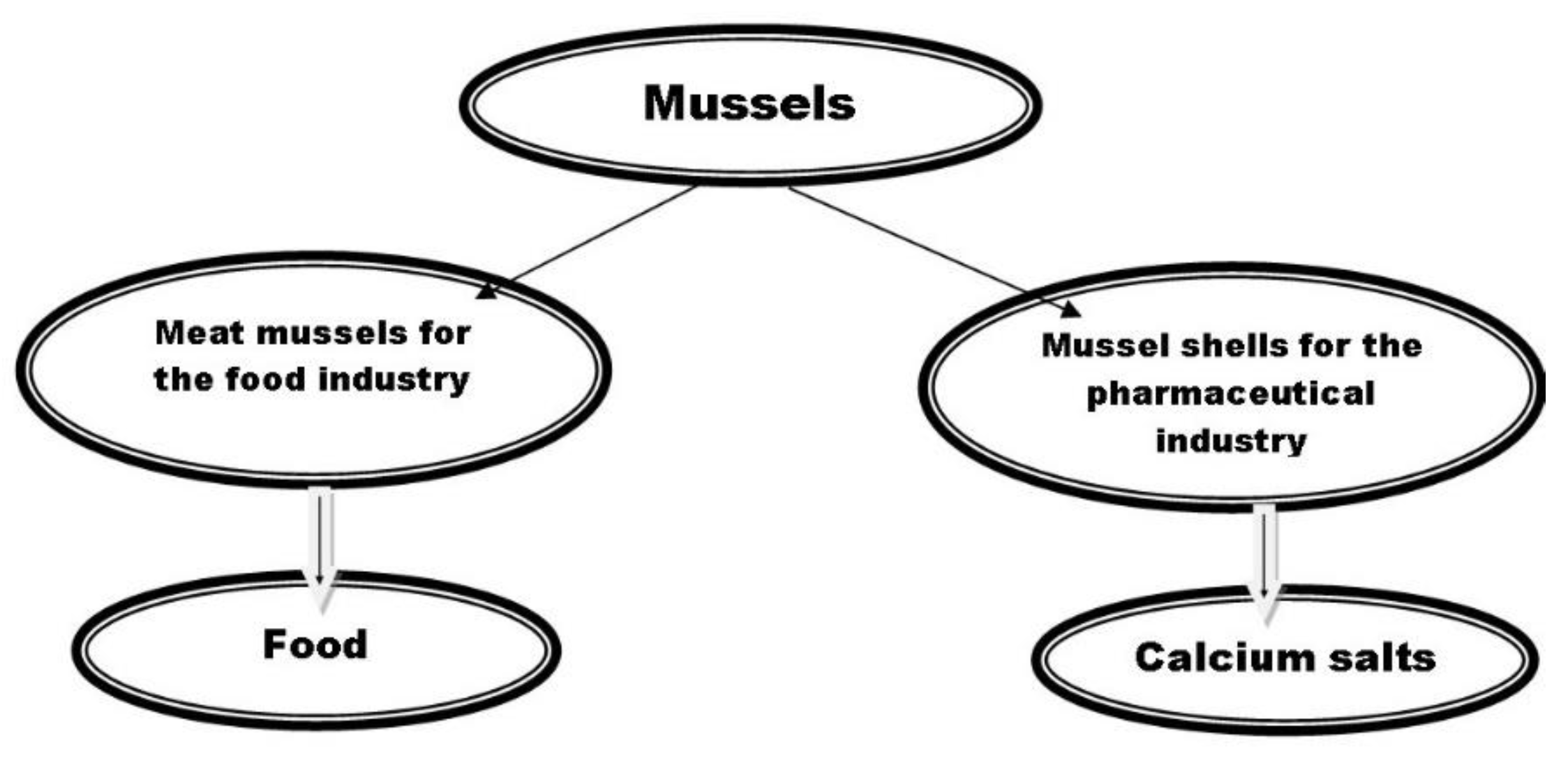
1. Introduction
2. Materials and Methods
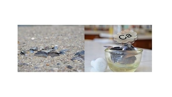
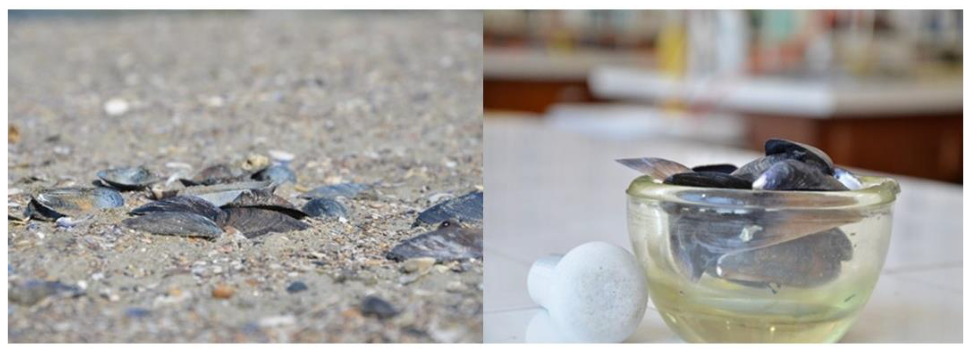
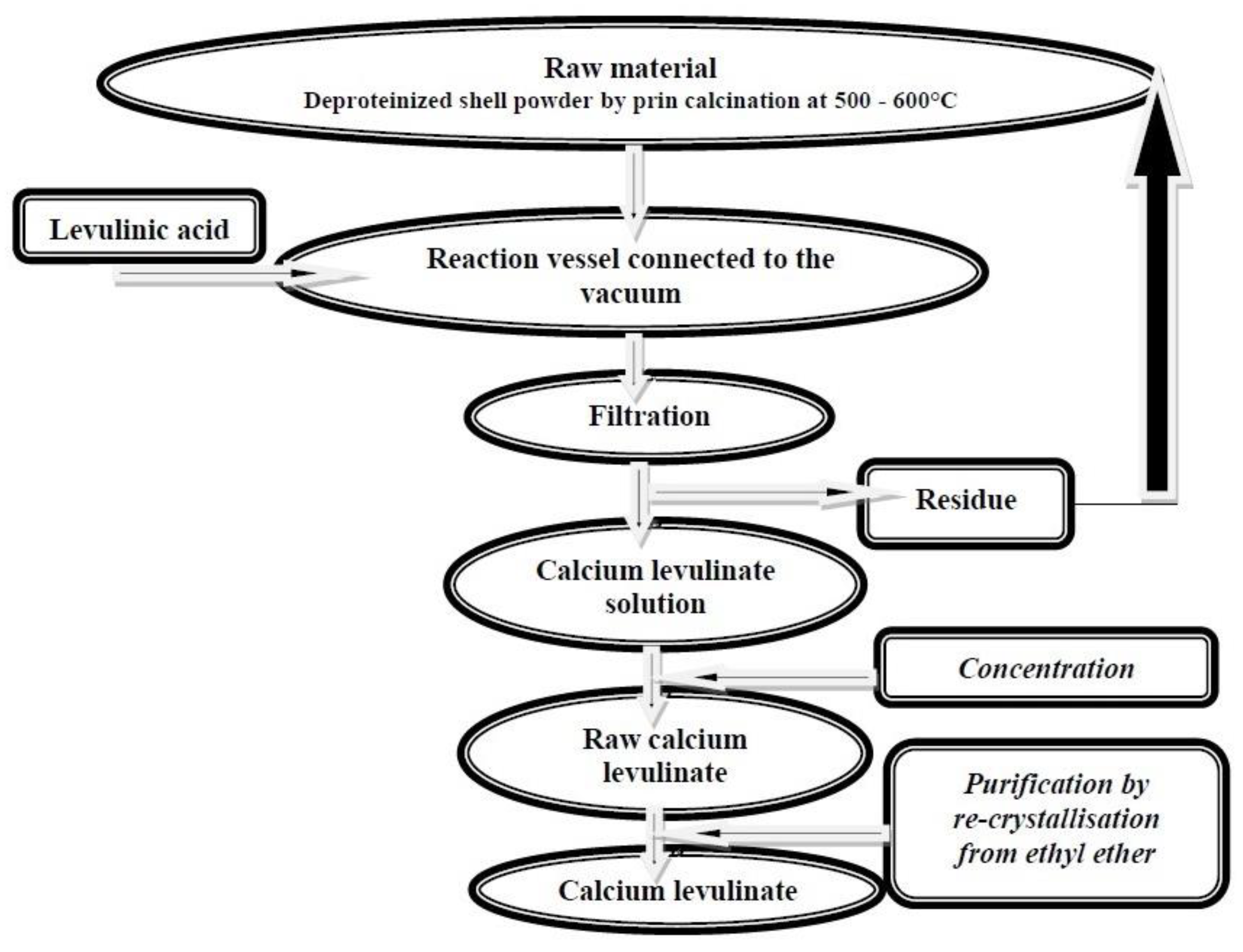
2.1. Extraction of Calcium Levulinate from Mussel Shell Waste
- The raw material is brought to powder to increase the contact area and thus the effectiveness of reagents used;
- The purpose of the treatment by boiling in 1% alkaline KOH solution is to release the organic component;
- The mineral cake (the major component) has to be rinsed under pH control to remove alkali traces, which, otherwise, in numbers, would contaminate the yields (calcium salts) and increase use of acid;
- Processes for the preparation of the various salts share the step of acid attack in a reaction vessel. This was designed in such a way as to allow gradual addition of the acid (by the funnel mounted on upper, tapped side), to enable thorough use of calcium carbonate in the shells and elimination of the resulting reaction CO2 from the system. Connection to a vacuum pump hastened the exhaust, also securing a certain level of foam;
- In particular, certain purification and recrystallisation means were applied for each type of salt.
2.2. Calcium Levulinate Anhydrous Toxicity Evaluation
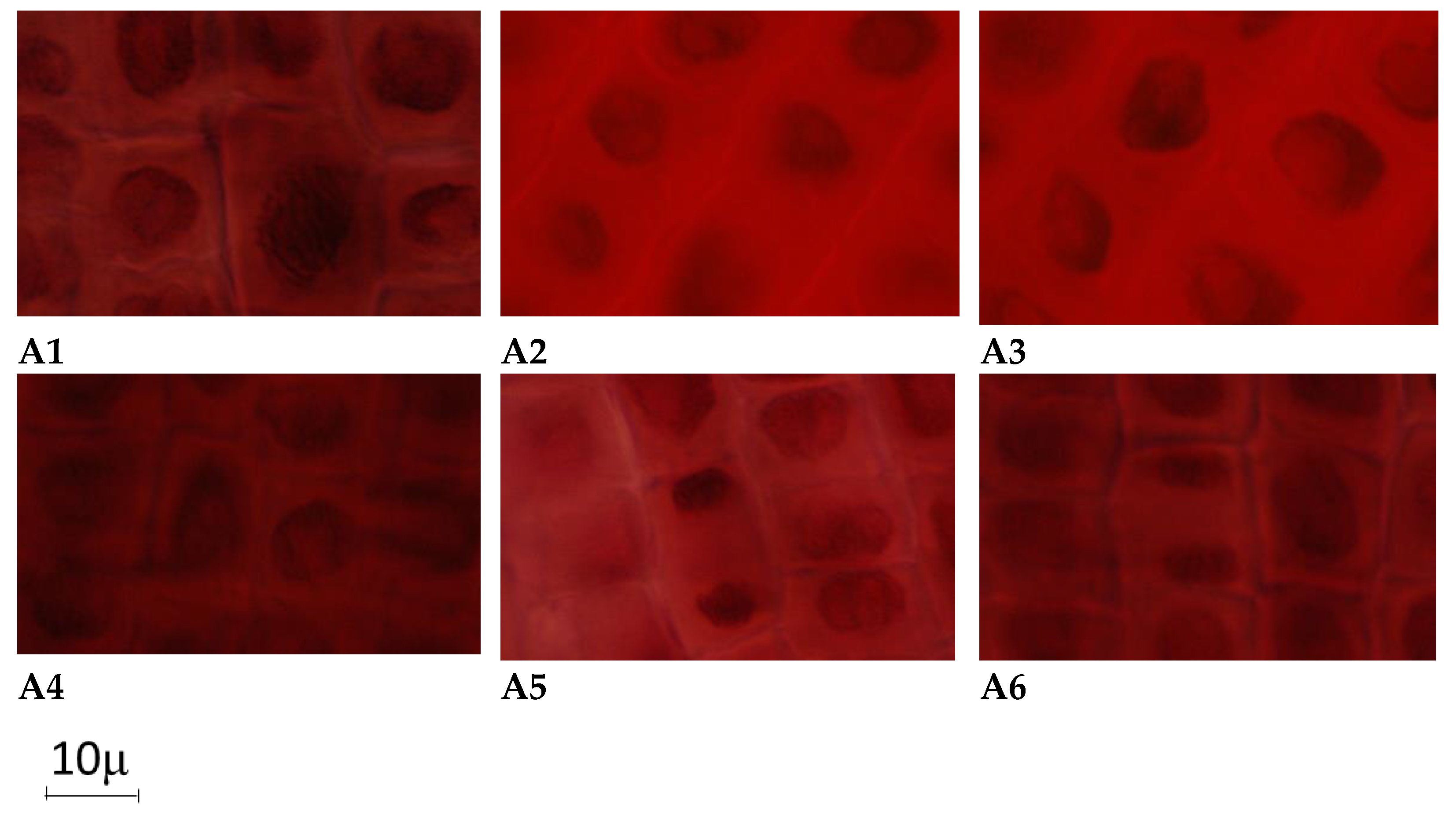
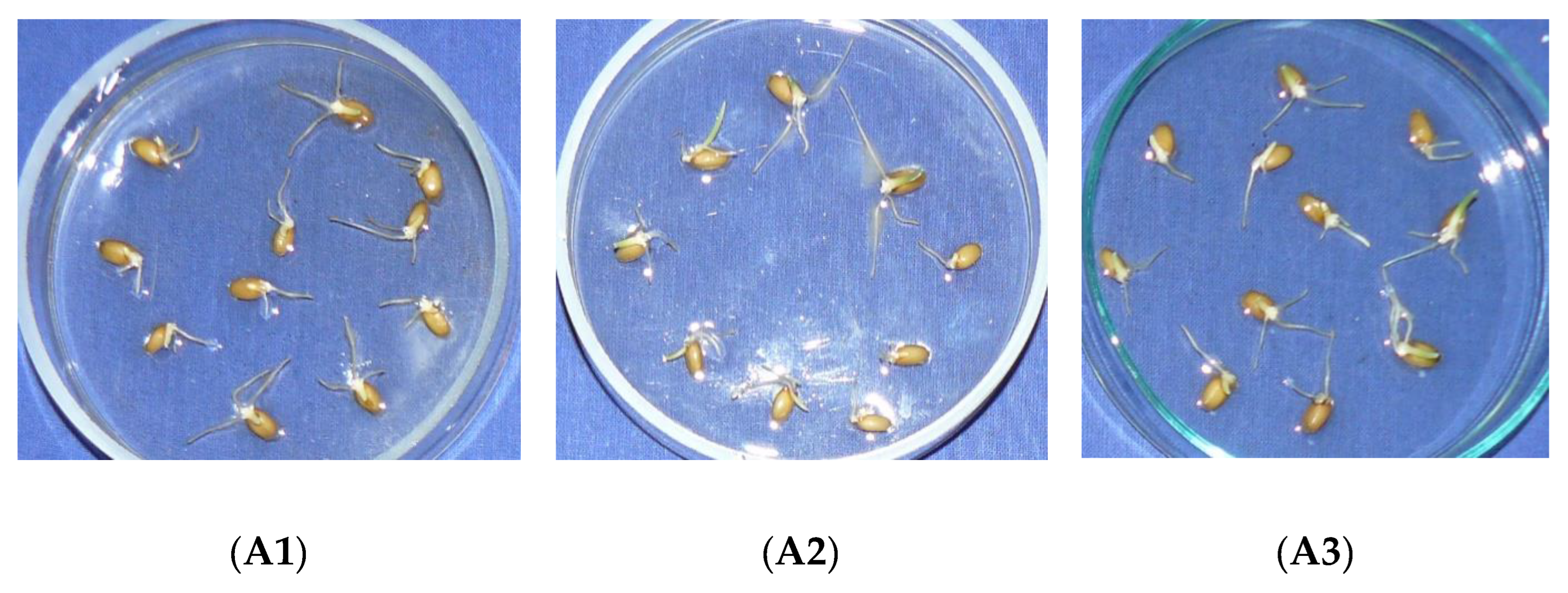
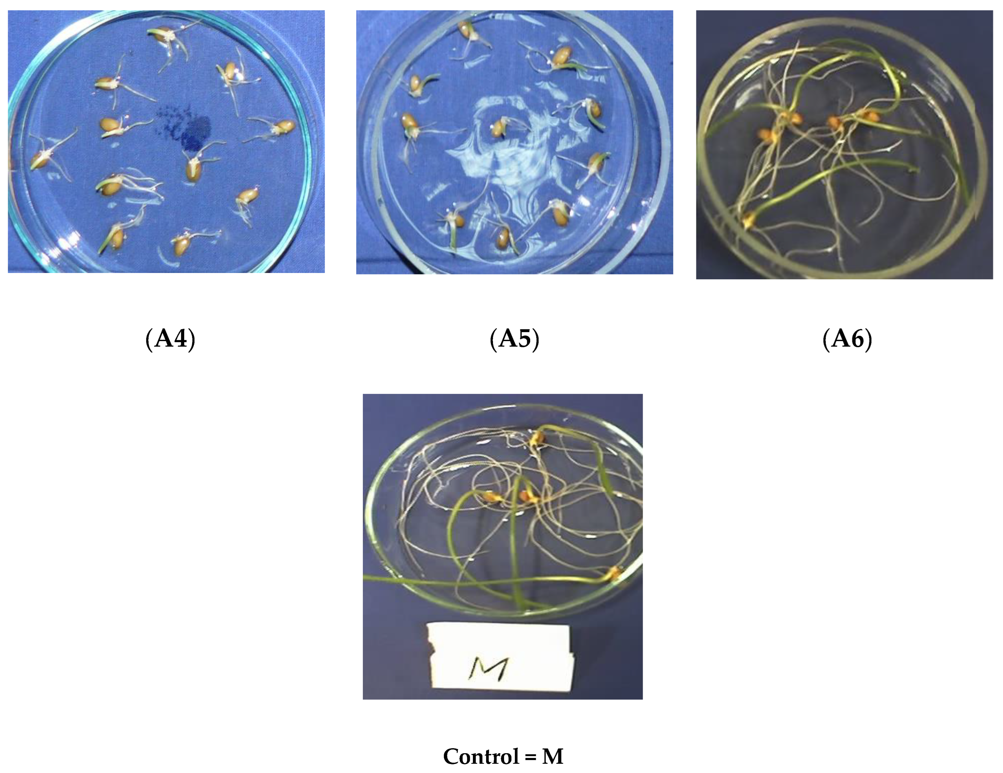
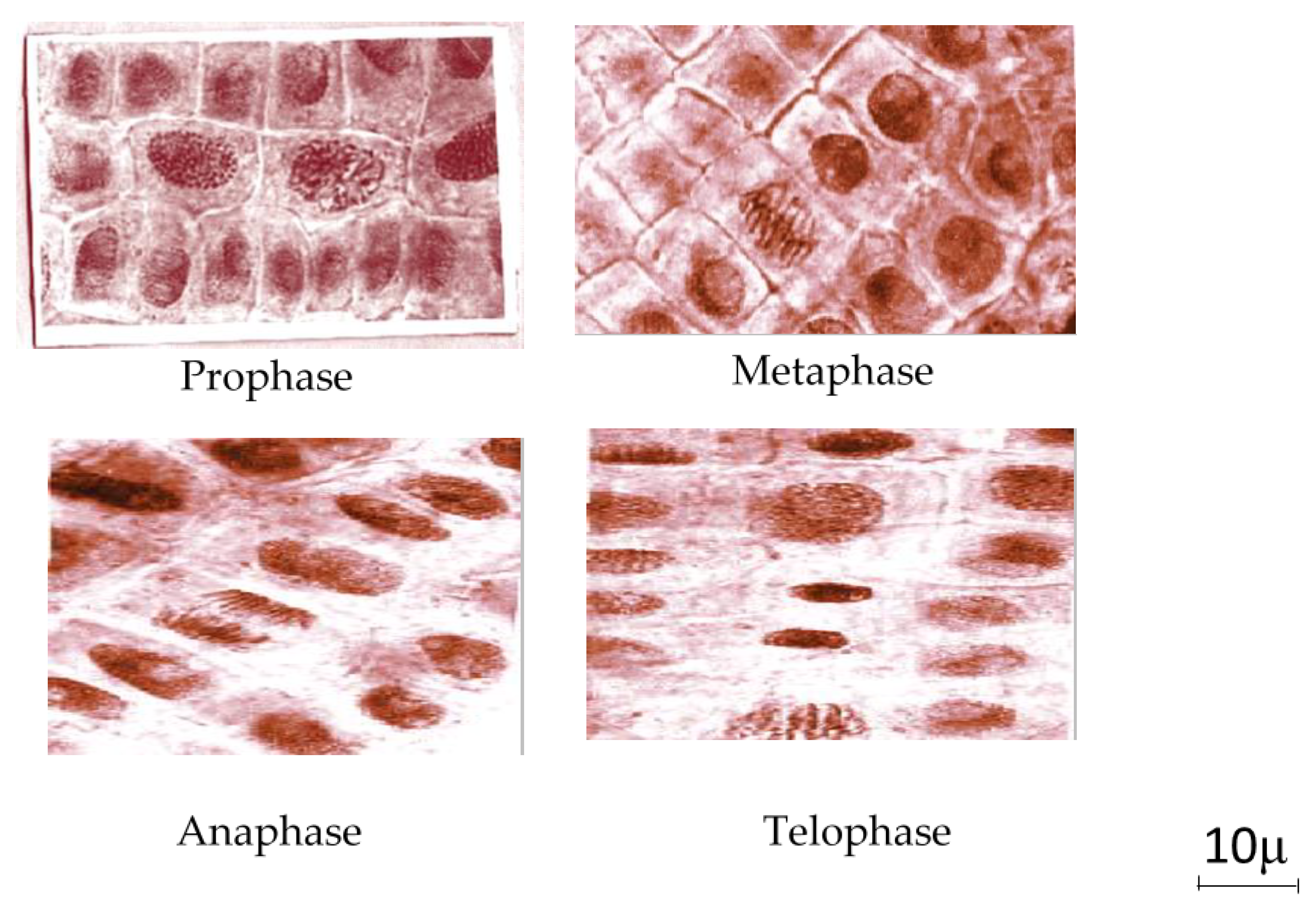
2.2.1. Acute Toxicity Evaluation of Calcium Levulinate on Plant Cells
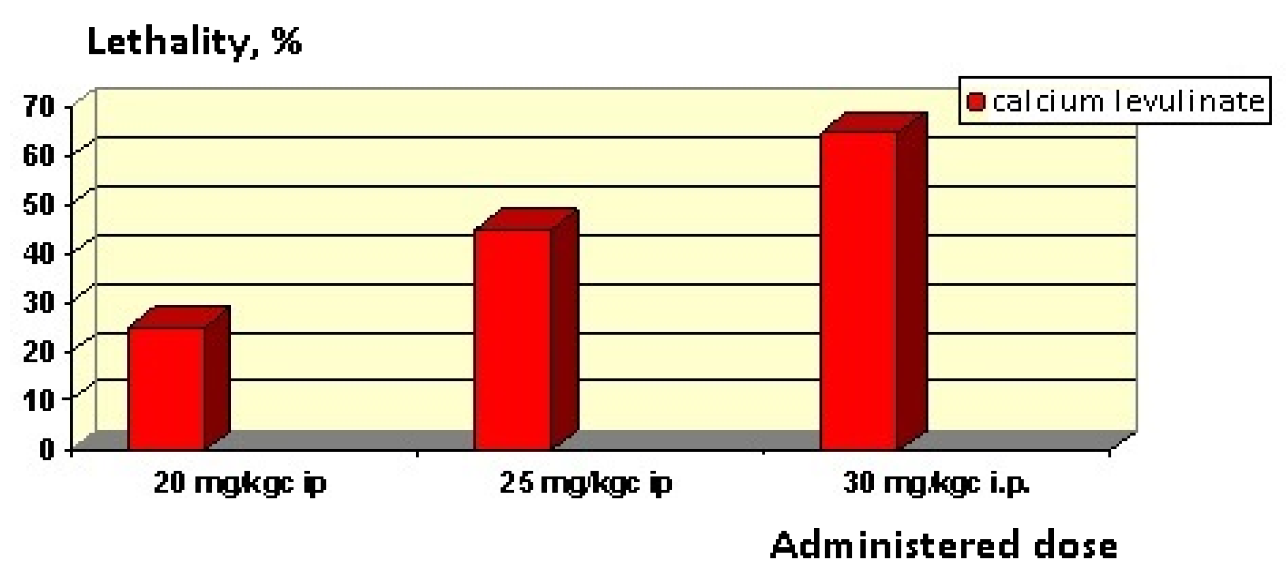
2.2.2. Acute Toxicity of Calcium Levulinate Evaluation on Animals
2.2.3. Analysis of Mineral Composition of Mussel Shells
3. Results and Discussion
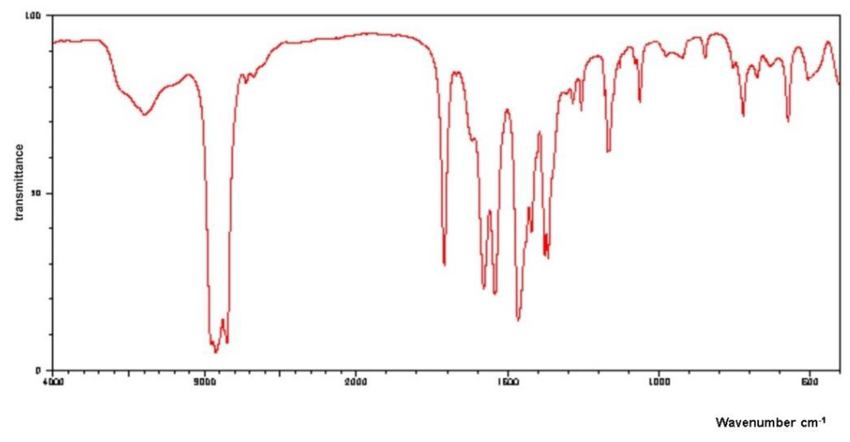
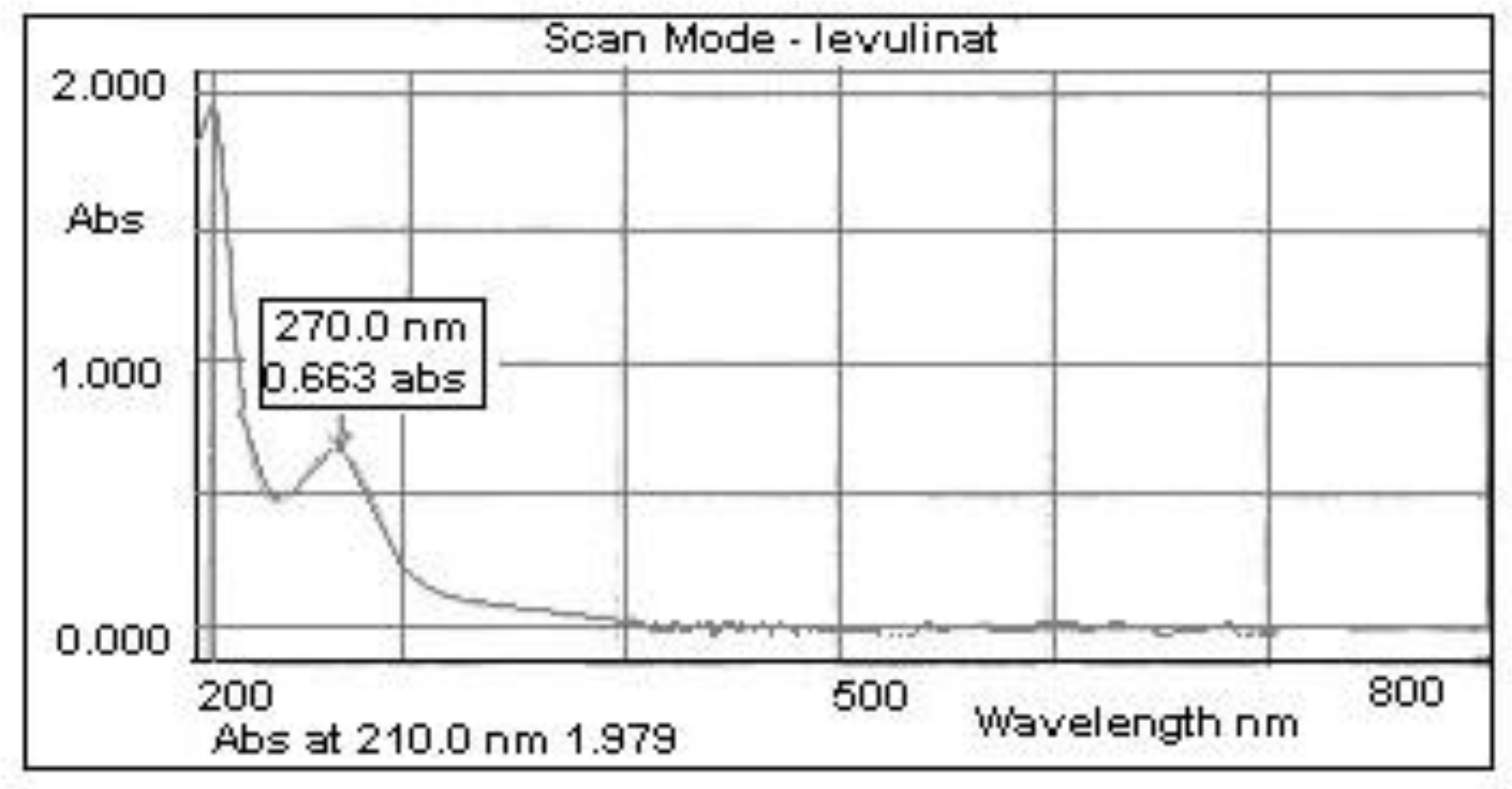
3.1. Characterization of Calcium Levulinate
3.2. Calcium Levulinate Toxicity Evaluation
3.2.1. Macroscopic Examination
3.2.2. Microscopic Examination
3.2.3. Evaluation of Acute Toxicity on Animals
Clinical Observations
3.3. Mineral Composition of Mussel Shells
3.4. Economic and Environmental Impact
4. Conclusions
Author Contributions
Funding
Institutional Review Board Statement
Data Availability Statement
Conflicts of Interest
References
- Costa, S.; Afonso, C.; Bandarra, N.M.; Gueifao, S.; Castanheira, I.; Carvalho, M.L.; Cardoso, C.; Nunes, M.L. The emerging farmed fish species meagre (Argyrosomus regius): How culinary treatment affects nutrients and contaminants concentration and associated benefit-risk balance. Food Chem. Toxicol. 2013, 60, 277–285. [Google Scholar] [CrossRef] [PubMed]
- Rittenschober, D.; Stadlmayr, B.; Nowak, V.; Du, J.; Charrondiere, U.R. Report on the development of the FAO/INFOODS user database for fish and shellfish (uFiSh)—Challanges and possible solutions. Food Chem. 2016, 193, 112–120. [Google Scholar] [CrossRef]
- Carrington, E.; Waite, J.H.; Sarà, G.; Sebens, K.P. Mussels as a model system for integrative echomecanics. Ann. Rev. Mar. Sci. 2015, 7, 443–469. [Google Scholar] [CrossRef] [PubMed] [Green Version]
- Galimany, E.; Wikfors, G.H.; Dixon, M.S.; Newell, C.R.; Meseck, S.L.; Henning, D.; Li, Y.Q.; Rose, J.M. Cultivation of the Ribbed mussel (Geukensia demissa) for nutrient bioextraction in an urban estuary. Environ. Sci. Technol. 2017, 51, 13311–13318. [Google Scholar] [CrossRef] [PubMed]
- Mititelu, M.; Moroşan, E.; Neacșu, S.M.; Ioniţă, E.I. Research regading the pollution degree from Romanian Black Sea coast. Farmacia 2018, 66, 1059–1063. [Google Scholar] [CrossRef]
- Newell, R.I.E.J. Ecosystem influences of natural and cultivated populations of suspension-feeding bivalve molluscs: A review. Shellfish Res. 2004, 23, 51–61. [Google Scholar]
- Çelik, M.Y.; Karayücel, S.; Karayücel, I.; Öztürk, R.; Eyüboglu, B.J. Meat yield, condition index, and biochemical composition of mussels (Mytilus galloprovincialis Lamarck, 1819) in Sinop, south of the Black Sea. Aquat. Food Prod. Technol. 2012, 21, 198–205. [Google Scholar] [CrossRef]
- Grienke, U.; Silke, J.; Tasdemir, D. Bioactive compounds from marine mussels and their effects on human health. Food Chem. 2014, 142, 48–60. [Google Scholar] [CrossRef]
- Holmer, M.; Thorsen, S.W.; Carlsson, M.S.; Kjerulf, P.J. Pelagic and benthic nutrient regeneration processes in mussels cultures (Mytilus edulis) in a Eutrophic Coastal Area (Skive Fjord, Denmark). Estuaries Coasts 2015, 38, 1629–1641. [Google Scholar] [CrossRef] [Green Version]
- Fuentes, A.; Fernández-Segovia, I.; Escriche, I.; Serra, J.A. Comparison of physico-chemical parameters and composition of mussels (Mytilus galloprovincialis Lmk.) from different Spanish origins. Food Chem. 2009, 112, 295–302. [Google Scholar] [CrossRef]
- Freites, L.; Fernández-Reiriz, M.J.; Labarta, U. Fatty acid profiles of Mytilus galloprovincialis (Lmk) mussels of subtidal and rocky shore origin. Comp. Biochem. Physiol. Part B Biochem. Mol. Biol. 2002, 132, 453–461. [Google Scholar] [CrossRef]
- Murphy, J.K.; Moone, D.B.; Mann, J.N.; Nichols, D.P.; Sinclair, J.A. Lipid, FA, and sterol composition of New Zealand green lipped mussel (Perna canaliculus) and Tasmanian blue mussel (Mytilus edulis). Lipids 2002, 37, 587–595. [Google Scholar] [CrossRef] [PubMed]
- Di Nunzio, M.; Valli, V.; Bordoni, A. Pro- and anti-oxidant effect of polyunsaturated fatty acid supplementation in HepG2 cells. Prostaglandins Leukot. Essent. Fat. Acids 2011, 85, 121–127. [Google Scholar] [CrossRef] [PubMed]
- Kesavulu, M.M.; Kameswararao, B.; Apparao, C.; Kumar, E.G.; Harinarayan, C.V. Effect of omega-3 fatty acids on lipid peroxidation and antioxidant enzyme status type 2 diabetic patients. Diabetes Metab. 2002, 28, 20–26. [Google Scholar] [PubMed]
- Saravanan, P.; Davidson, N.C.; Schmidt, E.B.; Calder, P.C. Cardiovascular effects of marine omega-3 fatty acids. Lancet 2010, 376, 540–550. [Google Scholar] [CrossRef]
- Hamester, M.R.R.; Balzer, P.S.; Becker, D. Characterization of calcium carbonate obtained from oyster and mussel shells and incorporation in polypropylene. Mater. Res. 2012, 15, 204–208. [Google Scholar] [CrossRef] [Green Version]
- Ituen, E.U. Mechanical and chemical properties of selected molluse shells in Nigeria. Int. J. Agric. Policy Res. 2015, 3, 53–59. [Google Scholar]
- Morris, J.P.; Thierry, B.; Gauthier, C. Shells from aquaculture: A valuable biomaterial, not a nuisance waste product. Rev. Aquac. 2019, 11, 42–57. [Google Scholar] [CrossRef] [Green Version]
- Mihele, D.; Mititelu, M. Process for obtaining calcium levulinate from mussels shells, involves deproteinisation, treatment with levulinic acid and purification of shell powder deproteinisatio. RO122478-B1, 2010. [Google Scholar]
- Onoda, H.; Nakanishi, H.; Takenaka, A.J. Preparation of calcium phosphates with Corbicula Shells. J. Ecotechnol. Res. 2012, 16, 85–89. [Google Scholar]
- Mititelu, M.; Ioniţă, A.C.; Moroşan, E. Research regarding integral processing of mussels from Black Sea. Farmacia 2014, 62, 625–632. [Google Scholar]
- The National Formulary XVIII, U.S.P. XXVIth ed.; United States Pharmacopoeia Convention, Inc.: Rockville, MA, USA, 2003; p. 254.
- Gordon, B.; Kough, O.S.; Proskouriakoff, A.J. Studies on calcium levulinate with special reference to the influence on edema. Lab. Clin. Med. 1933, 18, 507–511. [Google Scholar]
- Proskouriakoff, A. Some salts of levulinic acid. JACS 1933, 55, 2132–2134. [Google Scholar] [CrossRef]
- Sharath, B.O.; Tiwari, R.; Mal, S.S.; Dutta, S. Straightforward synthesis of calcium levulinate from biomass-derived levulinic acid and calcium carbonate in egg-shells. Mater. Today Proc. 2019, 17, 77–84. [Google Scholar] [CrossRef]
- Nuţă, D.; Dinu, M. Testarea actiunii fitobiologice a unor noi N-(2-dialchilaminoacetil)-benzanilide divers substituite. Farmacia 2005, 53, 116–123. [Google Scholar]
- AVMA. Guidelines for the Euthanasia of Animals; American Veterinary Medical Association: Schaumburg, IL, USA, 2013. [Google Scholar]
- OECD. Guidelines for Testing of Chemicals. In Acute Oral Toxicities Up and Down Procedure; OECD: Paris, France, 2001; Volume 425, pp. 1–26. [Google Scholar]
- Ahmed, M. Acute toxicity (lethal dose 50 calculation) of herbal drug Somina in rats and mice. Pharmacol. Pharm. 2015, 6, 185–189. [Google Scholar] [CrossRef] [Green Version]
- Gadaleta, D.; Vuković, K.; Toma, C.; Lavado, G.J.; Karmaus, A.L.; Mansouri, K.; Kleinstreuer, N.C.; Benfenati, E.; Roncaglioni, A. SAR and QSAR modeling of a large collection of LD50 rat acute oral toxicity data. J. Cheminform. 2019, 11, 58. [Google Scholar] [CrossRef] [Green Version]
- Raj, J.; Chandra, M.; Dogra, T.D.; Pahuja, M.; Raina, A. Determination of median lethal dose of combination of endosulfan and cypermethrin in wistar rat. Toxicol. Int. 2013, 20, 1–5. [Google Scholar]
- AOAC. Association of Official Analytical Chemists, Official Methods of Analysis, 15th ed.; AOAC: Washington, DC, USA, 1990. [Google Scholar]
- Al-Obaidi, F.A.; Mehdi, B.I.; Al- Shadeedi, S.M. Identification of inorganic elements in egg shell of some wild birds in Baghdad. Adv. Appl. Sci. Res. 2012, 3, 1454–1458. [Google Scholar] [CrossRef]
- Chang, F.; Li, G.; Haws, M.; Niu, T. Element concentrations in shell of Pinctada margaritifera from French Polynesia and evaluation for using as a food supplement. Food Chem. 2007, 104, 1171–1176. [Google Scholar] [CrossRef]
- Mair, P.; Wilcox, R. Robust statistical methods in R using the WRS2 package. Behav. Res. Meth. 2020, 52, 464–488. [Google Scholar] [CrossRef]
- Bojiţă, M.; Roman, L.; Săndulescu, R.; Oprean, R. Analiza şi Controlul Medicamentelor. Metode Instrumentale în Analiza şi Controlul Medicamentelor; Intelcredo, Deva: Bucharest, Romania, 2003; Volume 2, pp. 65, 306, 467, 469. [Google Scholar]
- FAO. The State of World Fisheries and Aquaculture—Meeting the SUSTAINABLE Development Goals. Rome. 2018. Available online: http://www.fao.org/3/i9540en/i9540en.pdf (accessed on 1 December 2021).
- Venugopal, V.; Gopakumar, K. Shellfish: Nutritive value, health benefits, and consumer safety. Compr. Rev. Food Sci. Food Saf. 2017, 16, 1219–1242. [Google Scholar] [CrossRef] [PubMed] [Green Version]
- Jacquet, J.; Sebo, J.; Elder, M. Seafood in the future: Bivalves are better. Solutions 2017, 8, 27. [Google Scholar]
- Suplicy, F.M. A review of the multiple benefits of mussel farming. Rev. Aquac. 2018, 12, 204–223. [Google Scholar] [CrossRef]


| Test Solutions * | VCalcium levulinate solution (mL) | VDistilled water (mL) | VTotal (mL) | Concentration (g% Calcium Levulinate) |
|---|---|---|---|---|
| A1 | 15.00 | - | 15.00 | 10.00% |
| A2 | 10.00 | 5.00 | 15.00 | 6.66% |
| A3 | 7.50 | 7.50 | 15.00 | 5.00% |
| A4 | 5.00 | 10.00 | 15.00 | 3.33% |
| A5 | 1.00 | 14.00 | 15.00 | 0.66% |
| A6 | 0.10 | 14.90 | 15.00 | 0.06% |
| Characteristics | Limits Accepted by USP | Obtained Values |
|---|---|---|
| Melting point (°C) | 119–125 | 122.5 |
| pH solution 10% | 7.8–8.5 | 7.9 |
| Loss on drying 60 °C, 5 mmHg | 10.5–12.0 | 10.8 |
| Chloride content (%) | max. 0.070 | 0.052 |
| Sulphate content (%) | max. 0.050 | 0.041 |
| Reducing sugars | Absent | Absent |
| Identification | ||
| Iodine/Iodinated Dinitrophenylhydrazine | CHI3 is formed Hydrazone is formed | CHI3 is formed Hydrazone is formed |
| Dosing with Na2EDTA | 97.5–100.5% | 98.73% |
| C% | H% | N% | S% | |
|---|---|---|---|---|
| Assay values | 44.04 | 5.37 | 0.34 | 0.14 |
| Theoretical values | 44.43 | 5.22 | 0.00 | 0.00 |
| Time (Hours) | Control Sample (mm) | A1 (mm) | A2 (mm) | A3 (mm) | A4 (mm) | A5 (mm) | A6 (mm) |
|---|---|---|---|---|---|---|---|
| 24 | 22.5 ± 0.45 | 11.5 ± 0.22 | 10.9 ± 0.93 | 12.5 ± 1.31 | 12.9 ± 0.26 | 13.4 ± 0.44 | 17.2 ± 0.48 |
| 48 | 42.7 ± 0.65 | 11.9 ± 0.82 | 12.6 ± 1.14 | 13.4 ± 1.06 | 13.5 ± 0.24 | 14.7 ± 0.42 | 24.2 ± 1.62 |
| 72 | 61.6 ± 0.55 | 12.6 ± 1.14 | 12.8 ± 1.71 | 14.5 ± 0.85 | 15.7 ± 0.62 | 15.4 ± 1.09 | 35.2 ± 1.02 |
| 96 | 84.2 ± 0.64 | 12.6 ± 0.78 | 12.8 ± 0.56 | 15. 2 ± 1.16 | 16.4 ± 1.24 | 17.2 ± 1.36 | 55.6 ± 0.78 |
| 120 | 105.3 ± 1.27 | 12.6 ± 0.71 | 12.8 ± 1.14 | 15.8 ± 1.76 | 16.9 ± 0.54 | 19.6 ± 1.18 | 86.4 ± 1.08 |
| E%/120 h | - | −88.03 | −87.84 | −84.99 | −83.95 | −81.38 | −17.94 |
| Element | Concentration, ppb |
|---|---|
| Ca | 35,452.65 ± 1883.07 |
| P | 595.46 ± 16.73 |
| Mg | 53.22 ± 2.60 * |
| Na | 467.21 ± 2.39 * |
| Fe | 255.38 ± 0.60 ** |
| Zn | 30.14 ± 0.13 ** |
| Mn | 27.07 ± 0.66 ** |
| Cr | 93.20 ± 1.62 * |
| Pb | 0.85 ± 1.12 * |
| Cd | BDL |
| Hg | BDL |
| Element | Concentration, % |
|---|---|
| Calcium | 95.88 ± 1.2 * |
| Phosphorus | 1.61 ± 0.2 * |
| Magnesium | 0.14 ± 0.04 ** |
| Natrium | 1.26 ± 0.3 * |
| Iron | 0.69 ± 0.02 ** |
| Others | 0.42 ± 0.03 ** |
Publisher’s Note: MDPI stays neutral with regard to jurisdictional claims in published maps and institutional affiliations. |
© 2021 by the authors. Licensee MDPI, Basel, Switzerland. This article is an open access article distributed under the terms and conditions of the Creative Commons Attribution (CC BY) license (https://creativecommons.org/licenses/by/4.0/).
Share and Cite
Mititelu, M.; Stanciu, G.; Drăgănescu, D.; Ioniță, A.C.; Neacșu, S.M.; Dinu, M.; Stefan-van Staden, R.-I.; Moroșan, E. Mussel Shells, a Valuable Calcium Resource for the Pharmaceutical Industry. Mar. Drugs 2022, 20, 25. https://doi.org/10.3390/md20010025
Mititelu M, Stanciu G, Drăgănescu D, Ioniță AC, Neacșu SM, Dinu M, Stefan-van Staden R-I, Moroșan E. Mussel Shells, a Valuable Calcium Resource for the Pharmaceutical Industry. Marine Drugs. 2022; 20(1):25. https://doi.org/10.3390/md20010025
Chicago/Turabian StyleMititelu, Magdalena, Gabriela Stanciu, Doina Drăgănescu, Ana Corina Ioniță, Sorinel Marius Neacșu, Mihaela Dinu, Raluca-Ioana Stefan-van Staden, and Elena Moroșan. 2022. "Mussel Shells, a Valuable Calcium Resource for the Pharmaceutical Industry" Marine Drugs 20, no. 1: 25. https://doi.org/10.3390/md20010025