Communication

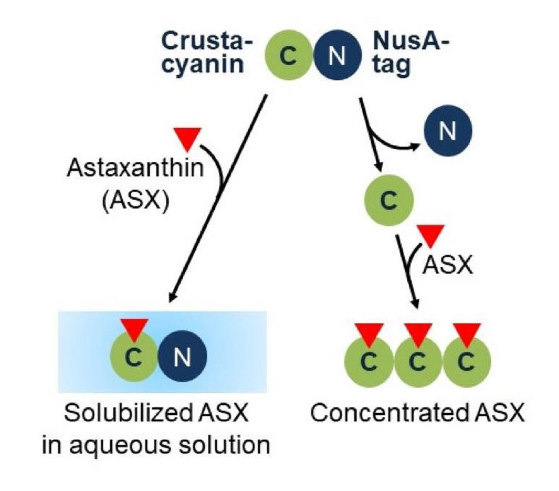
A Method of Solubilizing and Concentrating Astaxanthin and Other Carotenoids

1
Laboratory of Environmental Bioengineering, Graduate Division of Nutritional and Environmental Sciences, University of Shizuoka, 52-1 Yada, Suruga-ku, Shizuoka 422-8526, Japan
2
Department of Environmental and Life Sciences, School of Food and Nutritional Sciences, University of Shizuoka, 52-1 Yada, Suruga-ku, Shizuoka 422-8526, Japan
*
Author to whom correspondence should be addressed.
Both the authors contribute equally to this paper.
Mar. Drugs 2021, 19(8), 462; https://doi.org/10.3390/md19080462
Submission received: 23 July 2021 / Revised: 11 August 2021 / Accepted: 13 August 2021 / Published: 16 August 2021

Abstract

:
The valuable marine carotenoid, astaxanthin, is used in supplements, medicines and cosmetics. In this study, crustacyanin, an astaxanthin-binding protein, was used to solubilize and concentrate astaxanthin. The recombinant crustacyanin of European lobster spontaneously formed an inclusion body when it was over-expressed in Escherichia coli. In this study, fusing the NusA-tag to the crustacyanin subunits made it possible to express in a soluble fraction and solubilize astaxanthin in aqueous solution. By cutting off the NusA-tag, the crustacyanin subunits generated the pure insoluble form, and captured and concentrated astaxanthin. Overall, the attaching and releasing NusA-tag method has the potential to supply solubilized carotenoids in aqueous solution and concentrated carotenoids, respectively.

Graphical Abstract

1. Introduction

Carotenoids are used as red or orange pigments for foods, supplements, pharmaceuticals and cosmetics. Carotenoids are found as natural compounds in microorganisms, algae, plants and animals [1,2,3,4,5,6]. A valuable carotenoid, astaxanthin (3,3′-dihydroxy-β,β-carotene-4,4′-dione; C40H52O4), acts as a protective agent against oxidative damage to cells in vivo because it has high antioxidant activity [7,8,9]. Astaxanthin for human consumption is often produced by extraction from the microalgae, Haematococcus pluvialis [10]; generally extracted from H. pluvialis using super-critical CO2, a method which is not harmful with easy removal of the CO2 [11,12]. However, the efficiency of extracting astaxanthin from H. pluvialis cells has been reported to be less than 50%, even when carried out under high pressure (30 MPa) [13,14]. Repeating the super-critical extraction of astaxanthin several times also leads to very high running costs [15,16,17,18]. In addition, astaxanthin is generally extracted with organic solvents and can be concentrated by evaporation [19]. For a sustainable society in the future, environmentally-negative solvent discharge and energy-intensive evaporation processes are not desirable. Therefore, there is a need to develop an environmentally friendly astaxanthin concentrating method. Furthermore, providing astaxanthin in the form of an aqueous solution is important for developing applications of this valuable product, such as cosmetics and nutritional supplement drinks.
In the cells of the shells of crustaceans, astaxanthin exists with its binding protein, crustacyanin. Wald et al. first extracted crustacyanin from the shells of lobsters [20]. Crustacyanin subunits can be classified into two groups according to the composition of their amino acids, molecular size and peptide mapping. One group consists of the A1, C1 and C2 subunits, and the other consists of the A2 and A3 subunits [21]. Heterologous expression of the crustacyanin subunits in E. coli has also been carried out to clarify their three-dimensional structures [22]. One possible method of concentrating carotenoids is to use astaxanthin-binding proteins. However, the expression levels of these crustacyanin subunits are lower than those required for its application.
The current study has developed a simple method for solubilizing and concentrating astaxanthin using an easily-purified single crustacyanin protein. Using the attaching and releasing NusA-tag method, the crustacyanin protein was solubilized with the NusA-tag, followed by cleavage of the tag, which easily induces a pure precipitate for concentrating astaxanthin. It has been demonstrated that the method developed in this study can be used not only for astaxanthin, the original substrate of crustacyanin, but also for α-carotene and β-carotene.

2. Results and Discussion

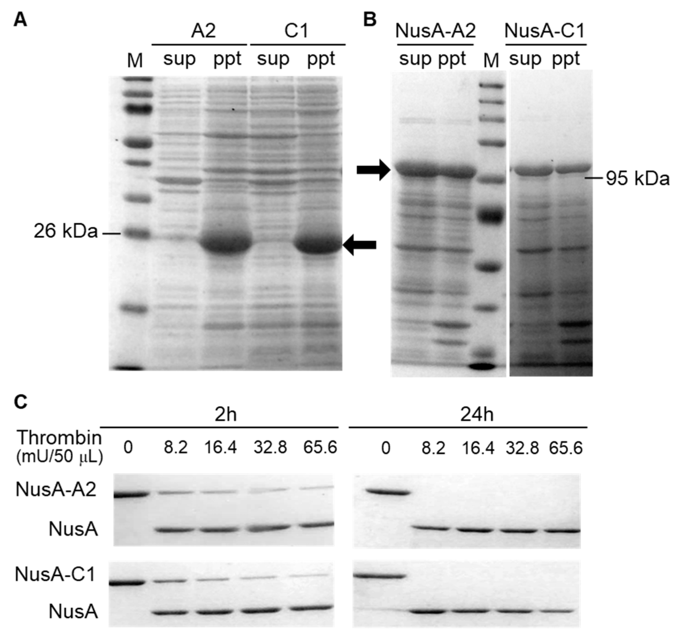
2.1. Optimization of Crustacyanin Preparation

For the first preparation step, native European lobster crustacyanin was produced in E. coli. The expression levels of A2 and C1 were higher than those of the other subunits [21], and A2C1 was a main subcomplex which forms H. gammarus crustacyanin [22]. Therefore, genes artificially synthesized with codon optimization for E. coli which encode the native H. gammarus crustacyanin A2 and C1 proteins were introduced into E. coli. Figure 1A shows that almost all the A2 (23 kDa) and C1 (26 kDa) proteins were expressed as insoluble inclusion bodies in E. coli. Therefore, their expressions in the inclusion bodies made the application of these proteins difficult. The NusA-tag is a protein known to increase the solubility of its conjugated target protein. In the present study, the NusA-tag (molecular weight calculated as 55 kDa and shown as 69 kDa in SDS-PAGE) [23] was fused to the N-terminal of the A2 and C1 proteins. As a result, the A2 and C1 proteins tagged with NusA were expressed in the soluble fraction at almost the same level as that of the inclusion bodies (Figure 1B).
In the second step, to construct a simple method to purify the crustacyanin proteins, the NusA-tag was removed from the NusA-tagged crustacyanin proteins. The protease, thrombin, was used to digest the spacer region between the NusA-tag and the crustacyanin proteins. To optimize the conditions for digestion, the NusA-A2 and NusA-C1 proteins (0.2 μg/mL) were digested using the biotinylated thrombin at different concentrations (0, 8.2, 16.4, 32.8 and 65.6 mU) at 4 °C for different reaction times (2, 4, 8 or 24 h). After this digestion reaction in the presence of 65.6 mU thrombin protease for 2 h, both the NusA-A2 and NusA-C1 proteins were almost completely digested to generate the A2 and C1 proteins (Figure 1C). In contrast, a reaction time of 24 h using less than 8.2 mU thrombin protease was sufficient for the complete digestion of NusA from the NusA-A2 and NusA-C1 proteins.

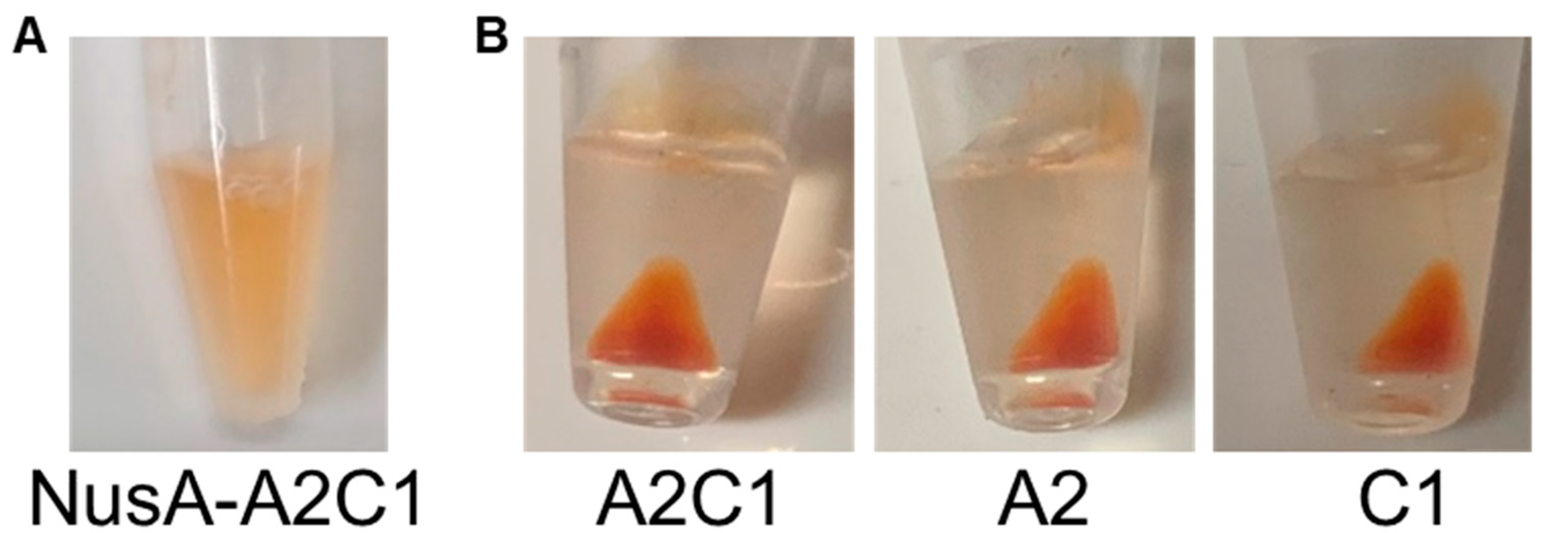
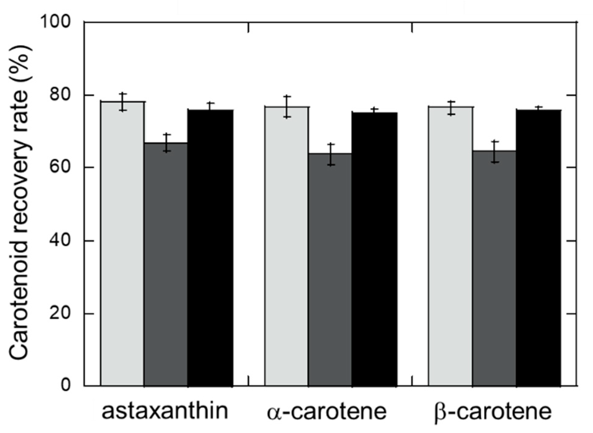
2.2. Carotenoid Recovery by Insoluble Crustacyanin

The crustacyanin A2C1 complex has been reported to have the ability to bind to astaxanthin [21]. The binding ability of A2C1 was compared when tagged with or without NusA. The NusA-A2C1 and NusA-free A2C1 complexes were constructed by mixing the NusA-A2/NusA-C1 proteins and NusA-free A2/C1 proteins, respectively. The astaxanthin was solubilized in buffer with the NusA-A2C1 complex and no precipitation was observed after centrifugation (Figure 2), which indicated that the astaxanthin had solubilized in the aqueous phase.
This method of solubilizing astaxanthin in aqueous solution could be applied in various industries, particularly for cosmetics and supplemental nutrition drinks. In contrast, the digestion of the NusA-tag from the NusA-A2 and NusA-C1 proteins and NusA-A2C1 complex constructed from the resulting NusA-free proteins generated a red-colored precipitate after binding to astaxanthin and centrifugation (Figure 2). These precipitates indicate the ability of the method to capture and concentrate astaxanthin of a high purity as the precipitated form of crustacyanin.
To evaluate whether the crustacyanin subunits could be used to concentrate carotenoids other than astaxanthin, the abilities of the A2 or C1 protein and their A2C1 complex to bind α-carotene and β-carotene were compared (Figure 3). The crustacyanin A2 protein individually bound all three carotenoids at a recovery rate (>75%) higher than that for the C1 protein, and almost equal to that for the A2C1 complex. These results indicated that the expression of both the A2 and C1 proteins and the formation of their complex is not required for the efficient capture of carotenoids. The crustacyanin A2 subunit alone provided a simple method for concentrating carotenoids. Figure 3 also shows no differences in the recovery rate for astaxanthin, α-carotene and β-carotene from the mixture. This wide-ranging ability to capture carotenoids indicates the potential of this method for application to any other type of carotenoid.

3. Materials and Methods

3.1. Strains, Plasmids and Transformation

Genes encoding the European lobster Homarus gammarus crustacyanin A2 and C1 subunits (Gene bank accession No. LC598211 and No. LC598212, respectively) were artificially synthesized with codon optimization for Escherichia coli (Eurofins Genomic, Tokyo, Japan). These genes were amplified using PCR, then subcloned into the pET-21d vector (Merck KGaA, Darmstadt, Germany) to construct pET21d-A2 (the forward primer and reverse primer used in this amplification were 5′-GTGCGGCCGCAAGCTTCGCGCGATACACGCATTCG-3′ and 5′-CGACAAGAGTCCGGGAGCTCTGGACGGCATTCCGTCCTTTG-3′, respectively) and pET21d-C1 (the forward primer and reverse primer used in this amplification were 5′-GTGCGGCCGCAAGCTTCAGCGTTTTCTGGGTATCATACGG-3′ and 5′-CGACAAGAGTCCGGGAGCTCTGGACAAGATTCCCGATTTTGTTG-3′, respectively). E. coli Rosetta2 (Merck KGaA) was then transformed using pET21d-A2 or pET21d-C1 to obtain E. coli strains which could over-express native A2 or C1 subunits induced by IPTG. The PCR fragments including the A2 or C1 genes were also subcloned into the pET43.1a vector (Merck KGaA) to construct pET43.1a-A2 and pET43.1a-C1. E. coli Origami B (Merck KGaA) was transformed by pET43.1a-A2 or pET43.1a-C1 to obtain E. coli strains which could over-express the A2 or C1 subunits with the NusA-tag (Nus⋅Tag) at their N-terminus. These E. coli Origami B mutant strains were then transformed using the pTF16 vector (Takara Bio, Shiga, Japan), which contains the gene-encoding Tig chaperone induced by L-arabinose, to construct E. coli Origami B (pET-43.1a-A2/pTF-16) and Origami B (pET-43.1a-C1/pTF-16).

3.2. Preparation of Soluble NusA-Crustacyanin

The E. coli Rosetta 2 and Origami B mutant strains were cultured in LB medium (10 g/L tryptone, 5 g/L yeast extract and 5 g/L sodium chloride) supplemented with 0.5 mg/mL L-arabinose and 100 μg/mL ampicillin in an Erlenmeyer flask, stirred at 100 rpm at 37 °C. When the cell concentration reached an OD600 value of 0.5, 1 mM IPTG was added and then cultured at 100 rpm for a further 3 h at 30 °C. After centrifugation (3000× g, 5 min, 4 °C), the supernatant was removed and the cell pellet was suspended in an appropriate volume of 50 mM sodium phosphate buffer (pH 7.0), then sonicated for a total of 45 s (on 5 s/off 5 s × 9 cycles) using an ultrasonic disruptor (Vibra Cell, Sonics & Materials Inc, Newtown, CT, USA). After centrifugation (1060× g, 5 min, 4 °C), ammonium sulfate (30% saturated concentration) was rapidly added to the disrupted cell extract, then incubated for 30 min at 4 °C. Fifteen milliliters of the resulting solution was centrifuged (1060× g, 5 min, 4 °C), then the supernatant was removed. The remaining pellet was resuspended in 3 mL of 50 mM sodium phosphate buffer (pH 7.0), dialyzed in the same buffer overnight, followed by replacement with 3 mL of fresh buffer and additional dialysis for 4 h. The dialyzed NusA-A2 and NusA-C1 proteins were centrifuged (1060× g, 5 min, 4 °C), then the concentration of each solubilized protein in the supernatant was measured using a Protein Assay BCA Kit (FUJIFILM Wako Pure Chemical Corp., Osaka, Japan). Equal volumes of the NusA-A2 and NusA-C1 solutions were mixed to form a NusA-A2C1 complex as described previously [22]. The chemicals were purchased from Nacalai Tesque (Kyoto, Japan) or FUJIFILM Wako Pure Chemical Corp.

3.3. Preparation of NusA-free Crustacyanin

NusA was digested from the NusA-A2 or NusA-C1 protein as described in the manual of the Novagen® Thrombin kits (Merk KGaA, Darmstadt, Germany). The biotinylated thrombin was used at different concentrations at 4 °C for different reaction times. Ten microliters of the reaction mixture were applied to SDS-PAGE to check the efficiency of digesting NusA from the NusA-A2 and NusA-C1 proteins. Under the optimized digestion conditions, large amounts (0.4 mg/mL) of NusA-A2 and NusA-C1 proteins were separately digested with 320 mU biotinylated thrombin at 4 °C for 3 days to prepare enough amounts of NusA-free crustacyanin for the next experiment described in Section 3.4. The NusA-free A2 and C1 proteins were collected through precipitation by centrifugation (1060× g, 5 min, 4 °C) and removing the supernatant. The proteins were concentrated by resuspension in the appropriate volume of 50 mM sodium phosphate buffer (pH 7.0).

3.4. Concentrating Carotenoids Using NusA-free Crustacyanin

Sixty microliters of a carotenoid mixture containing 20 μg/mL each of astaxanthin, α-carotene and β-carotene were dissolved in acetone, then gently mixed with 0.1 mL of each of the single NusA-free A2 or NusA-free C1 proteins (all at a concentration of 1.0 mg/mL) and 50 μL of their A2C1 complex generated from A2 and C1 proteins (at a concentration of 1.0 μg/μL). After centrifugation, the carotenoids precipitated with the A2 or C1 proteins or the A2C1 complex were suspended in 0.4 mL of acetone, vortexed and centrifuged (16,000× g, 5 min, 4 °C) to obtain the first supernatant for recovering carotenoids. To recover the remaining carotenoids from each protein, the precipitated protein was resuspended in 0.2 mL of acetone, vortexed and centrifuged (16,000× g, 5 min, 4 °C), and then the resulting second supernatant was mixed with the first supernatant (total 0.6 mL). The carotenoid concentrations were determined as described previously [24,25] using a high-performance liquid chromatography system (Shimadzu, Kyoto, Japan) equipped with a COSMOSIL Cholester Packed Column (4.6 i.d. × 250 mm, Nacalai Tesque, Kyoto, Japan). The operating conditions were: column temperature, 35 °C; mobile phase, methanol/tetrahydrofuran (80/20 (v/v)); flow rate, 1.0 mL/min; and detection of carotenoids at 470 nm using a UV detector (SPD-20AV, Shimadzu).

4. Conclusions

In this study, we developed an efficient method for producing crustacyanin subunits and used them to solubilize and concentrate carotenoids. Crustacyanin subunits with a NusA-tag attached to their N-terminus achieved soluble expression in E. coli and solubilized astaxanthin in aqueous solution. Astaxanthin could be concentrated by cleaving the NusA-tag from the crustacyanin subunit followed by its precipitation with astaxanthin. Furthermore, this method also could be applied to α-carotene and β-carotene. These results should indicate the potential of the attaching and releasing NusA-tag method to supply a solubilization process by attaching the NusA-tag, and a concentration process by releasing the NusA-tag, for the production of a broad range of carotenoids.

Author Contributions

Conceptualization, K.Y.H. and S.Y.; Methodology, K.Y.H., S.Y., Y.H.-H. and H.K.; Validation, K.Y.H. and S.Y.; Formal Analysis, S.Y.; Investigation, S.Y.; Data Curation, K.Y.H. and S.Y.; Writing—Original Draft Preparation, K.Y.H. and S.Y.; Writing—Review and Editing, Y.H.-H. and H.K.; Visualization, K.Y.H. and S.Y.; Project Administration, K.Y.H.; Funding Acquisition, K.Y.H. All authors have read and agreed to the published version of the manuscript.

Funding

This work was supported by JSPS KAKENHI Grant Numbers JP21H03645 and JP16K00616. This work was also supported by JST-Mirai Program Grant Number JPMJMI17EJ.

Institutional Review Board Statement

Not applicable.

Informed Consent Statement

Not applicable.

Data Availability Statement

The article contains all the data produced in this study.

Acknowledgments

We thank N. Tanzawa for her technical assistance.

Conflicts of Interest

The authors declare no financial or commercial conflict of interest.

Abbreviations

IPTG, isopropyl-β-D-thiogalactopyranoside; PCR, polymerase chain reaction; SDS-PAGE, sodium dodecyl sulfate-polyacrylamide gel electrophoresis.

References

  1. Vachali, P.; Bhosale, P.; Bernstein, P.S. Microbial carotenoids. Methods Mol. Biol. 2012, 898, 41–59. [Google Scholar]
  2. Mata-Gómez, L.C.; Montañez, J.C.; Méndez-Zavala, A.; Aguilar, C.N. Biotechnological production of carotenoids by yeasts: An overview. Microb. Cell Fact. 2014, 13, 12. [Google Scholar] [CrossRef] [Green Version]
  3. Yamamoto, K.; Hara, K.Y.; Morita, T.; Nishimura, A.; Sasaki, D.; Ishii, J.; Ogino, C.; Kizaki, N.; Kondo, A. Enhancement of astaxanthin production in Xanthophyllomyces dendrorhous by efficient method for the complete deletion of genes. Microb. Cell Fact. 2016, 15, 155. [Google Scholar] [CrossRef] [Green Version]
  4. Gervasi, T.; Santini, A.; Daliu, P.; Salem, A.Z.; Gervasi, C.; Pellizzeri, A.V.; Salem, Z.M.; Gervasi, C.; Pellizzeri, V.; Barrega, L.; et al. Astaxanthin production by Xanthophyllomyces dendrorhous growing on a low cost substrate. Agroforest Syst. 2020, 94, 1229–1234. [Google Scholar] [CrossRef]
  5. Villegas-Méndez, M.Á.; Aguilar-Machado, D.E.; Balagurusamy, N.; Montañez, J.; Morales-Oyervides, L. Agro-industrial wastes for the synthesis of carotenoids by Xanthophyllomyces dendrorhous: Mesquite pods-based medium design and optimization. Biochem. Eng. J. 2019, 150, 107260. [Google Scholar] [CrossRef]
  6. Korumilli, T.; Mishra, S.; Korukonda, J.R. Production of astaxanthin by Xanthophyllomyces dendrorhous on fruit waste extract and optimization of key parameters using Taguchi method. J. Biochem. Technol. 2020, 11, 25. [Google Scholar]
  7. Satoh, A.; Tsuji, S.; Okada, Y.; Murakami, N.; Urami, M.; Nakagawa, K.; Ishikura, M.; Katagiri, M.; Koga, Y.; Shirasawa, T. Preliminary clinical evaluation of toxicity and efficacy of a new astaxanthin-rich Haematococcus pluvialis extract. J. Clin. Biochem. Nutr. 2009, 44, 280–284. [Google Scholar] [CrossRef] [PubMed] [Green Version]
  8. Tripathi, D.N.; Jena, G.B. Intervention of astaxanthin against cyclophosphamide-induced oxidative stress and DNA damage: A study in mice. Chem.-Biol. Interact. 2009, 180, 398–406. [Google Scholar] [CrossRef] [PubMed]
  9. Hara, K.Y.; Morita, T.; Endo, Y.; Mochizuki, M.; Araki, M.; Kondo, A. Evaluation and screening of efficient promoters to improve astaxanthin production in Xanthophyllomyces dendrorhous. Appl. Microbiol. Biotechnol. 2014, 98, 6787–6793. [Google Scholar] [CrossRef] [PubMed]
  10. Panis, G.; Rosales, C.J. Commercial astaxanthin production derived by green alga Haematococcus pluvialis: A microalgae process model and a techno-economic assessment all through production line. Algal. Res. 2016, 18, 175–190. [Google Scholar] [CrossRef] [Green Version]
  11. Brunner, G.J. Supercritical fluids: Technology and application to food processing. Food Eng. 2005, 67, 21–33. [Google Scholar] [CrossRef]
  12. da Silva, R.P.F.F.; Rocha-Santos, T.A.P.; Duarte, A.C. Supercritical fluid extraction of bioactive compounds. TrAC Trends Anal. Chem. 2016, 76, 40–51. [Google Scholar] [CrossRef] [Green Version]
  13. Valderrama, J.O.; Perrut, M.; Majewski, W. Extraction of astaxantine and phycocyanine from microalgae with supercritical carbon dioxide. J. Chem. Eng. Data 2003, 48, 827–830. [Google Scholar] [CrossRef]
  14. Nobre, B.; Marcelo, F.; Passos, R.; Beiro, L.; Palavra, A.; Gouveia, L.; Mendes, R. Supercritical carbon dioxide extraction of astaxanthin and other carotenoids from the microalga Haematococcus pluvialis. Eur. Food Res. Technol. 2006, 223, 787–790. [Google Scholar] [CrossRef]
  15. Perrut, M. Supercritical fluid applications: Industrial developments and economic issues. Ind. Eng. Chem. Res. 2000, 39, 4531–4535. [Google Scholar] [CrossRef]
  16. Pan, J.L.; Wang, H.M.; Chen, C.Y.; Chang, J.S. Extraction of astaxanthin from Haematococcus pluvialis by supercritical carbon dioxide fluid with ethanol modifier. Eng. Life Sci. 2012, 12, 638–647. [Google Scholar] [CrossRef]
  17. Todd, R.; Baroutian, S.J. A techno-economic comparison of subcritical water, supercritical CO2 and organic solvent extraction of bioactives from grape marc. J. Clean. Prod. 2017, 158, 349–358. [Google Scholar] [CrossRef]
  18. Khoo, K.S.; Lee, S.Y.; Ooi, C.W.; Fu, X.; Miao, X.; Ling, T.C.; Show, P.L. Recent advances in biorefinery of astaxanthin from Haematococcus pluvialis. Bioresour. Technol. 2019, 288, 121606. [Google Scholar] [CrossRef]
  19. Park, J.Y.; Oh, Y.K.; Choi, S.A.; Kim, M.C. Recovery of astaxanthin-containing oil from Haematococcus pluvialis by nano-dispersion and oil partitioning. Appl. Biochem. Biotechnol. 2020, 190, 1304–1318. [Google Scholar] [CrossRef] [PubMed]
  20. Wald, G.; Nathanson, N. Crustacyanin, the blue carotenoid-protein of the lobster shell. Biol. Bull. 1948, 95, 249. [Google Scholar]
  21. Chayen, N.E.; Cianci, M.; Grossmann, J.G.; Habash, J.; Helliwell, J.R.; Nneji, G.A.; Raftery, J.; Rizkallah, P.J.; Zagalsky, P.F. Unravelling the structural chemistry of the colouration mechanism in lobster shell. Acta Crystallogr. 2003, 59, 2072–2082. [Google Scholar] [CrossRef] [PubMed]
  22. Ferrari, M.; Folli, C.; Pincolini, E.; McClintock, T.S.; Rössle, M.; Berni, R.; Cianci, M. Structural characterization of recombinant crustacyanin subunits from the lobster Homarus americanus. Acta Crystallogr. 2012, 68, 846–853. [Google Scholar]
  23. Davis, G.D.; Elisee, C.; Newham, D.M.; Harrison, R.G. New fusion protein systems designed to give soluble expression in Escherichia coli. Biotechnol. Bioeng. 1999, 65, 382–388. [Google Scholar] [CrossRef]
  24. Hara, K.Y.; Kageyama, Y.; Tanzawa, N.; Hirono-Hara, Y.; Kikukawa, H.; Wakabayashi, K. Development of astaxanthin production from citrus peel extract using Xanthophyllomyces dendrorhous. Environ. Sci. Pollut. Res. 2021, 28, 12640–12647. [Google Scholar] [CrossRef] [PubMed]
  25. Kikukawa, H.; Okaya, T.; Maoka, T.; Miyazaki, M.; Murofushi, K.; Kato, T.; Hirono-Hara, Y.; Katsumata, M.; Miyahara, S.; Hara, K.Y. Carotenoid Nostoxanthin production by Sphingomonas sp. SG73 isolated from deep sea sediment. Mar. Drugs 2021, 19, 274. [Google Scholar] [CrossRef] [PubMed]
Figure 1. Expression and digestion pattern of crustacyanin. (A) Native crustacyanin A2 and C1 are represented by the arrows; (B) NusA-tagged crustacyanin A2 and C1 proteins are represented by the arrows. The abbreviations “sup”, “ppt” and “M” represent supernatant, pellet and molecular marker, respectively; (C) Cleaving the NusA-tag from the A2 and C1 proteins tagged with NusA. Biotinylated thrombin was added to NusA-A2 and NusA-C1 crustacyanin in a 50 μL reaction mixture at different concentrations shown for 2 or 24 h. Concentration of acrylamide used in SDS-PAGE is 8.0%.
Marinedrugs 19 00462 g001
Figure 2. Solubilization and concentration of astaxanthin by crustacyanin subunits. (A) Addition of astaxanthin to crustacyanin A2 and C1 subunits attaching the NusA-tag; (B) Addition of astaxanthin to crustacyanin A2C1, A2 and C1 after digesting the NusA-tag.
Marinedrugs 19 00462 g002
Figure 3. Capturing carotenoids from a mixture using the crustacyanin subunits. The crustacyanin subunits were reacted with a mixture of carotenoids containing astaxanthin, α-carotene and β-carotene, each at a concentration of 20 μg/mL. The amount of each carotenoid captured in the crustacyanin was measured by high-performance liquid chromatography and their recovery rates were calculated from the amounts added. The light gray, dark gray and black bars represent the crustacyanin A2, C1 subunits and A2C1 complex, respectively. Error bars represent the standard deviations.
Marinedrugs 19 00462 g003
Publisher’s Note: MDPI stays neutral with regard to jurisdictional claims in published maps and institutional affiliations.

