Article

Glyceroglycolipid Metabolism Regulations under Phosphate Starvation Revealed by Transcriptome Analysis in Synechococcus elongatus PCC 7942

1
State Key Laboratory of Microbial Metabolism, School of Life Sciences and Biotechnology, Shanghai Jiao Tong University, 800 Dongchuan Road, Shanghai 200240, China
2
Joint International Research Laboratory of Metabolic & Developmental Sciences, Shanghai Jiao Tong University, Shanghai 200240, China
3
Biomass Energy Research Center, Shanghai Jiao Tong University, Shanghai 200240, China
*
Author to whom correspondence should be addressed.
Mar. Drugs 2020, 18(7), 360; https://doi.org/10.3390/md18070360
Submission received: 4 June 2020 / Revised: 4 July 2020 / Accepted: 10 July 2020 / Published: 13 July 2020
(This article belongs to the Special Issue Marine Carbohydrate-Based Compounds with Medicinal Properties)

Abstract

:
Glyceroglycolipids, abundant in cyanobacteria’s photosynthetic membranes, present bioactivities and pharmacological activities, and can be widely used in the pharmaceutical industry. Environmental factors could alter the contents and compositions of cyanobacteria glyceroglycolipids, but the regulation mechanism remains unclear. Therefore, the glyceroglycolipids contents and the transcriptome in Synechococcus elongatus PCC 7942 were analyzed under phosphate starvation. Under phosphate starvation, the decrease of monogalactosyl diacylglycerol (MGDG) and increases of digalactosyl diacylglycerol (DGDG) and sulfoquinovosyl diacylglycerol (SQDG) led to a decrease in the MGDG/DGDG ratio, from 4:1 to 5:3, after 12 days of cultivation. However, UDP–sulfoquinovose synthase gene sqdB, and the SQDG synthase gene sqdX, were down-regulated, and the decreased MGDG/DGDG ratio was later increased back to 2:1 after 15 days of cultivation, suggesting the regulation of glyceroglycolipids on day 12 was based on the MGDG/DGDG ratio maintaining glyceroglycolipid homeostasis. There are 12 differentially expressed transcriptional regulators that could be potential candidates related to glyceroglycolipid regulation, according to the transcriptome analysis. The transcriptome analysis also suggested post-transcriptional or post-translational regulations in glyceroglycolipid synthesis. This study provides further insights into glyceroglycolipid metabolism, as well as the scientific basis for glyceroglycolipid synthesis optimization and cyanobacteria glyceroglycolipids utilization via metabolic engineering.

1. Introduction

Glyceroglycolipids are widely distributed in plants, microalgae and cyanobacteria. Monogalactosyl diacylglycerol (MGDG), digalactosyl diacylglycerol (DGDG) and sulfoquinovosyl diacylglycerol (SQDG) are the three main glyceroglycolipids in the photosynthetic membrane, which are essential for photosynthesis [1]. In cyanobacteria, about 50% of the photosynthetic membrane lipids are MGDG, 20% are DGDG and 16% are SQDG [2]. Glyceroglycolipids present both bioactivities and pharmacological activities, and can be widely used in the pharmaceutical industry [3]. Microalgae and cyanobacteria are competitive sources of glyceroglycolipids because of their abundant glyceroglycolipids, their simple cell structure and their eco-friendly characteristic. Many glyceroglycolipids with pharmaceutical value have been isolated from microalgae and cyanobacteria. MGDG with pro-apoptotic activity is extracted from Phaeodactylum tricornutum [4]. MGDG from Tetraselmis chuii and Nannochloropsis granulate have anti-inflammatory activities [5]. MGDG and DGDG from Chlorella Vulgaris [6] and Phormidium tenue [7] also present anti-tumor activities. In addition, SQDG, with eukaryotic DNA polymerase inhibitory activity, has been extracted from Gigartina tenella [8]. The SQDG isolated from the cyanobacteria Lyngbya lagerheimii [9] and Phormidium tenue [10] have AIDS-antiviral activities. Recent studies have also identified the immuno-stimulatory activity and the potential against Alzheimer’s disease of SQDG derived from microalgae [5].
Environmental factors directly influence the accumulation of many metabolites. Phosphate—the main ingredient of nucleic acid, protein and phospholipids—is an essential element in organism growth. However, phosphate is often limited in natural environments [11]. To cope with phosphate limitation, organisms have acquired different strategies, including ultrastructural rearrangements, C reallocation, transcriptome reprograming, and metabolome and lipid remodeling [11]. Glyceroglycolipid accumulation could be strongly regulated by phosphate concentration. In plants, the synthesis of some kinds of glyceroglycolipids (like DGDG and SQDG) will be induced when phosphate is lacking during cultivation, in order to supplement the shortage of phospholipids so as to maintain both the structures and functions of membranes [12,13]. A similar phenomenon was also reported in the cyanobacteria Synechococcus elongatus PCC 7942, wherein phosphate starvation resulted in a decrease in phospholipid and an increase in SQDG [14]. In Chlamydomonas nivalis, DGDG increased while MGDG decreased under phosphate deprivation [15]. In addition, an increase in total glyceroglycolipids is common in microalgae and cyanobacteria when exposed to a phosphate deficiency condition [16,17,18].
Glyceroglycolipid-related synthases have been well researched for decades. SQDG synthases present a high homology between plants and microalgae. UDP-sulfoquinovose synthase (EC: 3.13.1.1) (SQD1) and SQDG synthase (EC: 2.4.1.-) (SQD2) are involved in the SQDG synthesis in both plants and eukaryotic microalgae [19,20]. In cyanobacteria, sqdB (EC: 3.13.1.1) and sqdX (EC: 2.4.1.-), responsible for SQDG synthesis [21,22], show high sequence similarity with SQD1 and SQD2, respectively [23]. However, genetic differences exist between MGDG and DGDG synthases in plants and microalgae. In plants, three MGDG synthases (EC: 2.4.1.46) (MGD1, MGD2 and MGD3) [24,25,26] and two DGDG synthases (EC: 2.4.1.241) (DGD1 and DGD2) [27,28] have been identified. MGD1 and DGD1 participate in the synthesis of the bulk of MGDG and DGDG, respectively, while DGD2 is involved in DGDG synthesis under specific growth conditions [28], with MGD2 and MGD3 providing MGDG as a precursor [29]. In Chlamydomonas reinhardtii, only orthologues of MGD1 and DGD1 were identified [20,30], but a second isoform of the DGDG synthase, resembling the plant DGD2, was additionally found in Ostreococcus tauri [20]. In cyanobacteria, no homolog for the plant-type DGDG synthase has been detected [31], and dgdA (EC: 2.4.1.241) (the DGDG synthase in cyanobacteria) is only distantly related to DGD1 [23,32]. MGDG synthesis in cyanobacteria is more complicated. All cyanobacteria are likely to synthesize MGDG through the epimerization of monoglucosyl diacylglycerol (MGlcDG) [33] by the MGlcDG synthase (EC: 2.4.1.336) (mgdA) [34,35] and the MGlcDG epimerase (EC 5.1.3.34) (mgdE) [36]. Some studies have reported that phosphate deficiency would stimulate the expression of glyceroglycolipid-related synthases [17,37,38,39], but the underlying regulation mechanism is still poorly understood.
Metabolic engineering could modify the metabolisms of an organism so as to produce specific metabolites. According to recent reports, overexpressing a bHLH transcription factor [40] and a bZIP transcription factor [41] could enhance biomass and lipid productivity in Nannochloropsis salina. Overexpressing a soybean transcription factor, GmDof4, significantly enhanced the lipid production in Chlorella ellipsoidea, without sacrificing biomass [42]. The available information regarding the key regulators involved in cyanobacteria glyceroglycolipid metabolism is currently still deficient, which limits the development of cyanobacteria glyceroglycolipids utilization.
Previous studies in our lab indicated a decrease in MGDG and increases in DGDG and SQDG in Synechococcus sp. under phosphate starvation [2]. To investigate the relationship between phosphate starvation and glyceroglycolipid metabolism, the transcriptome of the cyanobacteria model organism Synechococcus elongatus PCC 7942 under phosphate starvation was analyzed in this study, which provided further insights into glyceroglycolipid metabolism under phosphate starvation, and a scientific basis for cyanobacteria glyceroglycolipids utilization in metabolic engineering.

2. Results and Discussion

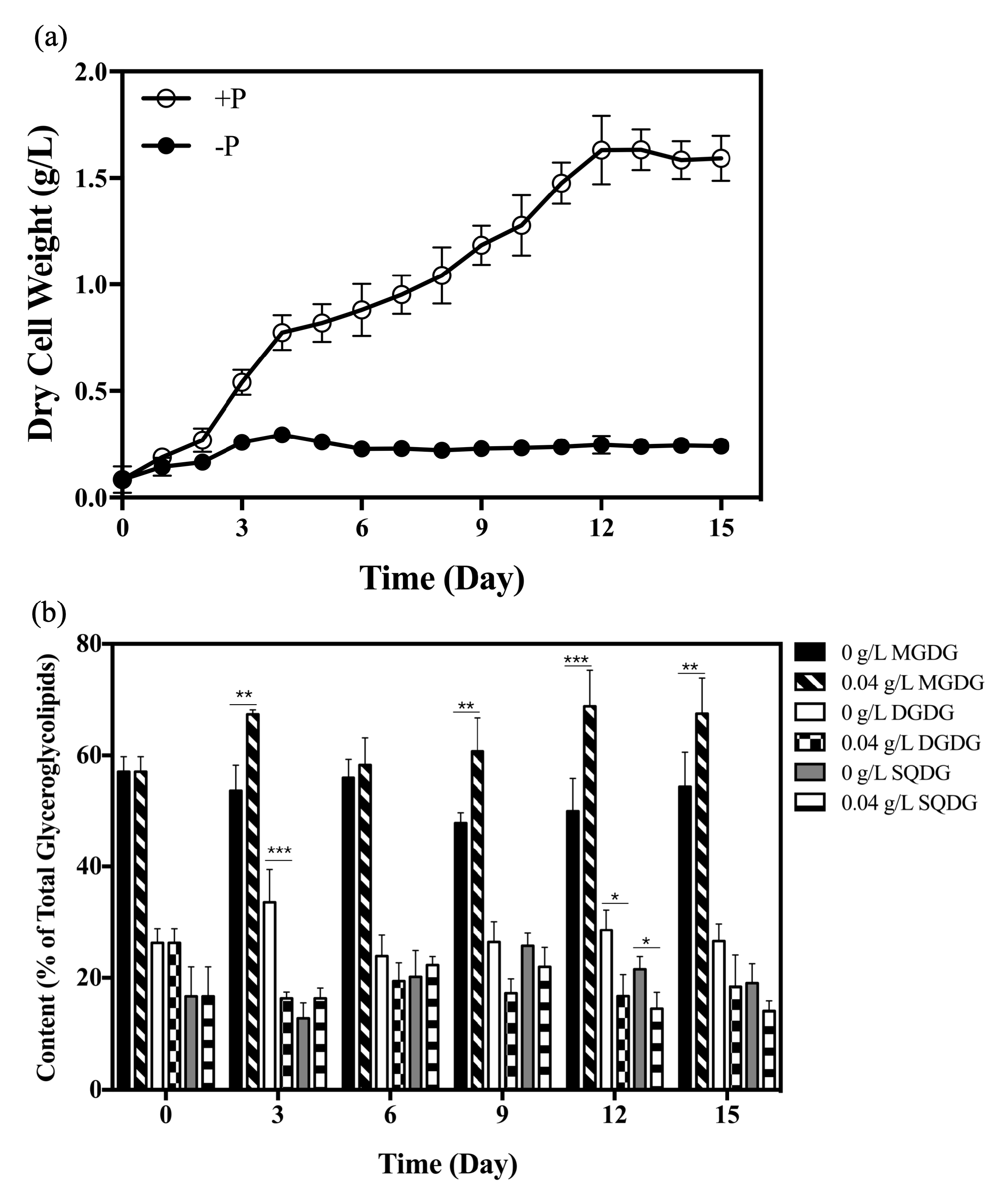
2.1. Changes in Glyceroglycolipid Composition in Synechococcus elongatus PCC 7942 under Phosphate Starvation

Our previous research demonstrated that the content of total glyceroglycolipids increased over all growth stages, and glyceroglycolipid composition changed in S. elongatus PCC 7942, under phosphate starvation, which helps cyanobacteria adapt to unfavorable conditions [43]. In order to gain more insight into glyceroglycolipid changes triggered by phosphate starvation, the growth, and dynamic variations in the composition, of three different glyceroglycolipids in S. elongatus PCC 7942 were investigated under the initial phosphate concentrations of 0.04 g/L and 0 g/L (Figure 1).
The growth of S. elongatus PCC 7942 under phosphate concentrations of 0.04 g/L reached a stationary phase after 12 days of cultivation (Figure 1a). The maximum biomass concentrations, with 0.04 g/L and 0 g/L phosphate, were 1.63 g/L and 0.25 g/L, respectively.
Under phosphate starvation, the difference in MGDG content was significant on day 3 (p = 0.0044), day 9 (p = 0.0067), day 12 (p = 0.0004) and day 15 (p = 0.006). The difference in DGDG content was significant on day 3 (p = 0.0009) and day 12 (p = 0.0145) under phosphate starvation. The difference in SQDG content was significant on day 12 (p = 0.0327). The largest difference in the composition of glyceroglycolipids in S. elongatus PCC 7942, caused by phosphate starvation, was observed on day 12 (Figure 1b). The MGDG content was 50% of the total glyceroglycolipids under phosphate starvation on day 12, which was 0.72 times lower than that in the phosphate-rich culture (69% of total glyceroglycolipids) (Figure 1b). DGDG and SQDG contents were 29% and 21% of total glyceroglycolipids, respectively, under phosphate starvation on day 12, which were respectively 1.70 and 1.50 times higher than that in the phosphate-rich culture (17% and 14% of total glyceroglycolipids) (Figure 1b), respectively. Many studies have already illustrated that phosphate starvation or limitation would cause lipid remodeling in many species, and found that the increased glyceroglycolipids could functionally substitute the degrading phospholipids [13,17,44]. This result demonstrated that the increase of total glyceroglycolipids content in S. elongatus PCC 7942 (Supplementary Figure S1) mainly resulted from the accumulation of DGDG and SQDG under phosphate starvation (Figure 1b), the same as Synechocystis sp. PCC 6803 [31], indicating their importance in adapting to phosphate stress [43].

2.2. Expressions of Glyceroglycolipid Synthase Genes in Synechococcus elongatus PCC 7942 under Different Phosphate Concentrations

To explore the changes in glyceroglycolipid composition at the transcriptional level, the expressions of glyceroglycolipid synthase genes in S. elongatus PCC 7942, cultivated under the initial phosphate concentrations of 0.04 g/L and 0 g/L for 12 days, were determined by qRT-PCR. In S. elongatus PCC 7942, mgdA (Synpcc7942_1083), mgdE (Synpcc7942_0861), dgdA (Synpcc7942_0986), sqdB (Synpcc7942_0578) and sqdX (Synpcc7942_0579) are the five glyceroglycolipid synthase genes.
The expression levels of mgdA and mgdE showed no significant differences under phosphate starvation (Figure 2a,b). The expression of mdgA is not regulated by the SphS–SphR two component system in response to inorganic phosphate [45]. The mgdA–mgdE system in cyanobacteria was replaced by MGD1 in eukaryotic microalgae and plants [33]. In Arabidopsis, the expression of MGD1 is not induced by phosphate deficiency [29]. Moreover, the expression of mdgA also showed no differences under heat stress [46]. Thus, the mgdA–mgdE system may not be regulated at the transcriptional level.
As mentioned above, the increased DGDG accounted for the majority of the increased total glyceroglycolipids, and DGDG levels were increased throughout the whole culture period (Figure 1b and Supplementary Figure S2). However, the expression level of dgdA showed no significant differences under phosphate starvation (Figure 2c). In plants, both DGD1 and DGD2 can be up-regulated under conditions of phosphate deficiency [47]. This result suggested that the regulation of DGDG synthases in cyanobacteria might be different from that in plants, since an evolutionary gap exists between DGDG synthases in plants and cyanobacteria [31].
SQDG has been regarded as a surrogate for phosphatidylglycerol (PG), and SQDG synthases will be specifically induced upon phosphate starvation [1,17]. Interestingly, the expression levels of sqdB and sqdX were down-regulated by 90% (Figure 2d,e). The degrees of down-regulation in sqdB and sqdX are comparable, since sqdB and sqdX are likely to form an operon called sqdBX [48]. In this study, SQDG content was increased under phosphate starvation on day 12. It showed a slightly decreased trend after 12 days of cultivation, although this was not statistically significant (Supplementary Figure S2).

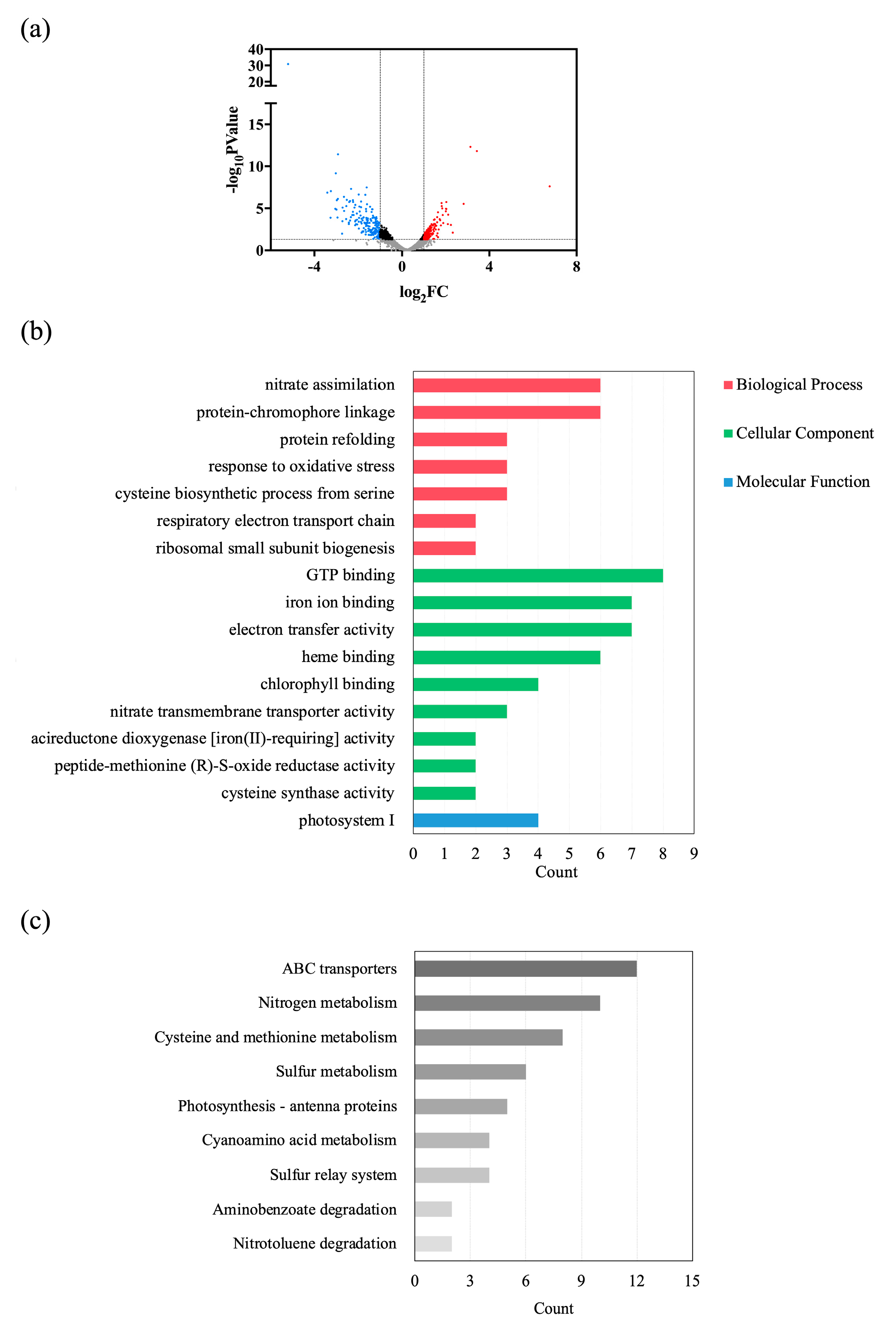
2.3. Global Transcriptomic Analysis under Different Phosphate Concentrations

To further investigate the regulatory mechanism of changes in glyceroglycolipid composition, the transcriptome of S. elongatus PCC 7942, cultivated under the initial phosphate concentrations of 0.04 g/L and 0 g/L, for 12 days, was analyzed. As shown in Figure 3a and Supplementary Table S1, 2,660 genes in total were analyzed, among which 165 genes (6.2%) were significantly up-regulated and 172 genes (6.5%) were significantly down-regulated under phosphate starvation conditions, compared with the levels under 0.04 g/L phosphate concentration.
The Gene Ontology (GO) enrichment of differentially expressed genes (DEGs), illustrated in Figure 3b and Supplementary Table S2, significantly enriched 17 GO terms. The most enriched GO term was GTP binding (Figure 3b), in which three DEGs were significantly down-regulated and five DEGs were significantly up-regulated (Supplementary Table S3), indicating the active signals transmitting under phosphate starvation. Notably, a gene coding the Escherichia coli Ras-like protein (era, Synpcc7942_0160) was significantly up-regulated, with a fold change value of 2.42 under phosphate starvation (Supplementary Table S3). In S. elongatus PCC 7942, an era overexpression strain exhibited significantly higher amounts of fatty acids compared to wild type [49]. Protein Era is highly conserved [50], and ERA-related GTPase (ERG) in plants is always related to chloroplast biogenesis [51,52,53], revealing its homologous function with cyanobacteria. Depletion of an Era-type GTP-binding protein resulted in abnormal chloroplasts lacking thylakoid membranes in rice, which indicated its importance in chloroplast development [54]. It could be speculated from this evidence in the literature that the accumulation of total glyceroglycolipids under phosphate starvation may be associated with the up-regulation of era.
The other two highly enriched GO terms were iron ion binding and electron transfer activity, suggesting electron transport was severely affected after 12 days of cultivation under phosphate starvation. The Kyoto Encyclopedia of Genes and Genomes (KEGG) enrichment of DEGs (Figure 3c and Supplementary Table S4) showed that nine metabolic pathways were significantly affected by phosphate starvation, of which the three most enriched were ABC transporters, nitrogen metabolism, and cysteine and methionine metabolism. Similar responses were commonly implicated when organisms were exposed to adverse conditions [55,56].
Besides, GTP-binding proteins play important roles in the cell cycle, cell division and ribosome maturation [57]. We analyzed the DEGs involved in the cell cycle, cell division and ribosome maturation. According to GO enrichment (Supplementary Table S2) and KEGG enrichment (Supplementary Table S4), these metabolisms were not significantly enriched (p-value > 0.05) under conditions of phosphate starvation on day 12, though one gene involved in the cell cycle, two genes involved in cell division and nine genes involved in ribosome maturation were differentially expressed under phosphate starvation.

2.4. Differential Expressions of Genes Involved in Glyceroglycolipid Synthesis

The glyceroglycolipid synthesis pathway, based on KEGG annotation, is shown in Figure 4a. In total, 12 genes were involved in this pathway. However, the UDP-glucose pyrophosphorylase (EC: 2.7.7.9) (ugp) responsible for transforming glucose-1-phosphate to UDP-glucose remained unidentified. In S. elongatus PCC 7942, a conserved hypothetical protein coded by Synpcc7942_0148 showed a percent identity of 49% with the ugp in Synechocystis sp. PCC 6803, coded by slr0207 [58], indicating Synpcc7942_0148 is possibly responsible for UDP-glucose synthesis. Moreover, the unique cyanobacteria UDP-glucose pyrophosphorylase (cugP), coded by sll1558, which is annotated as mannose-1-phosphate guanyltransferase (EC:2.7.7.13) (GMPP) but displays ugp activity, was identified in Synechocystis sp. PCC 6803 [59]. In S. elongatus PCC 7942, GMPP coded by Synpcc7942_1973 showed a percent identity of 79% with the product of sll1558, suggesting it is involved in UDP-glucose synthesis as a cugP.
According to transcriptome analysis, mgdA, mgdE and dgdA showed no significant differences in expression under conditions of phosphate starvation, while sqdB and sqdX were down-regulated, with fold changes of 0.63 and 0.43, respectively (Figure 4a and Supplementary Table S5), the same as the results of qRT-PCR. Moreover, the expressions of other genes related to glyceroglycolipid synthesis, and the two possible candidates responsible for UDP-glucose synthesis (Synpcc7942_0148 and Synpcc7942_1973), showed no significant differences under phosphate starvation as well (Figure 4a), except for pgm (Synpcc7942_0156), which is responsible for transforming glucose-6-phosphate into glucose-1-phosphate, and which was up-regulated with a fold change of 1.93 (Figure 4a and Supplementary Table S5). The up-regulation of pgm will lead to an accumulation of glucose-1-phosphate, the precursor of UDP-glucose, which provides glycosyl for glyceroglycolipid synthesis, thus accounting for the increase of glyceroglycolipids.
Besides the de novo synthesis of glyceroglycolipids, phosphate starvation could directly influence phospholipid degradation through phospholipases [17,44,60] to provide precursors of glyceroglycolipids. The phosphatidic acid (PA) produced by type D phospholipases (PLDs) would also be a candidate in the activation of MGDG synthesis, acting as a signal molecule [61]. Unlike various phospholipids existing in plants, eukaryotic microalgae and other photosynthetic organisms, PG is the only phospholipid in the thylakoid and cytoplasmic membranes of cyanobacteria [62], but the phosphatidylglycerophosphatase (EC: 3.1.3.27) (pgp) responsible for PG synthesis remains unidentified in most cyanobacteria. Whereas PG was degraded by phosphate starvation in our previous study [43], no significant differences were shown in the expressions of the two annotated genes related to the PG synthesis (Figure 4b) and Synpcc7942_0302 coding of the only identified PLD (Supplementary Table S5). In Nannochloropsis oceanica, phospholipid degradation mainly resulted from the up-regulation of type A phospholipases (PLAs) under conditions of phosphate limitation, since PLD genes were suppressed [17], although phosphatidylcholine (PC) hydrolyzation by PLDs is a main strategy for phospholipid degradation under cold stress [63]. In Synechocystis PCC6803, the remolding of PG involves reactions catalyzed by phospholipases A1 and A2, although the phospholipases have not yet been identified [64]. As yet, no homologs of PLA have been identified in S. elongatus PCC 7942.
The high catalytic rates of related enzymes may be a possible cause of this phenomenon. Besides, not all glyceroglycolipid-related genes respond to phosphate deficiency at the transcriptional level [29]. Meanwhile, the evidence in the literatures suggests that galactolipid synthesis in plants is not only modified at the transcriptional level, but also at the post-transcriptional [65] or post-translational level [61,66]. Selão et al. also demonstrated that lipid-synthesizing enzymes in both Synechococcus and Synechocystis must be regulated at the post-translational level, by temperature, rather than at the transcriptional level [45]. These results suggest that a more complicated regulatory mechanism exists in glyceroglycolipid synthesis.

2.5. Glyceroglycolipid Homeostasis in Glyceroglycolipid Synthesis under Phosphate Starvation

Microalgae, cyanobacteria and plants all possess mechanisms for establishing lipid homeostasis in thylakoid membranes [1]. The overexpressions of sqdB and sqdX would lead to up-regulations of mgdA, mgdE and dgdA, so as to maintain lipid homeostasis [67]. In this study, because DGDG increased and MGDG decreased under phosphate starvation, the MGDG/DGDG ratio fell to 5:3 on day 12 (4:1 in the phosphate-rich culture) (Figure 1b). This result was similar to the result for M. subterraneus, in which the MGDG/DGDG ratio was 2:1 in the control culture, which then decreased to about 1:1 under P-deprivation [68]. The MGDG/DGDG ratio appears stable under favorable controlled conditions [1], and it is crucial for the physical state of chloroplast membranes [68]. The control of the MGDG/DGDG ratio is a main feature of thylakoid lipid homeostasis [1]. Because of the decrease in the MGDG/DGDG ratio under phosphate starvation, more MGDG had to be synthesized in order to maintain the original MGDG/DGDG ratio. Besides, MGDG itself plays an important role in the proper development of thylakoid membranes in plants [65,69], and the synthesis of MGDG can be activated by lipid molecules (like PA, SQDG and PG) [45,66,70], indicating the importance of MGDG regulation in maintaining lipid homeostasis.
The expressions of sqdB and sqdX were down-regulated after 12 days of cultivation (Figure 2d,e and Figure 4a). It could be inferred that this phenomenon was associated with lipid homeostasis regulation. Because MGDG and SQDG are synthesized with the same precursors (diacylglycerol and UDP-glucose), the down-regulation of sqdB and sqdX could result in more precursors participating in MGDG synthesis. Besides, the down-regulation of sqdB and sqdX could also result in more precursors being available for DGDG synthesis. However, unlike DGDG synthesis, which is more strongly regulated at the transcriptional level, post-transcriptional and post-translational regulations are more important for MGDG synthesis in plants [65]. The increased level of SQDG under conditions of phosphate starvation could activate the activity of mgdA [45]. Thus, the available precursors provided by the down-regulation of sqdB and sqdX are likely to contribute more to MGDG synthesis. Moreover, the up-regulation of pgm (Figure 4a) could also provide an adequate precursor for MGDG synthesis. Under phosphate starvation, the MGDG/DGDG ratio increased back to 2:1 after 15 days of cultivation (Figure 1b). Thus, the down-regulation of sqdB and sqdX, together with the increased level of SQDG, contributed to the activating of MGDG synthesis under phosphate starvation.
Moreover, MGDG, DGDG and SQDG are essential for maintaining the stability of the photosystem [71,72]. Our previous study showed that the functions of the photosystem were nearly damaged when exposed to phosphate stress [43]. In this study, genes related to photosynthesis were repressed (Supplementary Table S6) by phosphate starvation. In plants, galactolipid biosyntheses are coordinated with photosynthetic protein synthesis [66]. Thus, the photosynthetic apparatus was likely to be disrupted, in part, by changes in the MGDG/DGDG ratio resulting from phosphate starvation, and a proper ratio of thylakoid membrane lipids was needed to help the photosynthetic apparatus recover.
In summary, in order to maintain the integrity of photosynthetic membranes and the photosynthetic apparatus, the regulation of glyceroglycolipid composition is based on the MGDG/DGDG ratio, which helps S. elongatus PCC 7942 maintain resiliency when exposed to favorable conditions in culture.

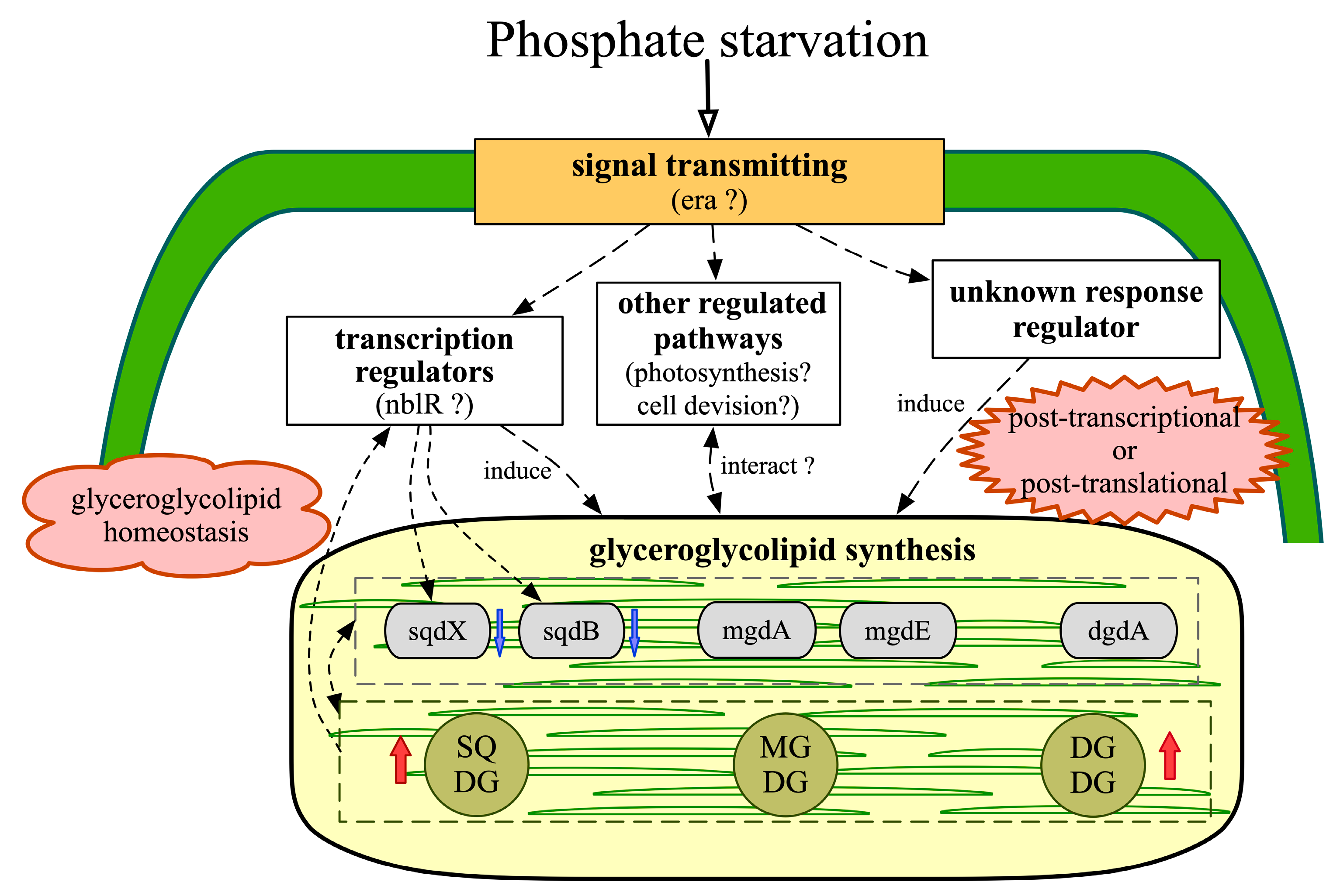
2.6. Regulatory Networks Involved in Glyceroglycolipid Synthesis

In plants and eukaryotic microalgae, some lipid-related transcription factors, like Dofs [73], are glyceroglycolipid-related. Nevertheless, transcriptional regulation is quite different between eukaryotes and prokaryotes. To determine the transcriptional regulators related to glyceroglycolipid metabolism under conditions of phosphate starvation in S. elongatus PCC 7942, 12 differentially expressed transcription regulators were selected, of which six were up-regulated and six were down-regulated (Table 1). These 12 transcription regulators can be divided into eight types: MarR family (1), MerR family (1), two component system (4), ArsR family (1), BadM/Rrf2 family (1), XRE family (2), GntR family (1) and DevT-like transcriptional factor (1). Most of these differentially expressed transcription regulators are involved in responses to various environmental stress conditions.
However, the genes coding the SphS–SphR phosphate sensing system and genes regulated by the system [74] showed no significant differences on day 12 of phosphate starvation, suggesting that these genes may respond to phosphate starvation at an early stage. Interestingly, an OmpR family response regulator gene nblR (Synpcc7942_2305), which regulates the degradation of phycobilisome (PBS) through the nbl pathway, as an activator of the PBS degradation protein gene’s (nblA) transcription under stresses [75], was down-regulated under phosphate starvation. However, nblA (Synpcc7942_2127) was significantly up-regulated (Supplementary Table S1), indicating that the down-regulation of nblR on day 12 was not to regulate PBS degradation. Sato et al. inferred that the sqdB might be involved in S-starvation-induced PBS degradation, particularly in Synechococcus [48]. It could be supposed that sqdB somehow belongs to the nbl pathway, and nblR is a probable signaling component in sqdBX regulation. In addition, some photosynthesis-associated transcription factors, like HY5 (a basic Leu zipper transcription factor) and GOLDEN2-LIKE (GLK), play pivotal roles in plant glyceroglycolipid regulation [66]. Therefore, glyceroglycolipid metabolism and the formation of photosynthetic machineries may be affected mutually. Besides, the up-regulated XRE family transcription regulator gene Synpcc7942_0110 was probably a hub gene under phosphate starvation, according to the protein–protein interaction analysis of DEGs (Supplementary Figure S3), which indicated that Synpcc7942_0110 played an important role in the global regulation of S. elongatus PCC 7942 under phosphate starvation.
The post-transcriptional and post-translational regulations are of great significance to glyceroglycolipid metabolism in plants. In the detached cotyledons of cucumber with impaired csMGD1 expression, light may activate MGDG biosynthesis in a post-transcriptional manner [76]. According to transcriptome analysis, three genes involved in sulfur relay system were significantly up-regulated (Supplementary Tables S1 and S4), suggesting that active tRNA modification under phosphate starvation contributed to metabolic regulation [77,78]. The activities of MGDG synthases can be modified by thioredoxins at the post-translational level [66]. In this study, a thioredoxin reductase gene (Synpcc7942_0623), a thioredoxin gene (Synpcc7942_1793) and a thioredoxin peroxidase gene (Synpcc7942_2309) were significantly up-regulated (Supplementary Table S1), all of which may contribute to activating MGDG synthesis. Like SQDG, PA and PG also play important roles in activating MGD1 [66,70]. Moreover, PG contributes to inducing MGDG synthesis by anchoring MGD1 and bringing substrates closer to the active site [66]. The information concerning the post-transcriptional and post-translational regulations involved in the glyceroglycolipid metabolism of cyanobacteria is still deficient, and thus more future studies are needed. In plants, auxin and cytokinin act as mediums between environmental conditions and the glyceroglycolipid metabolism [47,76]. Kobayashi et al. demonstrated that changes in plant membrane lipids during phosphate starvation are regulated by Pi signaling and auxin/cytokinin cross-talk [47]. In S. elongatus PCC 7942, increased SQDG, induced by overexpressions of sqdB and sqdX, would result in the abnormal expression of cell division-related genes and abnormal cell division [67]. The cell division protein gene FtsQ (Synpcc7942_2377) and the GroES protein gene (Synpcc7942_2314) involved in cell division were also important in global regulation under conditions of phosphate starvation (Supplementary Figure S3). Glyceroglycolipid synthesis and cell division could interact with each other. Taken together, it could be inferred that the glyceroglycolipid metabolism in cyanobacteria, under phosphate starvation, is regulated not only by Pi signaling, but also by other types of signaling and other metabolic pathways as well.

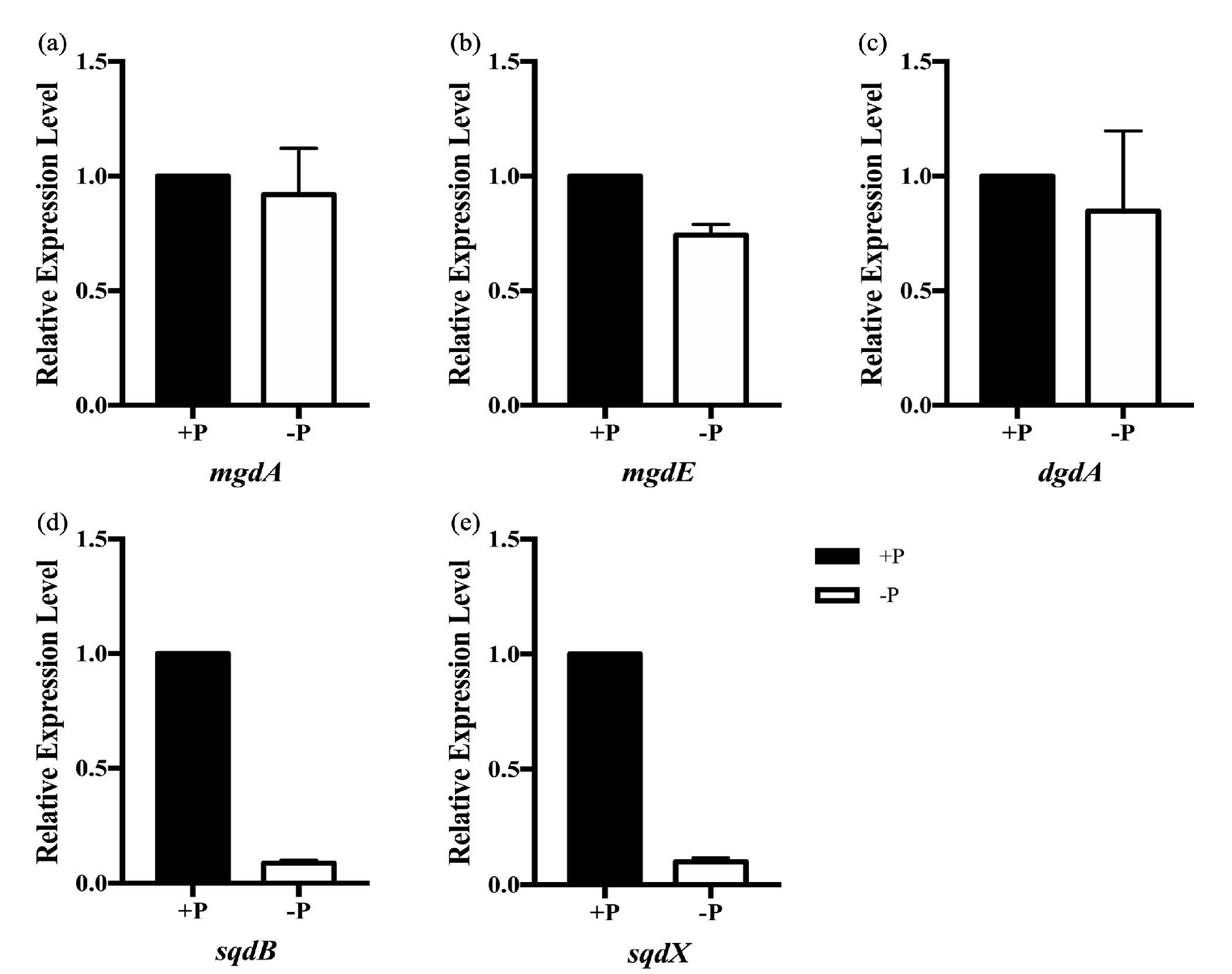
2.7. qRT-PCR Confirmations of Differentially Expressed Transcripts

qRT-PCR was used to confirm the accuracy of the transcriptomic analysis and measure the relative expression of selected transcripts. The results in Table 2 show that 82.4% of the measured transcripts (14 out of 17) followed the same trend as the RNA-Seq data, except for Synpcc7942_2416, Synpcc7942_1897 and Synpcc7942_1725. According to Celine, E. et al. [79], because of technical differences, over 80% of the measured genes having concordant expression represents a high concordance between RT-qPCR and RNA-seq. The expression of DEGs in transcriptomic analysis was reliable.

2.8. Reconstruction of Putative Glyceroglycolipid Regulatory Networks Based on Transcriptomic Evidence

Based on the transcriptomic evidence, a putative model for the glyceroglycolipids metabolism in S. elongatus PCC 7942, under phosphate starvation, was reconstructed (Figure 5).
Under phosphate starvation, the signals transmitting through GTP-binding proteins are active. The increase in total glyceroglycolipids mainly results from the accumulation of DGDG and SQDG as functional substitutes for phospholipids under phosphate starvation [43]. However, the changes in the composition of total glyceroglycolipids result in a sharp decrease of the MGDG/DGDG ratio, which disrupts the normal function of the plastid membrane [1]. To maintain glyceroglycolipid homeostasis, the down-regulations of sqdB and sqdX result in more precursors participating in MGDG synthesis, and the increased SQDG could act as an activator to enhance the activity of mgdA [45]. Transcriptional regulation, together with post-transcriptional and post-translational regulations, comprise an efficient strategy for glyceroglycolipid synthesis. Besides, glyceroglycolipids synthesis could interact with other metabolic pathways [66,67].
Hence, it could be proposed that the changes in glyceroglycolipid composition are emergency strategies of S. elongatus PCC 7942 adapting to phosphate starvation, and after 12 days of cultivation, glyceroglycolipid homeostasis plays a more important role in the recovery from phosphate starvation.

3. Materials and Methods

3.1. Cyanobacteria Species and Treatments

The cyanobacteria species in this study is Synechococcus elongatus PCC 7942 provided by Prof. Dingji Shi (Shanghai Ocean University, Shanghai, China).
S. elongatus PCC 7942 was cultivated in a 1-L Erlenmeyer flask with 500 mL working volume of modified BG-11 medium, under a temperature of 25 ± 2 °C and aeration rate of 140 µmol/m2/s. Standard BG-11 contains 0.04 g/L K2HPO4 concentration, while phosphate-starved BG-11 lacks K2HPO4. The light intensity was 8000 lx. The initial pH was 8.0.
The culture’s optical densities were measured at 730 nm by a UV-Vis spectrophotometer (Tianmei, Shanghai, China). Cell density was calculated with the equation: cell density (g/L) = 0.3349 × OD730 − 0.0129 (R2 = 0.9926). Standard curves are shown in Supplementary Material, Figure S4.

3.2. Glyceroglycolipid Analysis

Three different glyceroglycolipids were separated with a modified method of thin-layer chromatography (TLC) (Huanghai, Yantai, China) [43,80]. First, total lipids were extracted from cyanobacteria with a modified method [2,81]. Freeze-dried cyanobacteria powder (0.2 g) was broken by a cell crusher (Tissuelyser-24, Jingxin, Shanghai) at 50 Hz for 10 min. Then the broken cells were suspended in a 5-mL solvent mixture of chloroform/methanol (v/v 2:1). After being stirred for 20 min, the samples were centrifuged at 8000 rpm for 10 min. The procedure was repeated three times until the total lipids were fully extracted. The solvent phase was transferred and evaporated in a water bath (Shanghai YIHENG Technical Co. Ltd., Shanghai, China) at 65 °C and then dried in a drying oven (Jinghong, Shanghai, China) at 50 °C until the weight was stable. Then the total lipids were weighed with analytical balance (BS 124S, Sartorius, GöF6ttingen, Germany) and redissolved in chloroform/methanol (v/v 2:1) at a concentration of 10 µg/µL. The developing solvent was acetone/toluene/water (v/v/v 91:30:8). Different glyceroglycolipids were visualized in iodine vapor.
For quantitative analysis, fatty acid methyl esters of each glyceroglycolipid were prepared with 2 mL anhydrous 1 N methanolic HCl, and then incubated at 80 °C for 30 min [82]. The fatty acid profiles of different glyceroglycolipids were analyzed by AutoSystem XL GC/TurboMass MS (Perkin Elmer, Rodgau, Germany) [81]. The internal control was nonadecanoic acid.
The methods of calculating the content of each glyceroglycolipid were based on the method of Benning [80]. A total of six samples (two for each of the three cultures) per strain were analyzed, and means and standard deviations were calculated.

3.3. RNA Extraction, Library Preparation and Sequencing

Cyanobacteria cells were harvested in triplicate by centrifugation on day 12 under the phosphate concentrations of 0 and 0.04 g/L, respectively. Cells were immediately transferred to liquid nitrogen for later processing. Total RNA of each harvested sample was extracted with TRIzol reagent (Invitrogen Life Technologies) according to the manufacturer’s protocol. The sequencing library of each harvested sample was generated using a TruSeq RNA Sample Preparation Kit (Illumina, San Diego, CA, USA). The library was then sequenced on a HiSeq platform (Illumina) by Shanghai GeneFund Biotechnology Co. Ltd.

3.4. Transcript Quantification and Differential Expression Analysis

The clean reads were obtained by trimming raw reads from the sequencing with a Cutadapt tool [83] and a Trimmomatic tool [84]. The quality of clean reads was also assessed via FastQC tool [85]. Then, the high-quality trimmed reads were mapped to the reference genome by Bowtie2 [86]. Gene expression data were obtained and quantified with the fragments per kilobase of exon per million reads mapped method (FPKM) by HTSeq [87]. Empirical analysis of Digital Gene Expression in R (EdgeR) was applied for differential expression analysis [88]. Genes with p-value < 0.05 and fold change values (FC) ≥ 2 or ≤ 0.5 were regarded as DEGs.
GO enrichment analysis and KEGG enrichment analysis of DEGs were performed based on the hypergeometric distribution [89]. All DEGs were mapped to each GO term [90] and KEGG pathway [91]. The GO terms and the KEGG pathways of DEGs with a p-value < 0.05 were considered significantly enriched.

3.5. Experimental Validation of Gene Expression with qRT-PCR

The synthesis of cDNA was performed using FastKing RT Kit (With gDNase) (TIANGEN, Beijing, China). The gene-specific quantitative real-time PCR primers used in this study were documented in Supplementary Table S7. Real-time PCR was performed using a SuperReal PreMix Plus (SYBR Green) (TIANGEN, Beijing, China), and was carried out using an Eppendorf Mcep Realplex 4s System (Eppendorf, Hamburg, Germany). Reactions started at 95 °C for 15 min, followed by 40 cycles of 95 °C for 10 s and 60 °C for 25 s, and a melting curve step at 60–95 °C. Each qRT-PCR reaction was performed on three biological replicates. The relative expression levels were normalized to the glyceraldehyde-3-phosphate dehydrogenase (GAPDH) gene (gap3, Synpcc7942_1939), RNA polymerase sigma factor gene (rpoD, Synpcc7942_0649) [92] and phosphoenolpyruvate carboxylase gene (ppc, Synpcc7942_2252) [93], and were calculated using the 2-∆∆CT method [94].

4. Conclusions

The glyceroglycolipid composition of S. elongatus PCC 7942 changes to adapt to phosphate starvation. Glyceroglycolipid composition is regulated post-transcriptionally or post-translationally, allowing for more efficient adaptation to phosphate stress conditions. However, after 12 days of cultivation, the glyceroglycolipid composition is mainly regulated based on the MGDG/DGDG ratio in order to maintain the glyceroglycolipid homeostasis, which is beneficial in maintaining resilience when exposed to the preferred culture conditions again.

Supplementary Materials

The following are available online at https://www.mdpi.com/1660-3397/18/7/360/s1, Figure S1: Total glyceroglycolipids content under phosphate starvation. Values are the means ± standard deviations from the three separately grown cultures. Figure S2: Glycerolglycolipids relative content under phosphate starvation given by TLC-scanner. (a) MGDG. (b) DGDG. (c) SQDG. Values are the means ± standard deviations from the three separately grown cultures. Figure S3: PPI analysis of differentially expressed genes in Synechococcus elongatus PCC 7942 under different phosphate concentrations (0, 0.04g/L) after being cultivated for 12 days (Genes with annotation in String database are represented by abbreviation. Genes without annotation in String database are represented by gene number. Names of genes with degree less than 5 are not shown). Figure S4: Standard curve of OD730 and Dry Cell Weight of Synechococcus elongatus PCC 7942. Table S1: Differentially expressed genes under phosphate starvation in Synechococcus elongatus PCC 7942. Table S2: Enriched GO terms under phosphate starvation in Synechococcus elongatus PCC 7942. Table S3: Expression of genes in GO term GTP binding in Synechococcus elongatus PCC 7942 under phosphate starvation. Table S4: Enriched KEGG pathways under phosphate starvation in Synechococcus elongatus PCC 7942. Table S5: Expression of genes participating in glyceroglycolipid synthesis in Synechococcus elongatus PCC 7942 under phosphate starvation. Table S6: Expression of genes related to photosynthesis in Synechococcus elongatus PCC 7942 under phosphate starvation. Table S7: Primers used in qRT-PCR.

Author Contributions

Conceptualization, X.X. and X.M.; methodology, X.X. and X.M.; software, X.X.; validation, X.X. and X.M.; formal analysis, X.X.; investigation, X.X.; resources, X.X. and X.M.; data curation, X.X.; writing—original draft preparation, X.X.; writing—review and editing, X.X. and X.M.; visualization, X.X.; supervision, X.M.; project administration, X.M.; funding acquisition, X.M. All authors have read and agreed to the published version of the manuscript.

Funding

This research was financially supported by the National Natural Science Foundation of China (No.41476122). It was also supported by the National High Technology Research and Development Program (863 Program) of China (No. 2013AA065805).

Conflicts of Interest

The authors declare no conflict of interest.

References

  1. Boudiere, L.; Michaud, M.; Petroutsos, D.; Rebeille, F.; Falconet, D.; Bastien, O.; Roy, S.; Finazzi, G.; Rolland, N.; Jouhet, J.; et al. Glycerolipids in photosynthesis: Composition, synthesis and trafficking. Biochim. Biophys. Acta 2014, 1837, 470–480. [Google Scholar] [CrossRef]
  2. Wang, X.; Shen, Z.; Miao, X. Nitrogen and hydrophosphate affects glycolipids composition in microalgae. Sci. Rep. 2016, 6, 30145. [Google Scholar] [CrossRef] [PubMed] [Green Version]
  3. Zhang, J.; Li, C.; Yu, G.; Guan, H. Total synthesis and structure-activity relationship of glycoglycerolipids from marine organisms. Mar. Drugs 2014, 12, 3634–3659. [Google Scholar] [CrossRef] [PubMed] [Green Version]
  4. Andrianasolo, E.H.; Haramaty, L.; Vardi, A.; White, E.; Lutz, R.; Falkowski, P. Apoptosis-Inducing Galactolipids from a Cultured Marine Diatom, Phaeodactylum tricornutum. J. Nat. Prod. 2008, 71, 1197–1201. [Google Scholar] [CrossRef] [Green Version]
  5. Riccio, G.; De Luca, D.; Lauritano, C. Monogalactosyldiacylglycerol and Sulfolipid Synthesis in Microalgae. Mar. Drugs 2020, 18, 237. [Google Scholar] [CrossRef] [PubMed]
  6. Morimoto, T.; Nagatsu, A.; Murakami, N.; Sakakibara, J.; Tokuda, H.; Nishino, H.; Iwashima, A. Anti-tumour-promoting glyceroglycolipids from the green alga, chlorella vulgaris. Phytochemistry 1995, 40, 1433–1437. [Google Scholar] [CrossRef]
  7. Shirahashi, H.; Murakami, N.; Watanabe, M.; Nagatsu, A.; Sakakibara, J.; Tokuda, H.; Nishino, H.; Iwashima, A. Isolation and identification of anti-tumor-promoting principles from the fresh-water cyanobacterium Phormidium tenue. Chem. Pharm. Bull. 1993, 41, 1664–1666. [Google Scholar] [CrossRef] [PubMed] [Green Version]
  8. Mizushina, Y.; Watanabe, I.; Ohtaa, K.; Takemura, M.; Sahara, H.; Takahashi, N.; Gasa, S.; Sugawara, F.; Matsukage, A.; Yoshida, S.; et al. Studies on inhibitors of mammalian DNA polymerase α and β: Sulfolipids from a pteridophyte, Athyrium niponicum. Biochem. Pharmacol. 1998, 55, 537–541. [Google Scholar] [CrossRef]
  9. Gustafson, K.R.; Cardellina, J.H.; Fuller, R.W.; Weislow, O.S.; Kiser, R.F.; Snader, K.M.; Patterson, G.M.L.; Boyd, M.R. AIDS-Antiviral Sulfolipids From Cyanobacteria (Blue-Green Algae). J. Natl. Cancer Inst. 1989, 81, 1254–1258. [Google Scholar] [CrossRef]
  10. Loya, S.; Reshef, V.; Mizrachi, E.; Silberstein, C.; Rachamim, Y.; Carmeli, S.; Hizi, A. The Inhibition of the Reverse Transcriptase of HIV-1 by the Natural Sulfoglycolipids from Cyanobacteria: Contribution of Different Moieties to Their High Potency. J. Nat. Prod. 1998, 61, 891–895. [Google Scholar] [CrossRef]
  11. Alipanah, L.; Winge, P.; Rohloff, J.; Najafi, J.; Brembu, T.; Bones, A.M. Molecular adaptations to phosphorus deprivation and comparison with nitrogen deprivation responses in the diatom Phaeodactylum tricornutum. PLoS ONE 2018, 13, e0193335. [Google Scholar] [CrossRef] [PubMed]
  12. Murakami, H.; Nobusawa, T.; Hori, K.; Shimojima, M.; Ohta, H. Betaine Lipid Is Crucial for Adapting to Low Temperature and Phosphate Deficiency in Nannochloropsis. Plant Physiol. 2018, 177, 181–193. [Google Scholar] [CrossRef] [PubMed] [Green Version]
  13. Nakamura, Y. Phosphate starvation and membrane lipid remodeling in seed plants. Prog. Lipid Res. 2013, 52, 43–50. [Google Scholar] [CrossRef] [PubMed]
  14. Shimojima, M. Biosynthesis and functions of the plant sulfolipid. Prog. Lipid Res. 2011, 50, 234–239. [Google Scholar] [CrossRef] [PubMed]
  15. Lu, N.; Wei, D.; Chen, F.; Yang, S.T. Lipidomic profiling reveals lipid regulation in the snow alga Chlamydomonas nivalis in response to nitrate or phosphate deprivation. Process. Biochem. 2013, 48, 605–613. [Google Scholar] [CrossRef]
  16. Liang, K.; Zhang, Q.; Gu, M.; Cong, W. Effect of phosphorus on lipid accumulation in freshwater microalga Chlorella sp. J. Appl. Phycol. 2012, 25, 311–318. [Google Scholar] [CrossRef]
  17. Muhlroth, A.; Winge, P.; El Assimi, A.; Jouhet, J.; Marechal, E.; Hohmann-Marriott, M.F.; Vadstein, O.; Bones, A.M. Mechanisms of Phosphorus Acquisition and Lipid Class Remodeling under P Limitation in a Marine Microalga. Plant Physiol. 2017, 175, 1543–1559. [Google Scholar] [CrossRef] [Green Version]
  18. Gasparovic, B.; Godrijan, J.; Frka, S.; Tomazic, I.; Penezic, A.; Maric, D.; Djakovac, T.; Ivancic, I.; Paliaga, P.; Lyons, D.; et al. Adaptation of marine plankton to environmental stress by glycolipid accumulation. Mar. Environ. Res. 2013, 92, 120–132. [Google Scholar] [CrossRef]
  19. Yu, B.; Xu, C.; Benning, C. Arabidopsis disrupted in SQD2 encoding sulfolipid synthase is impaired in phosphate-limited growth. Proc. Natl. Acad. Sci. USA 2002, 99, 5732–5737. [Google Scholar] [CrossRef] [Green Version]
  20. Li-Beisson, Y.; Thelen, J.J.; Fedosejevs, E.; Harwood, J.L. The lipid biochemistry of eukaryotic algae. Prog. Lipid Res. 2019, 74, 31–68. [Google Scholar] [CrossRef]
  21. Frentzen, M. Phosphatidylglycerol and sulfoquinovosyldiacylglycerol: Anionic membrane lipids and phosphate regulation. Curr. Opin. Plant Biol. 2004, 7, 270–276. [Google Scholar] [CrossRef] [PubMed]
  22. Villanueva, L.; Bale, N.; Hopmans, E.C.; Schouten, S.; Damste, J.S. Diversity and distribution of a key sulpholipid biosynthetic gene in marine microbial assemblages. Environ. Microbiol. 2014, 16, 774–787. [Google Scholar] [CrossRef] [PubMed] [Green Version]
  23. Hölzl, G.; Dörmann, P. Chloroplast Lipids and Their Biosynthesis. Annu. Rev. Plant Biol. 2019, 70, 51–81. [Google Scholar] [CrossRef] [PubMed]
  24. Aronsson, H.; Schottler, M.A.; Kelly, A.A.; Sundqvist, C.; Dormann, P.; Karim, S.; Jarvis, P. Monogalactosyldiacylglycerol deficiency in Arabidopsis affects pigment composition in the prolamellar body and impairs thylakoid membrane energization and photoprotection in leaves. Plant Physiol. 2008, 148, 580–592. [Google Scholar] [CrossRef] [Green Version]
  25. Murakawa, M.; Shimojima, M.; Shimomura, Y.; Kobayashi, K.; Awai, K.; Ohta, H. Monogalactosyldiacylglycerol synthesis in the outer envelope membrane of chloroplasts is required for enhanced growth under sucrose supplementation. Front. Plant Sci. 2014, 5, 280. [Google Scholar] [CrossRef] [Green Version]
  26. Miège, C.; Maréchal, E.; Shimojima, M.; Awai, K.; Block, M.A.; Ohta, H.; Takamiya, K.; Douce, R.; Joyard, J. Biochemical and topological properties of type A MGDG synthase, a spinach chloroplast envelope enzyme catalyzing the synthesis of both prokaryotic and eukaryotic MGDG. Eur. J. Biochem. 1999, 265, 990–1001. [Google Scholar] [CrossRef] [Green Version]
  27. Froehlich, J.E.; Benning, C.; Dormann, P. The digalactosyldiacylglycerol (DGDG) synthase DGD1 is inserted into the outer envelope membrane of chloroplasts in a manner independent of the general import pathway and does not depend on direct interaction with monogalactosyldiacylglycerol synthase for DGDG biosynthesis. J. Biol. Chem. 2001, 276, 31806–31812. [Google Scholar] [CrossRef] [Green Version]
  28. Kelly, A.A.; Dormann, P. DGD2, an Arabidopsis gene encoding a UDP-galactose-dependent digalactosyldiacylglycerol synthase is expressed during growth under phosphate-limiting conditions. J. Biol. Chem. 2002, 277, 1166–1173. [Google Scholar] [CrossRef] [Green Version]
  29. Awai, K.; Marechal, E.; Block, M.A.; Brun, D.; Masuda, T.; Shimada, H.; Takamiya, K.; Ohta, H.; Joyard, J. Two types of MGDG synthase genes, found widely in both 16:3 and 18:3 plants, differentially mediate galactolipid syntheses in photosynthetic and nonphotosynthetic tissues in Arabidopsis thaliana. Proc. Natl. Acad. Sci. USA 2001, 98, 10960–10965. [Google Scholar] [CrossRef] [Green Version]
  30. Riekhof, W.R.; Sears, B.B.; Benning, C. Annotation of genes involved in glycerolipid biosynthesis in Chlamydomonas reinhardtii: Discovery of the betaine lipid synthase BTA1Cr. Eukaryot. Cell 2005, 4, 242–252. [Google Scholar] [CrossRef] [Green Version]
  31. Awai, K.; Watanabe, H.; Benning, C.; Nishida, I. Digalactosyldiacylglycerol is required for better photosynthetic growth of Synechocystis sp. PCC6803 under phosphate limitation. Plant Cell Physiol. 2007, 48, 1517–1523. [Google Scholar] [CrossRef] [PubMed] [Green Version]
  32. Maida, E.; Awai, K. Digalactosyldiacylglycerol is essential in Synechococcus elongatus PCC 7942, but its function does not depend on its biosynthetic pathway. Biochim. Biophys. Acta 2016, 1861, 1309–1314. [Google Scholar] [CrossRef] [PubMed]
  33. Sato, N.; Awai, K. Diversity in Biosynthetic Pathways of Galactolipids in the Light of Endosymbiotic Origin of Chloroplasts. Front. Plant Sci. 2016, 7, 117. [Google Scholar] [CrossRef] [PubMed] [Green Version]
  34. Yuzawa, Y.; Shimojima, M.; Sato, R.; Mizusawa, N.; Ikeda, K.; Suzuki, M.; Iwai, M.; Hori, K.; Wada, H.; Masuda, S.; et al. Cyanobacterial monogalactosyldiacylglycerol-synthesis pathway is involved in normal unsaturation of galactolipids and low-temperature adaptation of Synechocystis sp. PCC 6803. Biochim. Biophys. Acta 2014, 1841, 475–483. [Google Scholar] [CrossRef] [PubMed] [Green Version]
  35. Awai, K.; Kakimoto, T.; Awai, C.; Kaneko, T.; Nakamura, Y.; Takamiya, K.; Wada, H.; Ohta, H. Comparative genomic analysis revealed a gene for monoglucosyldiacylglycerol synthase, an enzyme for photosynthetic membrane lipid synthesis in cyanobacteria. Plant Physiol. 2006, 141, 1120–1127. [Google Scholar] [CrossRef] [Green Version]
  36. Awai, K.; Ohta, H.; Sato, N. Oxygenic photosynthesis without galactolipids. Proc. Natl. Acad. Sci. USA 2014, 111, 13571–13575. [Google Scholar] [CrossRef] [Green Version]
  37. Kobayashi, K.; Awai, K.; Nakamura, M.; Nagatani, A.; Masuda, T.; Ohta, H. Type-B monogalactosyldiacylglycerol synthases are involved in phosphate starvation-induced lipid remodeling, and are crucial for low-phosphate adaptation. Plant J. 2009, 57, 322–331. [Google Scholar] [CrossRef]
  38. Kelly, A.A.; Froehlich, J.E.; Dormann, P. Disruption of the two digalactosyldiacylglycerol synthase genes DGD1 and DGD2 in Arabidopsis reveals the existence of an additional enzyme of galactolipid synthesis. Plant Cell 2003, 15, 2694–2706. [Google Scholar] [CrossRef] [Green Version]
  39. Okazaki, Y.; Shimojima, M.; Sawada, Y.; Toyooka, K.; Narisawa, T.; Mochida, K.; Tanaka, H.; Matsuda, F.; Hirai, A.; Hirai, M.Y.; et al. A Chloroplastic UDP-Glucose Pyrophosphorylase from Arabidopsis Is the Committed Enzyme for the First Step of Sulfolipid Biosynthesis. Plant Cell 2009, 21, 892–909. [Google Scholar] [CrossRef] [Green Version]
  40. Kang, N.K.; Kim, E.K.; Sung, M.G.; Kim, Y.U.; Jeong, B.R.; Chang, Y.K. Increased biomass and lipid production by continuous cultivation of Nannochloropsis salina transformant overexpressing a bHLH transcription factor. Biotechnol. Bioeng. 2019, 116, 555–568. [Google Scholar] [CrossRef] [Green Version]
  41. Kwon, S.; Kang, N.K.; Koh, H.G.; Shin, S.E.; Lee, B.; Jeong, B.R.; Chang, Y.K. Enhancement of biomass and lipid productivity by overexpression of a bZIP transcription factor in Nannochloropsis salina. Biotechnol. Bioeng. 2018, 115, 331–340. [Google Scholar] [CrossRef] [PubMed] [Green Version]
  42. Zhang, J.; Hao, Q.; Bai, L.; Xu, J.; Yin, W.; Song, L.; Xu, L.; Guo, X.; Fan, C.; Chen, Y.; et al. Overexpression of the soybean transcription factor GmDof4 significantly enhances the lipid content of Chlorella ellipsoidea. Biotechnol. Biofuels 2014, 7, 128. [Google Scholar] [CrossRef] [Green Version]
  43. Peng, Z.; Feng, L.; Wang, X.; Miao, X. Adaptation of Synechococcus sp. PCC 7942 to phosphate starvation by glycolipid accumulation and membrane lipid remodeling. Biochim. Biophys. Acta 2019, 1864, 158522. [Google Scholar] [CrossRef]
  44. Abida, H.; Dolch, L.J.; Mei, C.; Villanova, V.; Conte, M.; Block, M.A.; Finazzi, G.; Bastien, O.; Tirichine, L.; Bowler, C.; et al. Membrane glycerolipid remodeling triggered by nitrogen and phosphorus starvation in Phaeodactylum tricornutum. Plant Physiol. 2015, 167, 118–136. [Google Scholar] [CrossRef] [PubMed] [Green Version]
  45. Selao, T.T.; Zhang, L.; Arioz, C.; Wieslander, A.; Norling, B. Subcellular localization of monoglucosyldiacylglycerol synthase in Synechocystis sp. PCC6803 and its unique regulation by lipid environment. PLoS ONE 2014, 9, e88153. [Google Scholar] [CrossRef] [PubMed] [Green Version]
  46. Shimojima, M.; Tsuchiya, M.; Ohta, H. Temperature-dependent hyper-activation of monoglucosyldiacylglycerol synthase is post-translationally regulated in Synechocystis sp. PCC 6803. FEBS Lett. 2009, 583, 2372–2376. [Google Scholar] [CrossRef] [PubMed] [Green Version]
  47. Kobayashi, K.; Masuda, T.; Takamiya, K.; Ohta, H. Membrane lipid alteration during phosphate starvation is regulated by phosphate signaling and auxin/cytokinin cross-talk. Plant J. 2006, 47, 238–248. [Google Scholar] [CrossRef]
  48. Sato, N.; Kamimura, R.; Kaneta, K.; Yoshikawa, M.; Tsuzuki, M. Species-specific roles of sulfolipid metabolism in acclimation of photosynthetic microbes to sulfur-starvation stress. PLoS ONE 2017, 12, e0186154. [Google Scholar] [CrossRef] [PubMed] [Green Version]
  49. Voshol, G.P.; Meyer, V.; van den Hondel, C.A. GTP-binding protein Era: A novel gene target for biofuel production. BMC Biotechnol. 2015, 15, 21. [Google Scholar] [CrossRef] [Green Version]
  50. Johnstone, B.H.; Handler, A.A.; Chao, D.K.; Nguyen, V.; Smith, M.; Ryu, S.Y.; Simons, E.L.; Anderson, P.E.; Simons, R.W. The widely conserved Era G-protein contains an RNA-binding domain required for Era function in vivo. Mol. Microbiol. 1999, 33, 1118–1131. [Google Scholar] [CrossRef] [Green Version]
  51. Cheng, P.; Li, H.; Yuan, L.; Li, H.; Xi, L.; Zhang, J.; Liu, J.; Wang, Y.; Zhao, H.; Zhao, H.; et al. The ERA-Related GTPase AtERG2 Associated with Mitochondria 18S RNA Is Essential for Early Embryo Development in Arabidopsis. Front. Plant Sci. 2018, 9, 182. [Google Scholar] [CrossRef] [Green Version]
  52. Suwastika, I.N.; Denawa, M.; Yomogihara, S.; Im, C.H.; Bang, W.Y.; Ohniwa, R.L.; Bahk, J.D.; Takeyasu, K.; Shiina, T. Evidence for lateral gene transfer (LGT) in the evolution of eubacteria-derived small GTPases in plant organelles. Front. Plant Sci. 2014, 5, 678. [Google Scholar] [CrossRef] [PubMed] [Green Version]
  53. Jeon, Y.; Ahn, C.S.; Jung, H.J.; Kang, H.; Park, G.T.; Choi, Y.; Hwang, J.; Pai, H.S. DER containing two consecutive GTP-binding domains plays an essential role in chloroplast ribosomal RNA processing and ribosome biogenesis in higher plants. J. Exp. Bot. 2014, 65, 117–130. [Google Scholar] [CrossRef] [PubMed] [Green Version]
  54. Sun, Y.; Tian, Y.; Cheng, S.; Wang, Y.; Hao, Y.; Zhu, J.; Zhu, X.; Zhang, Y.; Yu, M.; Lei, J.; et al. WSL6 encoding an Era-type GTP-binding protein is essential for chloroplast development in rice. Plant Mol. Biol. 2019. [Google Scholar] [CrossRef] [PubMed]
  55. Yan, Y.; Zheng, X.; Apaliya, M.T.; Yang, H.; Zhang, H. Transcriptome characterization and expression profile of defense-related genes in pear induced by Meyerozyma guilliermondii. Postharvest Biol. Technol. 2018, 141, 63–70. [Google Scholar] [CrossRef]
  56. Choi, S.Y.; Park, B.; Choi, I.G.; Sim, S.J.; Lee, S.M.; Um, Y.; Woo, H.M. Transcriptome landscape of Synechococcus elongatus PCC 7942 for nitrogen starvation responses using RNA-seq. Sci. Rep. 2016, 6, 30584. [Google Scholar] [CrossRef] [PubMed] [Green Version]
  57. Verstraeten, N.; Fauvart, M.; Versees, W.; Michiels, J. The universally conserved prokaryotic GTPases. Microbiol. Mol. Biol. Rev. 2011, 75, 507–542. [Google Scholar] [CrossRef] [PubMed] [Green Version]
  58. Du, W.; Liang, F.; Duan, Y.; Tan, X.; Lu, X. Exploring the photosynthetic production capacity of sucrose by cyanobacteria. Metab. Eng. 2013, 19, 17–25. [Google Scholar] [CrossRef]
  59. Maeda, K.; Narikawa, R.; Ikeuchi, M. CugP is a novel ubiquitous non-GalU-type bacterial UDP-glucose pyrophosphorylase found in cyanobacteria. J. Bacteriol. 2014, 196, 2348–2354. [Google Scholar] [CrossRef] [Green Version]
  60. Zavaleta-Pastor, M.; Sohlenkamp, C.; Gao, J.L.; Guan, Z.; Zaheer, R.; Finan, T.M.; Raetz, C.R.; Lopez-Lara, I.M.; Geiger, O. Sinorhizobium meliloti phospholipase C required for lipid remodeling during phosphorus limitation. Proc. Natl. Acad. Sci. USA 2010, 107, 302–307. [Google Scholar] [CrossRef] [Green Version]
  61. Boudiere, L.; Botte, C.Y.; Saidani, N.; Lajoie, M.; Marion, J.; Brehelin, L.; Yamaryo-Botte, Y.; Satiat-Jeunemaitre, B.; Breton, C.; Girard-Egrot, A.; et al. Galvestine-1, a novel chemical probe for the study of the glycerolipid homeostasis system in plant cells. Mol. Biosyst. 2012, 8, 2023–2035. [Google Scholar] [CrossRef]
  62. Wada, H.; Murata, N. Membrane Lipids in Cyanobacteria. In Lipids in Photosynthesis: Structure, Function and Genetics; Springer: Dordrecht, The Netherlands, 1998. [Google Scholar]
  63. Gu, Y.; He, L.; Zhao, C.; Wang, F.; Yan, B.; Gao, Y.; Li, Z.; Yang, K.; Xu, J. Biochemical and Transcriptional Regulation of Membrane Lipid Metabolism in Maize Leaves under Low Temperature. Front. Plant Sci. 2017, 8, 2053. [Google Scholar] [CrossRef] [PubMed] [Green Version]
  64. Laczko-Dobos, H.; Frycak, P.; Ughy, B.; Domonkos, I.; Wada, H.; Prokai, L.; Gombos, Z. Remodeling of phosphatidylglycerol in Synechocystis PCC6803. Biochim. Biophys. Acta 2010, 1801, 163–170. [Google Scholar] [CrossRef] [PubMed]
  65. Kobayashi, K.; Fujii, S.; Sasaki, D.; Baba, S.; Ohta, H.; Masuda, T.; Wada, H. Transcriptional regulation of thylakoid galactolipid biosynthesis coordinated with chlorophyll biosynthesis during the development of chloroplasts in Arabidopsis. Front. Plant Sci. 2014, 5, 272. [Google Scholar] [CrossRef] [PubMed] [Green Version]
  66. Kobayashi, K. Role of membrane glycerolipids in photosynthesis, thylakoid biogenesis and chloroplast development. J. Plant Res. 2016, 129, 565–580. [Google Scholar] [CrossRef] [PubMed] [Green Version]
  67. Sato, N.; Ebiya, Y.; Kobayashi, R.; Nishiyama, Y.; Tsuzuki, M. Disturbance of cell-size determination by forced overproduction of sulfoquinovosyl diacylglycerol in the cyanobacterium Synechococcus elongatus PCC 7942. Biochem. Biophys. Res. Commun. 2017, 487, 734–739. [Google Scholar] [CrossRef]
  68. Khozin-Goldberg, I.; Cohen, Z. The effect of phosphate starvation on the lipid and fatty acid composition of the fresh water eustigmatophyte Monodus subterraneus. Phytochemistry 2006, 67, 696–701. [Google Scholar] [CrossRef]
  69. Masuda, S.; Harada, J.; Yokono, M.; Yuzawa, Y.; Shimojima, M.; Murofushi, K.; Tanaka, H.; Masuda, H.; Murakawa, M.; Haraguchi, T.; et al. A Monogalactosyldiacylglycerol Synthase Found in the Green Sulfur Bacterium Chlorobaculum tepidum Reveals Important Roles for Galactolipids in Photosynthesis. Plant Cell 2011, 23, 2644–2658. [Google Scholar] [CrossRef] [Green Version]
  70. Dubots, E.; Botte, C.; Boudiere, L.; Yamaryo-Botte, Y.; Jouhet, J.; Marechal, E.; Block, M.A. Role of phosphatidic acid in plant galactolipid synthesis. Biochimie 2012, 94, 86–93. [Google Scholar] [CrossRef] [Green Version]
  71. Sakurai, I.; Mizusawa, N.; Wada, H.; Sato, N. Digalactosyldiacylglycerol is required for stabilization of the oxygen-evolving complex in photosystem II. Plant Physiol. 2007, 145, 1361–1370. [Google Scholar] [CrossRef] [Green Version]
  72. Kobayashi, K.; Narise, T.; Sonoike, K.; Hashimoto, H.; Sato, N.; Kondo, M.; Nishimura, M.; Sato, M.; Toyooka, K.; Sugimoto, K.; et al. Role of galactolipid biosynthesis in coordinated development of photosynthetic complexes and thylakoid membranes during chloroplast biogenesis in Arabidopsis. Plant J. 2013, 73, 250–261. [Google Scholar] [CrossRef] [PubMed]
  73. Manan, S.; Chen, B.; She, G.; Wan, X.; Zhao, J. Transport and transcriptional regulation of oil production in plants. Crit. Rev. Biotechnol. 2016, 37, 641–655. [Google Scholar] [CrossRef] [PubMed]
  74. Tiwari, B.; Singh, S.; Kaushik, M.S.; Mishra, A.K. Regulation of organophosphate metabolism in cyanobacteria. A review. Microbiology 2015, 84, 291–302. [Google Scholar] [CrossRef]
  75. Salinas, P.; Ruiz, D.; Cantos, R.; Lopez-Redondo, M.L.; Marina, A.; Contreras, A. The regulatory factor SipA provides a link between NblS and NblR signal transduction pathways in the cyanobacterium Synechococcus sp. PCC 7942. Mol. Microbiol. 2007, 66, 1607–1619. [Google Scholar] [CrossRef]
  76. Yamaryo, Y.; Kanai, D.; Awai, K.; Shimojima, M.; Masuda, T.; Shimada, H.; Takamiya, K.-I.; Ohta, H. Light and Cytokinin Play a Co-operative Role in MGDG Synthesis in Greening Cucumber Cotyledons. Plant Cell Physiol. 2003, 44, 844–855. [Google Scholar] [CrossRef] [Green Version]
  77. El Yacoubi, B.; Bailly, M.; de Crecy-Lagard, V. Biosynthesis and function of posttranscriptional modifications of transfer RNAs. Annu. Rev. Genet. 2012, 46, 69–95. [Google Scholar] [CrossRef]
  78. Wang, Y.; Pang, C.; Li, X.; Hu, Z.; Lv, Z.; Zheng, B.; Chen, P. Identification of tRNA nucleoside modification genes critical for stress response and development in rice and Arabidopsis. BMC Plant Biol. 2017, 17, 261. [Google Scholar] [CrossRef] [PubMed]
  79. Everaert, C.; Luypaert, M.; Maag, J.L.V.; Cheng, Q.X.; Dinger, M.E.; Hellemans, J.; Mestdagh, P. Benchmarking of RNA-sequencing analysis workflows using whole-transcriptome RT-qPCR expression data. Sci. Rep. 2017, 7, 1–11. [Google Scholar] [CrossRef] [Green Version]
  80. Hartel, H.; Dormann, P.; Benning, C. DGD1-independent biosynthesis of extraplastidic galactolipids after phosphate deprivation in Arabidopsis. Proc. Natl. Acad. Sci. USA 2000, 97, 10649–10654. [Google Scholar] [CrossRef] [Green Version]
  81. Wu, H.; Miao, X. Biodiesel quality and biochemical changes of microalgae Chlorella pyrenoidosa and Scenedesmus obliquus in response to nitrate levels. Bioresour. Technol. 2014, 170, 421–427. [Google Scholar] [CrossRef] [PubMed]
  82. Peng, Z.; Miao, X. Monoglucosyldiacylglycerol participates in phosphate stress adaptation in Synechococcus sp. PCC 7942. Biochem. Biophys. Res. Commun. 2020, 522, 662–668. [Google Scholar] [CrossRef] [PubMed]
  83. Martin, M. Cutadapt removes adapter sequences from high-throughput sequencing reads. EMBnet. J. 2011, 17, 10–12. [Google Scholar] [CrossRef]
  84. Bolger, A.M.; Lohse, M.; Usadel, B. Trimmomatic: A flexible trimmer for Illumina sequence data. Bioinformatics 2014, 30, 2114–2120. [Google Scholar] [CrossRef] [Green Version]
  85. Hwang, S.G.; Kim, K.H.; Lee, B.M.; Moon, J.C. Transcriptome analysis for identifying possible gene regulations during maize root emergence and formation at the initial growth stage. Genes Genom. 2018, 40, 755–766. [Google Scholar] [CrossRef] [PubMed]
  86. Langmead, B.; Salzberg, S.L. Fast gapped-read alignment with Bowtie 2. Nat. Methods 2012, 9, 357–359. [Google Scholar] [CrossRef] [PubMed] [Green Version]
  87. Anders, S.; Pyl, P.T.; Huber, W. HTSeq—A Python framework to work with high-throughput sequencing data. Bioinformatics 2015, 31, 166–169. [Google Scholar] [CrossRef]
  88. Zhou, L.; Cheng, D.; Wang, L.; Gao, J.; Zhao, Q.; Wei, W.; Sun, Y. Comparative transcriptomic analysis reveals phenol tolerance mechanism of evolved Chlorella strain. Bioresour. Technol. 2017, 227, 266–272. [Google Scholar] [CrossRef] [PubMed]
  89. Sun, X.; Shen, J.; Bai, F.; Xu, N. Transcriptome profiling of the microalga Chlorella pyrenoidosa in response to different carbon dioxide concentrations. Mar. Genom. 2016, 29, 81–87. [Google Scholar] [CrossRef]
  90. Ashburner, M.; Ball, C.; Blake, J.; Botstein, D.; Butler, H.; Cherry, J.M.; Davis, A.; Dolinski, K.; Dwight, S.; Eppig, J.; et al. Gene Ontology: Tool for the unification of biology. Nat. Genet. 2000, 25, 25–29. [Google Scholar] [CrossRef] [Green Version]
  91. Kanehisa, M.; Goto, S. KEGG: Kyoto encyclopedia of genes and genomes. Nucleic Acids Res. 2000, 28, 27–30. [Google Scholar] [CrossRef]
  92. Scherer, P.I.; Raeder, U.; Geist, J.; Zwirglmaier, K. Influence of temperature, mixing, and addition of microcystin-LR on microcystin gene expression in Microcystis aeruginosa. Microbiologyopen 2017, 6. [Google Scholar] [CrossRef] [PubMed]
  93. Woodger, F.J.; Badger, M.R.; Price, G.D. Inorganic carbon limitation induces transcripts encoding components of the CO2-concentrating mechanism in Synechococcus sp. PCC7942 through a redox-independent pathway. Plant Physiol. 2003, 133, 2069–2080. [Google Scholar] [CrossRef] [PubMed] [Green Version]
  94. Livak, K.J.; Schmittgen, T.D. Analysis of relative gene expression data using real-time quantitative PCR and the 2(-Delta Delta C(T)) Method. Methods 2001, 25, 402–408. [Google Scholar] [CrossRef] [PubMed]
Figure 1. Growth (a) and dynamic changes in glyceroglycolipid composition (b) in S. elongatus PCC 7942 under phosphate concentrations of 0.04 g/L (+P) and 0 g/L (−P). Values are the means ± standard deviations from the three separately grown cultures. 2-way ANOVA test (* p < 0.05, ** p < 0.01, *** p < 0.001).
Marinedrugs 18 00360 g001
Figure 2. Relative gene expressions of mgdA (a), mgdE (b), dgdA (c), sqdB (d) and sqdX (e) in S. elongatus PCC 7942 cultivated under phosphate concentrations of 0.04 g/L (+P) and 0 g/L (−P) for 12 days. Values are the means ± standard deviations from the three separately grown cultures. The expression level of each glyceroglycolipid synthase gene under phosphate concentration of 0.04 g/L (+P) was set to 1.
Marinedrugs 18 00360 g002
Figure 3. DEGs, and GO and KEGG enrichment of DEGs under phosphate starvation in S. elongatus PCC 7942. All DEGs were defined under p-value < 0.05 and fold change values (FC) ≥ 2 or ≤ 0.5. (a) Volcano plot of DEGs. Red and blue represent up-regulated and down-regulated DEGs, respectively. (b) Representative enriched GO terms of DEGs. Bars represent number of DEGs. (c) Representative enriched pathways of DEGs. Bars represent number of DEGs.
Marinedrugs 18 00360 g003
Figure 4. Metabolic pathway of glyceroglycolipid synthesis (a) and phospholipid synthesis (b) in Synechococcus elongatus PCC 7942. Pathways were reconstructed based on the KEGG annotation and the gene expression (colored rectangles) derived from transcriptome data of S. elongatus PCC 7942 (p-value < 0.05). Control groups (+P) are represented by rectangles with crosses. Genes up-regulated under phosphate starvation are indicated in red. Genes down-regulated are indicated in blue. Genes with no significant changes are indicated in black with diagonal lines in rectangles. xylR: xylose repressor (EC: 2.7.1.2); glk: glucokinase (EC: 2.7.1.2); pgm: phosphoglucomutase (EC: 5.4.2.2); galE: UDP-galactose epimerase (EC: 5.1.3.2); sqdB: UDP-sulfoquinovose synthase (EC: 3.13.1.1); sqdX: SQDG synthase (EC: 2.4.1.-); plsY: acyl-phosphate glycerol-3-phosphate acyltransferase (EC: 2.3.1.275); plsC: 1-acyl-sn-glycerol-3-phosphate acyltransferase (EC: 2.3.1.51); dgk: diacylglycerol kinase (EC: 2.7.1.107); mgdA: MGlcDG synthase (EC: 2.4.1.336); mgdE: MGlcDG epimerase (EC: 5.1.3.34); dgdA: DGDG synthase (EC: 2.4.1.241); cds: phosphatidate cytidylyltransferase (EC: 2.7.7.41); pgs: CDP-diacylglycerol-glycerol-3-phosphate 3-phosphatidyltransferase (EC: 2.7.8.5). Glu: glucose; Glu-6-P: glucose-6-phosphate; Glu-1-P: glucose-6-phosphate; UDP-Glu: UDP-glucose; UDP-Gal: UDP-galactose; UDP-Sq: UDP-sulfoquinovose; G-3-P: glycerol-3-phosphate; LPA: Lysophosphatidic acid; PA: phosphatidic acid; DAG: diacylglycerol; CDP-DAG: CDP-diacylglycerol; PGP: phosphatidylglycerophosphate; PG: phosphatidylglycerol; MGlcDG: monoglucosyl diacylglycerol; MGDG: monogalactosyl diacylglycerol; DGDG: digalactosyl diacylglycerol; SQDG: sulfoquinovosyl diacylglycerol.
Marinedrugs 18 00360 g004
Figure 5. Putative model for glyceroglycolipid metabolism in S. elongatus PCC 7942 under phosphate starvation, reconstructed from transcriptomic evidence in this study. Dotted arrows indicate the regulation between each part.
Marinedrugs 18 00360 g005
Table 1. Differentially expressed transcription regulators under phosphate starvation. Up-regulated genes (FC > 1) and down-regulated genes (FC < 1) were ordered by FC values.
Gene IDAnnotationFCp-Value
Synpcc7942_0938transcriptional regulator, ArsR family8.792818035.0074E-13
Synpcc7942_2585transcriptional regulator, BadM/Rrf2 family2.437514150.00232243
Synpcc7942_2416two component transcriptional regulator, winged helix family2.157396520.04603509
Synpcc7942_0110transcriptional regulator, XRE family2.133868520.02139503
Synpcc7942_1897putative transcription factor DevT-like2.125345890.00718275
Synpcc7942_1725transcriptional regulator, GntR family1.949071680.04567919
Synpcc7942_2305two component transcriptional regulator, winged helix family, nblR0.720207020.04515754
Synpcc7942_1739transcriptional regulator, MerR family0.611298140.02357565
Synpcc7942_0556two component transcriptional regulator, winged helix family0.610866840.01166383
Synpcc7942_2466two component transcriptional regulator, winged helix family0.586257720.03952819
Synpcc7942_1159transcriptional regulator, MarR family0.572966150.0038297
Synpcc7942_0764transcriptional regulator, XRE family0.479658350.00047173
Table 2. qRT-PCR analysis of different genes under phosphate starvation in Synechococcus elongatus PCC 7942.
Gene IDAnnotationFCqRT-PCR
Synpcc7942_0938transcriptional regulator, ArsR family8.79281823.18
Synpcc7942_2585transcriptional regulator, BadM/Rrf2 family2.43751423.82
Synpcc7942_2416two component transcriptional regulator, winged helix family2.1573972−0.84
Synpcc7942_0110transcriptional regulator, XRE family2.13386922.78
Synpcc7942_1897putative transcription factor DevT-like2.1253462−1.01
Synpcc7942_1725transcriptional regulator, GntR family1.9490722−1.58
Synpcc7942_1083a probable glycosyltransferase, mgdA1.337782−0.14
Synpcc7942_0861a conserved hypothetical protein, mgdE1.2055662−0.43
Synpcc7942_0986a probable glycosyltransferase, dgdA0.904922−0.32
Synpcc7942_2305two component transcriptional regulator, winged helix family0.7202072−2.79
Synpcc7942_0578UDP-sulfoquinovose synthase, sqdB0.6347082−3.54
Synpcc7942_1739transcriptional regulator, MerR family0.6112982−3.70
Synpcc7942_0556two component transcriptional regulator, winged helix family0.6108672−3.49
Synpcc7942_2466two component transcriptional regulator, winged helix family0.5862582−4.39
Synpcc7942_1159transcriptional regulator, MarR family0.5729662−4.38
Synpcc7942_0764transcriptional regulator, XRE family0.4796582−1.02
Synpcc7942_0579sulfolipid sulfoquinovosyl diacylglycerol biosynthesis protein, sqdX0.4255912−3.34

