Article

Hormaomycins B and C: New Antibiotic Cyclic Depsipeptides from a Marine Mudflat-Derived Streptomyces sp.

1
Natural Products Research Institute, College of Pharmacy, Seoul National University, Seoul 151-742, Korea
2
Department of Agricultural Biotechnology, College of Agriculture and Life Science, Seoul National University, Seoul 151-921, Korea
*
Author to whom correspondence should be addressed.
Mar. Drugs 2015, 13(8), 5187-5200; https://doi.org/10.3390/md13085187
Submission received: 29 June 2015 / Revised: 28 July 2015 / Accepted: 30 July 2015 / Published: 14 August 2015
(This article belongs to the Special Issue Marine Secondary Metabolites)

Abstract

:
Alterations in microbial culture conditions may trigger the production of diverse bioactive secondary metabolites. While applying various culture conditions and monitoring secondary metabolite profiles using LC/MS, hormaomycins B and C (1 and 2) were discovered from a marine mudflat-derived actinomycete, Streptomyces sp., collected in Mohang, Korea. The planar structures of the hormaomycins, which bear structurally-unique units, such as 4-(Z)-propenylproline, 3-(2-nitrocyclopropyl)alanine, 5-chloro-1-hydroxypyrrol-2-carboxylic acid and β-methylphenylalanine, were established as the first natural analogues belonging to the hormaomycin peptide class. The absolute configurations of 1 and 2 were deduced by comparing their CD spectra with that of hormaomycin. These hormaomycins exhibited significant inhibitory effects against various pathogenic Gram-positive and Gram-negative bacteria.

1. Introduction

Microbial secondary metabolites have been regarded as a major source of antibiotics over the past few decades [1,2]. However, the decline in the efficiency of discovering antibiotics from terrestrial microbes has increased natural products chemists’ interest in investigating the chemistry of marine microorganisms [3,4], particularly as antibiotic resistance emerges as a significant threat to human health [5]. Although only a small portion of marine microbes have been chemically studied because of limited accessibility and technical problems, the discovery of promising drug leads, including an anticancer drug candidate, salinosporamide A [6], and an antibiotic drug candidate, thiocoraline [7], from marine actinomycetes, has revealed that marine actinomycetes are particularly prolific in their production of structurally- and biologically-unique secondary metabolites with pharmaceutical potential.
As part of our efforts to search for new bioactive compounds, actinomycete strains were selectively isolated from uncommon marine environments, such as salterns, the Arctic sea and deep-sea sediments. The application of a chemical analysis-based discovery strategy using LC/MS chemical profiles allowed us to discover unique bioactive secondary metabolites in various structural class [8,9,10]. Based on genomic analyses of actinomycetes, the biosynthetic capacity of an actinomycete strain is considerably greater than the number of biosynthetic pathways turned on under a particular culture condition [11]. As the “OSMAC (One Strain Many Compounds)” strategy, which was developed to maximize microbial chemical diversity, suggests, altering the culture conditions of a microbial strain may trigger silent biosynthetic pathways [12]. In our previous report, the actinomycete strain SNM55, which was isolated from the intertidal zone mudflat in Mohang, Korea, was reported to produce the structurally-novel pseudodimeric peptides mohangamides, consisting of 14 amino acids and two unusual acyl chains [13]. The secondary metabolites of the strain SNM55 were further examined by changing the culture medium, time and other conditions. The chemical components of the SNM55 culture extracts were monitored using LC/MS almost every day. During a prolonged cultivation of the strain for 10 days, which is two-times longer than the cultivation time required for mohangamide production, another series of compounds, each bearing a chlorine atom, was detected by LC/MS. Scaling up the SNM55 culture and performing further chromatographic isolation yielded pure compounds. Subsequent spectroscopic analysis revealed that these compounds are new cyclic depsipeptides belonging to the hormaomycin class. Hormaomycin is a highly-modified antibiotic peptide that was originally identified from Streptomyces griseoflavus [14]. Despite the early report of its structure in approximately 1990, no natural congeners have been reported for hormaomycin. Here, we report hormaomycins B and C (1 and 2) (Figure 1), which were produced by altering the culture conditions of the marine Streptomyces strain SNM55, as the first new natural hormaomycin analogues and their antimicrobial activity against various pathogenic bacteria.

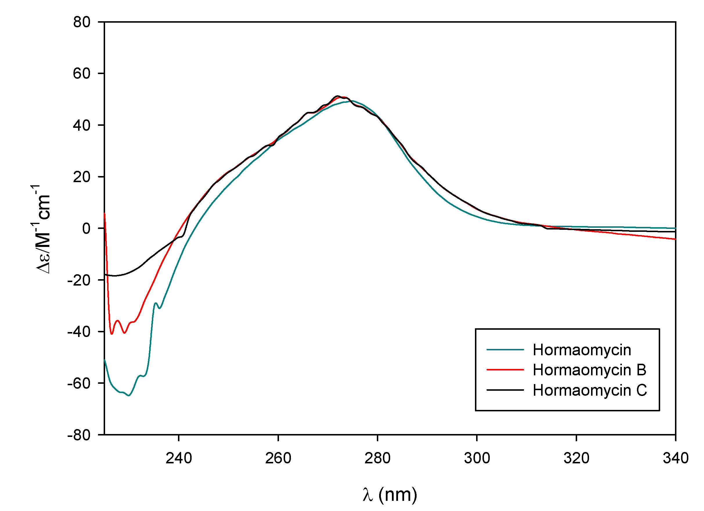
2. Results and Discussion

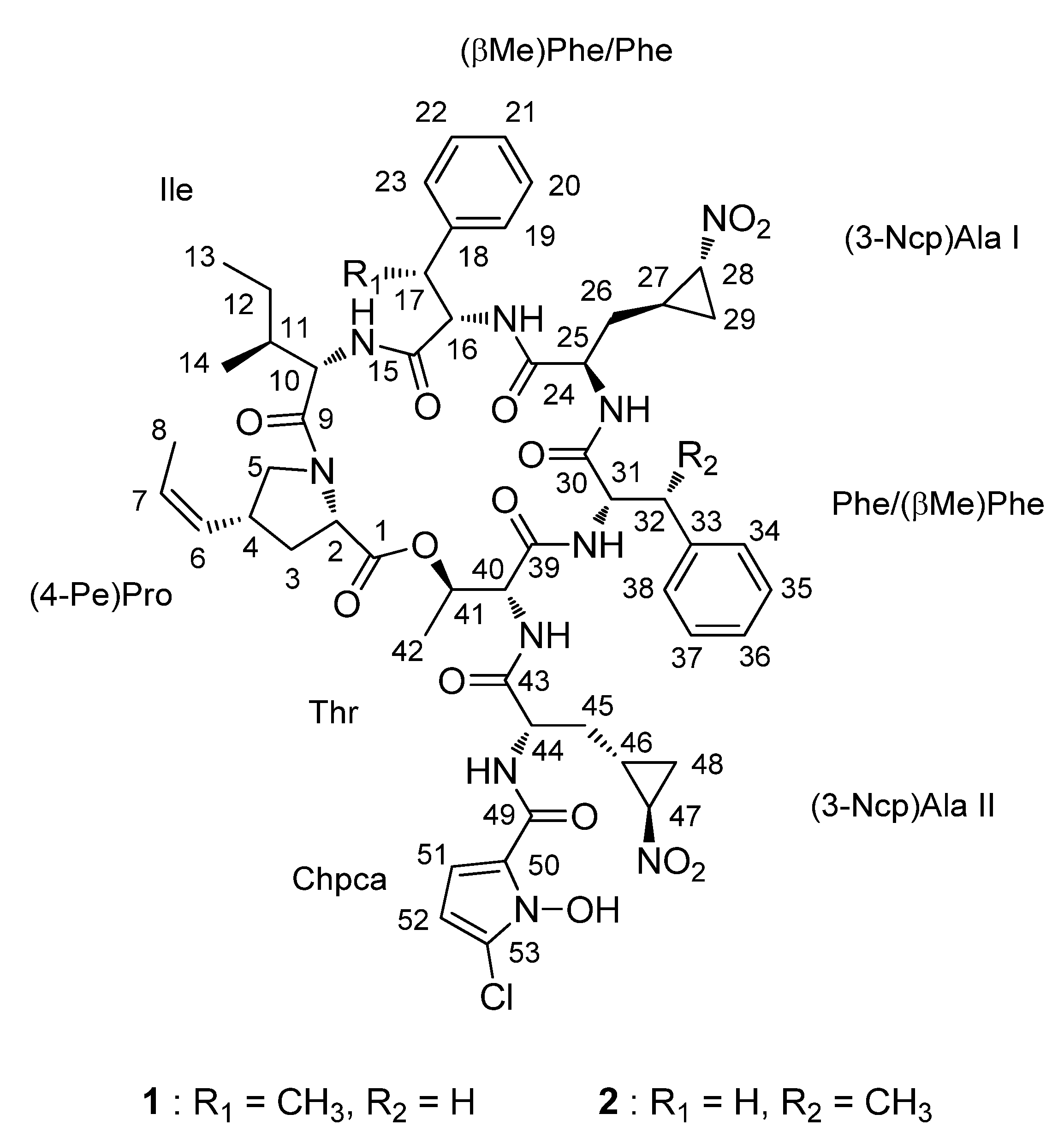
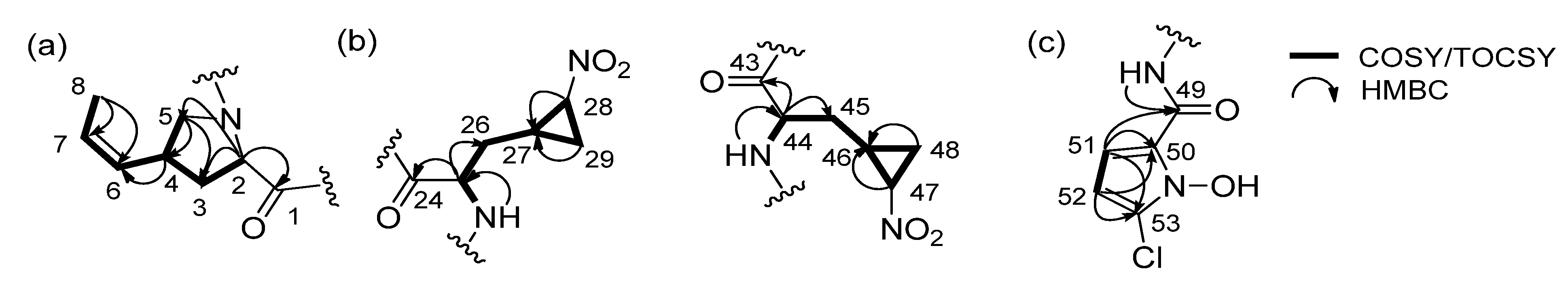
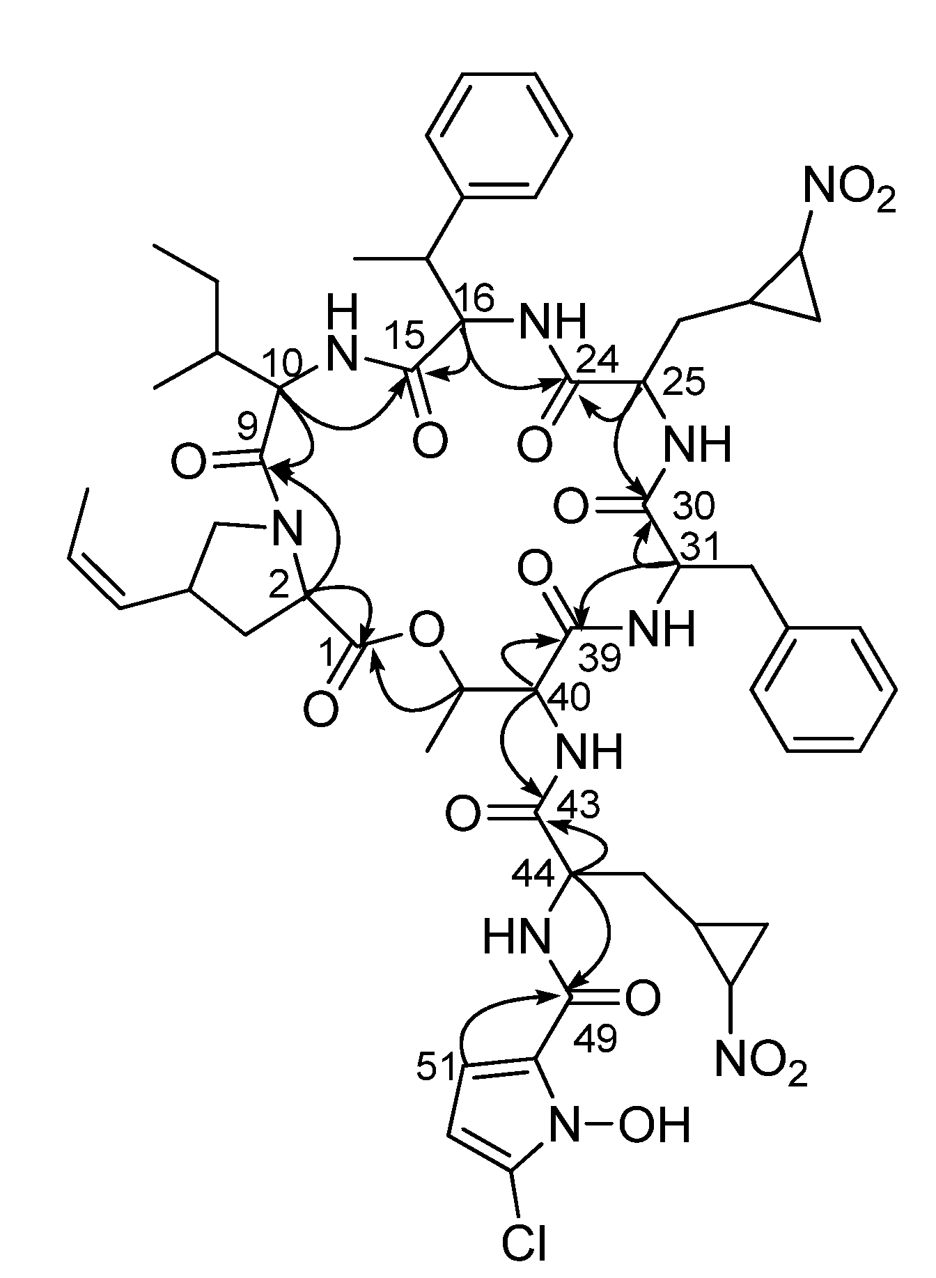
2.1. Structural Elucidation

Hormaomycin B (1) was isolated as a white powder with the molecular formula C54H67ClN10O14 on the basis of HRFABMS (obsd. [M + H]+ at m/z 1115.4611, calcd. for 1115.4605) coupled with 1H and 13C NMR spectroscopy (Table 1). The 1H spectrum of 1 in CDCl3 presented six amide protons at δH 8.98, 8.16, 7.31, 6.85, 6.79 and 6.26, indicating its peptide-derived nature. Consistently, the 13C NMR of 1 also showed seven amide/ester carbons at δC 172.2, 171.8, 171.5, 171.1, 169.3, 168.8 and 167.9. The analysis of 1H, 13C and HSQC NMR spectra clarified the peptide-derived features of hormaomycin B. As a peptide-derived molecule, hormaomycin B exhibited seven methine signals composing α-positions of amino acids (δHC 5.14/51.2, 3.52/52.2, 4.64/54.5, 4.55/58.4, 4.48/56.4, 4.33/60.8 and 4.26/61.7). Seven amide/ester carbons and seven α-position methines enabled us to deduce that hormaomycin B (1) bears seven or more amino acid units. Further analysis of one-bond correlations identified NMR signals that do not belong to ordinary amino acids units. In particular, two olefinic methines (δHC 5.64/128.8 and 5.26/127.8) and two methines (δHC 0.28/20.2 and −0.66/17.7) and one methylene (δHC 0.56 and −0.26/33.4) in the distinctive upfield suggested the existence of unusual units in the molecules. Additionally, two aromatic methines (δHC 6.68/109.8 and 6.08/103.5) with 4.5 Hz 1H-1H coupling indicated the existence of a five-membered aromatic ring, which does not correspond to any proteinogenic amino acid. Based on the overall analysis of the 1H, 13C NMR and HSQC spectra of 1, along with the existence of a chlorine atom in the molecular formula, hormaomycin B was expected to be a peptide-derived compound bearing several highly-modified amino acid residues.
Figure 1. The structures of hormaomycins B and C (1 and 2).
Marinedrugs 13 05187 g001
Further analysis of HSQC spectral data revealed all of the one-bond 1H-13C correlations in 1. Subsequent interpretation of the COSY, TOCSY and HMBC NMR spectra established eight partial structures dissected by amide bonds. These include a 4-(Z)-propenylproline ((4-Pe)Pro), a β-methylphenylalanine ((β-Me)Phe), two 3-(2-nitrocyclopropyl)alanines ((3-Ncp)Ala) and a 5-chloro-1-hydroxypyrrol-2-carboxylic acid (Chpca), as well as three common amino acids: an isoleucine (Ile), a phenylalanine (Phe) and a threonine (Thr).
Specifically, the COSY NMR spectrum exhibited correlations from H2-3 (δH 2.37, 1.80) to H-2 (δH 4.26) and H-4 (δH 3.27), showing the connectivity from C-2 (δC 61.7) to C-4 (δC 36.9) through C-3 (δC 34.9). Further COSY correlations of H-4 to H2-5 (δH 3.98, 3.31) and H-6 (δH 5.26) revealed that C-4 was located immediately adjacent to C-5 (δC 53.0) and C-6 (δC 127.8). The 1H-1H couplings from H-7 (δH 5.64) to H-6 and H-8 (δH 1.68) and HMBC correlations from H-8 to two olefinic carbons C-6 and C-7 (δC 128.8) established the propenyl substructure connected to C-4. The 1H-1H coupling constant (J = 9.5 Hz) between H-6 and H-7 determined the Z configuration of the C-6 double bond. The 13C NMR chemical shift of C-5 (δC 53.0) indicated that this carbon is bound to a nitrogen atom. Because there was no detected NH signal in this unit, a proline-type moiety was expected. The HMBC correlation from theα-proton (H-2; δH 4.26) to C-5 supported the hypothesis that H-2 and H-5 share one nitrogen to form a pyrrolidine. Furthermore, H-2 displayed an HMBC correlation to the C-1 carbonyl carbon (δC 171.8), completing a 4-(Z)-propenylproline ((4-Pe)Pro) unit (Figure 2a). This unit was further confirmed by TOCSY correlations from H-2 to H-8.
Figure 2. Determination of the unusual partial structures of 1. (a) 4-(Z)-propenylproline (b) 3-(2-nitrocyclopropyl)alanine I and II and (c) 5-chloro-1-hydroxypyrrol-2-carboxylic acid.
Marinedrugs 13 05187 g002
The next unusual unit was composed of distinctively-shielded protons of H2-26 (δH 0.56, −0.26), H-27 (δH 0.28) and H2-29 (δH 1.02, −0.66), along with 25-NH (δH 6.26), H-25 (δH 3.52) and H-28 (δH 2.92), based on COSY and TOCSY NMR spectra. In particular, strong COSY correlations among H-27, H-28 and H2-29 and their low chemical shifts revealed that this amino unit possesses a cyclopropane. Additionally, the 13C chemical shift (δC 58.7) of the C-28 methine in the cyclopropane indicated that this carbon is directly connected to a heteroatom. The IR spectrum exhibited a strong absorption band at 1543 cm−1, which corresponds to a nitro functional group. Therefore, C-28 was determined to bind to a nitro group. These assignments eventually elucidated a 3-(2-nitrocyclopropyl)alanine ((3-Ncp)Ala) unit (Figure 2b). Interestingly, the second (3-Ncp)Ala unit was also identified based on COSY and TOCSY correlations, even though the 1H chemical shifts of the second unit differ significantly from those of the first (3-Ncp)Ala residue (Figure 2b, Table 1).
Hormaomycin B (1) possesses eighteen sp2 carbons. Twelve of these were assigned in the two benzene rings in a phenylalanine and a β-methylphenylalanine, and two olefinic carbons were used in (4-Pe)Pro. Then, four sp2 carbons, including two methines, C-51 (δC 109.8) and C-52 (δC 103.5), and two quaternary carbons, C-50 (δC 119.8) and C-53 (δC 121.5), remained unassigned. These methine protons, H-51 (δH 6.68) and H-52 (δH 6.08), were coupled to each other (4.5 Hz). This coupling constant strongly suggested that these atoms are part of a five-membered aromatic ring. Furthermore, HMBC correlations from H-51 to C-50 and C-53 and from H-52 to C-50 and C-53 established the C-50-C-51-C-52-C-53 connectivity. The last position of the five-membered aromatic ring could be a nitrogen atom rather than an oxygen or sulfur atom based on the 13C chemical shifts of the four sp2 carbons, indicating a pyrrole ring [15]. This pyrrole ring is connected to C-49 (δC 159.7) at C-50 according to the HMBC correlation from H-51 to C-49. This downfield carbon at δC 159.7 was assigned as an amide carbonyl carbon based on the heteronuclear coupling from the amide proton 44-NH (δH 8.16) to this carbon. The remarkable shielding of C-49 (δC 159.7) in the 13C NMR spectrum compared to the chemical shifts of the other seven amide carbons (δC 172.2, 171.8, 171.5, 171.1, 169.3, 168.8 and 167.9) might be attributed to the conjugation effect of the pyrrole ring. Detailed examination of the mass and NMR data suggested that the quaternary carbon C-53 and the nitrogen atom in the pyrrole ring should be substituted with heteroatoms. Finally, partial structures dissected by amide bonds were established, and one chlorine atom and one hydroxy group in the molecular formula were not assigned. Considering the chemical shift of C-53 enabled a chlorine atom to be located at C-53. Subsequently, the hydroxy group was assigned to be bound to the nitrogen atom as an oxime functional group. Therefore, 5-chloro-1-hydroxypyrrol-2-carboxylic acid (Chpca) was eventually assembled (Figure 2c).
The sequence of eight partial structures ((4-Pe)Pro, Ile, Phe, (βMe)Phe, (3-Ncp)Ala I and II, Thr and Chpca) was determined on the basis of HMBC correlations. The long-range 1H-13C coupling from the α-proton of (4-Pe)Pro (H-2; δH 4.26) to the carbonyl carbon of Ile (C-9; δC 171.5) connected (4-Pe)Pro to Ile. The HMBC correlations from 10-NH (δH 7.31) and H-16 (δH 4.33) to C-15 (δC 167.9) clarified the attachment of (βMe)Phe to Ile. (3-Ncp)Ala I was directly connected to (βMe)Phe based on the HMBC correlations from 16-NH (δH 6.85) and H-25 (δH 3.52) to C-24 (δC 169.3), which belonged to (3-Ncp)Ala I. Phe was positioned next to (3-Ncp)Ala I by 1H-13C long-range coupling from NH-25 (δH 6.26) and H-31 (δH 4.48) to C-30 (δC 168.8). The HMBC correlations from 31-NH (δH 6.79) and H-40 (δH 4.55) to C-39 (δC 171.1) revealed the sequence from Phe to Thr. In addition, the NH proton (δH 8.98) of Thr and the α-proton (δH 5.14) of (3-Ncp)Ala II displayed HMBC signals linking them to the carbonyl carbon C-43 of (3-Ncp)Ala II, clarifying the connection between Thr and (3-Ncp)Ala II. Chpca was directly connected to (3-Ncp)Ala II on the basis of the HMBC correlations from 44-NH (δH 8.16) of (3-Ncp)Ala II and H-51 (δH 6.68) of Chpca to the carbonyl carbon C-49 (δC 159.7) belonging to Chpca. Finally the long-range HMBC correlation from H-41 (δH 5.40) to the carbonyl carbon at 171.8 ppm (C-1) closed a macrocyclic lactone ring, indicating the planar structure of hormaomycin B (1) in a cyclic depsipeptide (Figure 3).
Figure 3. Key HMBC correlations of hormaomycin B (1).
Marinedrugs 13 05187 g003
Hormaomycin C (2) was isolated as a white powder, and the molecular formula was deduced as C54H67ClN10O14 by analyzing HRFABMS (obsd. [M + Na]+ at m/z 1137.4432, calcd. [M + Na]+ 1137.4424) and 1H and 13C NMR data (Table 1). The molecular formula of 2 was identical to that of hormaomycin (1), and it was predicted that hormaomycin C (2) would be very similar to 1. Careful comparison of 1D and 2D NMR data revealed that all eight partial structures identified in hormaomycin B also exist in 2. However, the extensive analysis of the HMBC NMR spectra showed that the sequence of the units was different. Specifically, the methyl group (17-Me; δH 1.29) of (βMe)Phe and the methylene signal (H2-32; δH 2.79, 3.38) of Phe were replaced with a methylene (H2-17; δH 2.91, 2.97) and a methyl group (32-Me; δH 1.40) in 2, indicating the switched positions of (βMe)Phe and Phe between these compounds. The order of the other units was assigned based on further HMBC spectroscopic analysis, completing the planar structure of hormaomycin C (2).
During cultivation of the strain SNM55, hormaomycin, which was previously discovered from Streptomyces griseoflavus [14], was identified along with these new compounds. Hormaomycin is a cyclic depsipeptide that bears almost exclusively uncommon amino acids, such as (4-Pe)Pro and (3-Ncp)Ala, as well as Chpca. Because of its structural novelty and remarkable biological activity, chemical synthesis [16] and biosynthetic modifications [17] have been studied in recent decades. However, although artificial analogues have been reported during the total synthesis and biosynthetic engineering of hormaomycin [16,17], no natural analogues have been discovered. To our best knowledge, hormaomycins B and C are the first natural analogues of hormaomycin that have been obtained without chemical modification or gene cluster manipulation. Hormaomycin possesses two (βMe)Phe units, whereas both hormaomycins B and C bear one (βMe)Phe and one Phe. Therefore, hormaomycin was utilized to determine the absolute configurations of hormaomycins B and C (1 and 2). The 1H and 13C NMR data and CD spectrum of hormaomycin were carefully compared to those of 1 and 2. Based on the close similarity between the NMR data and CD spectra (Figure 4), as well as their common biosynthetic origin, the absolute configurations of hormaomycins B and C were determined to be identical to that of hormaomycin, which was determined by total synthesis [16].
Figure 4. The CD spectra of the hormaomycins in MeOH.
Marinedrugs 13 05187 g004
Table 1. 1H (600 MHz) and 13C NMR (150MHz) data for hormaomycins B and C (1 and 2) in CDCl3.
UnitC/H12
δH, mult (J in Hz)δCTypeδH, mult (J in Hz)δCType
(4-Pe)Pro1171.8C171.6C
24.26, 1H, m61.7CH4.26, 1H, dd (11.0, 6.5)61.8CH
32.37, 1H, m34.9CH22.36, 1H, m35.8CH2
1.80, 1H, m1.81, 1H, m
43.27, 1H, m36.9CH3.25, 1H, m37.0CH
53.98, 1H, m53.0CH23.98, 1H, m53.0CH2
3.31, 1H, m3.27, 1H, m
65.26, 1H, dd (9.5, 9.5)127.8CH5.26, 1H, dd (9.0, 9.0)127.9CH
75.64, 1H, dq (9.5, 7.0)128.8CH5.63, 1H, dq (9.0, 7.0)128.8CH
81.68, 1H, d (7.0)13.5CH31.66, 1H, d (7.0)13.5CH3
Ile9171.5C171.5C
104.64, 1H, m54.5CH4.64, 1H, m1q54.9CH
10-NH7.31, 1H, brs7.23, 1H, brs
111.88, 1H, m38.6CH1.90, 1H, m38.2CH
121.54, 1H, m25.0CH21.55, 1H, m25.0CH2
1.29, 1H, m1.30, 1H, m
130.90, 3H, d (7.0)10.8CH30.89, 3H, d (7.0)10.8CH3
141.04, 3H, d (7.0)15.2CH31.02, 3H, d (7.0)15.3CH3
(βMe)Phe/Phe15167.9C171.3C
164.33, 1H, m60.8CH4.58, 1H, m55.2CH
16-NH6.85, 1H, brs6.91, 1H, brs
173.02, 1H, m44.6CH2.91, 1H, m38.0CH2
17-Me1.29, 3H, d (6.5)18.0CH32.97, 1H, m
18141.9C136.5C
197.11, 1H, d (8.5)129.0CH7.11, 1H, d (8.0)129.5CH
207.13, 1H, t (8.5)127.5CH7.15, 1H, t (8.0)129.0CH
217.03, 1H, t (8.5)127.6CH7.09, 1H, t (8.0)127.7CH
227.13, 1H, t (8.5)127.5CH7.15, 1H, t (8.0)129.0CH
237.11, 1H, d (8.5)129.0CH7.11, 1H, d (8.0)129.5CH
(3-Ncp)Ala I24169.3C168.2C
253.52, 1H, m52.2CH3.55, 1H, m52.8CH
25-NH6.26, 1H, brs6.37, 1H, brs
260.56, 1H, m33.4CH20.78, 1H, m33.0CH2
−0.26, 1H, m0.13, 1H, m
270.28, 1H, m20.2CH0.58, 1H, m20.4CH
282.92, 1H, m58.7CH3.04, 1H, m58.6CH
29−0.66, 1H, m17.7CH2−0.34, 1H, m17.4CH2
1.02, 1H, m1.13, 1H, m
Phe/(βMe)Phe30168.8C168.3C
314.48, 1H, m56.4CH4.48, 1H, m60.6CH
31-NH6.79, 1H, brs6.79, 1H, brs
322.79, 1H, m38.7CH23.67, 1H, m37.1CH
3.38, 1H, m1.40, 3H, d (6.5)13.7CH3
33137.3C142.6C
347.22, 1H, m129.3CH7.24, 1H, m127.9CH
357.23, 1H, m127.8CH7.25, 1H, m128.8CH
367.15, 1H, t (8.5)127.2CH7.17, 1H, t (8.5)127.3CH
377.23, 1H, m127.8CH7.25, 1H, m128.8CH
387.22, 1H, m129.3CH7.24, 1H, m127.9CH
Thr39171.1C171.1C
404.55, 1H, m58.4CH4.52, 1H, m55.8CH
40-NH8.98, 1H, brs8.95, 1H, brs
415.40, 1H, m69.4CH5.38, 1H, m69.6CH
421.52, 3H, m17.2CH31.48, 3H, m17.3CH3
(3-Ncp)Ala II43172.2172.1
445.14, 1H, m51.2CH5.09, 1H, m51.5CH
44-NH8.16, 1H, brs7.96, 1H, brs
451.80, 1H, m35.5CH21.83, 1H, m35.2CH2
1.60, 1H, m1.61, 1H, m
461.93, 1H, m21.7CH1.91, 1H, m21.7CH
474.04, 1H, m59.8CH4.05, 1H, m59.6CH
481.95, 1H, m17.8CH21.95, 1H, m17.9CH2
1.02, 1H, m1.01, 1H, m
Chpca49159.7C160.1C
50119.8C119.8C
516.68, 1H, d (4.5)109.8CH6.81, 1H, d (4.5)109.8CH
526.08, 1H, d (4.5)103.5CH6.13, 1H, d (4.5)104.0CH
53121.5C120.5C
NOH10.8, 1H, brs10.8, 1H, brs

2.2. Bioactivities of the Hormaomycins

Hormaomycin was previously reported to exhibit remarkable antibacterial effects [18]. Therefore, the biological activities of the hormaomycins found here were evaluated with regard to their antimicrobial activities against various pathogenic bacterial strains, including Staphylococcus aureus ATCC 25923, Bacillus subtilis ATCC 6633, Kocuria rhizophila NBRC 12708, Streptococcus pyogenes ATCC 19615, Klebsiella pneumoniae ATCC10031, Salmonella enterica ATCC 14028, Proteus hauseri NBRC 3851 and Escherichia coli ATCC 25922 (Table 2). Hormaomycin exhibited more potent antibacterial activities against the tested Gram-positive bacteria than against the tested Gram-negative bacteria, particularly against S. aureus and K. rhizophila, exhibiting MIC values of 0.4 μM and 0.03 μM, respectively. Hormaomycin also exhibited significant antibacterial effects against Gram-negative P. hauseri. Hormaomycins B and C (1 and 2) displayed potent activity against K. rhizophila, but showed antibacterial activities against the tested bacteria that were generally 4–32-times weaker than those of hormaomycin. Accordingly, it was hypothesized that the existence of the methyl groups at C-17 and C-32, which form (βMe)Phe, play an important role in the antibacterial potency of the hormaomycins. Further investigations of the antifungal activity were conducted against the pathogenic fungi Aspergillus fumigatus, Trichophyton rubrum, T. mentagrophytes and Candida albicans, but no significant inhibitory activity was observed to result from the hormaomycin treatments (MIC > 100 μM).
Table 2. Antibacterial activity data of the hormaomycins.
CompoundMICs (μM)
Gram-PositiveGram-Negative
S. aureusB. subtilisK. rhizophilaS. pyogenesK. pneumoniaeS. entericaP. hauseriE. coli
Hormaomycin0.41.80.031.8>113>1130.9>113
Hormaomycin B7140.414>1152929>115
Hormaomycin C7560.238>11411414>114
Ampicillin<0.170.17<0.170.1745.93.7<0.1711.5

3. Experimental Section

3.1. General Experimental Procedures

Optical rotations were measured using a JASCO P-200 polarimeter with a 1-cm cell. IR spectra were obtained using a Thermo NICOLET iS10 spectrometer (sodium light source, JASCO, Easton, PA, USA). UV spectra were acquired using a Perkin Elmer Lambda 35 UV/VIS spectrometer (Perkin Elmer, Waltham, MA, USA). CD spectra were collected with an Applied Photophysics Chirascan-Plus (Applied Photophysics, Leatherhead Surrey, UK) with a 2-mm cell. Electrospray ionization (ESI) low-resolution LC/MS data were recorded on an Agilent Technologies 6130 Quadrupole mass spectrometer connected to an Agilent Technologies 1200 series high-performance liquid chromatography (HPLC) instrument (Agilent Technologies, Santa Clara, CA, USA). High-resolution fast atom bombardment (HR-FAB) mass spectra were recorded using a JEOL JMS-600W high-resolution mass spectrometer (Jeol, München, Germany) at the National Center for Inter-university Research Facilities at Seoul National University (NCIRF). 1H, 13C and 2D NMR spectra (See Supplementary Information Figures S1–S18) were recorded on Bruker Avance 600 MHz spectrometers (Bruker, Billerica, MA, USA) at the NCIRF.

3.2. Isolation, Cultivation and Extraction of Bacteria

A sediment sample was collected from the Mohang mudflat in Buan, Korea. The sample was dried at room temperature for 3 h. The dry sediment (1 g) was diluted in 4 mL of sterilized artificial seawater. The mixture was spread on actinomycete isolation agar, A4 medium (1 L of seawater, 18 g of agar and 100 mg/L cycloheximide) and A5 medium (750 mL of seawater, 250 mL of distilled water, 18 g of agar and 100 mg/L cycloheximide) by stamping and spreading. The single strain SNM55 was isolated on A5 medium. The strain SNM55 (GenBank Accession No. KP133063) was phylogenetically identified as a Streptomyces sp. (most closely related to Streptomyces javenis by 99% identity) based on the 16S rDNA sequence analysis. The bacterium was incubated in 50 mL of YEME medium (4 g of yeast extract, 10 g of malt extract and 4 g of glucose in 1 L of artificial seawater) in a 125-mL Erlenmeyer flask for 3 days on a rotary shaker at 160 rpm and 30 °C. Then, 10 mL of the culture were inoculated into 200 mL of YEME medium in a 500-mL Erlenmeyer flask. The strain SNM55 was further cultivated for 2 days, and 20 mL of the liquid culture were transferred to 1 L of YEME liquid medium in a 2.8-L Fernbach flask (12 ea × 1 L, total volume of 12 L). After incubating the bacterial culture for 10 days, the entire culture (12 L) was extracted with 16 L of ethyl acetate. The ethyl acetate layer was collected, and residual water in the organic layer was removed by adding anhydrous sodium sulfate. The extract was concentrated in vacuo. The entire procedure was repeated 6 times (72 L culture in total) to yield 4 g of dry material for chemical study and bioassays.

3.3. Isolation of the Hormaomycins

The extract of SNM55 (one sixth of the 4 g of dry material collected) was absorbed on Celite and loaded on a 2g Sep-Pak C18 cartridge. Then, the extract was fractionated with 20 mL each of 20%, 40%, 60%, 80% and 100% MeOH in water and 1:1 MeOH/dichloromethane. The hormaomycins were detected in the 80% and 100% MeOH/water fractions by LC/MS. The 80% and 100% fractions were combined into one vial and subjected to semi-preparative reversed-phase HPLC (Kromasil C18 (2): 250 × 10 mm, 5 μm) using a gradient solvent system (65% MeOH/H2O to 90% MeOH/H2O over 40 min and 100% MeOH from 40 min to 50 min, UV 280 nm detection, flow rate: 2mL/min). Three fractions containing the hormaomycins were collected at retention times of 43 min, 45 min and 52 min. Each of the hormaomycins was purified by an isocratic solvent system (75% acetonitrile/H2O, UV 280 nm detection, flow rate: 2mL/min) using a reversed-phase C18 column (Kromasil C18 (2): 250 × 10 mm, 5 μm). Hormaomycin B (1) (4 mg), hormaomycin C (2) (3 mg) and hormaomycin (6 mg) eluted as pure compounds at retention times of 14.5 min, 15.2 min and 18.4 min, respectively, under the final purification conditions.
Hormaomycin B (1): [α]D 21.9, (c 0.025, MeOH); UV (MeOH) λmax (log ε) 208 (4.57), 271 (4.08) nm; IR (neat) νmax 3309, 2958, 1626, 1543, 1369 cm−1; for 1H and 13C NMR data, see Table 1; HRFABMS m/z 1115.4611 [M + H]+ (calcd. for C54H68ClN10O14 1115.4605).
Hormaomycin C (2): [α]D 29.9, (c 0.025, MeOH); UV (MeOH) λmax (log ε) 208 (4.57), 271 (4.09) nm; IR (neat) νmax 3343, 2931, 1627, 1554, 1362 cm−1; for 1H and 13C NMR data, see Table 1; HRFABMS m/z 1137.4432 [M + Na]+ (calcd. for C54H67ClN10O14Na 1137.4424).

3.4. Antibacterial Activity Assay

Gram-positive bacteria (S. aureus ATCC 25923, B. subtilis ATCC 6633, S. pyogenes ATCC 19615 and K. rhizophila NBRC 12708) and Gram-negative bacteria (K. pneumoniae ATCC10031, S. enterica ATCC 14028, E. coli ATCC 25922 and P. hauseri NBRC 3851) were used for antimicrobial activity tests. Bacteria were grown overnight in Mueller Hinton (MH) broth at 37 °C, harvested by centrifugation and washed twice with sterile distilled water. Stock solutions of the hormaomycins were prepared in DMSO. Each stock solution was diluted with MH broth (5% lysed sheep blood for S. pyogenes) to give serial 2-fold dilutions in the range of 128 to 0.06 μg/mL. Aliquots (10 μL) of the broth containing approximately 5 ×10⁵ colony-forming units (cfu)/mL of the bacteria were added to each well of a 96-well microtiter plate. The plates were incubated for 24 h at 37 °C. The minimum inhibitory concentration (MIC) values were determined as the lowest concentration of test compound that inhibited bacterial growth. Ampicillin was used as a reference compound.

3.5. Antifungal Activity Assay

Potato dextrose agar (PDA) was used to cultivate C. albicans ATCC 10231. After incubation for 48 h at 28 °C, yeast cells were harvested by centrifugation and washed twice with sterile distilled water. A. fumigatus HIC 6094, T. rubrum NBRC 9185 and T. mentagrophytes IFM 40996 were plated on PDA and incubated for 2 weeks at 28 °C. Spores were harvested and washed twice with sterile distilled water. Fungal cells were resuspended in RPMI 1640 broth (Difco) to obtain an initial inoculum size of 105 spores/mL. In each well of a 96-well plate, 90 μL of cells (104 cells/mL) were mixed with the test compound solutions (hormaomycin and hormaomycins B and C) in 5% DMSO. A culture with DMSO (0.5%) was used as a solvent control, and a culture supplemented with amphotericin B was used as a positive control.

4. Conclusions

As one approach to maximize microbial chemical diversity, altering the culture conditions of a chemically-prolific marine actinomycete strain (SNM55) led us to discover hormaomycins B and C (1 and 2). These hormaomycins are structurally-unique cyclic depsipeptides that incorporate various unusual units, such as 4-(Z)-propenylproline, 3-(2-nitrocyclopropyl)alanine, β-methylphenylalanine and 5-chloro-1-hydroxypyrrol-2-carboxylic acid. The hormaomycins inhibited various pathogenic Gram-positive and Gram-negative bacteria. To the best of our knowledge, hormaomycins B and C are the first natural analogues of hormaomycin. The discovery of these new members of the hormaomycin family provides additional evidence that marine actinomycetes possess the potential to produce still-untapped bioactive secondary metabolites, which can be synthesized through diverse biosynthetic pathways under appropriate culture conditions.

Supplementary Files

Supplementary File 1

Acknowledgments

This work was supported by the National Research Foundation of Korea Grants funded by the Korean Government (Ministry of Science, ICT and Future Planning) (2014R1A2A1A11053477) and the HHMI International Early Career Scientist Program.

Author Contributions

Munhyung Bae, Jongheon Shin and Dong-Chan Oh designed all of the experiments and wrote the manuscript. Munhyung Bae also performed the experiments and collected spectroscopic data. Beomkoo Chung and Ki-Bong Oh contributed to performing antimicrobial tests.

Conflicts of Interest

The authors declare no conflicts of interest.

References

  1. Clardy, J.; Walsh, C. Lessons from natural molecules. Nature 2004, 432, 829–837. [Google Scholar] [CrossRef] [PubMed]
  2. Koehn, F.E.; Carter, G.T. The evolving role of natural products in drug discovery. Nat. Rev. Drug Discov. 2005, 4, 206–220. [Google Scholar] [CrossRef] [PubMed]
  3. Fenical, W.; Jensen, P.R. Developing a new resource for drug discovery: Marine actinomycete bacteria. Nat. Chem. Biol. 2006, 2, 666–673. [Google Scholar] [CrossRef] [PubMed]
  4. Hu, G.-P.; Yuan, J.; Sun, L.; She, Z.-G.; Wu, J.-H.; Lan, X.-J.; Zhu, X.; Lin, Y.-C.; Chen, S.-P. Statistical research on marine natural products based on data obtained between 1985 and 2008. Mar. Drugs 2011, 9, 514–525. [Google Scholar] [CrossRef] [PubMed]
  5. Arias, C.A.; Murray, B.E. A new antibiotic and the evolution of resistance. N. Engl. J. Med. 2015, 372, 1168–1170. [Google Scholar] [PubMed]
  6. Feling, R.H.; Buchanan, G.O.; Mincer, T.J.; Kauffman, C.A.; Jensen, P.R.; Fenical, W. Salinosporamide A: A highly cytotoxic proteasome inhibitor from a novel microbial source, a marine bacterium of the new genus Salinospora. Angew. Chem.-Int. Edit. 2003, 42, 355–357. [Google Scholar] [CrossRef] [PubMed]
  7. Perez Baz, J.; Canedo, L.M.; Fernandez Puentes, J.L.; Silva Elipe, M.V. Thiocoraline, a novel depsipeptide with antitumor activity produced by a marine Micromonospora. II. Physico-chemical properties and structure determination. J. Antibiot. 1997, 50, 738–741. [Google Scholar]
  8. Kim, S.-H.; Shin, Y.; Lee, S.-H.; Oh, K.-B.; Lee, S.K.; Shin, J.; Oh, D.-C. Salternamides A−D from a halophilic Streptomyces sp. actinobacterium. J. Nat. Prod. 2015, 78, 836–843. [Google Scholar] [CrossRef] [PubMed]
  9. Moon, K.; Ahn, C.-H.; Shin, Y.; Won, T.H.; Ko, K.; Lee, S.K.; Oh, K.-B.; Shin, J.; Nam, S.-I.; Oh, D.-C. New benzoxazine secondary metabolites from an Arctic actinomycete. Mar. Drugs 2014, 12, 2526–2538. [Google Scholar] [CrossRef] [PubMed]
  10. Moon, K.; Chung, B.; Shin, Y.; Rheingold, A.L.; Moore, C.E.; Park, S.J.; Park, S.; Lee, S.K.; Oh, K.-B.; Shin, J.; et al. Pentacyclic antibiotics from a tidal mud flat-derived actinomycete. J. Nat. Prod. 2015, 78, 524–529. [Google Scholar] [CrossRef] [PubMed]
  11. Zhu, H.; Sandiford, S. K.; van Wezel, G.P. Triggers and cues that activate antibiotic production by actinomycetes. J. Ind. Microbiol. Biotechnol. 2014, 41, 371–386. [Google Scholar] [CrossRef] [PubMed]
  12. Wang, W.-J.; Li, D.-Y.; Li, Y.-C.; Hua, H.-M.; Ma, E.-L.; Li, Z.-L. Caryophyllene sesquiterpenes from the marine-derived fungus Ascotricha sp. ZJ-M-5 by the one strain-many compounds strategy. J. Nat. Prod. 2014, 77, 1367–1371. [Google Scholar] [CrossRef] [PubMed]
  13. Bae, M.; Kim, H.; Moon, K.; Nam, S.-J.; Shin, J.; Oh, K.-B.; Oh, D.-C. Mohangamides A and B, new dilactone-tethered pseudo-dimeric peptides inhibiting Candida albicans isocitrate lyase. Org. Lett. 2015, 17, 712–715. [Google Scholar] [CrossRef] [PubMed]
  14. Roessner, E.; Zeeck, A.; Koenig, W.A. Structure determination of hormaomycin. Angew. Chem. 1990, 102, 84–85. [Google Scholar]
  15. Pretsch, E.; Bȕhlmann, P.; Affolter, C. Structure Determination of Organic Compounds-Tables of Spectral Data; Springer: New York, NY, USA, 2000; p. 104. [Google Scholar]
  16. Zlatopolskiy, B.D.; de Meijere, A. First total synthesis of hormaomycin, a naturally occurring depsipeptide with interesting biological activities. Chem. Eur. J. 2004, 10, 4718–4727. [Google Scholar] [CrossRef] [PubMed]
  17. Cai, X.; Teta, R.; Kohlhaas, C.; Cruesemann, M.; Ueoka, R.; Mangoni, A.; Freeman, M.F.; Piel, J. Manipulation of regulatory genes reveals complexity and fidelity in hormaomycin biosynthesis. Chem. Biol. 2013, 20, 839–846. [Google Scholar] [CrossRef] [PubMed]
  18. Andres, N.; Wolf, H.; Zähner, H. Hormaomycin, a new peptide lactone antibiotic effective in inducing cytodifferentiation and antibiotic biosynthesis in some Streptomyces species. Z. Naturforsch. 1990, 45, 851–855. [Google Scholar]

