Article

Anti-Mycobacterial Nucleoside Antibiotics from a Marine-Derived Streptomyces sp. TPU1236A

Faculty of Pharmaceutical Sciences, Tohoku Pharmaceutical University, Aoba-ku, Sendai 981-8558, Japan
*
Author to whom correspondence should be addressed.
Mar. Drugs 2014, 12(12), 6102-6112; https://doi.org/10.3390/md12126102
Submission received: 30 October 2014 / Revised: 9 December 2014 / Accepted: 10 December 2014 / Published: 17 December 2014

Abstract

:
Five new nucleoside antibiotics, named streptcytosines A–E (15), and six known compounds, de-amosaminyl-cytosamine (6), plicacetin (7), bamicetin (8), amicetin (9), collismycin B (10), and SF2738 C (11), were isolated from a culture broth of Streptomyces sp. TPU1236A collected in Okinawa, Japan. The structures of new compounds were elucidated on the basis of their spectroscopic data (HRFABMS, IR, UV, and 2D NMR experiments including 1H-1H COSY, HMQC, HMBC, and NOESY spectra). Streptcytosine A (1) belonged to the amicetin group antibiotics, and streptcytosines B–E (25) were derivatives of de-amosaminyl-cytosamine (6), 2,3,6-trideoxyglucopyranosyl cytosine. Compound 1 inhibited the growth of Mycobacterium smegmatis (MIC = 32 µg/mL), while compounds 25 were not active at 50 µg/disc. Bamicetin (8) and amicetin (9) showed the MICs of 16 and 8 µg/mL, respectively.

Graphical Abstract

1. Introduction

Tuberculosis (TB) caused by Mycobacterium tuberculosis is still one of the main infectious diseases all over the world including with the human immunodeficiency virus (HIV) and malaria [1,2,3]. It was estimated that there were about 9 million clinical cases, 1.3 million deaths from TB, and 0.3 million deaths from HIV-co-infected TB in 2012 [4]. The development of multidrug-resistant TB (MDR-TB) has been observed as resistance against potent first-line drugs (rifampicin and isoniazid), and 450,000 patients were revealed to be infected by MDR-TB in 2012. Moreover, approximately 9.6% of these drug-resistant strains of T. tuberculosis were extensive drug-resistant TB (XDR-TB), which additionally shows resistance to some second-line drugs such as fluoroquinones, amikacin, kanamycin, and capreomycin. More recently, totally drug resistant TB (TDR-TB), which is resistant to all second-line drugs, has often been reported in India and other Asian countries [1,2,3,4,5]. Although a new drug, delmanid (Deltyba®), which inhibits the cell wall biosynthesis of mycobacteria, was approved in 2014 for the treatment of MDR-TB in EU and Japan [6,7], continuous efforts to discover new anti-TB agents with novel mechanisms of action and structural features are the emergent global demand.
In the course of our studies on anti-TB metabolites from marine invertebrates and microorganisms, we have tested the culture broths of 50 marine-derived actinomycetes against Mycobacterium smegmatis NBRC 3207 and found that Streptomyces sp. strain TPU 1236A exhibited prominent activity. M. smegmatis has been used most widely in the search for anti-mycobacterial substances due to its fast-growing and non-pathogenic properties [8]. Bioassay-guided isolation from the culture broth of strain TPU1236A yielded five new compounds, designated as streptcytosines A–E (15) (Figure 1), together with six known compounds, de-amosaminyl-cytosamine (6) [9], plicacetin (7) [10], bamicetin (8) [11,12,13], amicetin (9) [9,11,12,13], collismycin B (10) [14,15], and SF2738 C (11) [15] (Figure 2). The isolation and anti-mycobacterial activities of compounds 111 have been described in this study.
Figure 1. Structures of new compounds 15 isolated from Streptomyces sp. TPU1236A.
Marinedrugs 12 06102 g001
Figure 2. Structures of known compounds 611 isolated from Streptomyces sp. TPU1236A.
Marinedrugs 12 06102 g002

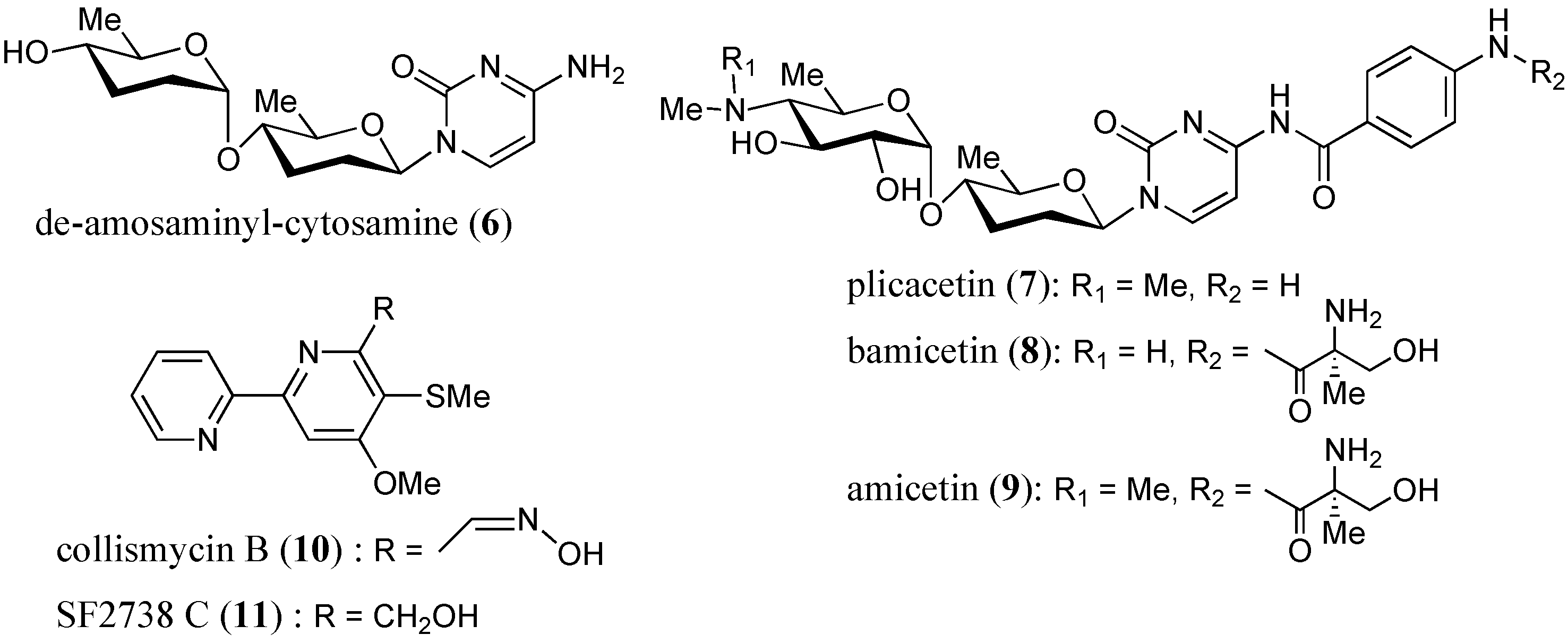
2. Results and Discussion

The strain TPU1236A was isolated from a seawater sample collected at Iriomote Island in Okinawa, Japan. The partial sequence of the 16S rRNA gene used for identification showed 100% identity with those of Streptomyces badius and Streptomyces sindenensis. The bacterium was cultured by shaking in a liquid medium for 7 days at 25 °C in the dark.
The extracts of broth filtrates and mycelia were subjected to ODS silica gel column chromatography followed by preparative HPLC separation, as described in the Experimental Section, to afford compounds 111 (Figure 1 and Figure 2). Six known compounds were identified by their structures by comparing the spectroscopic data for 611 with those of the reported values for de-amosaminyl-cytosamine (6), plicacetin (7), bamicetin (8), amicetin (9), collismycin B (10), and SF2738 C (11), respectively [9,10,11,12,13,14,15,16,17].

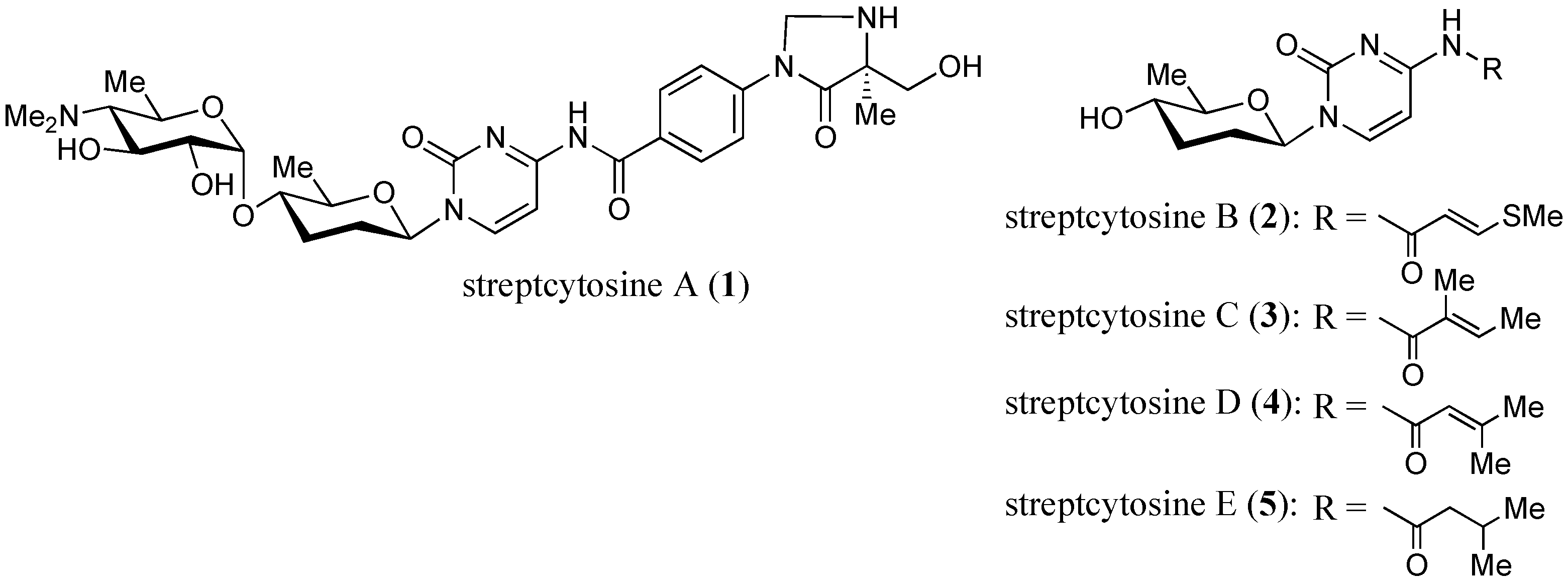
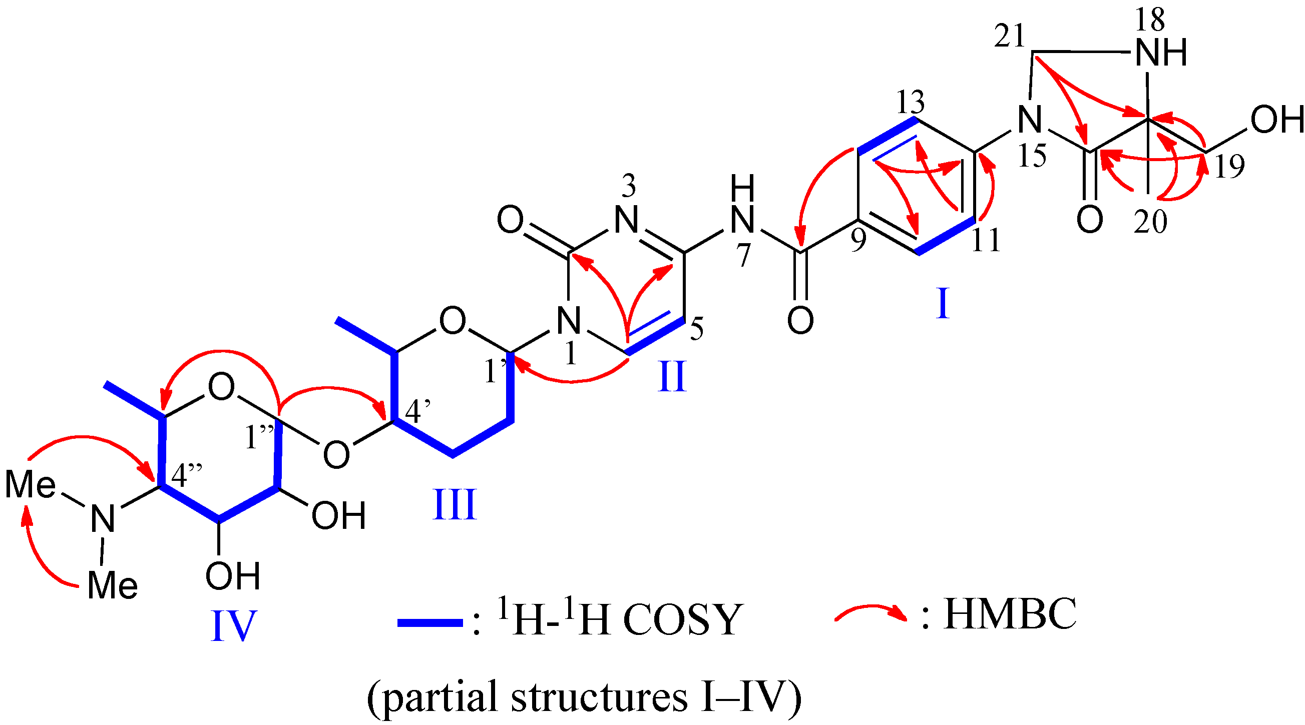
2.1. Structure of Streptcytosine A (1)

Compound 1 was obtained as a colorless oil. Its molecular weight and formula (630, C30H42N6O9) were determined from HRFABMS and NMR data. 1H and 13C NMR signals were assigned by an analysis of 1H-1H COSY, HMQC, and HMBC data (Table 1). Partial structures I–IV were established from 1H-1H COSY data, and connectivity between these partial structures were elucidated by the HMBC correlations (Figure 3).
The chemical shifts and couplings of two sugar moieties (amosamine and amicetose units) and two aromatic rings (cytosine and PABA units) were very similar to those of amicetin (9) [9]. Differences in the molecular formulas and weights of 1 and 9 were only one carbon atom and 12 Da, which was detected at δ 61.1 in the 13C NMR spectrum of 1. The protons attached to this carbon were observed as a broad singlet (2H) at δ 5.28, which showed HMBC correlations to C-16 and 17 (Figure 3). HMBC correlations from H3-19 to C-16, 17, and 20 and from H2-20 to C-16 and 17 along with the above data allowed to assign the partial structure of 4-imidazolidinone. Consequently, the skeletal structure of compound 1 was assigned as Figure 3 and named streptcytosine A.
Figure 3. 1H-1H COSY and key HMBC correlations for compound 1.
Marinedrugs 12 06102 g003
Table 1. 13C (100 MHz) and 1H (400 MHz) NMR data for streptcytosine A (1) (CD3OD).
C#δCδH, mult. (J in Hz)HMBC
2157.4
4165.0
599.17.58, d, (7.5)6
6146.78.21, d, (7.5)2, 4, 5, 1′
8168.4
9131.6
10130.88.10, d, (8.8)8, 11, 12, 14
11120.67.87, d, (8.8)10, 13
12142.0
13120.67.87, d, (8.8)11, 14
14130.88.10, d, (8.8)8, 10, 12, 13
16172.2
1768.3
1966.1(a) 3.83, d, (11.6)16, 17
(b) 4.03. d, (11.6)16
2017.61.57, s16, 17, 19
2161.15.28, br s16, 17
1′84.95.79, d, (7.8)
2′31.1(a) 2.19, br d, (9.0)4′
(b) 1.71, m4′
3′28.1(a) 1.71, m1′
(b) 2.41, m
4′76.73.46, m
5′78.43.77, dq, (9.0, 6.0)4′
6′19.31.39, d, (6.0)4′, 5′
1″96.85.03, d, (3.6)4′, 3″, 5″
2″74.03.56, dd, (9.1, 3.6)3″
3″68.13.98, dd, (11.0, 9.1)2″, 4″
4″72.13.12, dd (11.0, 10.0)3″, 5″, 6″, 7″, 8″
5″64.24.10, dq, (10.0, 6.2)
6″19.11.47, d, (6.2)4″, 5″
7″42.63.01, s4″, 8″
8″42.63.01, s4″, 7″
Since streptcytosine A (1) was obtained together with plicacetin (7), bamicetin (8), and amicetin (9) from the strain TPU1236A, the absolute configuration of the sugar moiety will be the same. The configuration of 1 at the C-17 position was also assigned to be the same as those of 8 and 9, since these compounds should be biosynthesized by the same pathway. Moreover, compounds 1 (+72), 7 (+96), 8 (+66), and 9 (+76) showed the same positive specific rotation. Therefore, streptcytosine A (1) may share the same configuration for whole asymmetric carbons as compounds 8 and 9, as depicted in Figure 1 and Figure 2.

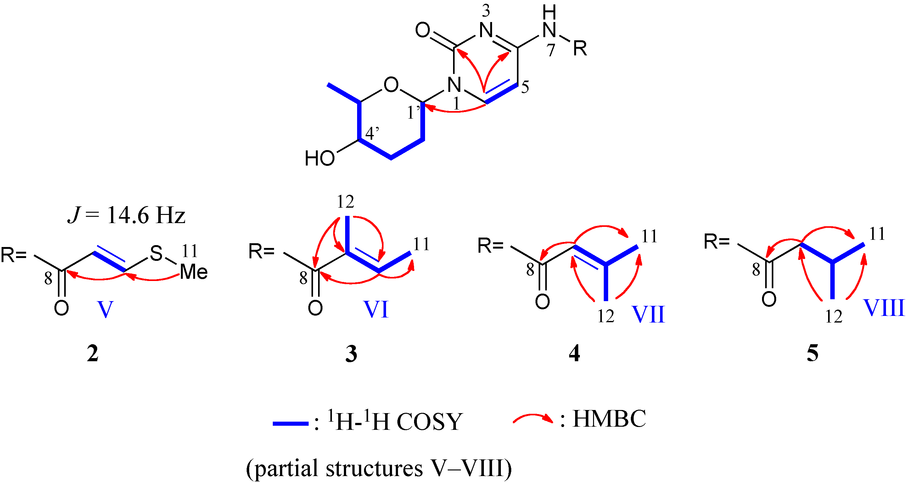
2.2. Structures of Streptcytosines B–E (25)

The molecular formulas and weights of compounds 2 (C14H19N3O4S, 325), 3 (C15H21N3O4, 307), 4 (C15H21N3O4, 307), and 5 (C15H23N3O4, 309) were determined from the HRFABMS and NMR data (Table 2). The presence of amicetose and cytosine moieties in 25 was assigned by the analysis of 1H-1H COSY and HMBC spectra (Figure 4). The 1H and 13C NMR signals due to two moieties in 25 were very similar to those in de-amosaminyl-cytosamine (6) [9]. Therefore, compounds 25 had the same amicetosylcytosine unit, and differences were observed in the amide moieties.
Streptcytosine B (2) had an S-Me group and 2-butenoyl unit. HMBC correlations from H3-11 to C-10, H-10 to C-8, and from H-9 to C-8 revealed the partial structure V as the amide moiety in 2. The orientation of the double bond was assigned as E from the coupling constant (14.6 Hz) between H-9 and 10.
The 2-methyl-2-butenoyl unit (partial structure VI) in streptcytosine C (3) was elucidated from 1H-1H COSY and HMBC data (Figure 4). An NOE correlation was observed between H-10 and H3-11, but no cross peak was detected between H-10 and H3-12. These observations on NOEs were the same as cytosaminomycin D, which has a tiglic acid (2-methyl-2E-butenoic acid) moiety attached to the PABA unit by an amide bond [18]. Moreover, chemical shifts due to C-11 (δ 14.7) and 12 (12.2) of 3 resembled those of cytosaminomycin D (14.5 and 12.2), which was compared the chemical shifts with those of tiglic acid (13.83 and 10.95) and angelic acid (15.93 and 20.22, 2-methyl-2Z-butenoic acid). Thus, the orientation of the double bond was assigned as E.
Streptcytosine D (4) had a 3-methyl-2-butenoyl moiety (partial structure VII), which was established from 1H-1H COSY and HMBC data as shown in Figure 4.
The 3-methylbutanoyl moiety (partial structure VIII) in streptcytosine E (5) was also assigned from 1H-1H COSY and HMBC correlations (Figure 4).
Figure 4. 1H-1H COSY and key HMBC correlations for compounds 25.
Marinedrugs 12 06102 g004
Table 2. 13C (100 MHz) and 1H (400 MHz) NMR data for streptcytosines B–E (25) (CD3OD).
2345
C#δCδH, mult. (J in Hz)δCδH, mult. (J in Hz)δCδH, mult. (J in Hz)δCδH, mult. (J in Hz)
2156.6 155.5 154.2 157.8
4164.6 164.2 164.3 164.8
598.47.34, d (7.5)98.37.30, d (7.6)98.27.26, d (7.5)99.27.47, br d (7.6)
6146.88.19, d (7.5)147.58.23, d (7.6)147.08.21, d (7.5)146.78.12, d (7.6)
8166.2 171.3 168.2 176.5
9115.76.07, d (14.6)133.5 118.75.95, qq (1.3, 1.3)47.32.32, d (7.2)
10150.78.01, d (14.6)137.46.71, q (7.0)161.3 27.12.10, m
1114.72.42, s14.71.89, d (7.0)28.01.99, d (1.2)22.70.99, d (6.7)
12––––12.21.90, s20.82.24, d (1.2)22.70.99, d (6.7)
1′85.05.71, dd (9.9, 2.1)85.05.71, dd (10.1, 2.2)84.95.71, dd (10.0, 2.2)84.95.70, dd (9.9, 1.8)
2′32.4(a) 2.15, m (b) 1.67, m32.4(a) 2.16, m (b) 1.67, m32.4(a) 2.15, m (b) 1.67, m32.4(a) 2.15, m (b) 1.66, m
3′31.7(a) 1.67, m (b) 2.13, m31.7(a) 1.67, m (b) 2.13, m31.6(a) 1.67, m (b) 2.12, m31.7(a) 1.66, m (b) 2.12, m
4′71.83.28, m71.73.28, m71.83.27, m71.83.28, m
5′80.73.51, dq (9.1, 6.1)80.73.51, dq (9.2, 6.2)80.63.51, dq (9.1. 6.1)80.63.50, dq (9.2, 6.2)
6′18.61.34, d (6.1)18.61.34, d (6.2)18.61.33, d (6.1)18.61.33, d (6.2)

2.3. Anti-Mycobacterial Activity of Compounds 111

The antibacterial activities of compounds 111 against M. smegmatis NBRC 3207 were evaluated using the paper disc method [19], and MICs were determined by the liquid microdilution method using 96-well plastic plates (Table 3).
Compounds 1, 79, and 11 showed activity against M. smegmatis at 5 µg/disc. Amicetin (9) was reported to inhibit the growth of M. tuberculosis [20] and showed strong activity against M. smegmatis in our experiment. The inhibition activity of compounds 1 and 7 (MIC = 32 µg/mL) was about a half of that of compounds 8 (MIC = 16 µg/mL). Therefore, the 2-methylserine moiety attached to the PABA unit will be important for the anti-mycobacterial activities of these compounds.
On the other hand, compounds 26 were not active against M. smegmatis at 50 µg/disc. Consequently, the amino sugar (amosamine) and/or PABA moieties will be essential for anti-mycobacterial activity.
Table 3. Anti-mycobacterial activities (inhibition zone: mm) of compounds 111 against Mycobacterium smegmatis NBRC 3207.
Compound5 μg/disc10 μg/discMIC (μg/mL)
191232
2–– a––n.d.b
3––––n.d.
4––––n.d.
5––––n.d.
6––––n.d.
791332
8182416
921268
10––12>64
1191264
streptomycin sulfate30380.50
a: An inhibition zone was not detected. b: Not determined.

3. Experimental Section

3.1. General Experimental Procedures

Optical rotations were measured with a JASCO P-2300 digital polarimeter (JASCO, Ltd., Tokyo, Japan). UV spectra were obtained on a Hitachi U-3310 UV-Visible spectrophotometer (Hitachi, Ltd., Tokyo, Japan) and IR spectra on a PerkinElmer Spectrum One Fourier transform infrared spectrometer (Waltham, MA, USA). NMR spectral data were obtained by a JEOL JNM-AL-400 NMR spectrometer (JEOL Ltd., Tokyo, Japan; 400 MHz for 1H and 100 MHz for 13C) in CD3OD (δH 3.31, δC 49.0). High-resolution FAB mass spectra were recorded on a JEOL JMS-MS 700 mass spectrometer (JEOL Ltd., Tokyo, Japan). Preparative HPLC was conducted using a Toyosoda CCPU instrument with a Tosoh UV-8010 detector.

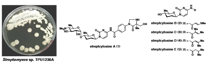
3.2. Isolation, Identification, and Fermentation of Producing Bacterium

The strain TPU1236A was isolated from a seawater sample collected at Iriomote Island in Okinawa, Japan in September, 2012. Approximately 1 mL of seawater was mixed with 25 mL of sterilized seawater with 1.0% SDS, and 50 μL of the mixture were developed on an agar plate (glycerol 0.6%, arginine 0.1%, K2HPO4 0.1%, MgSO4 0.05%, agar 1.5%, and antibiotics (cycloheximide 100 μg/mL and rifampin 5 μg/mL) in natural seawater) [21]. Identification of the strain TPU1236A was carried out by comparing the partial sequence of the 16S rRNA gene. S. badius (732/732, 100%) and S. sindenensis (730/730, 100%) showed the highest similarity. Therefore, this producing strain TPU1236A was identified as Streptomyces sp.
The culture of strain TPU1236A was maintained on a Waksman agar slant (glucose 1%, peptone 0.5%, meat extract 0.5%, NaCl 0.3%, and agar 1.2% in deionized water) at 4 °C. The mycelia grown on the slant culture were inoculated in a 100-mL Erlenmeyer flask containing 40 mL of a seed medium (ASW-A medium: soluble starch 2%, glucose 1%, peptone 0.5%, yeast extract 0.5%, and CaCO3 0.3% in natural seawater, pH 7.0), and the flasks were shaken (150 rpm) at 25 °C for 3 days. The seed culture (2 mL) was transferred into 500-mL Erlenmeyer flasks, each containing 200 mL of the same medium, and incubated at 25 °C for 7 days at 150 rpm.

3.3. Extraction and Isolation of Compounds 111

The whole broth (10.0 L) was filtered and the mycelia were extracted with MeOH (1 L). The broth filtrate was adsorbed on Diaion HP-20 (500 mL) as a column, and the resin was washed with water (2 L) and eluted with MeOH (2 L). The eluate from HP-20 was combined with the mycelial extract to give about 1.5 g of a solid material, which was separated on an ODS silica gel column by stepwise elution with MeOH in H2O to obtain 11 fractions. Collismycin B (10, 11.2 mg) was obtained as white crystals from the 70% MeOH fraction. The 70% MeOH fraction was purified by preparative HPLC (a PEGASIL ODS SP100 column, 250 mm × 10 mm, eluted with MeOH–H2O = 6:4 containing 0.05% TFA, 2.0 mL/min, detected at UV 210 nm), and streptcytosine A (1, 12.3 mg), plicacetin (7, 21.3 mg), and SF2738 C (11, 6.8 mg) were eluted at 9.9, 15.0, and 12.2 min, respectively. The 60% MeOH fraction was subjected to HPLC (ODS) with 45% MeOH in H2O containing 0.05% TFA at 2 mL/min to afford streptcytosines B (2, 3.2 mg, 16.8 min), C (3, 7.2 mg, 17.7 min), D (4, 8.1 mg, 19.3 min), and E (5, 5.1 mg, 20.2 min), de-amosaminyl-cytosamine (6, 12.0 mg, 20.1 min), and bamicetin (8, 20.5 mg, 5.8 min). Amicetin (9, 25.0 mg, 8.5 min) was isolated by HPLC with 60% MeOH in H2O (0.05% TFA) from the 80% MeOH fraction.
Streptcytosine A (1): obtained as a colorless oil; [α]23D +72 (c 0.1, MeOH); UV λmax (0.1 M HCl) nm (log ε): 269 (4.24), 276 (4.24), 317 (4.28); IR νmax (KBr) cm–1: 3500–3400, 2920, 1712, 1677, 1639, 1613, 1385, 1262, 1093, 802; HRFABMS (m/z) found: 631.3110, calcd: 631.3092 [M + H]+ for C30H43N6O9,; 1H and 13C NMR data, see Table 1.
Streptcytosine B (2): obtained as a pale yellow oil; [α]23D +24 (c 0.1, MeOH); UV λmax (0.1 M HCl) nm (log ε): 249 (3.94), 338 (4.09); IR νmax (KBr) cm–1: 3500–3400, 2927, 1683, 1646, 1616, 1576, 1493, 1385, 1247, 1092; HRFABMS (m/z) found: 326.1180, calcd: 326.1175 [M + H]+ for C14H20N3O4S; 1H and 13C NMR data, see Table 2.
Streptcytosine C (3): obtained as a pale yellow oil; [α]23D +36 (c 0.1, MeOH); UV λmax (0.1 M HCl) nm (log ε): 257 (3.95), 311 (4.18); IR νmax (KBr) cm–1: 3500–3400, 2928, 1683, 1646, 1613, 1561, 1489, 1385, 1262, 1092; HRFABMS (m/z) found: 308.1619, calcd: 308.1610 [M + H]+ for C15H22N3O4; 1H and 13C NMR data, see Table 2.
Streptcytosine (4): obtained as a pale yellow oil; [α]23D +68 (c 0.1, MeOH); UV λmax (0.1 M HCl) nm (log ε): 217 (3.96), 263 (4.16), 309 (4.32); IR νmax (KBr) cm–1: 3500–3400, 2937, 1735, 1674, 1634, 1561, 1492, 1396, 1272, 1095; HRFABMS (m/z) found: 308.1601, calcd: 308.1610 [M + H]+ for C15H22N3O4; 1H and 13C NMR data, see Table 2.
Streptcytosine (5): obtained as a pale yellow oil; [α]23D +62 (c 0.1, MeOH); UV λmax (0.1 M HCl) nm (log ε): 237 (3.61), 310 (4.04); IR νmax (KBr) cm–1: 3500–3400, 2963, 1720, 1677, 1624, 1571, 1494, 1383, 1276, 1095; HRFABMS (m/z) found: 310.1763, calcd: 310.1767 [M + H]+ for C15H24N3O4; 1H and 13C NMR data, see Table 2.

3.4. Anti-Mycobacterial Assay

The anti-mycobacterial assay was carried out using M. smegmatis NBRC 3207 with the paper disc method and liquid microdilution method. The strain NBRC 3207 was obtained from the Biological Resource Center (NBRC), NITE (Chiba, Japan) and maintained in 20% glycerol at −80 °C.
The test microorganism was cultured in Middlebook 7H9 broth (BD, Franklin Lakes, NJ, USA) containing 0.05% polysorbate 80 (BD), 0.5% glycerol, and 10% Middlebook OADC (BD) at 37 °C for 2 days and adjusted to 1.0 × 10⁶ CFU/mL. The inoculum was spread on the above medium containing 1.5% agar in a square plate. Each sample in MeOH was adsorbed to a sterile filter disc (6 mm, Advantec, Tokyo, Japan), and, after evaporation of MeOH, the disc was placed in a plate and incubated for 2 days at 37 °C. Streptomycin sulfate (5 and 10 µg/disc) and MeOH were used as positive and negative controls, respectively.
MICs were measured by the liquid microdilution method [22]. After 85 μL of Middlebrook 7H9 broth containing 0.05% polysorbate 80, 0.5% glycerol, and 10% Middlebook OADC was added to each well of a 96-well microplate (Corning Inc., Corning, NY, USA), test compound dissolved in MeOH (5.0 μL) was added to each well at the final concentration of 0.125 to 64 μg/mL, respectively. Finally, the test microorganism (10 μL) was added at a concentration of 1.0 × 10⁶ CFU/mL. Microplates were incubated at 37 °C for 2 days. MIC was defined as the lowest concentration of test compound where the test microorganism cannot grow.

4. Conclusions

Five new pyrimidine nucleoside antibiotics, named streptcytosines A–E (15), and six known compounds 611 were obtained from a marine-derived actinomycete, Streptomyces sp. TPU1236A, isolated from seawater collected at Iriomote Island in Okinawa, Japan. The anti-mycobacterial activity of the extract of the culture broth was reproduced by new compound 1 (streptcytosine A), plicacetin (7), bamicetin (8), amicetin (9), and SF2738 C (11). Compounds 8 and 9 were about two-fold more active than the other three compounds.

Acknowledgments

This work was supported in part by a Grant-in-Aid for Scientific Research (25870660) from the Ministry of Education, Culture, Sports, Science, and Technology (MEXT) of Japan to HY and the Foundation for Japanese Chemical Research to HY. We thank T. Matsuki and S. Sato for measuring mass spectra.

Authors Contribution

Y.-Y. Bu performed the fermentation, isolation, structure elucidation, and paper disc assay of compounds for his Ph.D. thesis. K. Ukai collected and isolated the actinomycetes. H. Yamazaki carried out the practical suggestions to Y.-Y. Bu through this project and evaluated the antimycobacterial activity by liquid microdilution method. M. Namikoshi, the supervisor of Y.-Y. Bu, designed this project and edited the manuscript.

Conflicts of Interest

The authors declare no conflict of interest.

References

  1. Zumla, A.; Raviglione, M.; Hafner, R.; von Reyn, C.F. Tuberculosis. N. Engl. J. Med. 2013, 368, 745–755. [Google Scholar] [CrossRef] [PubMed]
  2. Riccardi, G.; Pasca, M.R. Trends in discovery of new drugs for tuberculosis therapy. J. Antibiot. 2014, 67, 655–659. [Google Scholar] [CrossRef] [PubMed]
  3. Ntie-Kang, F.; Yong, J.N.; Owono, L.C.; Sippl, W.; Megnassan, E. Perspectives on tuberculosis pathogenesis and discovery of anti-tubercular drugs. Curr. Med. Chem. 2014, 21, 3466–3477. [Google Scholar] [CrossRef] [PubMed]
  4. World Health Organization (WHO). Global Tuberculosis Report 2013. Available online: http://www.who.int/tb/publications/global_report/en/ (accessed on 12 October 2014).
  5. Espinal, M.A. The global situation of MDR-TB. Tuberculosis 2003, 83, 44–51. [Google Scholar]
  6. Ryan, N.J.; Lo, J.H. Delamanid: first global approval. Drugs 2014, 74, 1041–1045. [Google Scholar] [CrossRef] [PubMed]
  7. Xavier, A.S.; Lakshmanan, M. Delamanid: A new armor in combating drug-resistant tuberculosis. J. Pharmacol. Pharmacother. 2014, 5, 222–224. [Google Scholar] [CrossRef] [PubMed]
  8. Reyrat, J.M.; Kahn, D. Mycobacterium smegmatis: An absurd model for tuberculosis? Trends Microbiol. 2001, 9, 472–473. [Google Scholar] [CrossRef] [PubMed]
  9. Chen, R.; Zhang, H.; Zhang, G.; Li, S.; Zhang, G.; Zhu, Y.; Liu, J.; Zhang, C. Characterizing amosamine biosynthesis in amicetin reveals AmiG as a reversible retaining glocosyltransferase. J. Am. Chem. Soc. 2013, 135, 12152–12155. [Google Scholar] [CrossRef] [PubMed]
  10. Haskell, T.H.; Ryder, A.; Frohardt, R.P.; Fusari, S.A.; Jakubowski, Z.L.; Bartz, Q.R. The isolation and characterization of three crystalline antibiotics from Streptomyces plicatus. J. Am. Chem. Soc. 1958, 80, 743–747. [Google Scholar] [CrossRef]
  11. DeBoer, C.; Caron, E.L.; Hinman, J.W. Amicetin, a new streptomyces antibiotic. J. Am. Chem. Soc. 1953, 75, 499–500. [Google Scholar] [CrossRef]
  12. Hinman, J.W.; Caron, E.L.; DeBoer, C. The isolation and purification of amicetin. J. Am. Chem. Soc. 1953, 75, 5864–5866. [Google Scholar] [CrossRef]
  13. Flynn, E.H.; Hinman, J.W.; Caron, E.L.; Woolf, D.O., Jr. The chemistry of Amicetin, a new antibiotic. J. Am. Chem. Soc. 1953, 75, 5867–5871. [Google Scholar] [CrossRef]
  14. Shindo, K.; Yamagishi, Y.; Okada, Y.; Kawai, H. Collismycins A and B, novel non-steroidal inhibitors of dexamethasone-glucocorticoid receptor binding. J. Antibiot. 1994, 47, 1072–1074. [Google Scholar] [CrossRef] [PubMed]
  15. Gomi, S.; Amano, S.; Sato, E.; Miyadoh, S.; Kodama, Y. Novel antibiotics SF2738A, B and C, and their analogs produced by Streptomyces sp. J. Antibiot. 1994, 47, 1385–1394. [Google Scholar] [CrossRef] [PubMed]
  16. Shammas, C.; Donarski, J.A.; Ramesh, V. NMR structure of the peptidyl transferase RNA inhibitor antibiotic amicetin. Magn. Reson. Chem. 2007, 45, 133–141. [Google Scholar] [CrossRef]
  17. Haskell, T.H. Amicetin, bamicetin and plicacetin. Chemical studies. J. Am. Chem. Soc. 1958, 80, 747–751. [Google Scholar] [CrossRef]
  18. Shiomi, K.; Haneda, K.; Tomoda, H.; Iwai, Y.; Omura, S. Cytosaminomycins, new anticoccidial agents produced by Streptomyces sp. KO-8119. II. Structure elucidation of cytosaminomycins A, B, C and D. J. Antibiot. 1994, 47, 782–786. [Google Scholar]
  19. Ericsson, H. The paper disc method for determination of bacterial sensitivity to antibiotics. Studies on the accuracy of the technique. Scand. J. Clin. Lab. Invest. 1960, 12, 408–413. [Google Scholar] [CrossRef] [PubMed]
  20. Amsterdam, D. Susceptibility testing of antimicrobials in liquid media. In Antibiotics in Laboratory Medicine, 4th ed.; Loman, V., Ed.; Williams & Wilkins: Baltimore, MD, USA, 1996; pp. 52–111. [Google Scholar]
  21. Mincer, T.J.; Jensen, P.R.; Kauffman, C.A.; Fenical, W. Widespread and persistent populations of a major new marine actinomycete taxon in ocean sediments. Appl. Environ. Microbiol. 2002, 68, 5005–5011. [Google Scholar] [CrossRef] [PubMed]
  22. Koyama, N.; Kojima, S.; Nonaka, K.; Masuma, R.; Matsumoto, M.; Omura, S.; Tomoda, H. Calpinactam, a new anti-mycobacterial agent, produced by Mortierella alpina FKI-4905. J. Antibiot. 2010, 63, 183–186. [Google Scholar] [CrossRef] [PubMed]

