Article

Oxygenated Polyketides from Plakinastrella mamillaris as a New Chemotype of PXR Agonists

1
Department of Pharmacy, University of Naples "Federico II", via D. Montesano 49, Naples 80131, Italy
2
Department of Clinical and Experimental Medicine, Faculty of Medicine, University of Perugia, via Gerardo Dottori 1, S. Andrea Delle Fratte, Perugia 06132, Italy
3
Department of Pharmacy, University of Salerno, via Ponte don Melillo, Fisciano (SA) 84084, Italy
*
Author to whom correspondence should be addressed.
Mar. Drugs 2013, 11(7), 2314-2327; https://doi.org/10.3390/md11072314
Submission received: 24 April 2013 / Revised: 27 May 2013 / Accepted: 28 May 2013 / Published: 2 July 2013
(This article belongs to the Special Issue Marine Compounds and Inflammation)

Abstract

:
Further purification of the apolar extracts of the sponge Plakinastrella mamillaris, afforded a new oxygenated polyketide named gracilioether K, together with the previously isolated gracilioethers E–G and gracilioethers I and J. The structure of the new compound has been elucidated by extensive NMR (1H and 13C, COSY, HSQC, HMBC, and ROESY) and ESI-MS analysis. With the exception of gracilioether F, all compounds are endowed with potent pregnane-X-receptor (PXR) agonistic activity and therefore represent a new chemotype of potential anti-inflammatory leads. Docking calculations suggested theoretical binding modes of the identified compounds, compatible with an agonistic activity on hPXR, and clarified the molecular basis of their biological activities.

Graphical Abstract

1. Introduction

Pregnane-X-receptor (PXR; NR1I2), a member of the nuclear receptor superfamily, primarily expressed in the liver and intestine [1], has a major role in the induction of genes involved in drug and xenobiotic transport and metabolism [2], thus preventing the accumulation of toxic chemicals in the body. In addition to its role as a xeno-sensor, PXR, through a complex, yet fully deciphered crosstalk with other nuclear receptors [3], such constitutive androstane receptor (CAR), farnesoid-X-receptor (FXR), liver-X-receptor (LXR), vitamin D receptor (VDR), peroxisome proliferator-activated receptors (PPARs), estrogen receptor (ER), glucocorticoid receptor (GR), COUP transcription factor I (COUP-TFI), and II, plays a relevant physiological role in the regulation of bilirubin clearance [4], bile acid detoxification [5,6,7], lipid, steroid [8], and glucose metabolism [9]. Moreover, recent studies demonstrated mutual inhibition between PXR and nuclear factor (NF)-κB signaling pathway, providing a potential molecular mechanism that links xenobiotic metabolism and inflammation [10,11]. These potential roles of PXR in disease pathogenesis suggest PXR as a novel target for drug discovery.
In recent years, several natural products from herbal medicine and marine sources were disclosed for their potentials to act as modulators of PXR [12,13,14]. In particular, studies from our laboratories disclosed some sterols [15,16,17,18] from the marine sponge Theonella swinhoei as a new class of PXR agonists that exerted potent immunomodulatory activity. Extensive in vivo and in vitro studies on solomonsterol A confirmed the therapeutic potential of PXR agonists in the treatment of inflammatory bowel disease (IBD) [19]. Further characterization of natural PXR agonists, and their biological activities, may hold the promise of developing novel PXR modulators for the treatment of metabolic diseases.
A recent report [20] described ginkgolides, terpene trilactones present in Ginkgo biloba extract, as a new chemotype for ligand activation of hPXR.
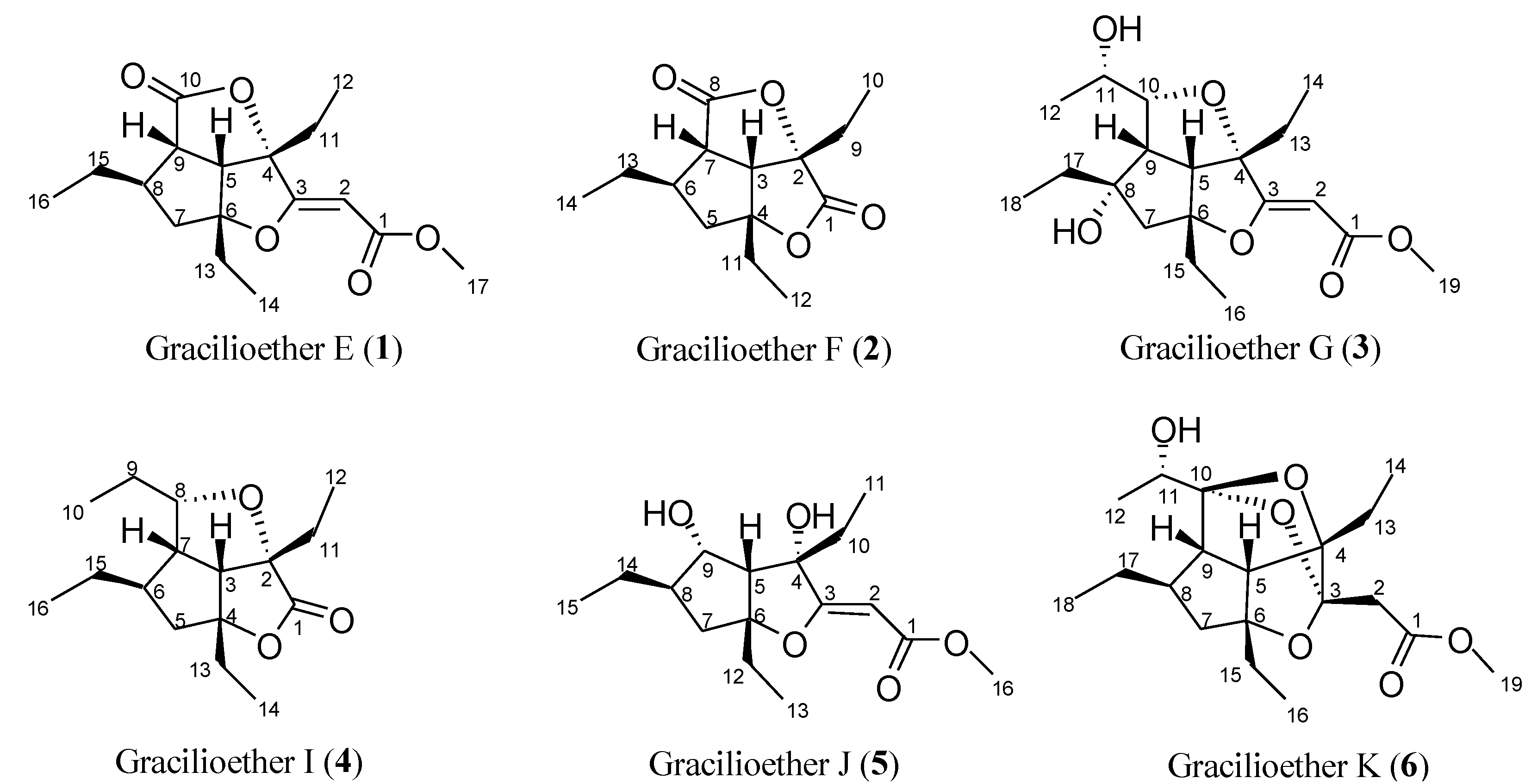
In connection with our interest toward the discovery of new ligands for nuclear receptors, we were intrigued by the structural resemblance between the polycyclic “cage-like” backbone of ginkgolides with the oxygenated polycyclic structure of gracilioethers E, F, G, I, and J (15, Figure 1), polyketides recently isolated by our group [21].
Therefore docking studies were performed to assess the accommodation of gracilioethers in the ligand binding domain (LBD) of hPXR, and to highlight the pharmacophoric parts able to establish weak interactions with the key residues of the investigated target.
In order to re-obtain the gracilioethers for pharmacological evaluations on the pure components of the apolar extracts of the sponge Plakinastrella mamillaris, a new oxygenated polyketide, named gracilioether K (6), was also isolated.
In the present study we reported the results of the docking analysis and the pharmacological evaluation indicating that, with the exception of gracilioether F (2), all compounds are endowed with potent PXR agonistic activity.
The structural characterization of the new polyketide derivative, gracilioether K, is also reported.
Figure 1. Gracilioethers from the sponge Plakinastrella mamillaris.
Marinedrugs 11 02314 g001

2. Results and Discussion

The starting hypothesis of considering gracilioethers 15, as characterized by structural requirements compatible with an agonistic activity on hPXR, was investigated by means of molecular docking experiments. As previously described [22,23,24], a putative PXR ligand should be able to interact with polar residues in the LBD. In this context, Ekins et al. proposed several agonist pharmacophoric models, characterized by the presence of a hydrogen bond acceptor and/or donor, and hydrophobes [25]. In particular Ser247 and other polar residues (namely Cys284, Gln285, His407) have been reported as key amino acids involved in hydrogen bond/polar interactions with the ligand counterpart.
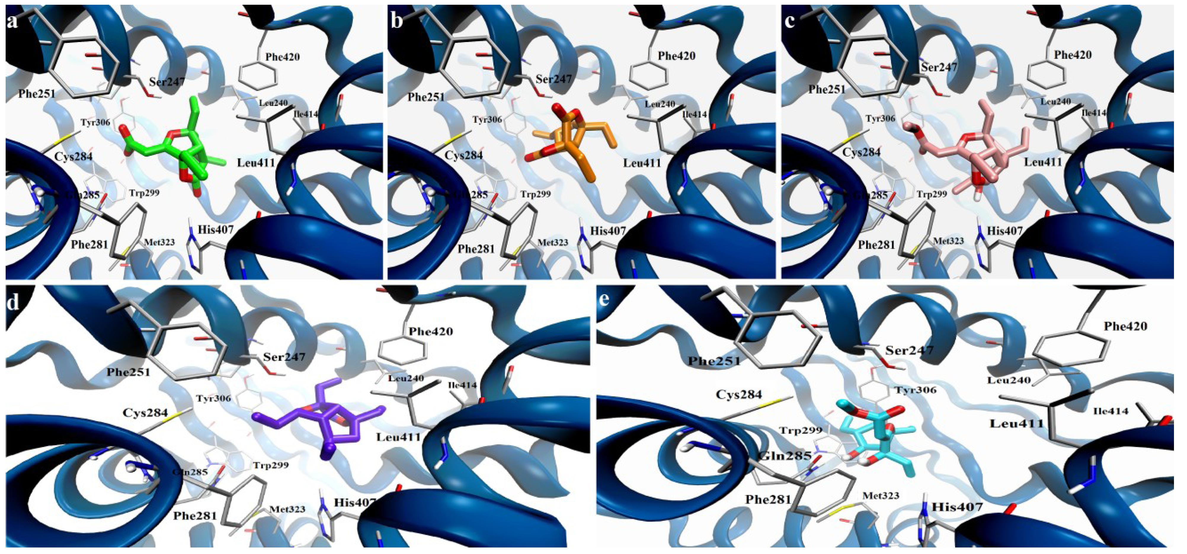
From a structural point of view, gracilioethers 15 are characterized by a well defined region with a high density of oxygens, potentially able to establish hydrogen bonds and polar interactions, and by a spatially distinct hydrophobic part. Starting from these assumptions, we have envisaged a binding mode, in which a part of these molecules is involved in polar interactions and/or hydrogen bonds with the key residues in the hPXR LBD, while the remaining part stabilizes the complex by van der Waals interactions (Figure 2). For each investigated compound, the docking analysis of the different binding poses was then conducted using a distance criterion, filtering and considering the number of poses interacting with key residues, Ser247, Cys284, Gln285, and His407.
Firstly, this analysis highlighted the ability of the investigated compounds to interact with the Ser247 [24] via polar interactions or hydrogen bonds in a high number of docking poses. In particular, for gracilioethers 1, 3, and 5 we found these fundamental interactions in more than 55% of the 50 docking poses saved for each compound. The significant number of polar groups in a defined part of their structures determines the stabilization of these putative complexes in the hPXR LBD through further hydrogen bonds or polar interactions (Gln285, Cys284, and His407), while the hydrophobic components are involved in van der Waals contacts (namely Leu240, Phe251, Phe281, Ile414).
If compared to compounds 1, 3, and 5, gracilioethers 2 and 4 are characterized by the absence of the unsaturated side chain at C-3, determining a minor number of interactions with the above reported key residues. Interestingly, a smaller percentage of poses interacting with Ser247 were obtained (30% for compound 2 and 36% for compound 4). A sensible minor number of matching poses were found for the interactions with Cys284 and His407, respecting the relative distributions between the different compounds.
Figure 2. Docking model of gracilioethers 15 in the ligand binding domain (LBD) of hPXR (secondary structure represented in blue): (a) 1 (pose n. 4, colored by atom types: C green, O red); (b) 2 (pose n. 27, colored by atom types: C orange, O red); (c) 3 (pose n. 2, colored by atom types: C pink, O red, H light grey); (d) 4 (pose n. 11, colored by atom types: C violet, O red); (e) 5 (pose n. 1, colored by atom types: C cyan, O red, H light grey).
Marinedrugs 11 02314 g002
The main difference between these two compounds is the presence of an additional ethyl group at C-8 in compound 4 replacing the γ-lactone moiety in compound 2. Analyzing the poses of 4 interacting with Ser247, we noticed that the presence of this aliphatic part in compound 4 determines the gain of favorable van der Waals interactions (Leu240, Phe251, Phe281, Leu411, Ile414) [24].
On the other hand, the analysis of the complexes of 2 interacting with Ser247 of hPXR revealed that the carbonyl part of the γ-lactone moiety is scarcely involved in hydrogen bonds or favorable polar interactions with the other key residues previously described. In addition, with respect to 4, the hPXR complex with gracilioether F (2) is characterized by poor hydrophobic contacts. An opposite situation was observed for gracilioether E (1), also featuring the γ-lactone moiety, where this chemical group contributes to the above polar pharmacophoric interactions in the most energetically favored poses.
These differences are depicted in Figure 2, where a representation of the binding modes of the different gracilioethers is reported. In these images, the poses in which the common chemical parts among these compounds occupy the same region in the hPXR LBD in a similar way are displayed; in such poses, the requirements of a putative agonistic activity, polar interactions with Ser247 for all compounds and with Gln285 for 13 and 5, are maintained. Moreover, the reported poses highlight that the punctual differences in the chemical structures of gracilioethers could determine the loss or the gain of additional hydrophobic (e.g., Phe251, Phe281, Ile414 found in compound 4, absent in 2) or polar interactions (His407), also influencing the stabilization of the corresponding hPXR LBD complexes and, consequently, the biological activity.

2.1. Isolation of Gracilioethers and Structural Characterization of Gracilioether K (6)

Prompted by good docking results, a re-isolation procedure was performed on the CHCl3 extract of the sponge Plakinastrella mamillaris. MPLC followed by HPLC purification afforded 15 in reasonable amounts for further pharmacological tests and the new oxygenated polyketide named gracilioether K (6).
Gracilioether K (6) was isolated as a white amorphous solid. The molecular formula of C19H30O6 was established by HR-ESIMS based on the pseudomolecular ion [M + Na]+ at m/z 377.1954, indicating an isomeric relationship with gracilioethers A [26] and G (3) [21] and five degrees of unsaturation.
Analysis of the 1H NMR and HSQC spectra revealed the presence of three ethyls, one methoxyl, one methyl, four methines (including one oxymethine), and two methylenes (Table 1). The 13C NMR spectrum further showed the presence of one acyl carbon at δC 171.1, two oxygenated quaternary carbons at δC 93.8 and 96.7, and two ketalic carbons at δC 110.49 and 110.51 (Table 1).
Table 1. 1H and 13C NMR data (700 and 175 MHz, CD3OD) of gracilioether K (6).
PositionδH aδCHMBCROESY b
1-171.1
22.63 d (14.2)
2.71 d (14.2)
40.2C1, C3H2-13, H3-16
3-110.49
4-96.7
52.80 d (10.5)54.6C3, C6, C9, C15H-7b, H2-13, H3-14, H2-15, H3-16
6-93.8
71.51 m
2.28 dd (7.6, 13.8)
47.9C5, C6, C8, C9H-5, H-9, H3-16
82.06 ovl39.4
92.74 dd (3.7, 10.5)59.0 H-7b, H3-12, H2-17, H3-18
10-110.51
113.88 q (6.5)66.0C10
121.22 d (6.5)17.7C10, C11H-9, H3-18
131.74 quint (7.5)
2.07 ovl
21.9C4, C14
C3, C4, C5
H2-2, H-5
H2-2
141.07 t (7.4)9.1C4H-5
151.55 m
1.65 quint (7.3)
32.6C5, C7H-5
H-5
160.93 t (7.5)9.8C6H2-2, H-5, H-7b
171.32 m
1.53 m
30.5C7, C8, C9, C18H-9
H-9
180.91 t (7.4)12.9C8H-9, H3-12
193.62 s52.2C1
a Coupling constants are in parentheses and given in hertz. 1H and 13C assignments aided by COSY, HSQC, and HMBC experiments; b ROESY experiment with mixing time of 200 ms; ovl: overlapped with other signals.
A detailed analysis of 2D NMR data indicated a carbon framework for gracilioether K (6) very similar to that of gracilioether A [26]. The most significant difference is the loss, in gracilioether K, of two sp2 carbons (δH/C 4.86/86.8 and δC 174.6), assigned respectively at C-2 and C-3 in gracilioether A [26].
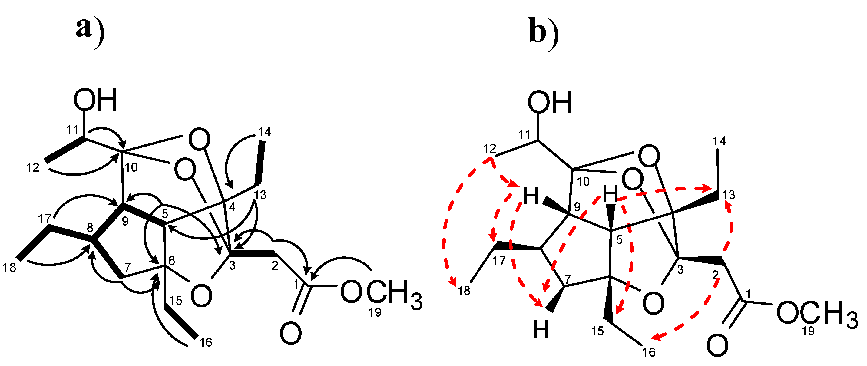
Analysis of COSY data allowed to the identify five spin systems: Two ethyl groups linked to quaternary carbons, a CH(5)CH(9)CH(8)(CH2CH3)CH2(7), a CH3(12)CH(11)OH, and one isolated CH2(2) (δH 2.63 and 2.71) spin systems (Figure 3a and Table 1). The HMBC correlations H3-14/C-4 and H3-16/C-6 connected the two ethyl units to C-4 and C-6 quaternary carbons, respectively. The hydroxyethyl group, CH3(12)CH(11)OH, was connected to the ketalic quaternary carbon C-10 (δC 110.51) on the basis of HMBC correlations H3-12/C-10 and H-11/C-10. The isolated AB spin system (δH 2.63 and 2.71) was connected to the acyl group at δC 171.1 and to the ketal carbon at δC 110.49 on the basis of HMBC correlations H-2/C-1 and H-2/C-3. A methyl ester group was easily inferred by the diagnostic long-range correlation between the methoxy protons at δH 3.62 and the acyl carbonyl at δC 171.1. Several diagnostic HMBC correlations (Figure 3a and Table 1) allowed us to secure the presence of the hexahydro-cyclopentanfurane system common to all gracilioethers, in which an ether bridge connecting C-3 and C-6 was placed by analogy with other gracilioethers and chemical shift considerations. Although no diagnostic heteronuclear long-range correlations were found to prove the C-9/C-10 linkage, the attachment of C-10/C-12 subunit to C-9 was supposed by analogy with gracilioether A, and confirmed by the strong dipolar couplings between H3-12/H-9 and H3-18 (Figure 3b).
Figure 3. (a) COSY connectivities (bold bonds), and key HMBC (black arrows) correlations; (b) Key ROE (red dashed arrows) correlations for gracilioether K (6).
Marinedrugs 11 02314 g003
Finally, two ether bridges between C-3 and C-10 and between C-4 and C-10 were assigned to satisfy the degrees of unsaturation and the molecular formula and to fulfil the functionalization degree of C-3, C-4, and C-10, as determined by 13C NMR chemical shifts value analysis.
The relative configuration of the tetracyclic system in 6 was established from ROESY correlations (Table 1), as shown in Figure 3b. The correlations between H-5/H-7b, H-13, H-14, H-15, H-16, and H-9/H-7b, H-17, H-18 suggested that the three ethyl groups, H-5 and H-9, were on the same face of the rings in a similar situation of all members of this family of marine oxygenated polyketides [21,26].
The analysis of the ROESY spectrum gave also information on the stereochemistry at C-3. In particular ROESY correlations H-2/H2-13 and H3-16 indicated that the carbomethoxy methyl substituent at C-3 and the ethyl group at C-4 were cis-oriented.
The stereochemical homology observed for all members of the gracilioethers family previously isolated from the sponge Plakinastrella mamillaris [21,26] suggested a common biogenetic origin. By analogy with well known biosynthetic pathway to PGE2, endoperoxy rearrangement in gracilioether A could afford hydroxyketone 7 that, through hemiacetal ring closure followed by 1,4 nucleophilic addition of the C-10 oxygen group to the unsaturated ester moiety, generated the complex polycyclic system of gracilioether K (Figure 4).
Therefore, from this biogenetic relationship, gracilioether K is likely to retain the same absolute configuration of all stereogenic centers of the hexahydro-cyclopentanfurane system as in gracilioether A [26].
Figure 4. Biosynthetic pathway to gracilioether K, involving endoperoxy rearrangement, hemiacetal formation and Michael-type attack on the α,β-unsaturated ester group.
Marinedrugs 11 02314 g004
The absolute stereochemistry of the secondary hydroxyl group at C-11 was determined by application of the modified Mosher’s method [27]. Treatment of 6 with R-()- or S-(+)-α-methoxy-α-(trifluoromethyl)phenylacetyl chloride (MTPACl) yielded S-()- and R-(+)-MTPA esters 6a and 6b, respectively. The ∆δ value distribution pattern clearly indicated 11S configuration (Figure 5a), the same found in gracilioether G (3) [21].
Finally, once fixed the configuration of all the other stereogenic centers, the analysis of the two possible diasteroisomeric structures presenting the 10R (Figure 5b) or 10S (Figure 5c) configuration revealed the presence of severe unfavorable distortions of the sp3 atoms involved in the interested rings for the S configuration at C-10 thus suggesting the R configuration as depicted in Figure 1.
Figure 5. (a) ∆δS-R values (ppm) of MTPA esters 6a and 6b; 3D model for (b) 10R configuration and (c) 10S configuration.
Marinedrugs 11 02314 g005

2.2. Pharmacological Evaluation

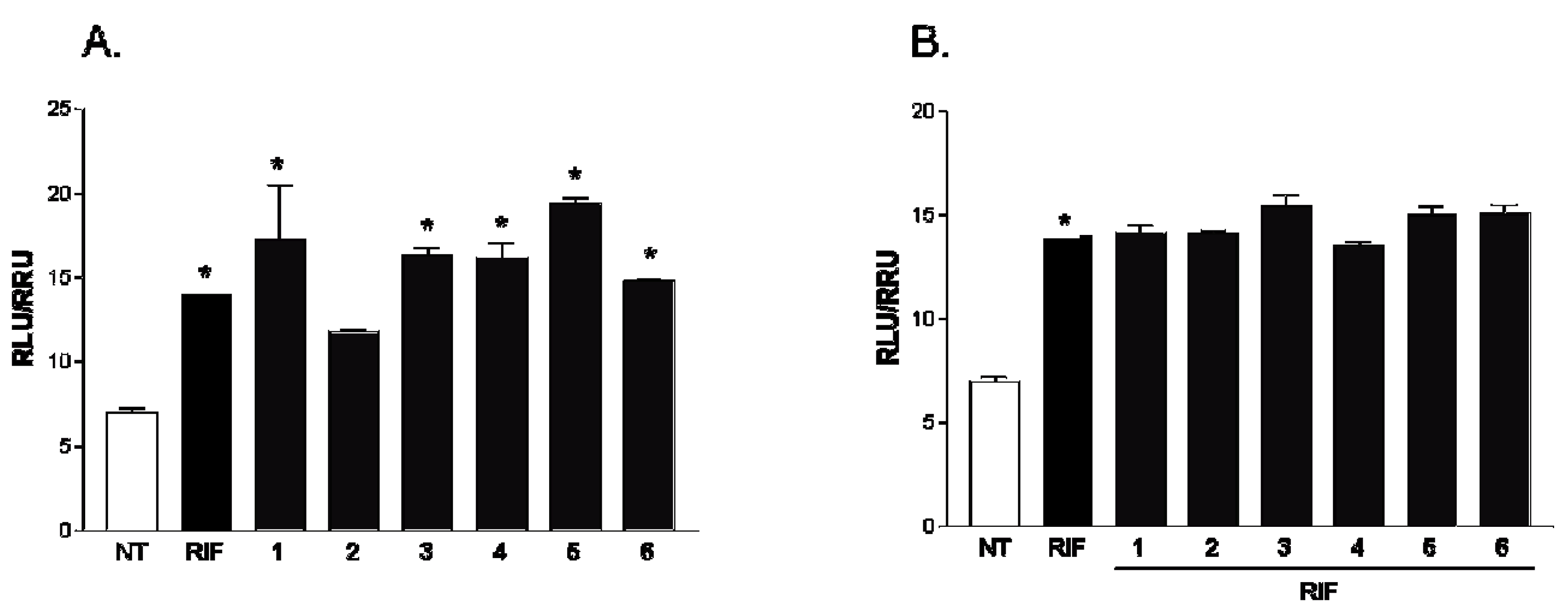
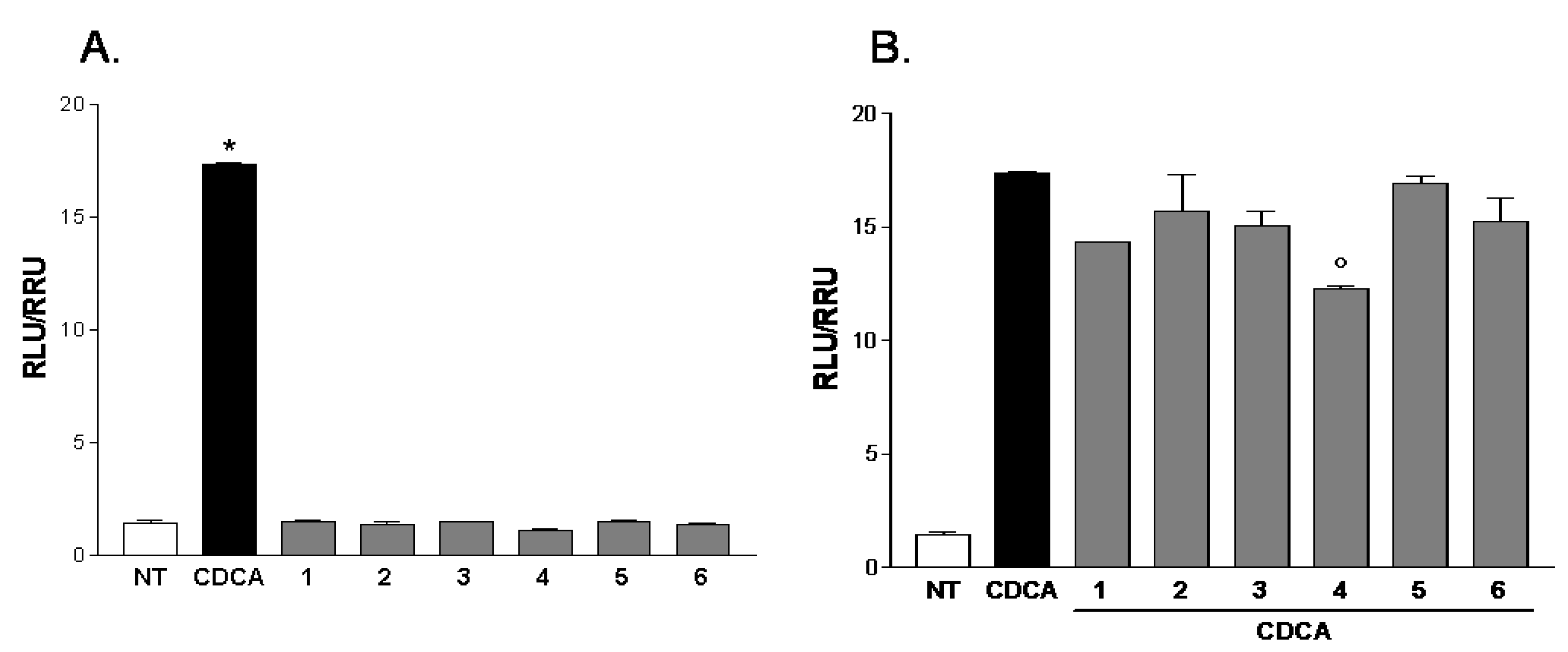
All compounds of this small series were challenged in a reporter gene assay using HepG2 cells, a human hepatoma cell line. As shown in Figure 6, all compounds, with the exception of gracilioether F (2), were PXR agonists without effect when administered in combination with rifaximin. However, when HepG2 cells transfected with FXR vectors were treated with gracilioethers 16, alone or in the presence of chenodeoxycholic acid (CDCA) 10 μM, no significant agonistic or antagonistic activities were observed, with the exception for the compound 4, which revealed a slight antagonistic activity on FXR (Figure 7).
Figure 6. Luciferase reporter assay performed in HepG2 transiently transfected with pSG5-PXR, pSG5-RXR, pCMV-bgal, p(cyp3a4)TKLUC vectors and stimulated 18 h with (A) Rifaximin (RIF), 10 μM, and of gracilioethers 16, 10 μM; and (B) Rifaximin (RIF), 10 μM, alone or in combination with gracilioethers 16, 50 μM. * p < 0.05 vs. not treated (NT).
Marinedrugs 11 02314 g006
Figure 7. Luciferase reporter assay performed in HepG2 transiently transfected with pSG5-FXR, pSG5-RXR, pCMV-bgal, p(hsp27)TKLUC vectors and stimulated 18 h with (A) CDCA, 10 μM, and gracilioethers 16, 10 μM; and (B) CDCA, 10 μM, alone or in combination with gracilioethers 16. * p < 0.05 vs. not treated (NT), ° p < 0.05 vs. CDCA.
Marinedrugs 11 02314 g007
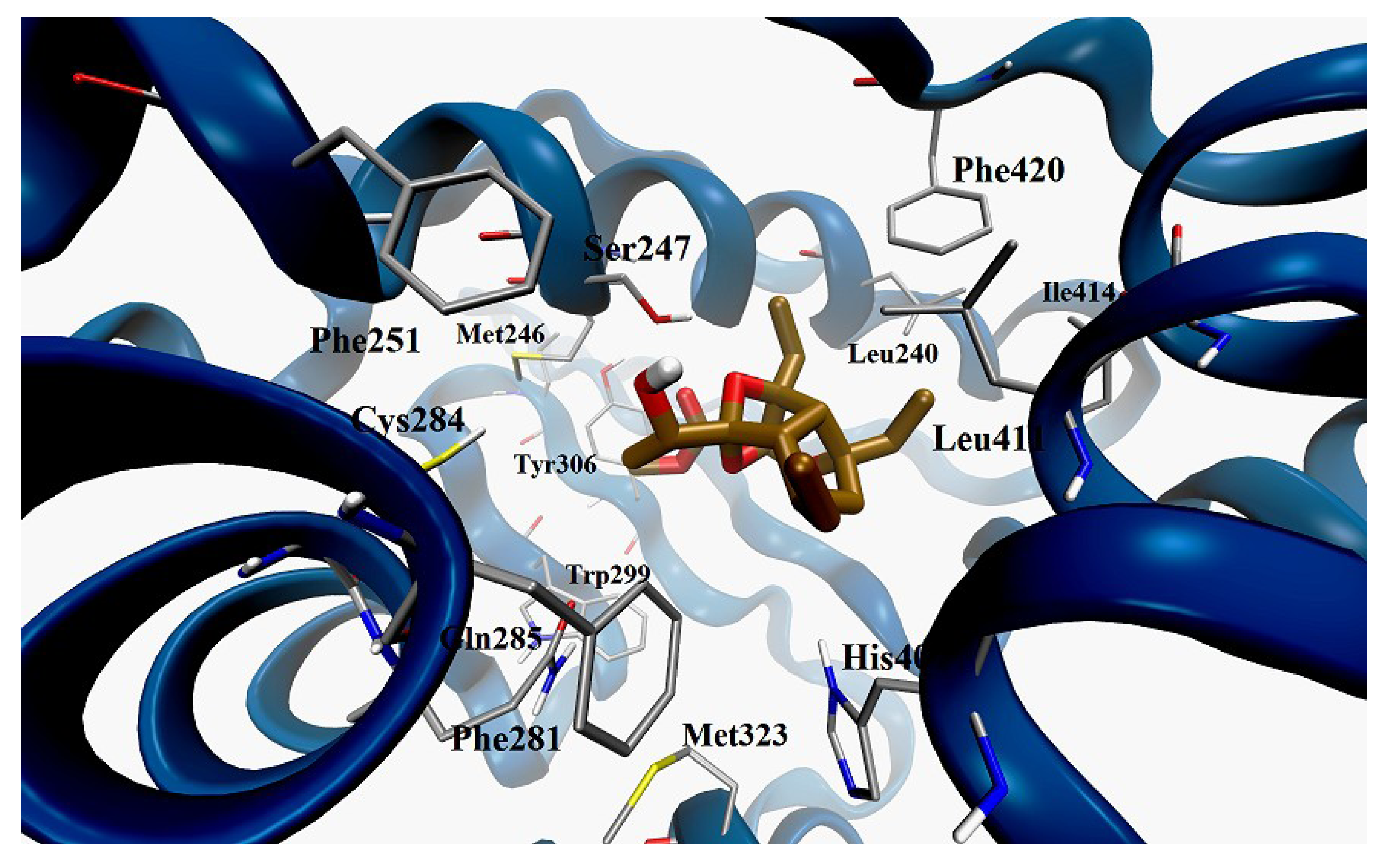
Docking calculations on graciolioether K (6) confirmed the observations highlighted for active gracilioethers 1, 3, 4 and 5. As shown in Figure 8, graciolioether K (6) establishes polar contacts with Ser247 and His407, and van der Waals interactions with Met243, Leu411, Ile414, and Phe420.
Figure 8. Docking model of gracilioether K (6) (pose n. 2, colored by atom types: C ochre, O red, H light grey); in the LBD of hPXR (secondary structure represented in blue).
Marinedrugs 11 02314 g008

3. Experimental Section

3.1. General Experimental Procedures

Specific rotations were measured on a Perkin-Elmer 243 B polarimeter. High-resolution ESI-MS measurements were performed with a Micromass QTOF Micromass spectrometer. ESI-MS experiments were performed on an Applied Biosystem API 2000 triple-quadrupole mass spectrometer. NMR spectra were obtained on a Varian Inova 700 MHz spectrometer (1H at 700 MHz, 13C at 175 MHz, respectively) equipped with Sun hardware, δ (ppm), J in Hz, spectra referred to CD3OD (δH 3.31, δC 49.0) as internal standards. HPLC was performed using a Waters Model 510 pump equipped with Waters Rheodyne injector and a differential refractometer, model 401. Through-space 1H connectivities were evidenced using a ROESY experiment with a mixing time of 200 ms. Silica gel (200–400 mesh) from Macherey-Nagel Company (Düren, Germany) was used for flash chromatography. The purities of compounds were determined to be greater than 95% by HPLC.

3.2. Sponge Material and Separation of Gracilioethers

Plakinastrella mamillaris Kirkpatrick, 1900 (order Homosclerophorida, family Plakinidae) was collected at the Fiji Islands, in May 2007. The sample was frozen immediately after collection and lyophilized to yield 171 g of dry mass. The sponge was identified by John Hooper, Queensland Museum, Brisbane, Australia, where a voucher specimen is deposited under the accessing number G322695.
The lyophilized material (171 g) was extracted with methanol (3 × 1.5 L) at room temperature and the crude methanolic extract (40 g) was subjected to a modified Kupchan’s partitioning procedure as follows. The methanol extract was dissolved in a mixture of MeOH/H2O containing 10% H2O and partitioned against n-hexane to give 17.3 g of the crude extract. The water content (% v/v) of the MeOH extract was adjusted to 30% and partitioned against CHCl3 to give 16.6 g of the crude extract.
The aqueous phase was concentrated to remove MeOH and then extracted with n-BuOH (2.4 g of crude extract). The CHCl3 extract (4.0 g) was chromatographed by silica gel MPLC using a solvent gradient system from CH2Cl2 to CH2Cl2:MeOH 1:1. Fractions eluted with CH2Cl2:MeOH 99:1 (370 mg) were further purified by HPLC on a Nucleodur 100-5 C18 (5 μm; 10 mm i.d. × 250 mm) with 65% MeOH:H2O as eluent (flow rate 3.5 mL/min) to give 5.3 mg of gracilioether K (6) (tR = 44.1 min); 70% MeOH:H2O as eluent (flow rate 3.5 mL/min) to give 3.3 mg of gracilioether F (2) (tR = 9.6 min), 6.1 mg of gracilioether E (1) (tR = 11.1 min), 11.0 mg of gracilioether A (tR = 26 min), 0.9 mg of gracilioether I (4) (tR = 27.9 min). Fractions eluted with CH2Cl2/MeOH 98:2 (326 mg) were further purified by HPLC on a Nucleodur 100-5 C18 (5 μm; 10 mm i.d. × 250 mm) with 60% MeOH/H2O as eluent (flow rate 3.5 mL/min) to give 4.3 mg of gracilioether J (5) (tR = 12.6 min) and 8.5 mg of gracilioether G (3) (tR = 21.9 min).

3.3. Characteristic Data for Gracilioether K (6)

Gracilioether K (6): White amorphous solid; [α]D25 −32.1 (c 0.06, MeOH); 1H and 13C NMR data in CD3OD given in Table 1; ESI-MS: m/z 377.2 [M + Na]+. HRMS (ESI): calcd. for C19H30NaO6: 377.1940; found 377.1954 [M + Na]+.

3.4. General Procedure for the Preparation of MTPA Esters (6a) and (6b)

Samples of 0.5 mg were dissolved in freshly distilled CH2Cl2 and treated with triethylamine (10 μL), (R)- or (S)-α-methoxy-α-(trifluoromethyl)phenylacetyl chloride (MTPA-Cl) (5 μL) and a catalytic amount of 4-(dimethylamino)pyridine to obtain respectively (S)- or (R)-MTPA esters. The mixture was left to stand at room temperature for 1 h, with the resulting mixture purified by HPLC on a Luna 5 μ Silica (2) column (5 μm; 4.6 mm i.d. × 250 mm) with 70% hexane:EtOAc as eluent (flow rate 1.0 mL/min).
[(S)-MTPA ester 6a]: Selected 1H NMR (400 MHz, CD3OD) δ (ppm): 1.29 (d, J = 6.3 Hz, H3-12), 1.51 (m, H-7a), 2.23 (dd, J = 7.9, 13.8 Hz, H-7b), 2.49 (dd, J = 3.6, 10.4 Hz, H-9), 2.81 (d, J = 10.4 Hz, H-5), 5.34 (q, J = 6.3 Hz, H-11).
[(R)-MTPA ester 6b]: Selected 1H NMR (400 MHz, CD3OD) δ (ppm): 1.38 (d, J = 6.5 Hz, H3-12), 1.48 (m, H-7a), 2.12 (dd, J = 8.1, 13.6 Hz, H-7b), 2.39 (dd, J = 3.6, 10.3 Hz, H-9), 2.75 (d, J = 10.3 Hz, H-5), 5.35 (q, J = 6.5 Hz, H-11).

3.5. Computation Details

The chemical structures of the compounds were built and processed with Macromodel 8.5 (Schrödinger, LLC, New York, NY, USA, 2003). Conformational search and Molecular Dynamics calculations were performed on a 4× AMD Opteron SixCore 2.4 GHz using Macromodel 8.5. Conformational search simulations were performed using the OPLS_2005 force field. The Monte Carlo multiple minimum (MCMM) method (105 steps) was used to allow a full exploration of the conformational space. Molecular Dynamics simulations were performed at 450 K and with a simulation time of 10 ns, after an equilibration phase of 1 ns. A constant dielectric term, mimicking the presence of the solvent, was used to reduce artifacts. Finally, the optimization (Conjugate Gradient, 0.0005 Å convergence threshold) of the structures was applied to identify the three-dimensional starting models for the subsequent steps of docking calculations.

Docking Calculation

Docking calculations were performed using the Autodock-Vina software [28]. In the configuration files of the crystallographic structure of hPXR (PDB code: 1M13) we specified the coordinate values for the targets, focusing the grid on the site of presumable pharmacological interest. In particular a grid box size of 30 × 36 × 32 and centered at 14.282 (x), 74.983 (y), and 0.974 (z) was used, with spacing of 1.0 Å between the grid points. The exhaustiveness value was set to 64, saving 50 conformations as maximum number of binding modes, and choosing 3.0 kcal/mol as maximum energy difference between the best and worst binding pose calculated. For all the investigated compounds, all open-chain bonds were treated as active torsional bonds. Docking results were analyzed with Autodock Tools 1.4.5. Illustrations of the 3D models were generated using VMD software [29].

3.6. In Vitro Transactivation

HepG2 cells were cultured at 37 °C in Minimum Essential Medium with Earl’s salts containing 10% fetal bovine serum (FBS), 1% l-glutamine and 1% penicillin/streptomycin. HepG2 cells were plated in a 24-wells plate at 1 × 10⁵ cells/well. The transfection experiments were performed using Fugene HD (Promega, Milan, Italy) according to manufacturer specifications. For FXR mediated transactivation, cells were transfected with 75 ng pCMVSPORT-FXR, 75 ng pSG5-RXR, 100 ng pGL4.70-Renilla and with 250 ng of the reporter vector p(hsp27)-TK-LUC containing the FXR response element IR1 cloned from the promoter of heat shock protein 27 (hsp27). For PXR mediated transactivation, cells were transfected with 75 ng pSG5-hPXRT1, 75 ng pSG5-RXR, 100 ng pGL4.70-Renilla, and with 250 ng of the reporter vector pGL3(henance)PXRE. At 24 h post-transfection, cells were treated for 2 h with compounds 16 10 μM (agonism) or 50 μM (antagonism) and then stimulated with CDCA 10 μM for 18 h. After treatments, cells were lysed in 100 μL diluted reporter lysis buffer (Promega) and 10 μL cellular lysate was assayed for Luciferase and Renilla activity using the Luciferase or Renilla Assay System (Promega). Luminescence was measured using GloMax™ 20/20 Luminometer (Promega). Luciferase activities were normalized for transfection efficiencies by dividing the Luciferase relative light units by Renilla relative lights units expressed from cells co-transfected with pGL4.70-Renilla.

4. Conclusions

The demonstration that gracilioethers, with the exception of gracilioether F, are PXR agonists, devoid of a significant activity on the bile acid sensor FXR, might have pharmacological and therapeutic relevance. Indeed, PXR agonists are being increasingly investigated for their ability to reduce inflammation in gastrointestinal tract and for their potential application in the treatment of a variety of human disorders including altered bone homeostasis, liver steatosis, and liver and lung fibrosis. PXR has, additionally, a multi-factorial impact on cancer, either by directly affecting cell proliferation and apoptosis, or by inducing chemotherapy resistance in colon, breast, prostate, and endometrial cancer, and in osteosarcoma, suggesting that identification of novel PXR ligands, agonists and antagonists, might have relevance for the treatment of these disorders. This study reaffirms sponges of Plakinastrella genus as an invaluable source of nuclear receptors ligands [30] endowed with unusual chemical scaffolds and useful for discovery of new pharmacological leads in several human diseases.

Acknowledgments

We thank the Fiji government for giving us the authorization to collect and work on Fijian sponges, the fisheries department and C. Payri (IRD) for their help. This work was supported by grants from MIUR (PRIN 2009) Sostanze ad attività antitumorale: Isolamento da fonti marine, sintesi di analoghi e ulteriore sviluppo della chemoteca “LIBIOMOL”. NMR spectra were provided by the CSIAS, Centro Interdipartimentale di Analisi Strumentale, Department of Pharmacy, Naples, Italy.

Conflict of Interest

The authors declare no conflict of interest.

References

  1. Lehmann, J.M.; McKee, D.D.; Watson, M.A.; Willson, T.M.; Moore, J.T.; Kliewer, S.A. The human orphan nuclear receptor PXR is activated by compounds that regulate CYP3A4 gene expression and cause drug interactions. J. Clin. Invest. 1998, 102, 1016–1023. [Google Scholar] [CrossRef]
  2. Kliewer, S.A.; Goodwin, B.; Willson, T.M. The nuclear pregnane X receptor: A key regulator of xenobiotic metabolism. Endocr. Rev. 2002, 23, 687–702. [Google Scholar] [CrossRef]
  3. Reschly, E.J.; Krasowski, M.D. Evolution and function of the NR1I nuclear hormone receptor subfamily (VDR, PXR, and CAR) with respect to metabolism of xenobiotics and endogenous compounds. Curr. Drug Metab. 2006, 7, 349–365. [Google Scholar] [CrossRef]
  4. Saini, S.P.; Mu, Y.; Gong, H.; Toma, D.; Uppal, H.; Ren, S.; Li, S.; Poloyac, S.M.; Xie, W. Dual role of orphan nuclear receptor pregnane X receptor in bilirubin detoxification in mice. Hepatology 2005, 41, 497–505. [Google Scholar] [CrossRef]
  5. Xie, W.; Radominska-Pandya, A.; Shi, Y.; Simon, C.M.; Nelson, M.C.; Ong, E.S.; Waxman, D.J.; Evans, R.M. An essential role for nuclear receptors SXR/PXR in detoxification of cholestatic bile acids. Proc. Natl. Acad. Sci. USA 2001, 98, 3375–3380. [Google Scholar] [CrossRef]
  6. Staudinger, J.L.; Goodwin, B.; Jones, S.A.; Hawkins-Brown, D.; MacKenzie, K.I.; LaTour, A.; Liu, Y.; Klaassen, C.D.; Brown, K.K.; Reinhard, J.; et al. The nuclear receptor PXR is a lithocholic acid sensor that protects against liver toxicity. Proc. Natl. Acad. Sci. USA 2001, 98, 3369–3374. [Google Scholar] [CrossRef]
  7. Fiorucci, S.; Cipriani, S.; Baldelli, F.; Mencarelli, A. Bile acid-activated receptors in the treatment of dyslipidemia and related disorders. Prog. Lipid Res. 2010, 49, 171–185. [Google Scholar] [CrossRef]
  8. Sonoda, J.; Chong, L.W.; Downes, M.; Barish, G.D.; Coulter, S.; Liddle, C.; Lee, C.H.; Evans, R.M. Pregnane X receptor prevents hepatorenal toxicity from cholesterol metabolites. Proc. Natl. Acad. Sci. USA 2005, 102, 2198–2203. [Google Scholar]
  9. Ihunnah, C.A.; Jiang, M.; Xie, W. Nuclear receptor PXR, transcriptional circuits and metabolic relevance. Biochim. Biophys. Acta 2011, 1812, 956–963. [Google Scholar] [CrossRef]
  10. Xie, W.; Tian, Y. Xenobiotic receptor meets NF-kappaB, a collision in the small bowel. Cell Metab. 2006, 4, 177–178. [Google Scholar] [CrossRef]
  11. Zhou, C.; Tabb, M.M.; Nelson, E.L.; Grun, F.; Verma, S.; Sadatrafiei, A.; Lin, M.; Mallick, S.; Forman, B.M.; Thummel, K.E.; et al. Mutual repression between steroid and xenobiotic receptor and NF-kappaB signaling pathways links xenobiotic metabolism and inflammation. J. Clin. Invest. 2006, 116, 2280–2289. [Google Scholar] [CrossRef]
  12. D’Auria, M.V.; Sepe, V.; Zampella, A. Natural ligands for nuclear receptors: Biology and potential therapeutic applications. Curr. Top. Med. Chem. 2012, 12, 637–669. [Google Scholar] [CrossRef]
  13. Gao, J.; Xie, W. Targeting xenobiotic receptors PXR and CAR for metabolic diseases. Trends Pharmacol. Sci. 2012, 33, 552–558. [Google Scholar] [CrossRef]
  14. Fiorucci, S.; Distrutti, E.; Bifulco, G.; D’Auria, M.V.; Zampella, A. Marine sponge steroids as nuclear receptor ligands. Trends Pharmacol. Sci. 2012, 33, 591–601. [Google Scholar] [CrossRef]
  15. De Marino, S.; Ummarino, R.; D’Auria, M.V.; Chini, M.G.; Bifulco, G.; Renga, B.; D’Amore, C.; Fiorucci, S.; Debitus, C.; Zampella, A. Theonellasterols and conicasterols from Theonella swinhoei. Novel marine natural ligands for human nuclear receptors. J. Med. Chem. 2011, 54, 3065–3075. [Google Scholar] [CrossRef]
  16. De Marino, S.; Ummarino, R.; D’Auria, M.V.; Chini, M.G.; Bifulco, G.; D’Amore, C.; Renga, B.; Mencarelli, A.; Petek, S.; Fiorucci, S.; et al. 4-Methylenesterols from Theonella swinhoei sponge are natural pregnane-X-receptor agonists and farnesoid-X-receptor antagonists that modulate innate immunity. Steroids 2012, 77, 484–495. [Google Scholar] [CrossRef]
  17. Festa, C.; de Marino, S.; D’Auria, M.V.; Bifulco, G.; Renga, B.; Fiorucci, S.; Petek, S.; Zampella, A. Solomonsterols A and B from Theonella swinhoei. The first example of C-24 and C-23 sulfated sterols from a marine source endowed with a PXR agonistic activity. J. Med. Chem. 2011, 54, 401–405. [Google Scholar] [CrossRef]
  18. De Marino, S.; Sepe, V.; D’Auria, M.V.; Bifulco, G.; Renga, B.; Petek, S.; Fiorucci, S.; Zampella, A. Towards new ligands of nuclear receptors. Discovery of malaitasterol A, an unique bis-secosterol from marine sponge Theonella swinhoei. Org. Biomol. Chem. 2011, 9, 4856–4862. [Google Scholar] [CrossRef]
  19. Sepe, V.; Ummarino, R.; D’Auria, M.V.; Mencarelli, A.; D’Amore, C.; Renga, B.; Zampella, A.; Fiorucci, S. Total synthesis and pharmacological characterization of solomonsterol A, a potent marine pregnane-X-receptor agonist endowed with anti-inflammatory activity. J. Med. Chem. 2011, 54, 4590–4599. [Google Scholar] [CrossRef]
  20. Lau, A.J.; Yang, G.; Yap, C.W.; Chang, T.K.H. Selective agonism of human pregnane X receptor by individual ginkgolides. Drug Metab. Dispos. 2012, 40, 1113–1121. [Google Scholar] [CrossRef]
  21. Festa, C.; de Marino, S.; D’Auria, M.V.; Deharo, E.; Gonzalez, G.; Deyssard, C.; Petek, S.; Bifulco, G.; Zampella, A. Gracilioethers E–J, new oxygenated polyketides from the marine sponge Plakinastrella mamillaris. Tetrahedron 2012, 68, 10157–10163. [Google Scholar] [CrossRef]
  22. Ekins, S.; Kortagere, S.; Iyer, M.; Reschly, E.J.; Lill, M.A.; Redinbo, M.R.; Krasowski, M.D. Challenges predicting ligand-receptor interactions of promiscuous proteins: The nuclear receptor PXR. PLoS Comput. Biol. 2009, 5. [Google Scholar] [CrossRef] [Green Version]
  23. Watkins, R.E.; Maglich, J.M.; Moore, L.B.; Wisely, G.B.; Noble, S.M.; Davis-Searles, P.R.; Lambert, M.H.; Kliewer, S.A.; Redinbo, M.R. 2.1 Å crystal structure of human PXR in complex with the St. John’s wort compound hyperforin. Biochemistry 2003, 42, 1430–1438. [Google Scholar]
  24. Xiao, L.; Nickbarg, E.; Wang, W.; Thomas, A.; Ziebell, M.; Prosise, W.W.; Lesburg, C.A.; Taremi, S.S.; Gerlach, V.L.; Le, H.V.; et al. Evaluation of in vitro PXR-based assays and in silico modeling approaches for understanding the binding of a structurally diverse set of drugs to PXR. Biochem. Pharmacol. 2011, 81, 669–679. [Google Scholar]
  25. Ekins, S.; Chang, C.; Mani, S.; Krasowski, M.D.; Reschly, E.J.; Iyer, M.; Kholodovych, V.; Ai, N.; Welsh, W.J.; Sinz, M.; et al. Human pregnane X receptor antagonists and agonists define molecular requirements for different binding sites. Mol. Pharmacol. 2007, 72, 592–603. [Google Scholar] [CrossRef]
  26. Ueoka, R.; Nakao, Y.; Kawatsu, S.; Yaegashi, J.; Matsumoto, Y.; Matsunaga, S.; Furihata, K.; van Soest, R.W.M.; Fusetani, N. Gracilioethers A–C, antimalarial metabolites from the marine sponge Agelas gracilis. J. Org. Chem. 2009, 74, 4203–4207. [Google Scholar] [CrossRef]
  27. Ohtani, I.; Kusumi, T.; Kashman, Y.; Kakisawa, H. High-field FT NMR application of Mosher’s method. The absolute configurations of marine terpenoids. J. Am. Chem. Soc. 1991, 113, 4092–4096. [Google Scholar] [CrossRef]
  28. Trott, O.; Olson, A.J. AutoDock Vina: Improving the speed and accuracy of docking with a new scoring function, efficient optimization and multithreading. J. Comput. Chem. 2010, 31, 455–461. [Google Scholar]
  29. Humphrey, W.; Dalke, A.; Schulten, K. VMD—Visual molecular dynamics. J. Mol. Graph. 1996, 14, 33–38. [Google Scholar] [CrossRef]
  30. Festa, C.; Lauro, G.; de Marino, S.; D’Auria, M.V.; Monti, M.C.; Casapullo, A.; D’Amore, C.; Renga, B.; Mencarelli, A.; Petek, S.; et al. Plakilactones from the marine sponge Plakinastrella mamillaris. Discovery of a new class of marine ligands of peroxisome proliferator-activated receptor γ. J. Med. Chem. 2012, 55, 8303–8317. [Google Scholar] [CrossRef]