Article

Core Oligosaccharide of Plesiomonas shigelloides PCM 2231 (Serotype O17) Lipopolysaccharide — Structural and Serological Analysis

by
Anna Maciejewska
1,*,
Jolanta Lukasiewicz
1,
Marta Kaszowska
1,
Aleksandra Man-Kupisinska
1,
Wojciech Jachymek
1 and
Czeslaw Lugowski
1,2
1
Department of Immunochemistry, Ludwik Hirszfeld Institute Immunology and Experimental Therapy, Polish Academy of Sciences, Rudolfa Weigla 12, 53-114 Wroclaw, Poland
2
Department of Biotechnology and Molecular Biology, University of Opole, Kardynala Kominka 6a, 45-032 Opole, Poland
*
Author to whom correspondence should be addressed.
Mar. Drugs 2013, 11(2), 440-454; https://doi.org/10.3390/md11020440
Submission received: 17 December 2012 / Revised: 8 January 2013 / Accepted: 22 January 2013 / Published: 6 February 2013

Abstract

:
The herein presented complete structure of the core oligosaccharide of lipopolysaccharide (LPS) P. shigelloides Polish Collection of Microorganisms (PCM) 2231 (serotype O17) was investigated by 1H, 13C NMR spectroscopy, mass spectrometry, chemical analyses and serological methods. The core oligosaccharide is composed of an undecasaccharide, which represents the second core type identified for P. shigelloides serotype O17 LPS. This structure is similar to that of the core oligosaccharide of P. shigelloides strains 302-73 (serotype O1) and 7-63 (serotype O17) and differs from these only by one sugar residue. Serological screening of 55 strains of P. shigelloides with the use of serum against identified core oligosaccharide conjugated with bovine serum albumin (BSA) indicated the presence of similar structures in the LPS core region of 28 O-serotypes. This observation suggests that the core oligosaccharide structure present in strain PCM 2231 could be the most common type among P. shigelloides lipopolysaccharides.

1. Introduction

Plesiomonas shigelloides is the only species of the genus Plesiomonas. This rod-shaped Gram-negative enterobacterium [1] is known as the causative agent of water- and food-borne outbreaks of gastrointestinal infections. It was ranked third as a cause of travellers’ diarrhoea in Japan and China [2]. P. shigelloides is responsible for rare incidents of extra-intestinal infections in humans; most notably, meningitidis in neonates, bacteremia, sepsis and septic shock were reported for this bacterium. Sepsis and meningitidis caused by P. shigelloides are associated with serious cases and high fatality rate [2]. As a Gram-negative bacterium, P. shigelloides has a lipopolysaccharide (LPS, endotoxin) as a major component of the cell envelope that is also a main virulence factor. LPS is an amphiphilic molecule built of an O-specific polysaccharide (O-specific PS, O-antigen), core oligosaccharide (core OS) and lipid A. All these regions are significant for biological activities of LPS and are involved in host-bacterium interactions. Studies on the lipopolysaccharide structures of P. shigelloides are relatively new. To date only seven LPS structures out of 102 O-serotypes of P. shigelloides were investigated. As first structures, the O-specific polysaccharides of P. shigelloides strains 22074 and 12254 were determined in 1995 by Linnerborg et al. [3]. So far, only two complete LPS molecules isolated from P. shigelloides CNCTC 113/92 (serotype O54) and CNCTC 144/92 (serotype O74) [4,5,6,7,8] were elucidated. Additionally the structures of the core oligosaccharide substituted with the O-specific chains from strain 302-73 (serotype O1) [9,10,11] and the O-specific polysaccharides from strains CNCTC 110/92 (serotype O51) [12] and AM36565 [13] were identified. Although the number of publications concerning the structure of P. shigelloides lipopolysaccharide has increased since the year 2000, published data are still limited to seven strains only. All these studies showed a few characteristic features of P. shigelloides LPSs, that is, the lack of phosphate groups, the presence of uronic acid residue in the core oligosaccharide and the unusual hydrophobicity of the O-specific polysaccharides.
Recently, Kubler-Kielb et al. reported for the first time on the structure of the core oligosaccharide substituted with one repeating unit (RU) of the O-specific PS isolated from LPS of P. shigelloides strain 7-63 (serotype O17) [14]. It was known that its O-antigen structure is identical to that of Shigella sonnei phase I [15,16]—a causative agent of dysentery. It consists of a disaccharide biological repeating unit α-L-AltpNAcA-(1→3)-β-D-FucpNAc4N [16]. Both species acquired virulence plasmid with gene cluster coding O-antigen of O17 serotype. Furthermore, strains of the serotype O17 of the genus Plesiomonas are the most frequently isolated from humans, mainly from cases of diarrhea [17].
We now present studies on the new type of core oligosaccharide identified for LPS of P. shigelloides strain Polish Collection of Microorganisms (PCM) 2231 classified as serovar O17:H11 [18]. Surprisingly, structures of the core OS of both strains differ in only one terminal sugar residue. For strain PCM 2231, terminal α-D-GlcpN was identified instead of terminal α-D-GalpN in strain 7-63. The structure of the core oligosaccharide substituted with one repeating unit of the O-specific PS was investigated by 1H, 13C NMR spectroscopy, mass spectrometry, and chemical analyses. The serotype O17 was verified and a distribution of newly identified core oligosaccharide type was investigated with the use of rabbit polyclonal sera anti-core OS conjugate with bovine serum albumin (BSA).

2. Results and Discussion

2.1. Isolation of Lipopolysaccharide and Core Oligosaccharide

The lipopolysaccharide of P. shigelloides strain PCM 2231 (serotype O17) was isolated by phenol/water extraction and purified as previously reported [7]. Yield of LPS was 1.8% of the dry bacterial mass. The SDS-polyacrylamide gel electrophoresis (SDS-PAGE) analysis showed the smooth character of LPS, with a characteristic ladder-like pattern indicating different degree of polymerization of the O-antigen. The O-specific polysaccharides and different oligosaccharides were released by mild acid hydrolysis of the LPS (200 mg) and separated by gel filtration on Bio-Gel P-10. Five fractions of polysaccharide region: PSI (31.2 mg), PSII (3.7 mg), PSIII (3.1 mg), PSIV (3.8 mg), PSV (11.6 mg), and three fractions of oligosaccharide region: OSI (5.4 mg), OSII (2.0 mg) and OSIII (≤0.5 mg) were obtained (Figure 1, inset structure). Analysis of the fractions, with the use of MALDI-TOF MS, showed that PSII, PSIII, PSIV, and PSV consisted of the core OS substituted with four, three, two, and one repeating units of the O-specific PS, respectively. The PSI consisted of the core OS substituted with at least five RUs of the O-specific PS. Fraction OSIII was a mixture of low molecular weight oligosaccharides containing 3-deoxy-D-manno-oct-2-ulosonic acid (Kdo) released during mild acid hydrolysis of the LPS.

2.2. Chemical Analysis of the Oligosaccharides PSV and OSI

Initial NMR analysis of the OSI indicated the presence of uronic acid, Kdo, and two non-acetylated hexosamine residues. Thus all subsequent sugar and methylation analyses were carried out on N-acetylated and carboxyl-reduced oligosaccharide to detect these residues. Compositional analysis of the N-acetylated core oligosaccharide (OSI) together with determination of the absolute configuration revealed the presence of L,D-Hep, D-Glc, D-Gal, D-GalA, and D-GlcN. Methylation analysis performed on the carboxyl-reduced and N-acetylated OSI indicated the presence of 2,3,7-trisubstituted L,D-Hepp, 3,7-disubstituted L,D-Hepp, 3,4-disubstituted L,D-Hepp, 7-substituted L,D-Hepp, terminal D-GlcpN, 6-substituted D-GlcpN, terminal D-Glcp, terminal D-Galp, 4-substituted D-GalpA, and 5-substituted Kdo. The PSV fraction consisted of all residues identified for OSI, and additionally 4-substituted D-GlcpN and terminal AltpNAcA were detected.

2.3. MALDI-TOF MS Analyses of the PSV, OSI and OSII

The MALDI-TOF mass spectrum of the OSI showed two main clusters of two pairs of ions at m/z 2014.7 [M − H] and 1996.7 [M − H2O − H] and m/z 1852.7 [M − Hex − H] and 1834.7 [M − Hex − H2O − H] (Figure 1B and inset structure). The ion at m/z 2014.7 corresponded to the complete core OS built of three Hep, Glc, Gal, two GalA, two GlcN, GlcNAc, and Kdo, which gave together a calculated monoisotopic mass of 2015.7 Da. The ion at m/z 1852.7 [M − Hex − H] corresponded to a decasaccharide devoid of one hexose residue in comparison with the complete core oligosaccharide. Less abundant ions at m/z 1811.6, m/z 1691.6, and m/z 1649.6 represent the complete core OS devoid of N-acetylohexosamine [M − HexNAc − H], both hexose and hexosamine [M − Hex − HexN − H], and both hexose and N-acetylohexosamine residues [M − Hex − HexNAc − H], respectively.
Figure 1. MALDI-TOF mass spectra of the core oligosaccharides OSII (A), OSI (B) and fraction PSV (C) isolated from LPS of P. shigelloides PCM 2231 (serotype O17). Complete structure of the core OS substituted with one RU of the O-specific PS was presented as the inset structure with marked OSII, OSI and PSV fractions. Heterogeneity related residues are marked with asterisk. Spectra were obtained in the negative reflectron mode with 2,4,6-trihydroxyacetophenone as a matrix. m/z values represent monoisotopic masses.
Marinedrugs 11 00440 g001
The mass spectrum of the oligosaccharide OSII was obtained for fraction analysed by NMR prior to mass spectrometry (Figure 1A and inset structure). m/z values of observed ions were ~1 Da higher than calculated masses of ions as a result of incomplete exchange of deuterium to hydrogen. Therefore major ions at m/z 1812.5 and 1650.5 corresponded to the core oligosaccharide devoid of one N-acetylohexosamine residue [M − HexNAc − H] and both N-acetylohexosamine and hexose residues [M − Hex − HexNAc − H]. Less abundant ions at m/z 1834.5 [M − HexNAc + Na − H], 1672.4 [M − Hex − HexNAc + Na − H], 1794.5 [M − HexNAc − H2O − H] and 1632.5 [M − Hex − HexNAc − H2O − H] represent the monosodiated or dehydrated molecules, respectively. The mass spectrum of the isolated PSV fraction (Figure 1C and inset structure) showed main ions at m/z 2417.9 [M − H], m/z 2399.9 [M − H2O − H] supported a tridecasaccharide structure built of one disaccharide repeating unit of the O-specific PS (mass difference of 403.2 Da) linked to the complete core OS. Ions at m/z 2255.9 and 2237.9 corresponded to the structure devoid of one hexose, resembling previously reported heterogeneity of the OSI and OSII. Finally, the presence of glycine was suggested by ions at m/z 2294.9, 2312.9, 2456.9, and 2474.9 separated by 57 Da with reference to corresponding ions at m/z 2237.9, 2255.9, 2399.9, and 2417.9.

2.4. NMR Analysis of the PSV

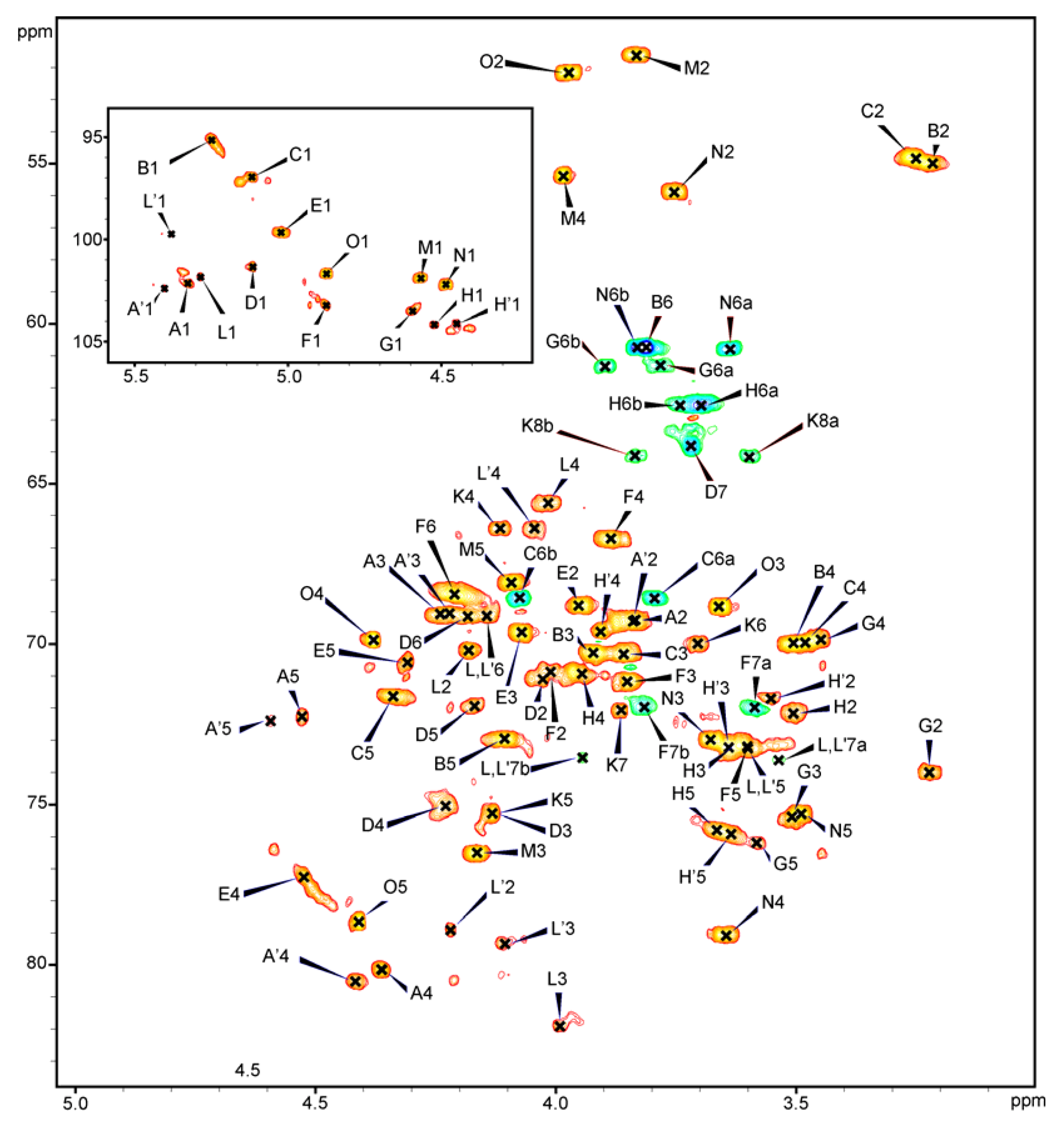
The fraction PSV consisted of the core oligosaccharide substituted with one repeating unit of the O-specific chain was analysed by 1D and 2D 1H, 13C-NMR spectroscopy. All the spin systems (Table 1) were assigned by COSY, TOCSY with different mixing times, HSQC-DEPT (Figure 2), HSQC-TOCSY, NOESY and HMBC spectra. Experimental chemical shift values were compared with previously published NMR data for respective monosaccharides and oligosaccharides [8,14,19].
Table 1. 1H and 13C NMR chemical shifts of the core oligosaccharide substituted with one RU of the O-specific PS of P. shigelloides PCM 2231 LPS (serotyp O17) (fraction PSV). Spectra were recorded for 2H2O solution at 27 °C. Acetone (δH 2.225, δC 31.05 ppm) was used as internal reference.
ResidueChemical shifts (ppm)
H-1H-2H-3a,bH-4H-5H-6a,bH-7a,bH-8a,b
C-1C-2C-3C-4C-5C-6C-7C-8, CH3CO
A→4)-α- D -Gal p A-(1→5.333.834.254.364.53
102.169.269.080.172.3175.6
A′→4)-α- D -Gal p A-(1→5.403.844.224.414.60
102.469.369.080.672.5175.6
Bα- D -Glc p N-(1→5.243.223.913.504.113.81 a
95.155.070.370.073.060.7
C→6)-α-D-GlcpN-(1→5.123.263.863.474.333.79, 4.08
97.054.970.370.071.668.6
D→3,4)-L-α-D-Hepp-(1→5.114.044.134.234.174.173.72 b
101.371.175.375.172.069.263.8
E→4)-α-D-GalpA-(1→5.023.954.074.524.31
99.768.869.777.370.6176.5
F→7)-L-α-D-Hepp-(1→4.884.003.843.893.604.213.58, 3.82
103.270.971.266.773.268.472.0
Gβ-D-Glcp-(1→4.593.223.513.453.593.78, 3.90
103.574.075.469.976.261.3
Hβ-d-Galp-(1→4.513.503.623.953.663.69-3.74 b
104.272.273.171.075.862.6
H′β-d-Galp-(1→4.453.553.643.913.643.69-3.74 b
104.171.773.269.676.062.6
K→5)-Kdo1.86, 2.224.124.133.703.863.60, 3.84
ndnd34.466.475.370.072.164.1
L→3,7)-L-α-D-Hepp-(1→5.284.183.994.013.604.143.55, 3.95
101.870.281.965.673.269.173.5
L′→2,3,7)-L-α-D-Hepp-(1→5.384.224.104.043.604.143.56, 3.94
99.778.979.366.473.269.173.6
M→3)-β-d-FucpNAc4N-(1→4.573.834.163.984.091.322.07
101.951.676.555.468.116.323.0, 174.7
N→4)-β-D-GlcpNAc-(1→4.483.753.683.653.493.63, 3.822.06
102.255.973.079.175.360.823.0, 175.4
Oα-l-AltpNAcA4.883.973.664.384.412.00
101.7 52.268.8 69.9 78.7175.423.0, 175.3
a, b Not resolved; nd: Not determined.
Figure 2. .600 MHz HSQC-DEPT spectrum of the core OS substituted with one RU of the O-specific PS of P. shigelloides PCM 2231 (serotype O17) (fraction PSV). The inset shows the anomeric region of the spectrum. The uppercase letters refer to designations of carbohydrate residues. The spectra were obtained for 2H2O suspensions at 27 °C.
Marinedrugs 11 00440 g002
The HSQC-DEPT spectrum obtained for PSV fraction contained signals for twelve major anomeric protons and carbons, and a Kdo spin system (Figure 2, Table 1).
Residue A with the H-1/C-1 signals at δ 5.33/102.1 ppm, JC-1,H-1 ~176 Hz as well as residue E with the H-1/C-1 signals at δ 5.02/99.7 ppm, JC-1,H-1 ~176 Hz, were assigned as the 4-substituted α-D-GalpA residues based on the characteristic five proton spin systems, the relatively high chemical shifts of the H-5, H-4, and H-3, the high 13C chemical shift of the C-4 (residue A: δ 80.1, residue E: δ 77.3) and C-6 (residue A: δ 175.6, residue E: δ 176.5) signals, large vicinal couplings between H-2, H-3 and the small vicinal couplings between H-3, H-4, and H-5 protons. Residue A′ was also identified due to heterogeneity (see text below) (Table 1).
Residue B with the H-1/C-1 signals at δ 5.24/95.1 ppm, JC-1,H-1 ~176 Hz, was assigned as the terminal α-D-GlcpN based on the chemical shift of the C-2 (δ 55.0), coupling constants between all ring protons.
Residue C with the H-1/C-1 signals at δ 5.12/97.0 ppm, JC-1,H-1 ~175 Hz, was recognized as the 6-substituted α-D-GlcpN based on the chemical shift of the C-2 (δ 54.9) and coupling constants between all ring protons and the characteristic downfield shift of the C-6 signal (δ 68.6).
Residue D with the H-1/C-1 signals at δ 5.11/101.3 ppm, JC-1,H-1 ~173 Hz, was recognized as a 3,4-disubstituted L-glycero-α-D-manno-Hepp based on coupling constants among H-1, H-2, and H-3 and the relative high chemical shift of the C-3 (δ 75.3) and C-4 (δ 75.1) signals.
Residue F with the H-1/C-1 signals at δ 4.88/103.2 ppm, JC-1,H-1 ~176 Hz, was recognized as the 7-substituted L-glycero-α-D-manno-Hepp from the 1H and 13C chemical shifts, the small vicinal couplings between H-1, H-2 and H-3 and similar chemical shifts as those for the monosaccharide L-α-D-Hepp and the relative high chemical shift of the C-7 (δ 72.0).
Residue G with the H-1/C-1 signals at δ 4.59/103.5 ppm, JC-1,H-1 ~163 Hz, was assigned as the terminal β-D-Glcp based on coupling constants between all protons in the sugar ring.
Residue H with the H-1/C-1 signals at δ 4.51/104.2 ppm, JC-1,H-1 ~163 Hz, was recognized as the terminal β-D-Galp due to the large coupling between H-1, H-2, and H-3 and the small vicinal couplings among H-3, H-4, and H-5. Residue H′ was also identified due to heterogeneity of the structure (see text below) (Table 1).
Residue K was identified as a 5-substituted Kdo based on the characteristic deoxy proton signals of H-3a (δ 1.86 ppm), H-3b (δ 2.22 ppm) and a high chemical shift of the C-5 signal (δ 75.3 ppm).
Residue L′ with the H-1/C-1 signals at δ 5.38/99.7 ppm, JC-1,H-1 ~176 Hz, was recognized as the 2,3,7-trisubstituted L-glycero-α-D-manno-Hepp based on 1H and 13C chemical shifts, the small vicinal couplings between H-1, H-2, and H-3, and the relatively high chemical shifts of the C-2 (δ 78.9), C-3 (δ 79.3), and C-7 (δ 73.6) signals. Residue L was also identified due to heterogeneity (see text below) (Table 1).
Residue M with the H-1/C-1 signals at δ 4.57/101.9 ppm, JC-1,H-1 ~168 Hz, was assigned as the 3-substituted β-D-FucpNAc4N based on the characteristic signal of the exocyclic CH3 group (δH 1.32, δC 16.3), the chemical shift of the C-2 (δ 51.6) and C-4 (δ 55.4) signals, the downfield shift of the C-3 signal (δ 76.5), and small vicinal couplings between H-3, H-4 and H-5. Residue M is a constituent of the O-specific PS disaccharide RU.
Residue N with the H-1/C-1 signals at δ 4.48/102.2 ppm, JC-1,H-1 ~163 Hz, was recognized as the 4-substituted β-D-GlcpNAc based on the chemical shift of the C-2 signal (δ 55.9), the relative high chemical shift of the C-4 signal (δ 79.1) and coupling constants between all ring protons.
Residue O with the H-1/C-1 signals at δ 4.88/101.7 ppm, JC-1,H-1 ~167 Hz, was assigned as the terminal α-L-AltpNAcA based on the chemical shift of the C-2 signal (δ 52.2), the high 13C chemical shift of the C-6 (δ 175.4) signal. Residue O is a constituent of the O-specific PS disaccharide RU.
Three additional anomeric signals and spin systems were present in all NMR spectra. Residue L with the H-1/C-1 signals at δ 5.28/101.8 ppm, JC-1,H-1 ~176 Hz, was recognized as the 3,7-disubstituted L-glycero-α-D-manno-Hepp from the 1H and 13C chemical shifts, the small vicinal couplings between H-1, H-2, and H-3, and the relatively high chemical shifts of the C-3 (δ 81.9) and C-7 (δ 73.5) signals. Residue L represented disubstituted variant of residue L′ (2,3,7-trisubstituted L-glycero-α-D-manno-Hepp) caused by the lack of the β-D-Glcp residue at O-2 of L′ residue. Residues A′ (H-1/C-1 signals at δ 5.40/102.4 ppm) and H′ (H-1/C-1 signals at δ 4.45/104.1 ppm) were recognized as variants of residue A (4-substituted α-D-GalpA) and H (terminal β-D-Galp), respectively, due to the absence of the β-D-Glcp residue.
The inter-residue connectivities between the adjacent monosaccharides were observed by NOESY and HMBC experiments (Table 2). Each disaccharide element in the PSV was identified, providing the sequence of monosaccharides. For PSV inter-residue Nuclear Overhauser Effects (NOEs) were found between H-1 of O and H-3 of M, H-1 of M and H-4 of N, H-1 of N and H-6a,b of C, H-1 of C and H-4 of A, H-1 of A and H-3 of L, H-1 of L and H-3 of D, H-1 of D and H-5 of K, H-1 of B and H-4 of E, H-1 of E and H-7a,b of F, H-1 of F and H-7a,b of L/L′, H-1 of G and H-2 of L′ and H-1 of H/H′ and H-4 of D.
Table 2. Selected 3JH,C-connectivities from the anomeric atoms of the core OS substituted with one RU of the O-specific PS of P. shigelloides PCM 2231 LPS (serotype O17).
ResidueAtom H-1/C-1 (ppm)Connectivities toInter-Residue atom/residue
δHδC
A→4)-α-D-GalpA-(1→5.33/102.13.99ndH-3 of L
Bα-D-GlcpN-(1→5.24/95.14.52ndH-4 of E
C→6)-α-D-GlcpN-(1→5.12/97.04.3680.1H-4, C-4 of A
D→3,4)-L-α-D-Hepp-(1→5.11/101.34.1375.3H-5, C-5 of K
E→4)-α-D-GalpA-(1→5.02/99.73.5871.9H-7a, C-7 of F
F→7)-L-α-D-Hepp-(1→4.88/103.23.9573.5H-7b, C-7 of L,L′
Gβ-D-Glcp-(1→4.59/103.5nd78.9C-2 of L
Hβ-D-Galp-(1→4.51/104.24.2375.1H-4, C-4 of D
L→3,7)-L-α-D-Hepp-(1→5.28/101.24.1375.3H-3, C-3 of D
M→3)-β-D-FucpNAc4N-(1→4.57/101.93.6579.1H-4, C-4 of N
N→4)-β-D-GlcpNAc-(1→4.48/102.23.79, 4.0868.6H-6a, H-6b, C-6 of C
Oα-l-AltpNAcA4.88/101.74.1676.5H-3, C-3 of M
nd: Not determined.
In the fraction PSV, glycine was identified by the presence of an additional carbonyl resonance at δC 168.7 ppm in the HMBC spectrum and a negative CH2 crosspeak (δH 3.98 ppm, δC 40.9 ppm) in the HSQC-DEPT spectrum. The presence of glycine in the PSV component was further supported by amino acid analysis and mass spectrometry; however connectivity between the glycine and the oligosaccharide was not determined.
These studies demonstrate the structure of the complete core oligosaccharide and linkage between the core region and O-specific PS of lipopolysaccharide P. shigelloides PCM 2231 (serotype O17). The structure of the complete core region of P. shigelloides PCM 2231 is similar to that of the core OSs of P. shigelloides strains 7-63 (serotype O17) [14] and 302-73 (serotype O1) [10]. The core OS of strain 7-63 (serotype O17) differs only by a single terminal residue of branched chain, that is, terminal α-D-GalpN was present instead of the terminal α-D-GlcpN (residue B) in strain PCM 2231 (serotype O17). The core OS of P. shigelloides 302-73 (serotype O1) contains Kdo as a residue linking the O-specific polysaccharide with the outer core region instead of β-D-GlcpNAc (residue N) present in strain PCM 2231 (serotype O17).

2.5. Serological Studies

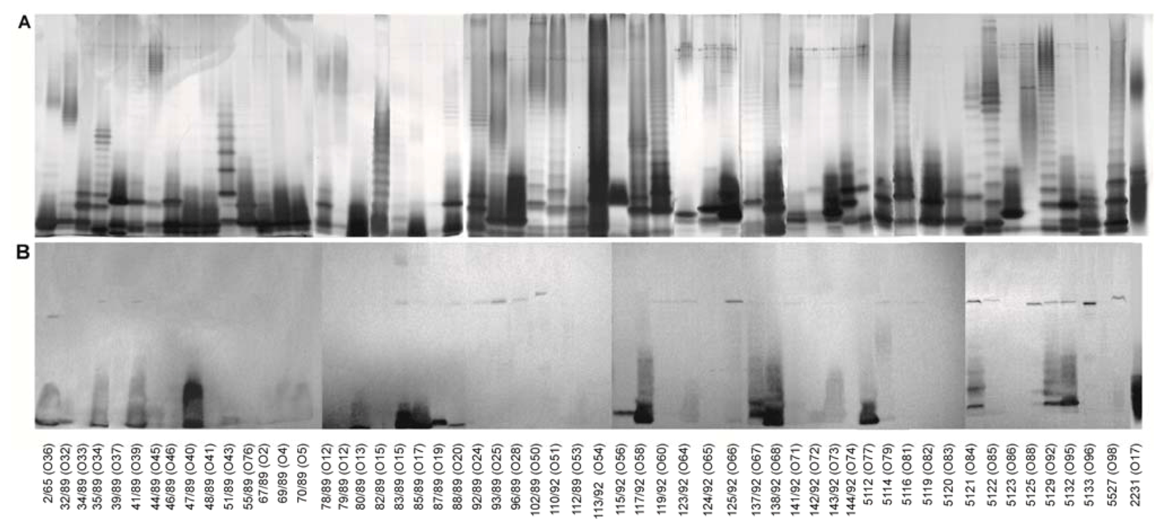
The core oligosaccharide (OSI) was conjugated with BSA (OSI-BSA) with the use of high temperature conjugation described by Boratynski et al. [20]. The polyclonal antisera obtained by immunization of rabbits with the conjugate were used to scan LPSs isolated from 55 strains of P. shigelloides for the presence of epitopes similar to those found in LPS of P. shigelloides PCM 2231 (serotype O17). The obtained conjugate was a good immunogen inducing high level of specific anti-core OS antibodies as was shown in a solid-phase ELISA with homologous LPS as an antigen (data not shown). LPSs were isolated by hot phenol/water extraction. Most of them were recovered from both the water and the phenol phase. LPSs were separated by SDS-PAGE (Figure 3A) and stained using the method of Tsai [21]. The reactivities of anti-conjugate sera with LPSs isolated from various P. shigelloides strains were examined with the use of immunoblotting (Figure 3B). Results for LPSs which were recovered from the phenol phases were identical in comparison with the water phase derived LPSs (data not shown). To simplify presented data, results for the water phase derived LPSs were shown with the exception of LPSs CNCTC 39/89, 102/89, 5112, recovered from the phenol phase.
The antibodies against the OSI-BSA conjugate reacted mainly with fast migrating LPS fractions, representing LPS with unsubstituted core oligosaccharide. However reactions with core OS epitopes present in smooth lipopolysaccharides were also observed. Serological screening of different strains of P. shigelloides indicates that similar epitopes might also be present in the core OS of the 28 out of 55 strains. Intensities of cross-reactions observed for different LPSs were diversified. Strong cross-reactions were observed with the fast migrating LPS fractions of strains CNCTC 2/65, 35/89, 41/89, 47/89, 80/89, 83/89, 85/89, 87/89, 88/89, 115/92, 117/92, 137/92, 138/92, 5112, 5121, 5129, and 5132 (Figure 3). Weak reactions were observed for LPSs P. shigelloides CNCTC 32/89, 51/89, 69/89, 70/89, 78/89, 123/92, 125/92, 142/92, 143/92, 5114, and 5527.
Figure 3. LPSs were analysed by SDS-PAGE (3 μg/lane), using a 15% separating gel, and visualized by the silver staining method (A). Reactivities of serum (200-fold diluted) against the OSI-BSA conjugate with P. shigelloides LPSs in immunoblotting (B).
Marinedrugs 11 00440 g003

3. Experimental Section

3.1. Bacteria

P. shigelloides strain PCM 2231 (serovar O17:H11) was obtained from the Polish Collection of Microorganisms (PCM) of the Institute of Immunology and Experimental Therapy, Wroclaw, Poland and 55 available different P. shigelloides strains CNCTC 2/65 (O36), 32/89 (O32), 34/89 (O33), 35/89 (O34), 39/89 (O37), 41/89 (O39), 44/89 (O45), 46/89 (O46), 47/89 (O40), 48/89 (O41), 51/89 (O43), 55/89 (O76), 67/89 (O2), 69/89 (O4), 70/89 (O5), 78/89 (O12), 79/89 (O12), 80/89 (O13), 82/89 (O15), 83/89 (O15), 85/89 (O17), 87/89 (O19), 88/89 (O20), 92/89 (O24), 93/89 (O25), 96/89 (O28), 102/89 (O50), 110/92 (O51), 112/92 (O53), 113/92 (O54), 115/92 (O56), 117/92 (O58), 119/92 (O60), 123/92 (O64), 124/92 (O65), 125/92 (O66), 137/92 (O67), 138/92 (O68), 141/92 (O71), 142/92 (O72), 143/92 (O73), 144/92 (O74), 5112 (O77), 5114 (O79), 5116 (O81), 5119 (O82), 5120 (O83), 5121 (O84), 5122 (O85), 5123 (O86), 5125 (O88), 5129 (O92), 5132 (O95), 5133 (O96), 5527 (O98) were obtained from the collection of the Institute of Hygiene and Epidemiology, Prague, Czech Republic. Bacteria were grown and harvested as described previously [22].

3.2. Lipopolysaccharide and Core Oligosaccharide

The LPS was extracted from bacterial cells by the hot phenol/water method [23] and purified as previously reported [7]. The yield of LPS was ~1.8% of the dry bacterial mass. LPS (200 mg) was degraded by treatment with 1.5% acetic acid containing 2% SDS at 100 °C for 15 min. The reaction mixture was freeze-dried, the SDS removed by extraction with 96% ethanol, and the residue suspended in water and centrifuged. The poly- and oligosaccharides were separated by gel permeation chromatography, performed on Bio-Gel 10 column, equilibrated with 0.05 M pyridine/acetic acid buffer of pH 5.6. Eluates were monitored with a Knauer differential refractometer and all fractions were freeze-dried and checked by MALDI-TOF mass spectrometry.

3.3. Analytical Methods

Monosaccharides were analysed as their alditol acetates by GC-MS [24]. Absolute configurations of the monosaccharides were determined as described by Gerwig et al. [25,26] using (−)-2-butanol for the formation of 2-butyl glycosides. The trimethylsilylated butyl glycosides produced from authentic samples were used as standards. Carboxyl reduction of the core oligosaccharide fractions was carried out according to the method of Taylor et al. [27] as previously described [22]. Methylation analyses were performed on both native and carboxyl reduced oligosaccharides according to the method of Hakomori [28]. Alditol acetates and partially methylated alditol acetates were analysed with a Hewlett-Packard 5972 system using the HP-1 fused-silica capillary column (0.2 mm × 12.5 m), He as carrier gas, flow rate 1 mL·min−1 and a temperature program 150 → 270 °C at 8 °C·min−1.

3.4. Mass Spectrometry

MALDI-TOF MS spectra (reflectron negative-ion mode) were obtained on a MDS SCIEX 4800 MALDI TOF/TOF and Bruker Autoflex III Tof/Tof instruments. Samples were dissolved in water (0.5 mg/mL) and desodiated with Dowex 50 W × 8 (H+). 2,4,6-Trihydroxyacetophenone (25 mg/mL in acetonitrile:water, 1:1, v/v) was used as a matrix.

3.5. NMR Spectroscopy

All NMR spectra were obtained on Bruker 600 MHz spectrometer (Laboratory of Structural Analyses, Wroclaw University of Technology, Wroclaw, Poland). The fraction PSV containing the core oligosaccharides substituted with one repeating unit of the O-specific chain was first repeatedly exchanged with 2H2O (99.9%) with intermediate lyophilisation. NMR spectra were obtained for 2H2O solutions at 27 °C using acetone (δH 2.225 ppm, δC 31.05 ppm) as internal reference. The signals were assigned by one- and two-dimensional experiments (COSY, TOCSY, NOESY, HMBC, HSQC-DEPT and HSQC-TOCSY). The JC-1,H-1 values were obtained from non-decoupled HSQC-DEPT experiment. In the TOCSY experiments the mixing times were 60, 90 and 120 ms. The delay time in the HMBC was 60 ms and the mixing time in the NOESY experiment was 60 ms.
All spectra were acquired and processed using standard Bruker software. The processed 2D spectra were assigned using the SPARKY program [29].

3.6. Preparation of Oligosaccharide Conjugate with BSA

The core oligosaccharide (OSI) was oxidised and purified as describe previously [30]. Briefly, the core oligosaccharide OSI (10 mg) was oxidised with 0.1 M NaIO4 (1 mL) at 21 °C in the dark for 60 min. Then ethylene glycol (10 μL) was added, and the solution was incubated at 21 °C for an additional 60 min. The reaction mixture was then applied directly on the Sephadex G-10 column, equilibrated with 0.05 M pyridine/acetic acid buffer of pH 5.6, and the first eluted fraction was lyophilised. The conjugation was carried out as described previously [20]. The oxidised core oligosaccharide OSI (3.8 mg) dissolved in H2O (200 μL) was mixed with an equal volume of BSA solution in H2O (2 mg/200 μL). Dimethylformamide was added to a final concentration of 2%, and the mixture was freeze-dried. Dry preparation was heated at 105 °C for 30 min, dissolved in H2O (1 mL), and dialyzed against H2O. The antigenic properties of the product were analysed by immunoblotting, using polyclonal rabbit serum against P. shigelloides PCM 2231.

3.7. Immunization Procedures

Rabbits were housed at the animal facility of the Institute of Immunology and Experimental Therapy (Wroclaw, Poland), and all the experiments were carried out according to the procedures approved by the Local Ethical Commission. Rabbits were immunized with the OSI-BSA conjugate, suspended in a complete Freund’s adjuvant, and polyclonal antibodies against the conjugate were obtained by the procedures previously described [31].

3.8. ELISA

Enzyme-Linked immunosorbent assay (ELISA), using LPS as solid-phase antigen, was performed with a modification [30] of the method described by Voller et al. [32]. The detection system consisted of a goat anti-rabbit IgG conjugated with alkaline phosphatase (Bio-Rad, Hercules, CA, USA) as a second antibody and p-nitrophenyl phosphate as a substrate.

3.9. SDS-PAGE

The LPS was analysed by SDS-PAGE according to the method of Laemmli [33] with modifications described previously [24] and LPS bands were visualised by the silver staining method [21].

3.10. Immunoblotting

Immunoblotting was performed on the SDS-PAGE-separated LPS fractions as previously described [34]. A goat anti-rabbit IgG conjugated with alkaline phosphatase (Bio-Rad) was used as the secondary antibody, and 5-bromo-4-chloro-3-indolyl phosphate/nitroblue tetrazolium was applied as a detection system.

4. Conclusions

Here we presented the structure of a complete core oligosaccharide composed of an undecasaccharide, which represents the second core type for serotype O17 of P. shigelloides. The core OS of P. shigelloides PCM 2231 is heterogeneous. The heterogeneity corresponded to the absence of terminal β-D-Glcp residue. Three minor glycoforms represent the complete core OS devoid of β-D-GlcpNAc residue, both terminal β-D-Glcp and hexosamine residues, and both terminal β-D-Glcp and β-D-GlcpNAc residues.
Serological screening of different strains of P. shigelloides indicates that identical or similar epitopes to P. shigelloides PCM 2231 might also be present in the core region of 28 out of 55 strains (51%). This observation suggests that the core structure in LPS of strain PCM 2231 (serotype O17) could be the most common type in P. shigelloides.
Previously published serological studies did not indicate the presence of a common lipopolysaccharide core type of P. shigelloides. Similar studies with the use of the antibodies against core OS conjugate were reported for strain P. shigelloides 113/92 (serotype O54) indicating serological cross-reactivity was limited to only three strains [8]. This is therefore the first report of the most common core OS structure within P. shigelloides LPS.

Acknowledgments

The authors wish to thank Piotr Jakimowicz, Faculty of Biotechnology, University of Wroclaw for help and assistance with the MALDI-TOF measurement. This work was supported by Grant N N401 1812 33 from the Ministry of Science and Higher Education, Poland. This paper describes the revised version of the structure previously presented at 25th International Carbohydrate Symposium, 01-06.08.2010, Tokyo, Japan.

Conflict of Interests

The authors declare no conflict of interest.

References

  1. Garrity, G.M.; Bell, J.A.; Lilburn, T.G. Bergey’s Taxonomic Outline. Available online: http://dx.doi.org/10.1007/bergeysoutline200310 (accessed on 31 January 2013).
  2. Stock, I. Plesiomonas shigelloides: An emerging pathogen with unusual properties. Rev. Med. Microbiol. 2004, 15, 129–139. [Google Scholar] [CrossRef]
  3. Linnerborg, M.; Widmalm, G.; Weintraub, A.; Albert, M.J. Structural elucidation of the O-antigen lipopolysaccharide from two strains of Plesiomonas shigelloides that share a type-specific antigen with Shigella flexneri 6, and the common group 1 antigen with Shigella flexneri spp. and Shigella dysenteriae 1. Eur. J. Biochem. 1995, 231, 839–844. [Google Scholar] [CrossRef]
  4. Czaja, J.; Jachymek, W.; Niedziela, T.; Lugowski, C.; Aldova, E.; Kenne, L. Structural studies of the O-specific polysaccharide from Plesiomonas shigelloides strain CNCTC 113/92. Eur. J. Biochem. 2000, 267, 1672–1679. [Google Scholar] [CrossRef]
  5. Lukasiewicz, J.; Dzieciatkowska, M.; Niedziela, T.; Jachymek, W.; Augustyniuk, A.; Lugowski, C.; Kenne, L. Complete lipopolysaccharide of Plesiomonas shigelloides O74:H5 (strain CNCTC 144/92) 2. Lipid A, its structural variability, the linkage to the core oligosaccharide, and the biological activity of lipopolysaccharide. Biochemistry 2006, 45, 10434–10447. [Google Scholar] [CrossRef]
  6. Lukasiewicz, J.; Niedziela, T.; Jachymek, W.; Kenne, L.; Lugowski, C. Structure of the lipid A-inner core region and biological activity of Plesiomonas shigelloides O54 (strain CNCTC 113/92) lipopolysaccharide. Glycobiology 2006, 16, 538–550. [Google Scholar] [CrossRef]
  7. Niedziela, T.; Dag, S.; Lukasiewicz, J.; Dzieciatkowska, M.; Jachymek, W.; Lugowski, C.; Kenne, L. Complete lipopolysaccharide of Plesiomonas shigelloides O74:H5 (strain CNCTC 144/92). 1. Structural analysis of the highly hydrophobic lipopolysaccharide, including the O-antigen, its biological repeating unit, the core oligosaccharide, and the linkage between them. Biochemistry 2006, 45, 10422–10433. [Google Scholar]
  8. Niedziela, T.; Lukasiewicz, J.; Jachymek, W.; Dzieciatkowska, M.; Lugowski, C.; Kenne, L. Core oligosaccharides of Plesiomonas shigelloides O54:H2 (strain CNCTC 113/92)—Structural and serological analysis of the lipopolysaccharide core region, the O-antigen biological repeating unit, and the linkage between them. J. Biol. Chem. 2002, 277, 11653–11663. [Google Scholar]
  9. Pieretti, G.; Corsaro, M.M.; Lanzetta, R.; Parrilli, M.; Canals, R.; Merino, S.; Tomás, J.M. Structural studies of the O-chain polysaccharide from Plesiomonas shigelloides strain 302–73 (serotype O1). Eur. J. Org. Chem. 2008, 2008, 3149–3155. [Google Scholar]
  10. Pieretti, G.; Corsaro, M.M.; Lanzetta, R.; Parrilli, M.; Vilches, S.; Merino, S.; Tomás, J.M. Structure of the core region from the lippolysaccharide of Plesiomonas shigelloides strain 302–73 (serotype O1). Eur. J. Org. Chem. 2009, 2009, 1365–1371. [Google Scholar] [CrossRef]
  11. Pieretti, G.; Carillo, S.; Lindner, B.; Lanzetta, R.; Parrilli, M.; Jimenez, N.; Regue, M.; Tomas, J.M.; Corsaro, M.M. The complete structure of the core of the LPS from Plesiomonas shigelloides 302–73 and the identification of its O-antigen biological repeating unit. Carbohydr. Res. 2010, 345, 2523–2528. [Google Scholar] [CrossRef]
  12. Maciejewska, A.; Lukasiewicz, J.; Niedziela, T.; Szewczuk, Z.; Lugowski, C. Structural analysis of the O-specific polysaccharide isolated from Plesiomonas shigelloides O51 lipopolysaccharide. Carbohydr. Res. 2009, 344, 894–900. [Google Scholar] [CrossRef]
  13. Sawen, E.; Ostervall, J.; Landersjo, C.; Edblad, M.; Weintraub, A.; Ansaruzzaman, M.; Widmalm, G. Structural studies of the O-antigenic polysaccharide from Plesiomonas shigelloides strain AM36565. Carbohydr. Res. 2012, 348, 99–103. [Google Scholar] [CrossRef]
  14. Kubler-Kielb, J.; Mocca, C.; Vinogradov, E. The elucidation of the structure of the core part of the LPS from Plesiomonas shigelloides serotype O17 expressing O-polysaccharide chain identical to the Shigella sonnei O-chain. Carbohydr. Res. 2008, 343, 3123–3127. [Google Scholar] [CrossRef]
  15. Batta, G.; Liptak, A.; Schneerson, R.; Pozsgay, V. Conformational stabilization of the altruronic acid residue in the O-specific polysaccharide of Shigella sonnei/Plesiomonas shigelloides. Carbohydr. Res. 1997, 305, 93–99. [Google Scholar] [CrossRef]
  16. Kenne, L.; Lindberg, B.; Petersson, C.; Katzenellenbogen, E.; Romanowska, E. Structural studies of the O-specific side chains of the Shigella sonnei phase I lipopolysaccharide. Carbohydr. Res. 1980, 78, 119–126. [Google Scholar] [CrossRef]
  17. Aldova, E. Serovars of Plesiomonas shigelloides. Zentralbl. Bakteriol. 1994, 281, 38–44. [Google Scholar]
  18. Aldova, E. The importance of serotyping Plesiomonas shigelloides. Epidemiol. Mikrobiol. Immunol. 1995, 44, 147–154. [Google Scholar]
  19. Gorin, P.A.J.; Mazurek, M. Further studies on the assignment of signals in 13C magnetic resonance spectra of aldoses and derived methyl glycosides. Can. J. Chem. 1975, 53, 1212–1223. [Google Scholar] [CrossRef]
  20. Boratynski, J.; Roy, R. High temperature conjugation of proteins with carbohydrates. Glycoconj. J. 1998, 15, 131–138. [Google Scholar] [CrossRef]
  21. Tsai, C.M.; Frasch, C.E. A sensitive silver stain for detecting lipopolysaccharides in polyacrylamide gels. Anal. Biochem. 1982, 119, 115–119. [Google Scholar] [CrossRef]
  22. Petersson, C.; Niedziela, T.; Jachymek, W.; Kenne, L.; Zarzecki, P.; Lugowski, C. Structural studies of the O-specific polysaccharide of Hafnia alvei strain PCM 1206 lipopolysaccharide containing D-allothreonine. Eur. J. Biochem. 1997, 244, 580–586. [Google Scholar]
  23. Westphal, O.; Jann, K. Bacterial lipopolysacharides: Extraction with phenol-water and further applications of the procedure. Methods Carbohydr. Chem. 1965, 5, 83–89. [Google Scholar]
  24. Niedziela, T.; Petersson, C.; Helander, A.; Jachymek, W.; Kenne, L.; Lugowski, C. Structural studies of the O-specific polysaccharide of Hafnia alvei strain 1209 lipopolysaccharide. Eur. J. Biochem. 1996, 237, 635–641. [Google Scholar]
  25. Gerwig, G.J.; Kamerling, J.P.; Vliegenthart, J.F.G. Determination of the D and L configuration of neutral monosaccharides by high-resolution capillary GLC. Carbohydr. Res. 1978, 62, 349–357. [Google Scholar] [CrossRef]
  26. Gerwig, G.J.; Kamerling, J.P.; Vliegenthart, J.F.G. Determination of the absolute configuration of monosaccharides in complex carbohydrates by capillary GLC. Carbohydr. Res. 1979, 77, 1–7. [Google Scholar] [CrossRef]
  27. Taylor, R.L.; Shively, J.E.; Conrad, H.E. Stoichiometric reduction of uronic acid carboxyl groups in polysaccharides. Methods Carbohydr. Chem. 1976, 7, 149–151. [Google Scholar]
  28. Hakomori, S. A rapid permethylation of glycolipid and polysaccharide catalyzed by methylsulphinyl carbanion in dimethyl sulphoxide. J. Biochem. 1964, 55, 205–208. [Google Scholar]
  29. Goddard, T.D.; Kneller, D.G. Sparky, 3rd ed; University of California: San Francisco, CA, USA, 2001. [Google Scholar]
  30. Jennings, H.J.; Lugowski, C. Immunochemistry of groups A, B, and C meningococcal polysaccharide-tetanus toxoid conjugates. J. Immunol. 1981, 127, 1011–1018. [Google Scholar]
  31. Lugowski, C.; Romanowska, E. Enterobacterial common antigen: Isolation from Shigella sonnei, purification and immunochemical characterization. Eur. J. Biochem. 1978, 91, 89–97. [Google Scholar] [CrossRef]
  32. Voller, A.; Draper, C.; Bidwell, D.E.; Bartlett, A. Microplate enzyme-linked immunosorbent assay for chagas’ disease. Lancet 1975, 1, 426–428. [Google Scholar]
  33. Laemmli, U.K. Cleavage of structural proteins during the assembly of the head of bacteriophage T4. Nature 1970, 227, 680–685. [Google Scholar] [CrossRef]
  34. Lugowski, C.; Jachymek, W.; Niedziela, T.; Rowinski, S. Serological characterisation of anti-endotoxin sera directed against the conjugates of oligosaccharide core of Escherichia coli type R1, R2, R3, J5 and Salmonella Ra with tetanus toxoid. FEMS Immunol. Med. Microbiol. 1996, 16, 21–30. [Google Scholar]