Article

Cyclic Bis-1,3-dialkylpyridiniums from the Sponge Haliclona sp.

1
Natural Products Research Institute, College of Pharmacy, Seoul National University, San 56-1, Sillim, Gwanak, Seoul 151-742, Korea
2
Department of Agricultural Biotechnology, College of Agriculture & Life Science, Seoul National University, San 56-1, Sillim, Gwanak, Seoul 151-921, Korea
3
Department of Biological Science, College of Life Science and Nano Technology, Hannam University, 461-6 Jeonmin, Yuseong, Daejeon 305-811, Korea
*
Authors to whom correspondence should be addressed.
Mar. Drugs 2012, 10(9), 2126-2137; https://doi.org/10.3390/md10092126
Submission received: 12 July 2012 / Revised: 9 August 2012 / Accepted: 14 September 2012 / Published: 24 September 2012

Abstract

:
Eight novel cyclic bis-1,3-dialkylpyridiniums, as well as two known compounds from the cyclostellettamine class, were isolated from the sponge Haliclona sp. from Korea. Structures of these novel compounds were determined using combined NMR and FAB-MS/MS analyses. Several of these compounds exhibited moderate cytotoxic and antibacterial activities against A549 cell-line and Gram-positive strains, respectively. The structure-activity relationships of cyclostellettamines are discussed based on their bioactivities.

Graphical Abstract

1. Introduction

The 1,3-dialkylpyridiniums (with both cyclic and linear frameworks) are widely distributed cytotoxic compounds that play a role in the antimicrobial properties of marine sponges of the order Haplosclerida (genera Amphimedon, Haliclona, and Xestospongia) [1,2,3,4]. Previous studies have evaluated 1,3-dialkylpyridinium inhibition of histone deacetylase (HDAC) [5,6] and epidermal growth factor (EGF) receptor-mediated mitogenesis [7]. These compounds are also known to block the interaction of chemokines [8] and the binding of methyl quinuclidinyl benzilate (QNB) to the muscarinic receptor [9,10]. In addition to their chemical and bioactivity properties, these metabolites are considered to be the biogenetic precursors of a wide variety of compounds with complicated carbon skeletons and diverse functionalities, such as halicyclamines, ingenamines, manzamines, and sarains [2].
When evaluating bioactive metabolites from Korean marine invertebrates, we recently identified haliclonin A, a cytotoxic macrocylic bisamide from the sponge Haliclona sp. [11]. The novel carbon skeleton of this compound was highly reminiscent of bis-1,3-dialkylpyridiniums, especially cyclostellettamines isolated from Haliclona sp. [9,12]. Thus, we searched for similar 1,3-dialkylpyridinium metabolites in the polar fractions of extracts from diverse collections of this sponge. Here, we report eight novel derivatives of cyclostellettamines with diverse alkyl chains. Several of these compounds exhibited moderate cytotoxicity against the lung cancer A549 cell-line and antibacterial activities against Gram-positive strains. Additional information on their structure-activity relationships were deduced based on their cytotoxicity and antibacterial activity.

2. Results and Discussion

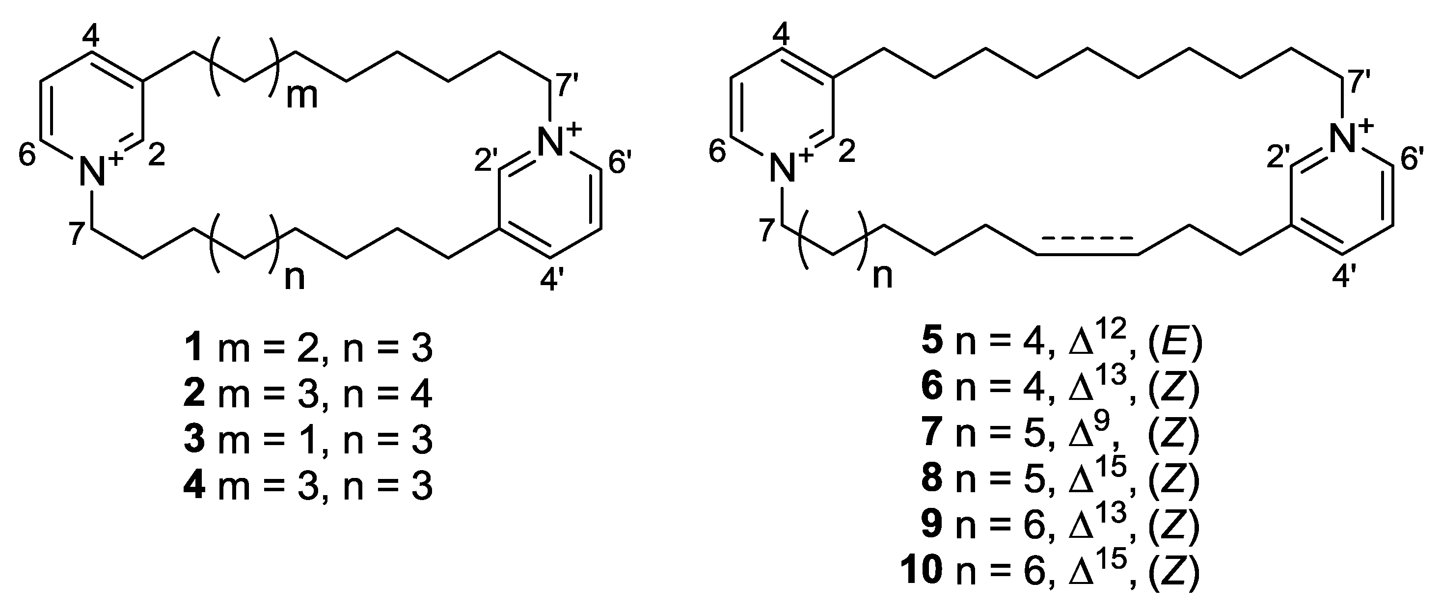
The sponge Haliclona sp. specimens were lyophilized, macerated, and repeatedly extracted with CH2Cl2 and MeOH. The combined extracts were separated by solvent-partitioning followed by ODS vacuum flash chromatography. The highly polar H2O-MeOH (50:50) chromatographic fraction was further fractionated by Sephadex LH-20 gel-permeation chromatography and separated by ODS-HPLC to yield 10 compounds (Figure 1). The structures of compounds 1 and 2 were identified as cyclostellettamine N and Q, respectively, based on spectroscopic analyses and a comparison of NMR and FAB-MS/MS data [13,14].
Figure 1. Structures of compounds 110.
Marinedrugs 10 02126 g001
The molecular formula of compound 3, with the shortest retention time in reversed-phase HPLC, was C28H42Cl2N2 based on the quasi-molecular ion clusters at m/z 407.3428 [M − H]+ and 443.3195 [M + Cl]+ in the HR-FAB-MS data. The spectroscopic data of this compound were very similar to those of 1 and 2 (Table 1 and Table 2).
Table 1. 13C NMR (ppm, mult) assignments for compounds 310 in MeOH-d4.
Position345678910
2145.5, CH145.6,CH145.3, CH145.3, CH145.2, CH145.6, CH145.2, CH145.3, CH
3145.3, C145.3, C145.7, C145.5, C146.7, C145.3, C145.7, C145.5, C
4147.0, CH146.9, CH146.9, CH147.0, CH146.9, CH146.9, CH146.9, CH147.0, CH
5129.2, CH129.2, CH129.2, CH128.8, CH129.2, CH129.1, CH129.1, CH129.2, CH
6143.4, CH143.4, CH143.4, CH143.5, CH143.5, CH143.4, CH143.5, CH143.4, CH
762.7, CH262.8, CH262.8, CH262.8, CH262.9, CH262.9, CH262.9, CH262.8, CH2
832.2, CH232.1, CH232.2, CH232.3, CH224.8, CH232.3, CH232.3, CH232.1, CH2
926.7, CH226.5, CH226.2, CH226.7, CH2137.6, CH26.9, CH226.8, CH226.8, CH2
1029.5 a, CH229.6 b, CH229.6, CH230.0 d, CH2124.7, CH29.7 f, CH230.5, CH230.1 h, CH2
1129.8 a, CH230.0 b, CH232.7, CH230.2, CH228.1, CH230.3 f, CH230.7, CH230.2 h, CH2
1229.9 a, CH230.0 b, CH2132.3, CH28.0, CH230.5, CH230.1 f, CH228.0, CH230.2 h, CH2
1330.2 a, CH230.1 b, CH2130.7, CH133.3, CH29.2 e, CH230.9, CH2131.1, CH30.7, CH2
1429.2, CH229.2, CH232.4, CH2128.0, CH29.2 e, CH228.3, CH2130.4, CH28.2, CH2
1531.3, CH231.2, CH231.1, CH229.2, CH229.6 e, CH2135.7,CH27.9, CH2133.5, CH
1633.2, CH233.1, CH233.2, CH233.2, CH231.1, CH2125.1,CH29.7, CH2127.8, CH
17- - - - 33.1, CH231.0, CH231.3, CH229.1, CH2
18- - - - - - 33.3,CH33.2, CH2
2′145.5,CH145.6, CH145.3, CH145.5, CH145.2, CH144.6, CH145.2,CH145.5, CH
3′145.4, C145.3, C145.5, C144.9, C145.7, C144.7, C145.7, C145.0, C
4′146.9, CH146.9, CH146.9, CH147.2, CH147.0, CH146.7, CH146.9, CH147.3, CH
5′129.2,CH129.2, CH129.1, CH129.2, CH129.2, CH129.2, CH129.1, CH128.9, CH
6′143.5,CH143.4, CH143.5, CH143.5, CH143.5, CH143.7, CH143.5, CH143.4, CH
7′62.7, CH262.8, CH262.8, CH262.9, CH262.8, CH262.8, CH262.8, CH262.9, CH2
8′31.9, CH232.1, CH232.2, CH232.4, CH232.0, CH232.2, CH232.3, CH232.6, CH2
9′26.4,CH226.5, CH226.6, CH226.8, CH226.5, CH226.6, CH226.7, CH227.0, CH2
10′30.3 a, CH229.6 b, CH229.9 c, CH229.7 d, CH229.7 e, CH229.1 f, CH229.6 g, CH230.3 h, CH2
11′30.4 a, CH230.0 b, CH229.9 c, CH229.9 d, CH230.1 e, CH229.9 f, CH229.9 g, CH230.4 h, CH2
12′29.0, CH230.0 b, CH230.0 c, CH230.0 d, CH230.1 e, CH229.9 f, CH230.0 g, CH230.5 h, CH2
13′30.9, CH230.1 b, CH230.0 c, CH230.4 d, CH230.2 e, CH230.0 f, CH230.1 g, CH230.7 h, CH2
14′33.0, CH229.2, CH229.2,CH228.7, CH229.1, CH229.1, CH229.4, CH229.1, CH2
15′- 31.2, CH231.2, CH230.7, CH230.9, CH230.4, CH231.3, CH231.1, CH2
16′- 33.1, CH233.0, CH232.9, CH233.2, CH233.1, CH233.3, CH233.1, CH2
a–h: Interchangeable signals.
Table 2. 1H MMR (δ, mult, J in Hz) assignments for compounds 36 in MeOH-d4.
Position3456
28.96 (1H, s)8.98 (1H, s)8.95 (1H, s)8.99 (1H, s)
3----
48.45 (1H, d, 7.6)8.46 (1H, d, 8.0)8.46 (1H, d, 8.2)8.45 (1H, d, 8.2)
58.02 (1H, dd, 7.7, 6.2)8.02 (1H, dd, 7.7, 6.4)8.02 (1H, dd, 7.4, 6.1)8.01 (1H, dd, 7.4, 6.6)
68.83 (1H, dd, 6.0)8.85 (1H, dd, 6.0)8.83 (1H, br s)8.84 (1H, dd, 6.2)
74.64 (1H, t, 6.8)4.65 (2H, t, 6.8)4.65 (2H, t, 7.2)4.64 (2H, t, 6.9)
82.00 (2H, quint, 7.3)2.01 (2H, tt, 7.4, 6.8)2.03 (2H, m)2.00 (2H, m)
91.26 (2H, m)1.25 (2H, m)1.29 (2H, m)1.23 (2H, m)
101.25 ~ 1.40 (2H, m)1.20 ~ 1.36 (2H, m)1.42 (2H, m)1.26 (2H, m)
111.25 ~ 1.40 (2H, m)1.20 ~ 1.36 (2H, m)2.01 (2H, m)1.24 (2H, m)
121.25 ~ 1.40 (2H, m)1.20 ~ 1.36 (2H, m)5.41 (1H, br t, 3.6)1.74 (2H, m)
131.25 ~ 1.40 (2H, m)1.20 ~ 1.36 (2H, m)5.41 (1H, br t, 3.6)5.38 (2H, m)
141.30 (2H, m)1.28 (2H, m)2.04 (2H, m)5.38 (2H, m)
151.72 (2H, quint, 7.4)1.74 (2H, tt, 7.3, 7.3)1.78 (2H, m)2.46 (2H, dd, 12.9, 6.5)
162.89 (2H, 7.3)2.90 (2H, t, 7.3)2.88 (2H, t, 7.6)2.95 (2H, t, 6.9)
17----
18----
2′8.96 (1H, s)8.98 (1H, s)8.98 (1H, s)8.95 (1H, s)
3′----
4′8.44 (1H, d, 7.7)8.46 (1H, d, 8.0)8.45 (1H, d, 6.8)8.45 (1H, d, 8.2)
5′8.02 (1H, dd, 7.7, 6.2)8.02 (1H, dd, 7.7, 6.4)8.01 (1H, dd, 7.4, 6.1)8.02 (1H, dd, 7.4, 6.6)
6′8.82 (1H, dd, 6.0)8.85 (1H, dd, 6.0)8.84 (1H, br s)8.85 (1H, dd, 6.2)
7′4.64 (1H, t, 6.5)4.65 (2H, t, 6.8)4.63 (2H, t, 6.7)4.63 (2H, t, 7.5)
8′2.02 (2H, tt, 7.8, 6.9)2.01 (2H, tt, 7.4, 6.8)2.00 (2H, m)1.98 (2H, m)
9′1.20 (2H, m)1.25 (2H, m)1.28 (2H, m)1.24 (2H, m)
10′1.25 ~ 1.40 (2H, m)1.20 ~ 1.36 (2H, m)1.27 ~ 1.40 (2H, m)1.23 ~ 1.35 (2H, m)
11′1.25 ~ 1.40 (2H, m)1.20 ~ 1.36 (2H, m)1.27 ~ 1.40 (2H, m)1.23 ~ 1.35 (2H, m)
12′1.20 (2H, m)1.20 ~ 1.36 (2H, m)1.27 ~ 1.40 (2H, m)1.23 ~ 1.35 (2H, m)
13′1.76 (2H, tt, 7.4, 7.2)1.20 ~ 1.36 (2H, m)1.27 ~ 1.40 (2H, m)1.23 ~ 1.35 (2H, m)
14′2.91 (2H, t, 7.0)1.28 (2H, m)1.30 (2H, m)1.25 (2H, m)
15′-1.74 (2H, tt, 7.3, 7.3)1.73 (2H, m)1.77 (2H, m)
16′-2.90 (2H, t, 7.3)2.89 (2H, t, 7.0)2.92 (2H, t, 6.9)
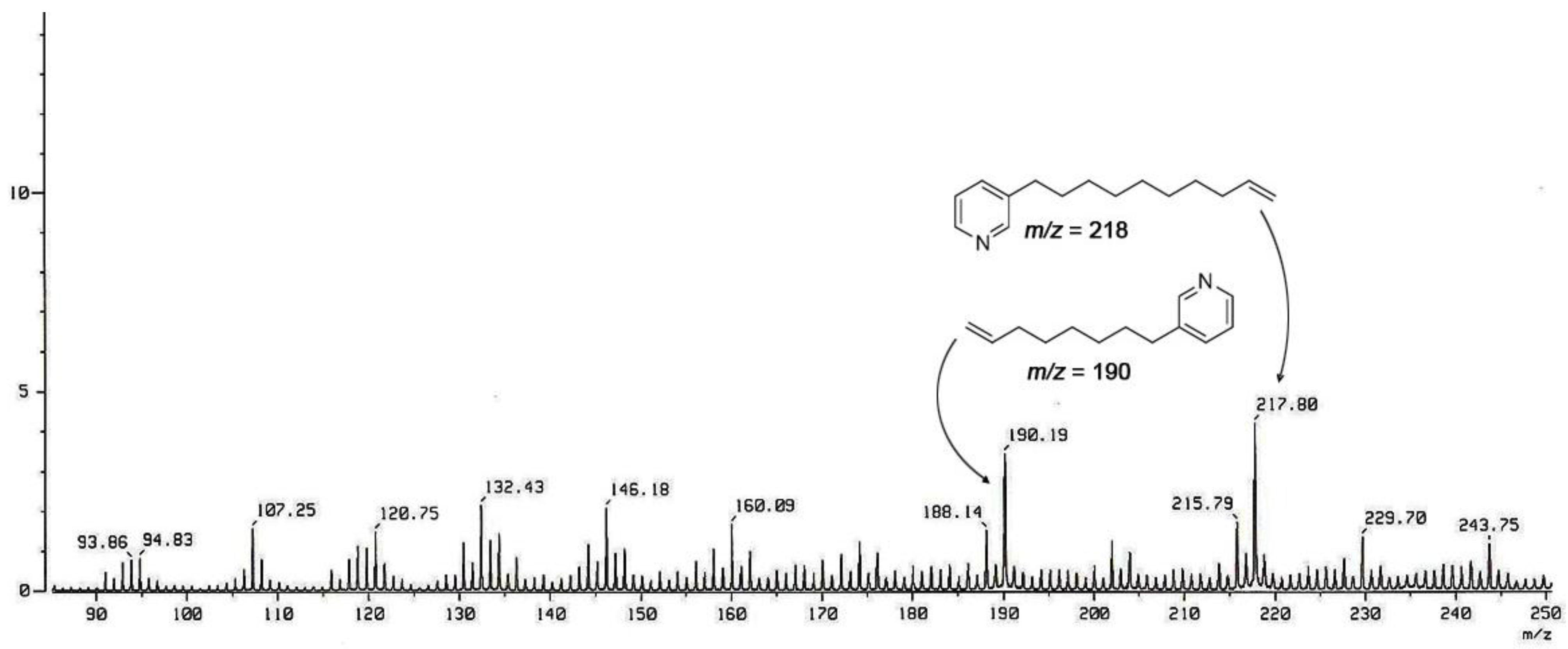
The UV absorption at 267 nm was reminiscent of two di-substituted pyridine rings, which was corroborated by aromatic proton signals [δH, 8.96 (2 H, br s, H-2, H-2′), 8.45 (1 H, d, J = 7.6 Hz, H-4), 8.44 (1 H, d, J = 7.7 Hz, H-4′), 8.02 (2 H, dd, J = 7.7, 6.2 Hz, H-5, H-5′), 8.83 (1 H, dd, J = 6.0 Hz, H-6), 8.82 (1 H, dd, J = 6.0 Hz, H-6′)] and carbon signals [δC 145.5 (C-2, C-2′), δC 145.3 (C-3), δC 145.4 (C-3′), δC 147.0 (C-4), δC 146.9 (C-4′), δC 129.2 (C-5, C-5′), δC 143.4 (C-6), δC 143.5 (C-6′)] in the NMR data. Although the two and three linearly contiguous methylene units attached to the pyridinium (N-1, N-1′, C-3, and C-3′) were identified based on 1H COSY and gHMBC, only a large methylene envelope at δH 1.25–1.40 was observed in the 1H NMR spectrum, indicating that 3 was composed of two di-substituted pyridine rings and long aliphatic chains, forming a highly symmetric cyclic framework. The lengths of the methylene chains were determined based on the FAB-MS/MS data, which yielded two intense peaks at m/z 190 and 218, as shown in Figure 2. Since these daughter ions were attributable to a pair of monomeric products generated via Hoffmann-type elimination7 from their parent ion, 3 was structurally defined to be a cyclic hetero-dimer composed of C8 and C10 chains connecting the N-1 and C-3′ (also N-1′ and C-3) of pyridiniums.
Figure 2. Positive FAB-MS/MS spectrum of compound 3.
Marinedrugs 10 02126 g002
The molecular formula of compound 4 was C30H46Cl2N2 based on HR-FAB-MS data with ion clusters at m/z 435.3741 [M − H]+ and 471.3503 [M + Cl]+. Thus, the structure consisted of two CH2 units larger than 3. The NMR data of this compound were very similar to those of 3, indicative of the same cycloalkylpyridinium nature. The FAB-MS/MS spectrum exhibited two intense daughter ions at m/z 218, confirming the symmetrical structure of 4, which contained two C10 linear alkyl chains connecting the N-1 and C-3 between two pyridiniums.
The molecular formulas of compounds 5 and 6 were both C30H44Cl2N2 based on HR-FAB-MS analyses. The 1H NMR spectra of these compounds were almost identical and consisted of two 1,3-disubstituted pyridiniums, two methylene chains, and two olefinic protons [5: δH 5.41 (2 H, br t, J = 3.6 Hz, H-12, H-13); 6: δH 5.38 (2 H, m, H-13, H-14)]. Two prominent daughter ions at m/z 216 and 218 in the FAB-MS/MS data of 5 and 6 showed a C10-saturated chain and a C10-chain with one double bond. The gHMBC data of 5 and 6 showed that these compounds had the same 1,3-dialkylpyridinium substitution patterns as 14.
The remaining alkyl substructure and the locations of the double bonds were determined by combined NMR analyses. The 1H COSY (5 and 6: H-7-H-16) and gHMBC (5: H-11/C-12, C-13, H-12/C-11, C-14, H-13/C-11, C-14, H-14/C-12, C-13, 6: H-12/C-13, C-14, H-13/C-12, C-15, H-14/C-12, C-15, H-15/C-13, C-14) data indicated that 5 and 6 contained a double bond at C-12 and C-13, respectively. Since the olefinic and allylic protons of the double bonds were chemically equivalent, the NOESY analysis could not be used to determine the configurations of the double bonds. Consequently, the configuration of the double bond in 5 was assigned as E based on the downfield shifts of the allylic methylene [δC 32.7 (C-11) and 32.4 (C-14)], while the Z-configuration was deduced for 6 from the upfield chemical shifts of the allylic methylene [δC 28.0 (C-12) and 29.2 (C-15)] [15].
The molecular formulas of 7 and 8 were C31H47Cl2N2 based on HR-FAB-MS analyses. As found for 5 and 6, the 1H NMR spectra of these compounds revealed characteristic features of cyclostellettamines: 1,3-disubstituted pyridines, methylene chains, and two olefinic protons [7: 5.37 (1 H, ddd, J = 11.4, 6.7, 2.0 Hz, H-9), 5.36 (1 H, ddd, J = 11.4, 6.7, 2.0 Hz, H-10); 8: δH 5.71 (1 H, dt, J = 10.8, 7.3 Hz, H-15), 5.66 (1 H, dt, J = 10.8, 7.3 Hz, H-16)] (Table 1 and Table 3). The FAB-MS/MS analyses of 7 and 8 supported the presence of a C10-saturated chain and a C11-chain with one double bond. The 1H COSY (7: H-7–H-12, H-16–H-17, 8: H-7–H-9, H-13–H-17) and gHMBC (7: H-8/C-9, C-10, H-9/C-8, C-11, H-10/C-8, C-11, H-11/C-8, C-10, 8: H-14/C-15, C-16, H-15/C-14, C-17, H-16/C-14, C-17, H-17/C-15, C-16) data placed the double bonds at C-9 and C-15 for 7 and 8, respectively. The configurations of these double bonds were both determined to be Z based on the chemical shifts of the allylic methylene carbons [7: 24.8 (C-8) and 28.1 (C-11), 8: δC 28.3 (C-14) and 31.0 (C-17)] [15].
Table 3. 1H MMR (δ, mult, J in Hz) assignments for compounds 710 in MeOH-d4.
Position78910
28.99 (1H, s)8.87 (1H, s)8.99 (1H, s)8.93 (1H, s)
3----
48.43 (1H, d, 8.0)8.46 (1H, d, 7.6)8.45 (1H, d, 8.0)8.45 (1H, d, 8.1)
58.00 (1H, dd, 7.8, 6.3)8.03 (1H, dd, 7.8, 6.1)8.02 (1H, dd, 7.7, 6.2)8.02 (1H, dd, 7.9, 6.3)
68.80 (1H, dd, 6.2)8.85 (1H, dd, 6.0)8.82 (1H, dd, 6.1)8.82 (1H, dd, 6.5)
74.62 (2H, t, 7.1)4.65 (2H, t, 7.0)4.61 (2H, t, 7.1)4.64 (2H, t, 6.9)
82.10 (2H, m)1.98 (2H, m)2.00 (2H, m)2.00 (2H, m)
95.37 (1H, ddd, 11.4, 6.7, 2.0)1.26 (2H, m)1.29 (2H, m)1.24 (2H, m)
105.36 (1H, ddd, 11.4, 6.7, 2.0)1.20 ~ 1.35 (2H, m)1.32 (2H, m)1.20 ~ 1.32 (2H, m)
111.94 (2H, m)1.20 ~ 1.35 (2H, m)1.28 (2H, m)1.20 ~ 1.32 (2H, m)
121.36 (2H, m)1.20 ~ 1.35 (2H, m)1.99 (2H, m)1.20 ~ 1.32 (2H, m)
131.23 ~ 1.35 (2H, m)1.36 (2H, m)5.34 (1H, m)1.20 ~ 1.32 (2H, m)
141.23 ~ 1.35 (2H, m)2.15 (2H, m)5.32 (1H, m)1.70 (2H, m)
151.23 ~ 1.35 (2H, m)5 71 (1H, dt, 10.8, 7.3)2.06 (2H, m)5.39 (1H, dt, 10.6, 6.7)
161.78 (2H, m)5.66 (1H, dt, 10.8, 7.3)1.38 (2H, m)5.38 (1H, dt, 10.6, 6.7)
172.88 (2H, t, 7.2)3.68 (2H, t, 7.3)1.72 (2H, m)2.46 (2H, dt, 13.5, 6.6)
18--2.90 (2H, t, 7.5) 2.95 (2H, t, 6.8)
2′8.90 (1H, s)8.79 (1H, s)8.98 (1H, s)8.89 (1H, s)
3′----
4′8.45 (1H, d, 8.0)8.47 (1H, d, 7.6)8.45 (1H, d, 8.0)8.43 (1H, d, 8.1)
5′8.02 (1H, dd, 7.8, 6.3)8.03 (1H, dd, 7.8, 6.1)8.01 (1H, dd, 7.7, 6.2)8.01 (1H, dd, 7.9, 6.3)
6′8.82 (1H, dd, 6.2)8.83 (1H, dd, 6.0)8.83 (1H, dd, 5.8)8.83 (1H, dd, 6.5)
7′4.60 (2H, t, 6.7)4.63 (2H, t, 7.5)4.63 (2H, t, 6.8)4.61 (2H, t, 6.9)
8′1.99 (2H, m)2.02 (2H, m)2.01 (2H, m)1.98 (2H, m)
9′1.24 (2H, m)1.28 (2H, m)1.27 (2H, m)1.25 (2H, m)
10′1.23 ~ 1.38 (2H, m)1.26 ~ 1.35 (2H, m)1.26 ~ 1.36 (2H, m)1.25 ~ 1.32 (2H, m)
11′1.23 ~ 1.38 (2H, m)1.26 ~ 1.35 (2H, m)1.26 ~ 1.36 (2H, m)1.25 ~ 1.32 (2H, m)
12′1.23 ~ 1.38 (2H, m)1.26 ~ 1.35 (2H, m)1.26 ~ 1.36 (2H, m)1.25 ~ 1.32 (2H, m)
13′1.23 ~ 1.38 (2H, m)1.26 ~ 1.35 (2H, m)1.26 ~ 1.36 (2H, m)1.25 ~ 1.32 (2H, m)
14′1.32 (2H, m)1.34 (2H, m)1.30 (2H, m)1.29 (2H, m)
15′1.73 (2H, m)1.74 (2H, m)1.73 (2H, m)1.75 (2H, m)
16′2.88 (2H, t, 7.0)2.90 (2H, t, 7.2)2.89 (2H, t, 7.3)2.91 (2H, t, 7.0)
The molecular formulas of 9 and 10 were both C32H49Cl2N2 based on HR-FAB-MS analyses. Similar to compounds 58, the 1H NMR spectra of 9 and 10 revealed a 1,3-dialkyl pyridinium structure with two olefinic protons [9: δH 5.34 (1 H, m, H-13), 5.32 (1 H, m, H-14); 10: 5.39 (1 H, dt, J = 10.6, 6.7 Hz, H-15), 5.38 (1 H, dt, J = 10.6, 6.7 Hz, H-16)]. The FAB-MS/MS analyses of these compounds confirmed the presence of a C10-saturated chain and a C12-chain with one double bond. The COSY (9: H-7–H-18, 10: H-7–H-9, H-14–H-18) and gHMBC (9: H-12/C-13, C-14, H-13/C-12, C-15, H-13/C-12, C-15, H-15/C-13, C-14, 10: H-14/C-15, C-16, H-15/C-14, C-17, H-16/C-14, C-17, H-17/C-15, C-16) data were indicative of a double bond at C-13 and C-15 for 9 and 10, respectively. The configurations of these double bonds were both assigned as Z based on the upfield shifts of the allylic methylene carbons [9: δC 28.0 (C-12), 27.9 (C-15), 10: 28.2 (C-14), 29.1 (C-17)].
A comparison of the 13C and 1H NMR data revealed almost identical chemical shifts for both carbons and protons among all cyclic 1,3-dialkylpyridinium dimers 110 (Table 1Table 3). These compounds were structurally similar to cyclostellettamines reported previously with differences in the length of the alkyl chains, as well as the presence of double bonds [9]. Compounds 510 were dehydrocyclostellettamines containing one double bond in the alkyl chains. Subsequently, the carbon chemical shifts of allylic methylenes and coupling constants between the olefinic protons were used to assign E or Z configurations to these double bonds.
The 1,3-dialkylpyridinium metabolites possess diverse cytotoxic and antimicrobial activities [16,17]. Thus, we evaluated the cytotoxic and antimicrobial activities of compounds 110. Since 7 was isolated in a TFA salt form, we evaluated the influence of the counter ion on bioactivity. Thus, the TFA salt of 4 was prepared using reversed phase HPLC (H2O-MeOH, 80:20, 0.1% TFA) on 4. Compounds 110 and 4a exhibited moderate cytotoxicity against the A549 lung cancer cell line, comparable to doxorubicin (Table 4). On the other hand, they also displayed a diverse range of antibacterial activities against Gram-positive strains (compounds 2, 4, 8, 9, and 10 were the most potent). These results are partly consistent with the previous report that the bioactivities of cyclostellettamines were significantly influenced by the length of the alkyl chains [16]. In addition, as shown for 6 and 9, the bioactivities were influenced by the existence and location of the double bond, although the alkyl chains were the same length. This suggests a mechanism-of-action based on the distances between the charged moieties and electron-rich sites, possibly through an interaction with ionic membrane sites. We also observed that the antibacterial activities of these compounds were generally weaker against Gram-negative than Gram-positive strains. Also, the influence of pyridinium counterions was negligible since there was no significant difference between the bioactivities of 4 and 4a. Overall, these results increase our understanding of the structure-activity relationships of cyclostellettamines.
In addition to the antimicrobial and cytotoxic activity assays, compounds 110 were also tested against diverse pathogenic fungal strains and microbial enzymes, such as isocitrate lyase, sortase A, and Na+/K+-ATPase, and failed to display any bioactivity (LC50 > 100 μg/mL for fungi, ED50 > 100 μM for enzymes).
Table 4. Results of Bioactivity Tests.
CompoundMIC (μg/mL)LC50 (μM) a
Gram (+) BacteriumGram (−) BacteriumA549
ABCDEF
15010050100100>10022.1
212.5253.1252525>10015.3
350501005025>10024.0
4501006.25100100>10026.3
525100255050>10025.9
65010050100100>10028.9
75010010010050>10089.4
812.5253.1255050>10015.7
912.5253.1252525>10014.7
1012.5253.1252550>10014.8
4a2510050100100>10021.9
Ampicillin0.390.780.390.780.396.25
Doxorubicin 3.37
A: Staphylococcus aureus (ATCC 6538p); B: Bacillus subtilis (ATCC 6633); C: Micrococcus luteus (IFO 12708); D: Salmonella typhimurium (ATCC 14028); E: Proteus vulgaris (ATCC 3851); F: Escherichia coli (ATCC 35270); a The results of 48 h treatment were chosen.

3. Experimental Section

3.1. General Experimental Procedures

Optical rotation was measured on a JASCO digital polarimeter using a 1 cm cell. IR spectra were recorded on a JASCO FT/IR 4200 spectrophotometer. UV spectra were recorded on a Hitachi U-3010 spectrophotometer. Proton and carbon NMR spectra were measured at 600 and 150 MHz (3) on Bruker Avance 600 and 500 and 125 MHz (1, 2 and 410) on Bruker Avance 500, respectively. Mass spectrometric data were obtained from the Korea Basic Science Research Institute (Daegu) on a JEOL JMS-700 mass spectrometer. All solvents used were spectral grade or distilled from glass prior to use.

3.2. Collection and Taxonomic Identification

Haliclona sp. specimens were collected by hand using SCUBA equipment (20 m depth) at Sagyeri off the shore of Jeju Island, Korea, in November 1999. The pale-purple sponge measured 65 × 55 mm with a thickness of 27 mm and produced slime strands. The surface had many mamillate forms of a hard but fragile consistency. The size of the oxea spicule was (170–198) × (8–10) μm. A voucher specimen (registry No. Spo. 63) was deposited at the Natural History Museum, Hannam University, Korea, under the curatorship of C.J.S. Collections of the same specimens were made at the nearby Chuja-do in November 2003, and at Sagyeri in September 2009 and June 2010. Since the morphological features and 1H NMR spectra of crude extracts from these collections were identical to those of the 1999 collection, chemical investigation was performed on the combined specimens.

3.3. Extraction and Isolation

The freshly collected specimens were frozen immediately and kept at −25 °C until use. The specimens (dried wt. 1.2 kg) were lyophilized, macerated, and extracted repeatedly with MeOH (3 L × 3) and CH2Cl2 (3 L × 2). The combined organic extract (940.3 g) was partitioned between n-BuOH and H2O, and the former (158.18 g) was repartitioned between 15% aqueous MeOH (24.43 g) and n-hexane (130.25 g). The aqueous MeOH layer was subjected to C18 reversed-phase vacuum flash chromatography using sequential mixtures of MeOH and H2O as eluents (six fractions in gradient, H2O–MeOH, from 50:50 to 0:100), and finally acetone. The H2O–MeOH (50:50) fraction (1.64 g) was gel-filtered on a Sephadex LH-20 column with MeOH. The bioactive fractions were separated by C18 reversed-phase semi-preparative HPLC (YMC ODS-A column, 1 × 25 cm, H2O–MeOH, 80:20) to yield, in order of elution, compounds 3, 4, 1, 7, 8, 2, 6, 5, 10, and 9. Final purification of these metabolites was then accomplished by reversed-phase HPLC (YMC-Pack CN column, 1 × 25 cm, H2O–MeOH (85:15) for 14, H2O–MeOH (90:10) for 5, 6, and 810, and gradient solvents of H2O–MeOH (85:15) to (40:60) and 0.1% TFA for 30 min for 7). The overall yields were 14.9, 19.8, 7.5, 187.4, 11.3, 22.5, 6.0, 72.4, 12.6, and 10.5 mg for 110, respectively.
Compound 3: Yellow, amorphous solid; UV (MeOH) λmax 267 (log ε = 3.7), IR (ZnSe) νmax 2927, 1506, 1461 cm−1; 1H and 13C NMR data, see Table 1 and Table 2, respectively; HR-FAB-MS m/z 407.3428 [M − H]+ (calculated for C28H43N2, 407.3426) and 443.3195 [M + Cl]+ (calculated for C28H44N2Cl, 443.3193); FAB-MS/MS fragments: m/z 190.19 [M − C15H24N]+ and 217.80 [M − C13H20N]+.
Compound 4: White, amorphous solid; UV (MeOH) λmax 266 (log ε = 3.9), IR (ZnSe) νmax 2924, 1505, 1467 cm–1; 1H and 13C NMR data, see Table 1 and Table 2, respectively; HR-FAB-MS m/z 435.3741 [M − H]+ (calculated for C30H47N2, 435.3739) and 471.3503 [M + Cl]+ (calculated for C30H48N2Cl, 471.3506); FAB-MS/MS fragments: m/z 218.05 [M − C15H24N]+ and 218.05 [M − C15H24N]+.
Compound 5: Yellow, amorphous solid; UV (MeOH) λmax 267 (log ε = 3.8), IR (ZnSe) νmax 2926, 1505, 1459 cm−1; 1H and 13C NMR data, see Table 1 and Table 2, respectively; HR-FAB-MS m/z 433.3586 [M − H]+ (calculated for C30H45N2, 433.3583), 469.3351 [M + Cl]+ (calculated for C30H46N2Cl, 469.3350); FAB-MS/MS fragments: m/z 216.06 [M − C15H24N]+, 218.04 [M − C15H22N]+.
Compound 6: Yellow, amorphous solid; UV (MeOH) λmax 266 (log ε = 3.8), IR (ZnSe) νmax 2927, 1505, 1458 cm−1; 1H and 13C NMR data, see Table 1 and Table 2, respectively; HR-FAB-MS m/z 433.3584 [M − H]+ (calculated for C30H45N2, 433.3583) and 469.3352 [M + Cl]+ (calculated for C30H46N2Cl, 469.3350); FAB-MS/MS fragments: m/z 216.07 [M − C15H24N]+ and 218.04 [M − C15H22N]+.
Compound 7: Yellow, amorphous solid; UV (MeOH) λmax 267 (log ε = 3.9), IR (ZnSe) νmax 2927, 1631, 1507, 1460, 1203, 1129 cm−1; 1H and 13C NMR data, see Table 1 and Table 3 respectively; HR-FAB-MS m/z 447.3736 [M − H]+ (calculated for C31H47N2, 447.3739) and 561.7414 [M + CF3COO]+ (calculated for C30H48N2CF3COO, 561.7416); FAB-MS/MS fragments: m/z 217.92 [M − C16H24N]+ and 229.76 [M − C15H24N]+.
Compound 8: Yellow, amorphous solid; UV (MeOH) λmax 267 (log ε = 3.8), IR (ZnSe) νmax 2924, 1506, 1467 cm−1; 1H and 13C NMR data, see Table 1 and Table 3, respectively; HR-FAB-MS m/z 447.3742 [M − H]+ (calculated for C31H47N2, 447.3739) and 483.3504 [M + Cl]+ (calculated for C31H48N2Cl, 483.3506); FAB-MS/MS fragments: m/z 218.11 [M − C16H24N]+ and 229.87 [M − C15H24N]+.
Compound 9: Yellow, amorphous solid; UV (MeOH) λmax 266 (log ε = 3.7), IR (ZnSe) νmax 2925, 1505, 1459 cm−1; 1H and 13C NMR data, see Table 1 and Table 3, respectively; HR-FAB-MS m/z 461.3898 [M − H]+ (calculated for C32H49N2, 461.3896) and 497.3665 [M + Cl]+ (calculated for C32H50N2Cl, 497.3663); FAB-MS/MS fragments: m/z 218.19 [M − C17H26N]+ and 243.75 [M − C15H24N]+.
Compound 10: Yellow, amorphous solid; UV (MeOH) λmax 266 (log ε = 3.9), IR (ZnSe) νmax 2925, 1505, 1458 cm−1; 1H and 13C NMR data, see Table 1 and Table 3, respectively; HR-FAB-MS m/z 461.3899 [M − H]+ (calculated for C32H49N2, 461.3896) and 497.3665 [M + Cl]+ (calculated for C32H50N2Cl, 497.3663); FAB-MS/MS fragments: m/z 218.19 [M − C17H26N]+ and 243.75 [M − C15H24N]+.

3.4. Preparation of TFA Salt of Compound 4 (4a)

A small amount of compound 4 (6.0 mg) was injected into the HPLC (YMC-Pack CN column, 1 × 25 cm, H2O–MeOH, 80:20, 0.1% TFA) to yield 4a (4.8 mg): UV (MeOH) λmax 266 (log ε = 3.8), IR (ZnSe) νmax 2928, 1631, 1506, 1464, 1203, 1129 cm–1; HR-FAB-MS m/z 435.3741 [M − H]+ (calculated for C30H47N2, 435.3739) and 549.7306 [M + CF3COO]+ (calculated for C30H48N2CF3COO, 549.7309); FAB-MS/MS fragments: m/z 218.05 [M − C15H24N]+ and 218.05 [M − C15H24N]+.

3.5. Biological Assays

Cytotoxicity assays, antimicrobial assays, and isocitrate lyase, sortase A, and Na+/K+-ATPase inhibition assays were performed as described previously [18,19,20,21,22]. For the cytotoxicity test, an MTT viability assay was performed as previously described with slight modifications [18]. MTT was first prepared as a stock solution of 5 mg/mL in phosphate buffered saline (PBS, pH 7.2) and was filtered. At the end of the treatment period (24 h, 48 h, and 72 h), with three different test drug concentrations in triplicate, MTT solution (20 μL) was added to each well and then incubated for 4 h at 37 °C; then solubilizing buffer (10% sodium dodecyl sulfate dissolved in 0.01 N HCl, 100 μL) was added to each well. After overnight incubation, the 96-well plate was read by an enzyme-linked immunosorbent assay (ELISA) reader at 570 nm for absorbance to determine the cell (A549 cell line) viability. The viable cells produced a dark blue formazan product, whereas no such staining was formed by dead cells [19]. The LC50 value was defined as the concentration that resulted in a 50% decrease in cell viability compared to that of control reactions in the absence of an inhibitor. The values (mean ± SD) were calculated from the dose–response curves of three concentrations of each test sample in three independent experiments (n = 3).

4. Conclusion

Eight novel cyclic bis-1,3-dialkylpyridiniums (310), as well as two known compounds (1 and 2) belong to the cyclostellettamine class, were isolated from the sponge Haliclona sp. from Korea. These compounds were structurally different from the previous reported cyclostelletamines in the lengths of the alkyl chains, as well as the presence of double bonds in the chains. Several of these new compounds exhibited moderate cytotoxicity against A549 cell-line and inhibitory activities against Gram-positive bacterial strains. As shown in 6 and 9, the bioactivities were influenced by the existence and the location of a double bond. Overall, these results provide us better understanding of the structure-activity relationships of the cyclostellettamine class compounds.

Supplementary Files

  • Supplementary File 1::

    PDF-Document (PDF, 746 KB)

  • Acknowledgments

    We thank the Basic Science Research Institute in Daegu, Korea, for providing mass data. This study was partially supported by National Research Foundation of Korea (NRF) grants, funded by the Korean government (MEST, Ministry of Education, Science and Technology) (No. 20120006709).

    References

    1. Andersen, R.J.; van Soest, R.W.M.; Kong, F. Alkaloids: Chemical and Biological Perspectives; Pelletier, S.W., Ed.; Pergamon Press/Elsevier Science: New York, NY, USA, 1996; pp. 301–355. [Google Scholar]
    2. Matzanke, N.; Gregg, R.J.; Weinreb, S.M. Biomimetic and synthetic approaches to marine sponge alkaloids derived from bis-pyridine macrocycles. Org. Prep. Proc. Int. 1998, 30, 3–51. [Google Scholar]
    3. Turk, T.; Sepčic, K.; Mancini, I.; Guella, G. 3-Akylpyridinium and 3-alkylpyridine compounds from marine sponges, their synthesis, biological activities and potential use. Stud. Nat. Prod. Chem. 2008, 35, 355–397. [Google Scholar] [CrossRef]
    4. Blunt, J.W.; Copp, B.R.; Keyzers, R.A.; Munro, M.H.G.; Prinsep, M.R. Marine natural products. Nat. Prod. Rep. 2012, 29, 144–222. [Google Scholar] [CrossRef]
    5. Oku, N.; Nagai, K.; Shindoh, N.; Terada, Y.; van Soest, R.W.M.; Matsunaga, S.; Fusetani, N. Three new cyclostellettamines, which inhibit histonedeacetylase, from a marine sponge of the genus Xestospongia. Bioorg. Med. Chem. Lett. 2004, 14, 2617–2620. [Google Scholar] [CrossRef]
    6. Pérez-Balado, C.; Nebbioso, A.; Rodríguez-Graña, P.; Minichiello, A.; Miceli, M.; Altucci, L.; de Lera, Á.R. Bispyridinium dienes: Histone deacetylase inhibitors with selective activities. J. Med. Chem. 2007, 50, 2497–2505. [Google Scholar] [CrossRef]
    7. Davies-Coleman, M.T.; Faulkner, D.J.; Dubowchik, G.M.; Roth, G.P.; Polson, C.; Fairchild, C. A new EGF-active polymeric pyridinium alkaloid from the sponge Callyspongia fibrosa. J. Org. Chem. 1993, 58, 5925–5930. [Google Scholar] [CrossRef]
    8. Ondeykal, J.G.; Herath, K.B.; Jayasuriya, H.; Polishook, J.D.; Bills, G.F.; Dombrowski, A.W.; Mojena, M.; Koch, G.; DiSalvo, J.; DeMartino, J.; Guan, Z.; Nanakorn, W.; Morenberg, C.M.; Balick, M.J.; Stevenson, D.W.; Slattery, M.; Borris, R.P.; Singh, S.B. Discovery of structurally diverse natural product antagonists of chemokine receptor CXCR3. Mol. Divers. 2005, 9, 123–129. [Google Scholar] [CrossRef]
    9. Fusetani, N.; Asai, N.; Matsunaga, S.; Honda, K.; Yasumuro, K. Cyclostellettamines A–F, pyridine alkaloids which inhibit binding of methyl quinuclidinylbenzilate (QNB) to muscarinic acetylcholine receptors, from the marine sponge, Stelletta maxia. Tetrahedron Lett. 1994, 35, 3967–3970. [Google Scholar] [CrossRef]
    10. Anan, H.; Seki, N.; Noshiro, O.; Honda, K.; Yasumuro, K.; Ozasa, T.; Fusetani, N. Total synthesis of cyclostellettamine C, a bispyridiniummacrocyclic alkaloid having muscarinic acetylcholine receptor antagonistic activity. Tetrahedron 1996, 52, 10849–10860. [Google Scholar] [CrossRef]
    11. Jang, K.H.; Kang, G.W.; Jeon, J.; Lim, C.; Lee, H.-S.; Sim, C.J.; Oh, K.-B.; Shin, J. Haliclonin A, a new macrocyclicdiamide from the sponge Haliclona sp. Org. Lett. 2009, 11, 1713–1716. [Google Scholar]
    12. Casapullo, A.; Pinto, O.C.; Marzocco, S.; Autore, G.; Riccio, R. 3-Alkylpyridinium alkaloids from the pacific sponge Haliclona sp. J. Nat. Prod. 2009, 72, 301–303. [Google Scholar] [CrossRef]
    13. Schmidt, G.; Timm, C.; Köck, M. New haliclamines E and F from the arctic sponge Haliclona viscosa. Org. Biomol. Chem. 2009, 7, 3061–3064. [Google Scholar] [CrossRef]
    14. Grube, A.; Timm, C.; Köck, M. Synthesis and mass spectrometric analysis of cyclostellettamines H, I, K and L. Eur. J. Org. Chem. 2006, 5, 1285–1295. [Google Scholar]
    15. de Haan, J.W.; van de Ven, L.J.M. Configurations and conformations in acyclic, unsaturated hydrocarbons. A 13C NMR study. A 13C NMR study. Org. Magn. Reson. 1973, 5, 147–153. [Google Scholar]
    16. de Oliveira, J.H.H.L.; Seleghim, M.H.R.; Timm, C.; Grube, A.; Köck, M.; Nascimento, G.G.F.; Martins, A.C.T.; Silva, E.G.O.; de Souza, A.O.; Minarini, P.R.R.; et al. Antimicrobial and antimycobacterial activity of cyclostellettamine alkaloids from sponge Pachychalina sp. Mar. Drugs 2006, 4, 1–8. [Google Scholar] [CrossRef]
    17. de Oliveira, J.H.H.L.; Grube, A.; Kock, M.; Berlinck, R.G.; Macedo, M.L.; Ferreira, A.G.; Hajdu, E. Ingenamine G and cyclostellettamines G–I, K, and L from the new brazilian species of marine sponge Pachychalina sp. J. Nat. Prod. 2004, 67, 1685–1689. [Google Scholar] [CrossRef] [Green Version]
    18. Mosmann, T. Rapid colorimetric assay for cellular growth and survival: Application to proliferation and cytotoxicity assays. J. Immunol. Methods 1983, 65, 55–63. [Google Scholar] [CrossRef]
    19. Ulukaya, E.; Ozdikicioglu, F.; Oral, A.Y.; Demirci, M. The MTT assay yields a relatively lower result of growth inhibition than the ATP assay depending on the chemotherapeutic drugs tested. Toxicol. In Vitro 2008, 22, 232–239. [Google Scholar] [CrossRef]
    20. Oh, K.-B.; Lee, J.H.; Chung, S.-C.; Shin, J.; Shin, H.J.; Kim, H.-K.; Lee, H.-S. Antimicrobial activities of the bromophenols from the red alga Odonthalia corymbifera and some synthetic derivatives. Bioorg. Med. Chem. Lett. 2008, 18, 104–108. [Google Scholar] [CrossRef]
    21. Lee, H.-S.; Lee, T.-H.; Yang, S.H.; Shin, H.J.; Shin, J.; Oh, K.-B. Sesterterpene sulfates as isocitratelyase inhibitors from tropical sponge Hippospongia sp. Bioorg. Med. Chem. Lett. 2007, 17, 2483–2486. [Google Scholar] [CrossRef]
    22. Johansson, M.; Karlsson, L.; Wennergren, M.; Jansson, T.; Powell, T.L. Activity and protein expression of Na+/K+ ATPase are reduced in microvilloussyncytiotrophoblast plasma membranes isolated from pregnancies complicated by intrauterine growth restriction. J. Clin. Endocrinol. Metab. 2003, 88, 2831–2837. [Google Scholar] [CrossRef]

    Share and Cite

    MDPI and ACS Style

    Lee, Y.; Jang, K.H.; Jeon, J.-e.; Yang, W.-Y.; Sim, C.J.; Oh, K.-B.; Shin, J. Cyclic Bis-1,3-dialkylpyridiniums from the Sponge Haliclona sp. Mar. Drugs 2012, 10, 2126-2137. https://doi.org/10.3390/md10092126

    AMA Style

    Lee Y, Jang KH, Jeon J-e, Yang W-Y, Sim CJ, Oh K-B, Shin J. Cyclic Bis-1,3-dialkylpyridiniums from the Sponge Haliclona sp. Marine Drugs. 2012; 10(9):2126-2137. https://doi.org/10.3390/md10092126

    Chicago/Turabian Style

    Lee, Yoonyeong, Kyoung Hwa Jang, Ju-eun Jeon, Woo-Young Yang, Chung J. Sim, Ki-Bong Oh, and Jongheon Shin. 2012. "Cyclic Bis-1,3-dialkylpyridiniums from the Sponge Haliclona sp." Marine Drugs 10, no. 9: 2126-2137. https://doi.org/10.3390/md10092126

    APA Style

    Lee, Y., Jang, K. H., Jeon, J. -e., Yang, W. -Y., Sim, C. J., Oh, K. -B., & Shin, J. (2012). Cyclic Bis-1,3-dialkylpyridiniums from the Sponge Haliclona sp. Marine Drugs, 10(9), 2126-2137. https://doi.org/10.3390/md10092126

    Article Metrics

    Back to TopTop