Article

A New Dibenz[b,e]oxepine Derivative, 1-Hydroxy-10-methoxy-dibenz[b,e]oxepin-6,11-dione, from a Marine-Derived Fungus, Beauveria bassiana TPU942

1
Department of Natural Product Chemistry, Tohoku Pharmaceutical University, Sendai 981-8558, Japan
2
Faculty of Agriculture, Indonesia Development University, Manado 95361, Indonesia
*
Author to whom correspondence should be addressed.
Mar. Drugs 2012, 10(12), 2691-2697; https://doi.org/10.3390/md10122691
Submission received: 28 September 2012 / Revised: 19 October 2012 / Accepted: 14 November 2012 / Published: 27 November 2012

Abstract

:
1-Hydroxy-10-methoxy-dibenz[b, e]oxepin-6,11-dione (1) was obtained from the culture broth of a marine-derived fungus, Beauveria bassiana TPU942, isolated from a marine sponge collected at Iriomote Island in Okinawa, together with two known compounds, chrysazin (2) and globosuxanthone A (3). The structure of 1 was elucidated on the basis of its spectroscopic data (HREIMS, 1D and 2D NMR experiments including 1H–1H COSY, HMQC and HMBC spectra). Dibenz[b, e]oxepines are rare in nature, and only six natural products have been reported. Therefore, compound 1 is the seventh natural product in this class. Compounds 2 and 3 showed an antifungal activity against Candida albicans, and 3 inhibited the cell growth against two human cancer cell lines, HCT-15 (colon) and Jurkat (T-cell lymphoma). Compound 1 did not show an apparent activity in the same bioassays.

Graphical Abstract

1. Introduction

Microorganisms from marine environments, especially marine-derived fungus, have proven to be an attractive source of new bioactive secondary metabolites [1,2,3,4,5]. Many of the metabolites possess unique chemical structures and interesting biological activities, such as cytotoxicity against cancer cell lines, antimicrobial activity, inhibition of several enzyme activities and so on [1,2,3,4,5].
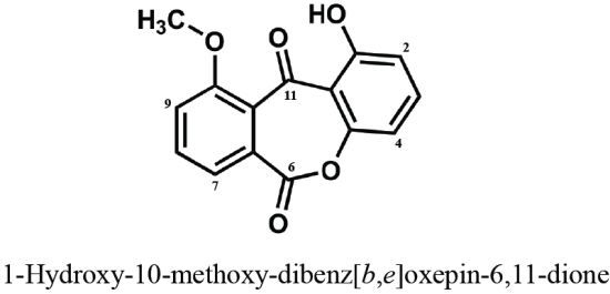
In the course of our studies on bioactive components produced by marine-derived fungi isolated from tropical and sub-tropical coral reefs, we found that the EtOAc extract of the culture broth from a marine-derived fungus, Beauveria bassiana TPU942, isolated from a marine sponge collected at Iriomote Island, Okinawa Prefecture, Japan, showed cytotoxicity against a human T-cell lymphoma Jurkat cells. Bioassay-guided separation from the EtOAc extract led to the isolation of a new dibenz[b, e]oxepine derivative (1) and two known anthraquinone and xanthone derivatives, chrysazin (2) and globosuxanthone A (3). The structure of compound 1 was elucidated as 1-hydroxy-10-methoxy-dibenz[b, e]oxepin-6,11-dione (Figure 1) by the analysis of its spectroscopic data. Natural products possessing a dibenz[b, e]oxepine structure are quite rare, and only six compounds have been reported as natural products in scientific journals [6,7,8,9]. Antifungal and cytotoxic activities were exhibited by two known compounds (2 and 3).
We describe herein the isolation and structure elucidation of compound 1 and biological activities of isolated compounds.
Figure 1. Structures of compounds 13 isolated from Beauveria bassiana TPU942.
Marinedrugs 10 02691 g001

2. Results and Discussion

The producing fungus, B. bassiana TPU942, was isolated from a piece of an unidentified marine sponge collected at Iriomote Island and cultured as described in Experimental Section. The culture broth and the EtOAc extract of strain TPU942 showed cytotoxicity in the screening bioassay against Jurkat cells and was separated into seven fractions (Fr. 1–Fr. 7) using a silica gel column. Compounds 1 and 2 were isolated from Fr. 1 (eluted with 100% CHCl3) and 3 from Fr. 2 (CHCl3–MeOH = 100:1) by HPLC (ODS).
Compound 2 was identified as chrysazin [10], a ubiquitous anthraquinone derivative isolated from several organisms, from the spectroscopic data. The structure of compound 3 was assigned on the basis of its spectral data and comparison of these data with those of the reported values for globosuxanthone A [11]. Globosuxanthone A (3) was originally isolated from Chaetominum globosum, and a cytotoxicity against seven human solid tumor cell lines, accumulation of cells at either G2/M or S phase and induction of apoptosis have been described [11].
Compound 1 showed a molecular ion peak at m/z 270 [M]+ in the EIMS, and the molecular formula C15H10O5, 11 degrees of unsaturation, was determined from HREIMS [m/z 270.0531 [M]+, Δ +0.3 mmu] and NMR data. IR absorptions of 1 at 1735 and 3435 cm−1 suggested the presence of carbonyl and hydroxyl groups in the molecule. The 13C NMR spectrum showed 15 resolved signals, which were classified into one oxygenated methyl, six sp2 methine, three sp2 quaternary, three oxygenated sp2 quaternary and two carbonyl carbons by the analysis of 1D and 2D NMR spectra (Table 1). The 1H NMR spectrum displayed 10 proton signals, and two signals at δ 4.03 and 12.2 were assigned as a hydroxy proton (1-OH) and methoxy protons (10-OMe), respectively (Table 1). The connectivity of carbons and protons was established by the HMQC correlations.
Table 1. 13C (100 MHz) and 1H NMR (400 MHz) data for compound 1 (CDCl3).
No.δCδH (J in Hz)HMBC
1161.8 -1, 2, 11a
2111.0 6.82 dd (8.3, 1.0)1, 4
3137.1 7.61 t (8.5)1, 4a
4106.9 6.94 dd (8.3, 1.0)2, 4a
4a155.8 --
5
6156.1 --
6a133.7 --
7119.5 7.56 dd (8.3, 1.0)6, 9, 10a
8135.0 7.77 dd (8.5, 7.1)6, 6a
9122.7 7.33 dd (7.3, 1.0)10, 10a
10169.5 --
10a117.5 --
11181.0 --
11a109.0 --
1-OH-12.2 s1, 2, 11a
10-OCH353.1 4.03 s10
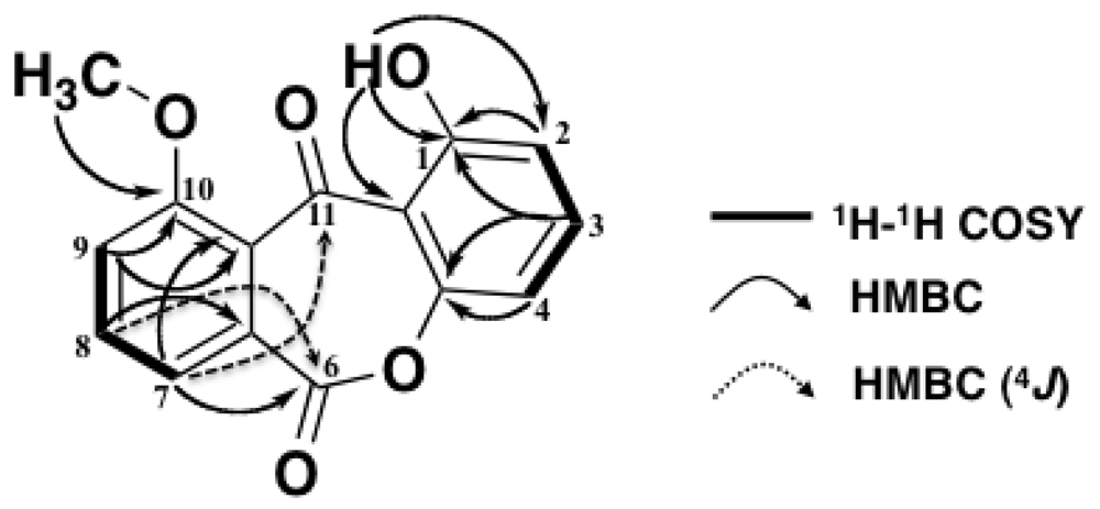
The presence of two 1,2,3-trisubstituted aromatic rings was assigned by the 1H–1H COSY and HMBC correlations of the 1H NMR signals at δ 6.82 (H-2), 7.61 (H-3), 6.94 (H-4), 7.56 (H-7), 7.77 (H-8) and 7.33 (H-9) (Figure 2). HMBC correlations were observed from 1-OH (δ 12.2) to C-1 (δ 161.8), C-2 (δ 111.0) and C-11a (δ 109.0) and from 10-OMe (δ 4.03) to C-10 (δ 169.5). Therefore, an OH group was attached at the C-1 position and an OMe group at C-10. An HMBC correlation from H-7 (δ 7.56) to C-6 (δ 156.1) and from H-8 to C-6 (4J), and the 13C chemical shift of C-4a (δ 155.8), an oxygenated sp2 carbon, revealed that C-4a and C-6a were connected via C-6 by an ester bond. Therefore, one remaining carbonyl group (δ 181.0, C-11) was inevitably attached between C-10a and C-11a [6,7,8,9]. This assignment was supported by an HMBC correlation from H-7 to C-11 (4J). Thus, the structure of compound 1 was assigned as 1-hydroxy-10-methoxy-dibenz[b, e]oxepin-6,11-dione.
Figure 2. 1H-1H COSY and HMBC correlations of compound 1.
Marinedrugs 10 02691 g002
Dibenz[b, e]oxepines are rare metabolites, and, thus far, only six natural products (49, Figure 3) have been reported [6,7,8,9]. Compound 1 is, therefore, the seventh example of this class of natural products. 13C and 1H NMR data for 1 were very similar to those of the reported values for 5 [7].
Figure 3. Structures of related compounds 49.
Marinedrugs 10 02691 g003
Compounds 13 were tested for their antimicrobial activity against four test microorganisms (fungus Mucor hiemalis, yeast Candida albicans, Gram-positive bacterium Staphylococcus aureus and Gram-negative bacterium Escherichia coli) using the paper disk method and for their cytotoxicity against two human cancer cell lines (colon adenocarcinoma HCT-15 and T-cell leukemia Jurkat cells) using the MTT method [12] (Table 2). Compounds 2 and 3 showed inhibition zone of 7 mm against C. albicans at the concentration of 10 μg/disk. Compound 3 inhibited the cell proliferation against HCT-15 and Jurkat cells with IC50 values of 10.7 and 2.3 μM, respectively. On the other hand, compound 1 did not show apparent activity in these bioassays. Further study on biological activity of compound 1 is now in progress.
Table 2. Biological activities of compound 13.
CompoundIC50 (μM) Inhibition zone (mm)
Cytotoxicity Antimicrobial activity (10 μg/disk)
HCT-15 Jurkat S. aureusE. coliC. albicansM. hiemalis
1> 30 > 30 a a a a
2> 30 > 30 a a 7 a
3 10.7 2.3 a a 7 a
a Not active.

3. Experimental Section

3.1. General

EI mass spectra were obtained by a JEOL JMS-MS 700 mass spectrometer (Tokyo, Japan). 1H and 13C NMR spectra were recorded on a JEOL JNM-AL-400 NMR spectrometer (400 MHz for 1H and 100 MHz for 13C) in DMSO-d6H 2.49, δC 39.5) or CDCl3H 7.26, δC 77.0). UV spectra were measured on a Hitach U-3310 UV-Visible spectrophotometer (Tokyo, Japan) and IR spectra on a PerkinElmer Spectrum One Fourier transform infrared spectrometer (Waltham, MA, USA). Preparative HPLC was carried out with a Hitachi L-6200 system.

3.2. Materials

Fetal bovine serum (FBS) and other culture materials were purchased from Invitrogen (Carlsbad, CA, USA). 3-(4,5-Dimethylthiazol-2-yl)-2,5-diphenyltetrazolium bromide (MTT) was purchased from Sigma-Aldrich (St. Louis, MO, USA). All other chemicals and organic solvents were purchased from Wako Pure Chemical Industries Ltd. (Osaka, Japan).

3.3. Fermentation and Isolation

Three pieces of an unidentified marine sponge collected in the coral reef of Iriomote Island in Okinawa, Japan were incubated on a 1/2 PDA plate (Difco Laboratories, Detroit, MI, USA). The strain TPU942 was grown from the sponge body and inoculated into a slant (1/10 YSA). The fungus was identified as Beauveria bassiana by the comparison of 217 bp ITS1 rDNA sequence (100% match).
A slant culture of strain TPU942 grown on 1/10 YSA (0.020% yeast extract, 0.10% soluble starch, and 1.5% agar; dissolved in 90% sea water and adjusted to pH 6.0 before sterilization) was inoculated into a 500-mL Erlenmeyer flask containing 100 mL of the seed medium (2.0% glucose, 0.50% polypeptone, 0.050% MgSO4·7H2O, 0.20% yeast extract, 0.10% KH2PO4 and 0.10% agar; adjusted to pH 6.0 before sterilization). The flask was shaken reciprocally for three days at 27 °C to obtain the seed culture, which was then transferred to the production medium (3.0% sucrose, 3.0% soluble starch, 1.0% malt extract, 0.30% Ebios (Asahi Food & Healthcare Co. Ltd., Tokyo, Japan), 0.50% KH2PO4 and 0.050% MgSO4·7H2O; adjusted to pH 6.0 before sterilization). The production culture was carried out at 27 °C for seven days under the agitation condition. The seven-day-old whole broth (2.0 L) was extracted with 2.0 L of acetone. The extract was filtered and concentrated to remove acetone, and the aqueous solution was extracted with ethyl acetate. The EtOAc extract was dried over Na2SO4 and concentrated in vacuo to dryness to yield a red brown material (1088.3 mg), and the residue was suspended in CHCl3 and adsorbed on a silica gel column (100 g). The silica gel column was eluted stepwise with each 500 mL of CHCl3, a mixture (v/v) of CHCl3–CH3OH (10:1, 5:1 and 1:1) and CH3OH into seven fractions (Fr. 1–Fr. 7). An active Fr. 1 (CHCl3 eluate) was concentrated in vacuo to dryness to give a brown oil (112.9 mg). A portion (40 mg) of Fr. 1 was purified by a preparative HPLC [column; PEGASIL ODS (Senshu Scientific. Co. Ltd. Tokyo, Japan), 10 × 250 mm; solvent, 90% CH3OH; detection, UV at 254 nm; flow rate, 2.0 mL/min] to give compounds 1 (eluted at 11.0 min) and 2 (eluted at 17.8 min) as a pale yellow solid (3.0 mg) and an orange solid (24.1 mg), respectively. The second active Fr. 2 (CHCl3-CH3OH = 100:1) was concentrated to yield a brown oil (368.9 mg), and 50 mg of residue was purified by a preparative HPLC (same conditions as Fr. 1) to yield compound 3 (eluted at 7.6 min) as a white solid (4.7 mg).
1-Hydroxy-10-methoxy-dibenz[b, e]oxepin-6,11-dione (1): obtained as a pale yellow solid; UV λmax (MeOH) nm (ε): 230 (39200), 254 (36900), 282 (10800), 300 (10200), 363 (3600); IR νmax (KBr) cm−1: 3435, 1735, 1650, 1604, 1490 1290; HREIMS (m/z) found: 270.0531, calcd: 270.0528 [M]+ for C15H10O5; 1H and 13C NMR data, see Table 1.
Chrysazin (2): obtained as an orange solid; EIMS (m/z): 240 [M]+; 1H NMR (CDCl3) δ 7.26 (2H, dd, J = 8.5, 1.2 Hz), 7.65 (2H, t, J = 8.0 Hz), 7.78 (2H, dd, J = 7.5, 1.2 Hz), 12.0 (2H, s, 2 × OH); 13C NMR (CDCl3) δ 115.8, 120.0, 124.6, 133.5, 137.2, 162.5, 181.5, 193.0.
Globosuxanthone A (3): obtained as a white solid; EIMS (m/z): 304 [M]+; 1H NMR (DMSO-d6) δ 4.28 (1H, d, J = 4.4 Hz), 6.49 (1H, d, J = 10.1 Hz), 6.61 (1H, dd, J = 9.9, 4.1 Hz), 6.82 (1H, d, J = 8.2 Hz), 7.07 (1H, d, J = 8.7 Hz), 7.66 (1H, t, J = 8.2 Hz), 12.4 (1H, s, OH); 13C NMR (DMSO-d6) δ 51.7, 71.4, 75.0, 107.3, 110.1, 111.2, 114.6, 119.6, 136.0, 141.3, 154.8, 159.7, 159.9, 171.6, 180.6.

3.4. Antimicrobial Assay

The growth inhibitory activity was examined by the paper disk method against Mucor hiemalis IAM 6088 (fungus), Candida albicans IFM 4954 (yeast), Staphylococcus aureus IAM 12544T (Gram-positive bacterium) and Escherichia coli IAM 12119T (Gram-negative bacterium) as test microorganisms.

3.5. Cytotoxicity Assay

HCT-15 and Jurkat cells were obtained from the Center for Biomedical Research, Institute of Development, Aging and Cancer, Tohoku University (Miyagi, Japan). The cell lines were cultured in RPMI-1640 medium. The medium was supplemented with 10% fetal bovine serum, 100 units/mL penicillin, and 100 μg/mL streptomycin. Exponentially growing cells, cultured in a humidified chamber at 37 °C containing 5.0% CO2, were used for experiments.
Cytotoxic activity was evaluated using the colorimetric MTT assay [12]. HCT-15 (1.0 × 10⁴ cells in 100 μL) or Jurkat cells (2.0 × 10⁴ cells in 100 μL) were added to each well of a 96-well plastic plate (Corning Inc., Corning, NY, USA). A sample (1.0 μL in CH3OH) was added to each well to make the final concentration from 0 to 30 μM, and the cells were incubated for 48 hr at 37 °C. MTT (10 μL of 5.5 mg/mL stock solution), and a cell lysate solution (90 μL, 40% N, N-dimethylformamide, 20% sodium dodecyl sulfate, 2.0% CH3COOH and 0.030% HCl) were added to each well and the plate was shaken thoroughly by agitation at room temperature overnight. The optical density of each well was measured at 570 nm using an MTP-500 microplate reader (Corona Electric Co., LTD., Ibaraki, Japan).

4. Conclusions

A new 1-hydroxy-10-methoxy-dibenz[b, e]oxepine-6,11-dione (1) and two known compounds, chrysazin (2) and globosuxanthone A (3), were obtained from a marine-derived fungus, B. bassiana, strain TPU942, isolated from a marine sponge collected in Iriomote Island, Okinawa. Bioactivities observed by the extract of culture broth were reproduced by two known compounds, but compound 1 was not active in the same bioassays. Compound 1 had a rare dibenz[b, e]oxepine structure, and only six natural products have thus far been reported.

Acknowledgments

This work was supported in part by a Grant-in-aid for Scientific Research (21603012) from the Ministry of Education, Culture, Sports, Science, and Technology (MEXT) of Japan to MN. We are grateful to Center for Biomedical Research, Institute of Development, Aging and Cancer, Tohoku University for kindly providing human cancer cell lines. We express our thanks to T. Matsuki and S. Sato for measuring mass spectra.
  • Samples Availability: Not available.

References

  1. Faulkner, D.J. Marine natural products. Nat. Prod. Rep. 2002, 19, 1–48. [Google Scholar]
  2. Blunt, J.W.; Copp, B.R.; Munro, M.H.G.; Northcote, P.T.; Prinsep, M.R. Marine natural products. Nat. Prod. Rep. 2012, 29, 144–222. [Google Scholar] [CrossRef]
  3. Robert, A.H. Marine natural products. Annu. Rep. Prog. Chem. 2012, 108, 131–146. [Google Scholar]
  4. Mostafa, E.R.; Rainer, E. Secondary metabolites of fungi from marine habitats. Nat. Prod. Rep. 2011, 28, 290–344. [Google Scholar]
  5. Namikoshi, M. Biologically Active Natural Products from Marine Fungi. In Biomaterials from Aquatic and Terrestrial Organism; Fingerman, M., Nagabhushanam, R., Eds.; Science Publishers: Enfield, NH, USA, 2006; pp. 285–359. [Google Scholar]
  6. Fujimoto, H.; Inagaki, M.; Satoh, Y.; Yoshida, E.; Yamazaki, M. Monoamine oxidase-inhibitory components from an ascomycete, Coniochaeta tetraspora. Chem. Phram. Bull. 1996, 44, 1090–1092. [Google Scholar] [CrossRef]
  7. Carvalhoa, M.R.; de Almeida Barbosab, L.C.; de Queiroz, J.H.; Howarthc, O.W. Novel lactones from Aspergillus versicolor. Tetrahedron Lett. 2001, 42, 809–811. [Google Scholar] [CrossRef]
  8. Rosario, M.A.; Rodrigues, F.E.; Moitinho, M.L.; Santos, L.S. Biologically active polyketides produced by Penicillium janthinellum isolated as an endophytic fungus from fruits of Melia azedarach. J. Braz. Chem. Soc. 2005, 16, 280–283. [Google Scholar] [CrossRef]
  9. Huang, H.; Li, Q.; Feng, X.; Chen, B.; Wang, J.; Liu, L.; She, Z.; Lin, Y. Structural elucidation and NMR assignments of four aromatic lactones from amangrove endophytic fungus (No. GX4-1B). Magn. Reson. Chem. 2010, 48, 496–499. [Google Scholar]
  10. Muhlemann, H. Anthrachinons and anthrachinon glycosides. Pharm. Acta Helv. 1952, 27, 9–10. [Google Scholar]
  11. Wijeratne, E.M.; Turbyville, T.J.; Fritz, A.; Whitesell, L.; Gunatilaka, A.A. A new dihydroxanthenone from a plant-associated strain of the fungus Chaetomium globosum demonstrates anticancer activity. Bioorg. Med. Chem. 2006, 14, 7917–7923. [Google Scholar] [CrossRef]
  12. Mosmann, T. Rapid colorimetric assay for cellular growth and survival: Application to proliferation and cytotoxicity assays. J. Immunol. Methods 1983, 65, 55–63. [Google Scholar] [CrossRef]