Article

Endophytic Bacteria Can Replace the Need for Synthetic Auxin during In Vitro Rooting of Pyrus communis

by
Michele Carla Nadal
1,*,
Gustavo Magno dos Reis Ferreira
1,
Gracielle Vidal Silva Andrade
1,
Victor Hugo Buttrós
1,
Filipe Almendagna Rodrigues
1,
Caroline Marcela da Silva
2,
Adalvan Daniel Martins
1,
Léo Rufato
3,
José Magno Queiroz Luz
4,
Joyce Dória
1 and
Moacir Pasqual
1
1
Department of Agriculture, Federal University of Lavras, P.O. Box 3037, Lavras 37200-900, MG, Brazil
2
Department of Biology, Federal University of Lavras, P.O. Box 3037, Lavras 37200-900, MG, Brazil
3
Agro-Veterinary Center, Santa Catarina State University, Av. Luiz de Camões, 2090, Bairro Conta Dinheiro, Lages 88520-000, SC, Brazil
4
Institute of Agricultural Sciences, Federal University of Uberlândia, BR 050, km 78, Uberlândia 38410-337, MG, Brazil
*
Author to whom correspondence should be addressed.
Agronomy 2022, 12(5), 1226; https://doi.org/10.3390/agronomy12051226
Submission received: 16 April 2022 / Revised: 14 May 2022 / Accepted: 17 May 2022 / Published: 19 May 2022
(This article belongs to the Special Issue In Vitro Multiplication and Seed Production Technology)

Abstract

:
This study aimed to investigate the presence of beneficial microorganisms in the micropropagation of Pyrus communis rootstocks through the isolation, identification, and selection of auxin-producing bacteria. The selected microorganisms were also inoculated in vitro to evaluate their effect on the plant rooting process and their behavior during the acclimatization phase. The results showed the presence of endophytic microorganisms in plant tissue cultures of the ‘OH×F87′ and ‘PDW’ selections. A low diversity was observed in the population of microorganisms isolated from the in vitro culture of the ‘OH×F87’and ‘PDW’ selections, with a predominance of the genera Acinetobacter, Bacillus, and Buttiauxella. The selection of promoting microorganisms was performed based on the auxin production test, in which 30.36% of the microorganisms tested positive. In the in vitro inoculation, it was possible to observe the promotion of growth and emission of roots in the pear rootstocks, from the inoculation with bacteria identified as capable of producing auxin. This process can be used to replace the inclusion of synthetic auxin in the productive chain of woody fruit trees propagated by vegetative means, such as pears. None of the bacterial isolates was notably more promising, but the general similarity of treatments containing the A. septicus and A. ursingii strains, with the synthetic auxin treatment, suggests the possibility of its use on a large scale allowing the adoption of the cheaper method. of rooting. This work opens the door for further research using new, more promising microbial isolates, and also for lower-cost microorganism cultivation techniques, such as low-cost media obtained from agro-industrial residues.

1. Introduction

The pear is a temperate fruit tree belonging to the Rosaceae family, being the most common species of the Pyrus genus. Despite Brazil being a large market in the production and consumption of fruits in general, the country does not stand out in the cultivation of temperate climate fruits, such as pears. The expansion of this fruit in the country, and the improvement of productivity rates are limited mainly by the lack of genetic material, which includes adequate rootstocks [1], in addition to technical problems such as low effective fructification of the crop in the south of the country [2].
The rootstocks are fundamental in the process of formation of the fruit tree since they interfere in the development and vigor of the canopy, precocity of production, quantity, and quality of production, as well as the ability to adapt to unfavorable soil and climate conditions, in addition to resistance to pests and diseases [3,4]. New cultivars have been studied in Brazilian plant breeding programs, such as the rootstocks ‘OHxF87′ and ‘PDW’, which are promising in high-density orchards.
The OH×F (Old Home × Farmingdale) clone series of Pyrus communis is widely used in North America, due to its precocity, yield, and fruit quality similar to European pear cultivars [5,6]. ‘OH×F87′ is considered one of the best in the series, having as particular characteristics a semi-dwarf size and compatibility with most European and Asian pear varieties [7]. Similarly, the clone ‘PDW’ or Pyrodwarf (Old Home × Bonne Luise d’Avranches) also of Pyrus communis, has good compatibility with European and Asian pear varieties and low susceptibility to iron deficiency chlorosis [8].
Regarding the propagation of plant species, in vitro micropropagation has become increasingly common. Unlike other methods of vegetative propagation, in vitro propagation allows the control of variables responsible for plant development, such as root formation. The technique also provides a fast and efficient mass production of seedlings, while also maintaining the agronomic characteristics derived from the original plant. The final stage of micropropagation is the acclimatization phase, in which the plants are gradually exposed to the natural environment, for later transfer to the field.
In general, the in vitro propagation of woody species of the Rosaceae family is less adaptable to tissue culture, especially in the rooting process, decreasing the success rate of micropropagated seedlings and its further adoption as the main form of propagation of the crop [9].
The use of plant hormones such as auxin in the form of indole-3-acetic acid (IAA) or indole-3-butyric acid (IBA) has typically a high success rate in the rooting of this species [10,11] being an old object of study [12]. Considering that plant-associated microorganisms are able to modulate plant development and can be used to increase the efficiency of the micropropagation and in vitro rooting of explants [13], the use of microorganisms as a source of auxin or as a rooting mediator of woody Rosaceae rootstocks, such as apple and pear, can be a major tool for a more sustainable agriculture [14,15].
Given that synthetic auxins are classified as biochemical pesticides by the US Environmental Protection Agency and United States Department of Agriculture, and their use is controlled in some countries, the use of microorganisms instead of synthetic hormones is an alternative to reduce the use of synthetic auxins in micropropagation [16,17,18,19,20]. Moreover, synthetic hormones are expensive, and the use of microorganisms can help reduce the cost of seedling production.
In addition to the difficulties related to rootstocks rooting, a persistent presence of unidentified endogenous microorganisms is common in micropropagated plants, even with good practices in asepsis, a rigorous establishment, and several successive in vitro transfers [21,22]. This phenomenon was also observed in Prunus avium (plum) and Malus sp. (apple), both belonging to the Rosaceae family [23,24,25], and in forestry species such as Pinus sylvestris. The isolation and identifications of endogenous microorganisms create opportunities for the discovery of new strains capable of promoting plant growth in several cultivated species, increasing yield, and/or reducing the dependence on synthetic reagents [26,27].
The synthesis of auxin by microbial metabolism usually requires the presence of the amino acid L-tryptophan, which can be converted to auxin via different pathways, such as IPyA, with indole-3-pyruvic acid as the final product, the TAM pathway, with tryptamine as the final product, and the IAN pathway, with indole-3-acetonitrile as the final product [28,29,30]. In addition, other compounds that are actively involved in IAA synthesis in bacteria have been reported to possess auxin activity, such as indole-3-acetamide, indole-3-pyruvate, and indole-3-acetaldehyde [31,32].
Several soil-borne bacteria are known to produce hormones that can regulate plant development [33,34]. Bacterial IAA is a reciprocal signaling molecule in plant-microorganism interactions, where the plant provides exudates containing nutrients and shelter for microorganisms, and the microorganisms provide auxin, which is essential for root development [35,36].
Important plant growth-promoting bacteria (PGPB), such as Azospirillum brasilense or Bacillus sp., are widely used in agriculture due to their abilities to promote plant growth [37,38,39,40]. However, many other microorganisms, such as Proteus vulgaris, P. mirabilis, Klebsiella pneumoniae, Escherichia coli, and some bacteria of the genus Acinetobacter, can also stimulate plant growth.
Plant growth promotion by beneficial bacteria can occur either via indirect mechanisms such as solubilization of phosphate in the soil, production of siderophores, or biological control, or via direct routes, such as the production of plant growth regulators including auxin, cytokinins, gibberellins, or abscisic acid [41,42,43].
Despite their recognized capacity for promoting plant development, the use of microorganisms in a controlled manner in vegetative micropropagation in tissue culture is still incipient [23]. However, some studies have shown that it is possible to develop the rhizogenic potential in the presence of microorganisms, avoiding the use of synthetic auxins [44,45]. Thus, the use of plant growth-promoting microorganisms may shorten the rooting period and increase the growth of shoots and number of sprouts during the acclimatization process, which represents a decrease in cultivation time, reduced costs, and an environmentally friendly approach [46,47].
The main objective of this work was first to verify the existence of endophytic microbial populations capable of promoting plant growth in pear explants. From the isolates obtained, the second stage was conducted with the aim of observing the effects of the manipulation of the endophytic microbial population, through the re-inoculation of only the microorganisms considered beneficial to plant development, due to their ability to produce auxin. Two promising pear cultivars (P. communis) were used, which were inoculated with the isolates during in vitro micropropagation, and then the rhizogenic potential and the behavior of the transplants during acclimatization were evaluated.

2. Materials and Methods

2.1. Plant Material and In Vitro Growth

To carry out this experiment Pyrus communis rootstocks from the ‘OH×F87′ (Old Home × Farmingdale) and ‘PDW’ (Old Home × Bonne Luise d’Avranches) selections were used, which were provided by the Agro-veterinary Center, Santa Catarina State University, Brazil. The plant material was established in QL (Leblay) medium [48] at the Laboratory of Plant Micropropagation at Santa Catarina State University. For the multiplication protocol, where the clones used to install the experiment are generated, MS (Murashige and Skoog) culture medium [49] was used, containing 30 g L−1 sucrose and 5.5 g L−1 agar, supplemented with 1.5 mg L−1 BAP (6-benzylaminopurine) and 0.1 mg L−1 IBA (indole-3-butyric acid), plant hormones used for plant multiplication, the pH of the culture was set to 5.8. A total of 20 mL of the nutrient solution was added to the test tubes, which were then placed in a growth room (26 °C) with a 16 h photoperiod (40–56 μmol m−2 s−1) for 45 days.

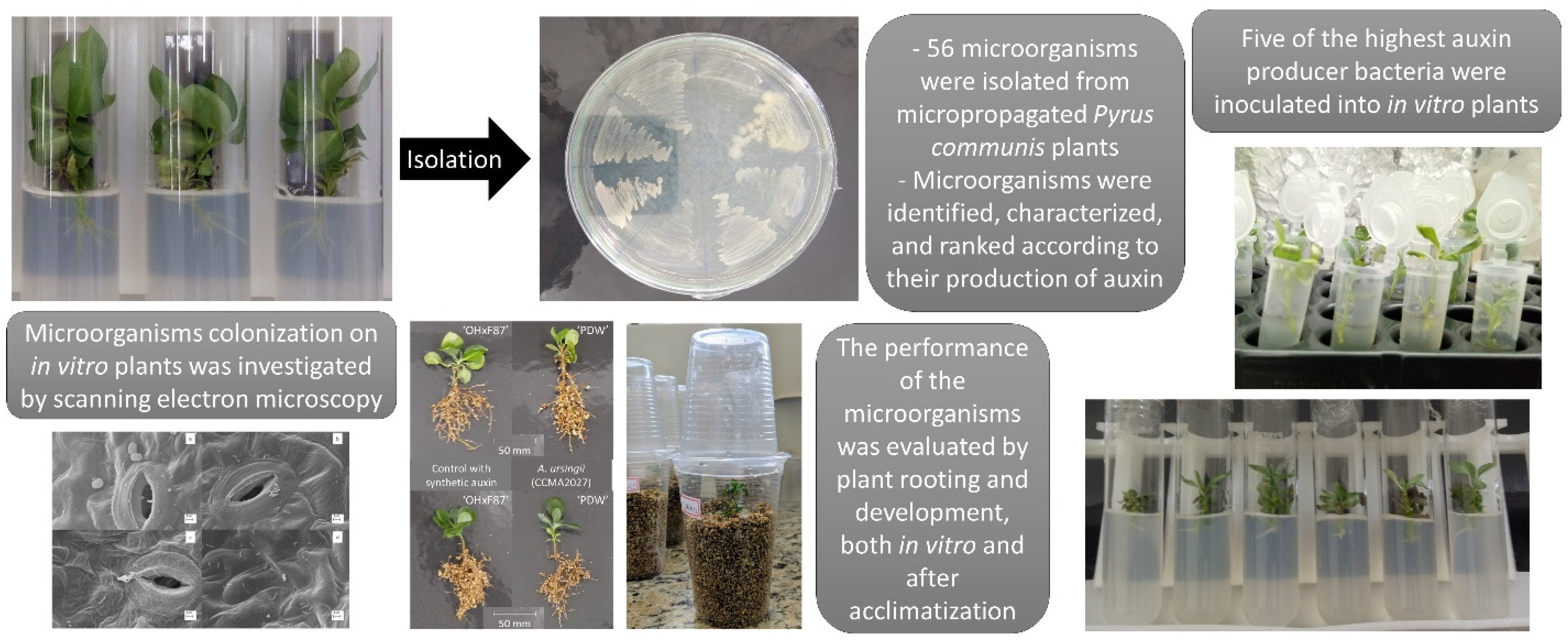
2.2. Isolation and Identification of Endophytic Bacteria in Pyrus communis

The microorganisms were isolated from rootstocks multiplied in vitro. Surface sterilization of the segments of leaves and stems was performed. For this process, the material was washed in distilled water and then 70% alcohol alcohol for 30 s, followed by rinsing in sterile water, this process was repeated three times, and immersion in a 1% hypochlorite solution for 1 min. Plating was performed by the dilution method (1:10) in sterile peptone water (0.1% peptone). The plant material was macerated together with the peptone water and incubated in a shaker at 150 rpm for 30 min at 28 °C. Subsequently, three serial dilutions were performed (1:10), and using the surface plating method, a 0.1 mL aliquot was inoculated into the culture media nutrient agar (NA) (5 g L−1 peptone, 3 g L−1 yeast extract, 15 g L−1 agar), M9 (10 g L−1 Na2HPO4, 3 g L−1 KH2PO4, 0.6 g L−1 NaCl, 20 g L−1 NH4Cl, 5 g L−1 glucose, 15 g L−1 agar), and Luria Bertani (LB) (10 g L−1 tryptone, 5 g L−1 yeast extract, 10 g L−1 NaCl, 15 g L−1 agar). The plates were incubated for 36 h in a biochemical oxygen demand incubator at 28 °C. After this period, dilutions with 30 to 300 colonies were selected and separated by different morphotypes. Three biological replicates were used, each with three technical replicates.
All bacterial isolates found were identified using the ion-assisted matrix/flight time laser desorption technique (MALDI-TOF). The isolates were subjected to Matrix-Assisted Laser Desorption/Ionization–Time of Flight Mass Spectrometer (Bruker Daltonics®, Bremen, Germany) analysis for the identification according to the methodology described by Carvalho et al. [50]. For this analysis, three biological replicates were used, each with three technical replicates.

2.3. Selection of IAA-Promoting Bacteria

To determine the production of the auxin IAA, the strains were grown in nutrient broth (NB) (5 g L−1 peptone, 3 g L−1 yeast extract) supplemented with 0.1 g L−1 L-tryptophan and incubated in a shaker at 150 rpm for 24 h at 28 °C. One-milliliter aliquots were taken and centrifuged for 5 min at 5000× g, and 0.5 milliliters of the supernatant were mixed with 0.5 mL of Salkowski reagent [51]. The mixture was incubated in the dark at room temperature for 20 min, and the absorbance was measured by a mass spectrophotometer (Multiskan GO) using the software SkanIt 5.0 Microplate Readers RE, version 5.0.0.42. To determine the auxin concentration, a standard curve of auxin concentrations of 0, 5, 10, 15, and 20 mg L−1 was obtained, and a linear regression equation was fitted. Three biological replicates were used, each with three technical replicates.

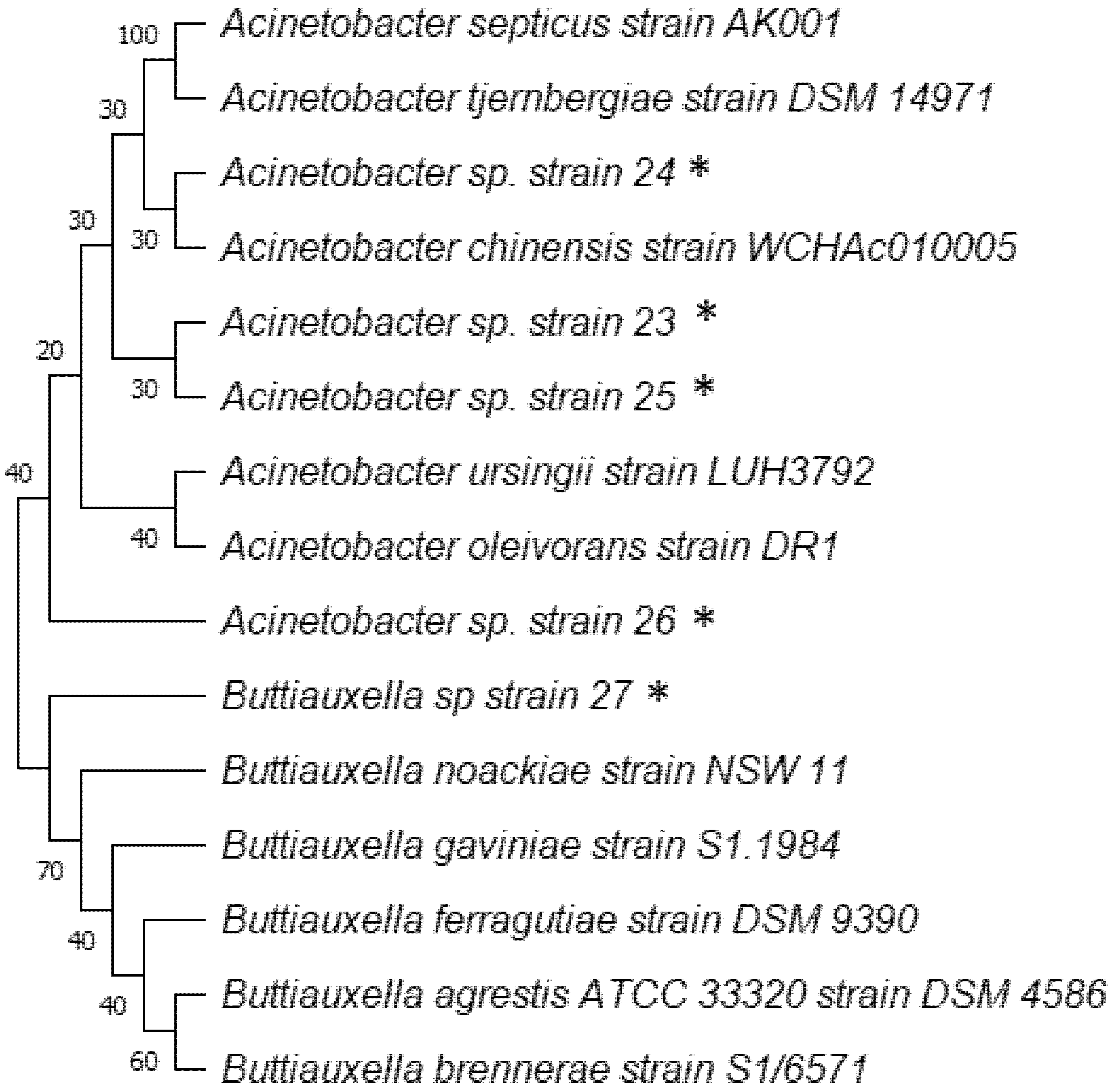
2.4. 16S DNA Gene Sequencing of Isolated Microorganisms with Higher IAA Production

The isolates selected due to the higher production of auxin were identified by the 16SrDNA sequencing technique. In total, five microorganisms were identified using this technique. The genomic DNA was extracted with the Wizard® Genomic DNA Purification Kit, from 10 mL of the bacterial isolates added in a nutrient broth medium for 48 h, under agitation. Two forward primers, 27f (AGA GTT TGA TCM TGG CTC AG) and 515f (GTG CCA GCM GCC GCG GTA A), and a reverse primer 1492r (CGG TTA CCT TGT TAC GAC TT) were used for amplification of the 16S ribosomal gene [52]. Each 30 µL of the final volume of each reaction was composed of: 6.0 µL of the 5x FIREPol® Master Mix (12.5 mM MgCl2, 0.4 M Tris-HCL, 0.1 M. (NH4)2SO4, 0.1% w/v Tween−20 and 1 mM dNTPs of each nucleotide), 0.9 µL of the forward primer (10 mM) and 0.9 µL of the reverse primer (10 mM), 19.7 µL of H2O nuclease-free and 2.5 µL of gDNA (10 ng µL−1). The reactions were distributed in 0.2 mL microtubes and conducted to the SimpliAmp™ thermal cycler, programmed with the following conditions: 95 °C for 2 min, 30 cycles of 95 °C for 30 s, 55 °C for 30 s and 72 °C for 1 min and 40 s, ending at 72 °C for 5 min and 4 °C until electrophoresis on 1% agarose gel.
The products resulting from the PCR were purified with 3.0 M sodium acetate and 95% ethanol, in the proportions of 0.1 and 2.5× the reaction volume used, respectively. The Sanger reaction, from the purified PCR products, was done with the BigDye terminator (PE Applied Biosystems) and the samples were read with ABI 3730xL DNA ANALYZER 48 capillary sequencer (PE Applied Biosystems).
The sequences obtained were aligned in irrigated the Bioedit software and compared with the sequences deposited in the GenBank database, using the BLAST tool from the National Center for Biotechnological Information (NCBI). The multiple sequences obtained in BLAST were aligned in the MEGA-X software using the ClustalW package tool. The nucleotide replacement method was Hasegawa-Kishino-Yano, and the phylogenetic analysis was performed using the maximum likelihood method with 500 bootstrap replications.

2.5. In Vitro Inoculation

After selection of the most promising bacteria, they were evaluated according to their ability to promote plant growth in vitro, especially regarding their potential to induce rooting. In order for this test to be carried out, the bases of the explants were dipped for 30 min in NB solution (5 g L−1 peptone and 3 g L−1 yeast extract) containing bacteria at a population density of approximately 108 CFU mL−1. After 30 min, the bases of the explants were dried in sterile absorbent paper and transferred to tubes containing 20 mL of MS medium, with 30 g L−1 sucrose, 5.5 g L−1 agar, pH adjusted to 6.0 [49]. The material was transferred to a growth chamber with a controlled temperature of 26 °C, and a 16 h photoperiod (40–56 μmol m−2 s−1) where it remained for 35 days. Subsequently, the following parameters were evaluated: number of leaves and roots, length of shoots and roots, and total fresh weight and dry weight.

2.6. Acclimatization

After 35 days of in vitro growth, both rooted and unrooted plants from all treatments were removed from the test tubes, and their base was washed with tap water to remove the agar remnants. Subsequently, the plants were transferred to plastic cups (200 mL) containing vermiculite. The acclimatization process occurred at a temperature of 24 ± 2 °C, and a photoperiod of 16 h (40–56 μmol m−2 s−1) for 60 days. The substrate was irrigated periodically with sterile distilled water. After this period, the following parameters were evaluated: number of leaves and roots, length of shoots and roots, and total fresh weight and dry weight.

2.7. Ultrastructural Analysis by Scanning Electron Microscopy

Scanning electron microscopy was used to observe the presence of bacteria in the plants after treatment in vitro. Plants were fixed in Karnovsky solution (2.5% glutaraldehyde, 2.0% paraformaldehyde in 0.05 M sodium cacodylate buffer, 0.001 M CaCl2, pH 7.2). After fixation, the standard protocol described by Bozzola & Russel [53] was used with some modifications. The samples were collected from the fixative and placed in glycerol for 30 min. Subsequently, they were cut in liquid nitrogen (cryofracture), followed by three washes in distilled water and dehydration in acetone series (25%, 50%, 75%, 90% once, and 100% 3 times). After dehydration, the samples were taken to a Balzers CPD 030 critical point dryer to replace the acetone with CO2 and to complement drying. The samples were mounted on stubs with carbon tape on aluminum foil and covered with gold in a Balzers SCD 050 sputter coater, for later observation under an LEO EVO 40 scanning electron microscope [54].

2.8. Experimental Design and Statistical Analyses

A completely randomized experimental design was used, with eight treatments containing 20 in vitro replicates and 20 replicates evaluated during the acclimatization period. The plants were inoculated with treatments that consisted of the five bacterial isolates with the highest IAA production identification through the DNA 16S gene (Acinetobacter septicus strain 23—CCMA2026, Buttiauxella sp. strain 27—CCMA2030, A. ursingii strain 24—CCMA2027, A. ursingii strain 25—CCMA2028, A. ursingii strain 26—CCMA2029) and two controls (synthetic auxin and zero control). The R software was used to perform the statistical analyses, through which the Shapiro-Wilk test was performed to assess the normality of the data at a significance level of 5%. ANOVA was applied to the data that exhibited a normal distribution, comparing the means by the Tukey test (5%). For data that did not have a normal distribution, the Kruskal-Wallis test was used and a comparison was performed using the Nemenyi test.

2.9. Experiment Workflow

Figure 1 shows the experiment workflow.

3. Results

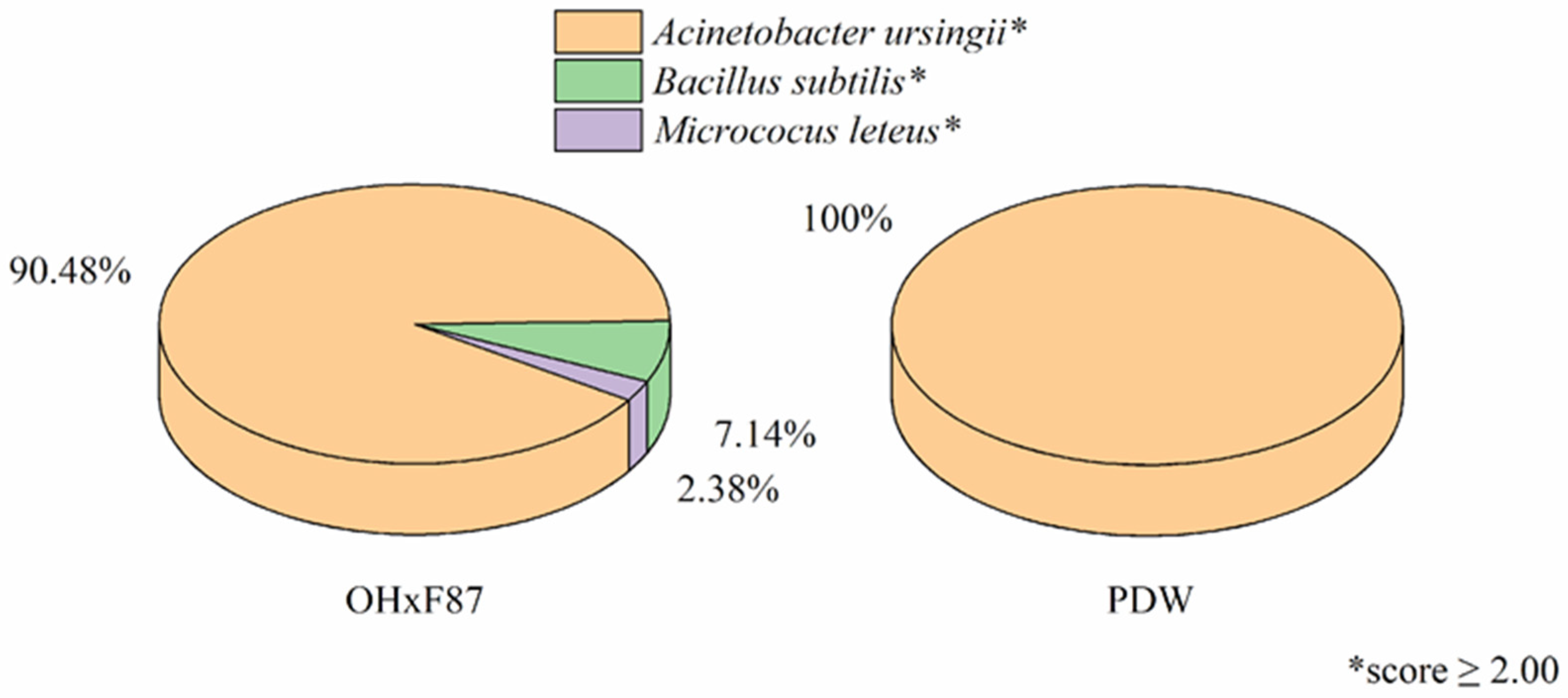
3.1. Isolation and Identification Using MALDI-TOF MS of Endophytic Bacteria in Pyrus communis Rootstocks

In total, 56 isolates were identified belonging to the following species: Acinetobacter ursingii, Bacillus subtilis, and Micrococcus luteus. A low diversity of microorganisms cultivable in culture medium was observed, with a predominance of the genus Acinetobacter. For the ‘OHxF87′ rootstock, 94.48% of the microorganisms isolated were identified as being Acinetobacter ursingii, a species that also corresponded to 100% of the microorganisms isolated from the ‘PDW’ rootstock. (Figure 2).

3.2. Selection of IAA-Promoting Bacteria

Among the 56 microorganisms identified in total, 17 strains were able to produce auxin to some extent, corresponding to 30.35% of the total isolates. The five strains with the highest production were then selected for in vitro inoculation. The IAA concentrations on the selected ones ranged from 8.60 to 19.48 mg L−1 after 24 h of culture (Table 1).

3.3. 16S DNA Gene Sequencing of Isolated Microorganisms with Higher AIA Production

Among the microorganisms analyzed, those that showed the highest production of IAA in vitro were identified, using the 16S rDNA gene sequencing technique. Comparing the data obtained in the blast with the NCBI database, a phylogenetic analysis was performed to assist in the identification and classification of microorganisms. The bacteria identified and their respective percentages of homology were A. septicus strain 23 (97.79%); A. ursingii strain 24 (99.43%); A. ursingii strain 25 (94.40%); A. ursingii strain 26 (98.74%) and Buttiauxella sp. strain 27 (84.77%) (Figure 3).

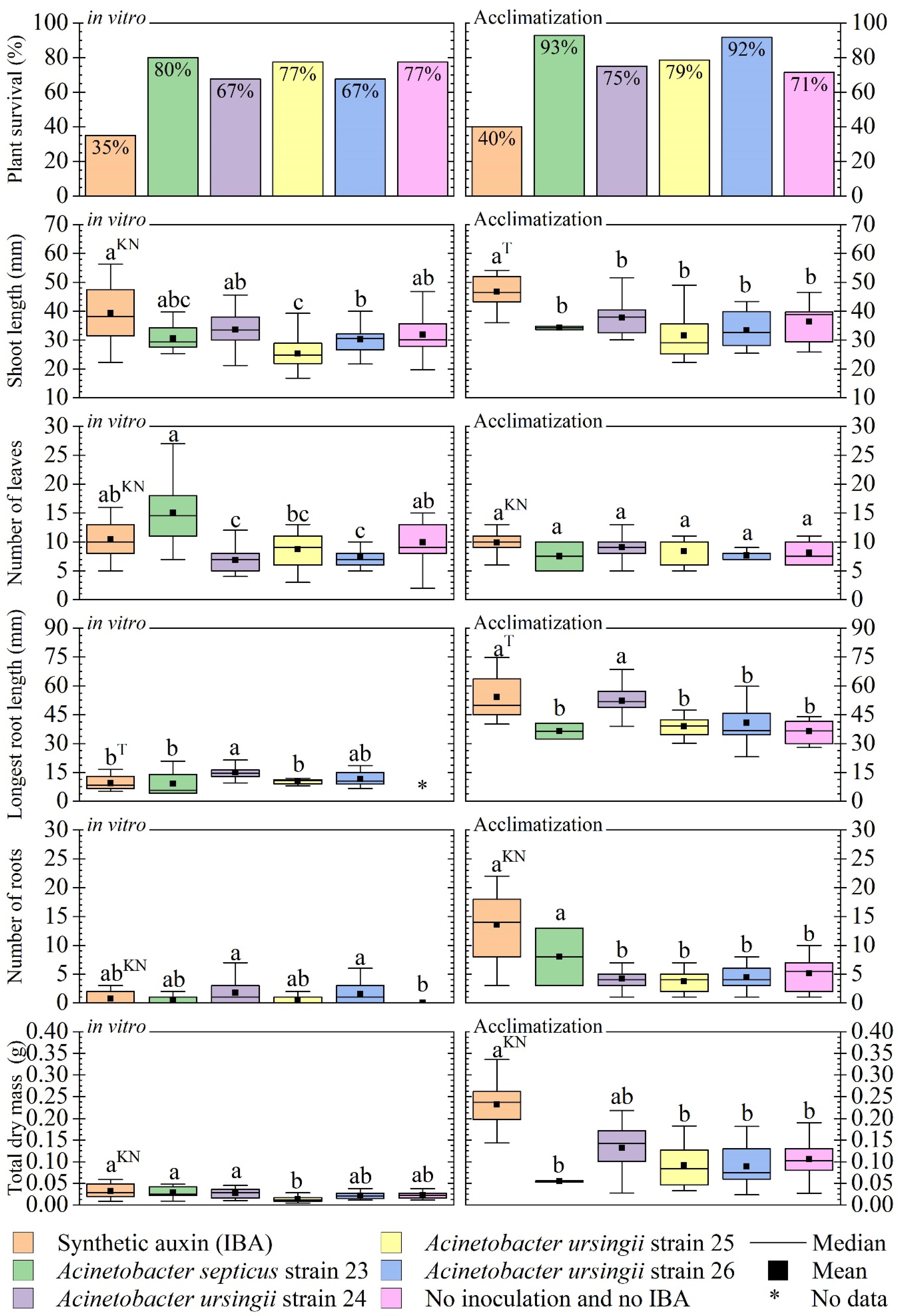
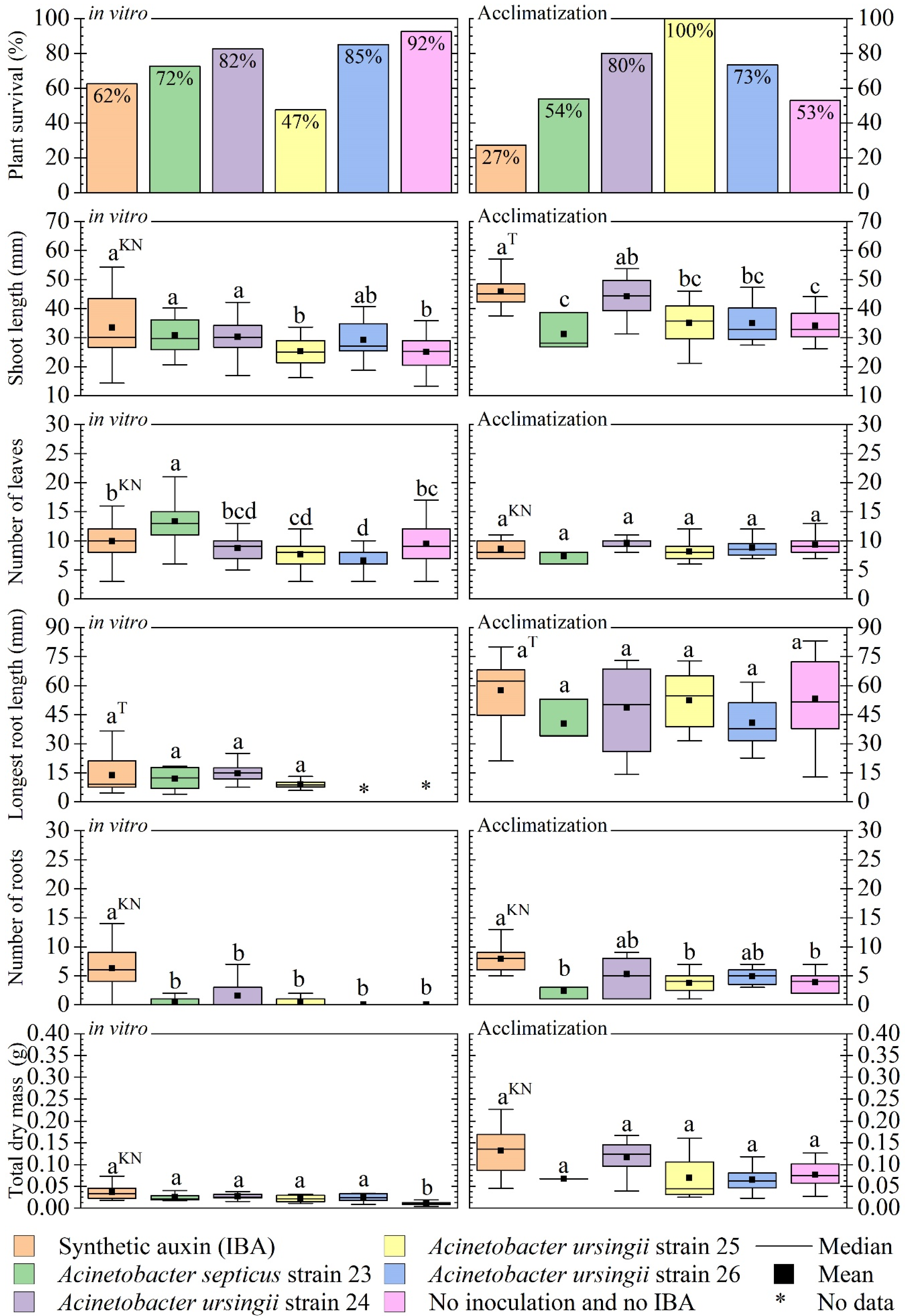
3.4. Evaluation of In Vitro Inoculation: In Vitro Development and Acclimatization

Considering the results obtained for the rootstock ‘OHxF87′, there were differences in the percentage of survival with and without inoculation of microorganisms. The survival of plants with in vitro inoculation of A. septicus strain 23 and A. ursingii strains 24, 25, and 26 was higher than the rate observed in the treatment with synthetic auxin. The bacterial strains 23 and 25 resulted in 80% in vitro survival, while strains 24 and 26 showed an in vitro survival rate of around 70%. For synthetic auxin, the in vitro survival rate was 35% (Figure 4).
The high survival rates persisted during the acclimatization period, where strains 23, 24, 25, and 26 of Acinetobacter sp. showed higher survival rates than the treatment composed of synthetic auxin (40% survival rate), especially strains 23 and 26, with a 90% rate of seedling survival.
The in vitro shoot length was longer in plants treated with a synthetic auxin, with an average of 38 mm. The plants inoculated with A. septicus (strain 23) and A. ursingii (strain 24) were very similar to the treatment with IBA, with averages of 30 and 35 mm, respectively. During the acclimatization phase, the plants treated with IBA had a longer shoot length, with an average of 46 mm. The bacterial strains did not differ from each other in terms of shoot length, with averages between 34 and 38 mm (Figure 4).
For the number of leaves emitted during in vitro cultivation, strain 23 (A. septicus) stood out with the highest average among treatments, where it presented an average of 15 leaves, while inoculation with strains 24, 25, and 26 of A. ursingii, as well as the zero control, produced only around 10 leaves per seedling. During the acclimatization phase, there was no statistical difference in the number of leaves emitted between treatments (Figure 4).
Regarding in vitro root system formation, A. ursingii strain 24 promoted the longest root length, ranging between 10 and 15 mm, statistically differing from the other bacterial strains (23, 25, and 26), the treatment with IBA, and the zero control for the rootstock ‘OHxF87′. During the acclimatization phase, treatments submitted to contact with synthetic auxin and colonization by strain 24 of A. ursingii showed longer roots, with an average of 48 mm. Strains 23 (A. septicus), 25, 26 (A. ursingii), and the zero control did not differ from each other in the statistics, reaching an average length of 38 mm. (Figure 4).
During the in vitro cultivation, the number of leaves between treatments did not differ in the statistics, with two roots emitted per seedling, on average. Considering the highest variation observed in the number of roots for strains 24 and 26 of A. ursingii, it is possible to observe the appearance of up to seven roots per seedling. In the acclimatization phase, the treatment containing synthetic auxin and the one inoculated with strain 23 of A. septicus produced the best results, with an average of 14 and 8 leaves respectively, values that do not differ in statistics (Figure 4).
As for the accumulation of dry mass, during in vitro cultivation, there was no statistical difference between any of the treatments, all with an average of 0.025 g. In the acclimatization phase, the treatment consisting of the application of synthetic auxin produced better results, but it was closely followed by the treatment in which the ‘OHxF87′ rootstocks were colonized by strain 24 of A. ursingii, with averages of 0.24 and 0.14 g, respectively. Disregarding the variations between repetitions, strain 23 (A. septicus) presented the worst result, because, in none of the treatment repetitions, the dry mass exceeded 0.05 g. The treatments that correspond to the contact with strains 25 and 26 of A. ursingii and the zero control were also statistically identical, with means varying between 0.10 and 0.15 g (Figure 3).
Regarding the rootstock ‘PDW’, the addition of synthetic auxin and microorganisms significantly reduced the survival rate of seedlings in the in vitro cultivation phase. A. ursingii strain 25 presented the worst survival rate at this stage, with only 50%, showing the opposite effect of that observed for the same strain in the acclimatization phase. A. ursingii strain 25 induced 100% survival of pear seedlings submitted to acclimatization, contrasting both with the 25% achieved by the addition of synthetic auxin and with the survival rates below 80% observed in the other isolates (Figure 5).
As for the in vitro shoot length, the application of synthetic auxin and the inoculation of strains 23 of A. septicus and 24 of A. usringii produced slightly better results, around 30 mm, than the zero control and strains 25 and 26 of A. ursingii, with averages around 26 mm. In the acclimatization phase, synthetic auxin and strain 24 of A. ursingii showed longer shoots, with an average of 45 mm, contrasting with the values close to 30 mm obtained in the zero control and the inoculation with strains 23 of A. septicus, 25, and 26 of A. ursingii (Figure 5).
The A. septicus strain 23 provided seedlings with more leaves, averaging around 14 per seedling, while the application of synthetic auxin and inoculation with A. usringii strains 24, 25, and 26 did not produce seedlings with a number of leaves greater than 10 in the in vitro cultivation phase. There was no statistical difference in the number of leaves obtained in the acclimatization, for any of the treatments to which the ‘PDW’ rootstock was submitted (Figure 5).
For the longest root length, there was no statistical difference between the values obtained in each treatment, both in in vitro cultivation and in the acclimatization phase. In both phases evaluated, the number of roots was higher in plants submitted to treatment with a synthetic auxin, both with an average of eight roots per seedling. In in vitro cultivation, none of the bacterial isolates showed a statistical difference regarding the number of roots emitted, when compared to each other, with emission of up to two roots per seedling. In the acclimatization phase, strains 24 and 26 of A. ursingii produced seedlings slightly more similar to those produced by direct contact with synthetic auxin. For the dry mass, in none of the phases, there was a significant difference between the treatments to which the ‘PDW’ rootstock was submitted (Figure 5).
None of the evaluated rootstocks supported colonization by Buttiauxella sp. (strain 27), since there was no survival of plants that came into direct contact with a pure population of this microorganism.

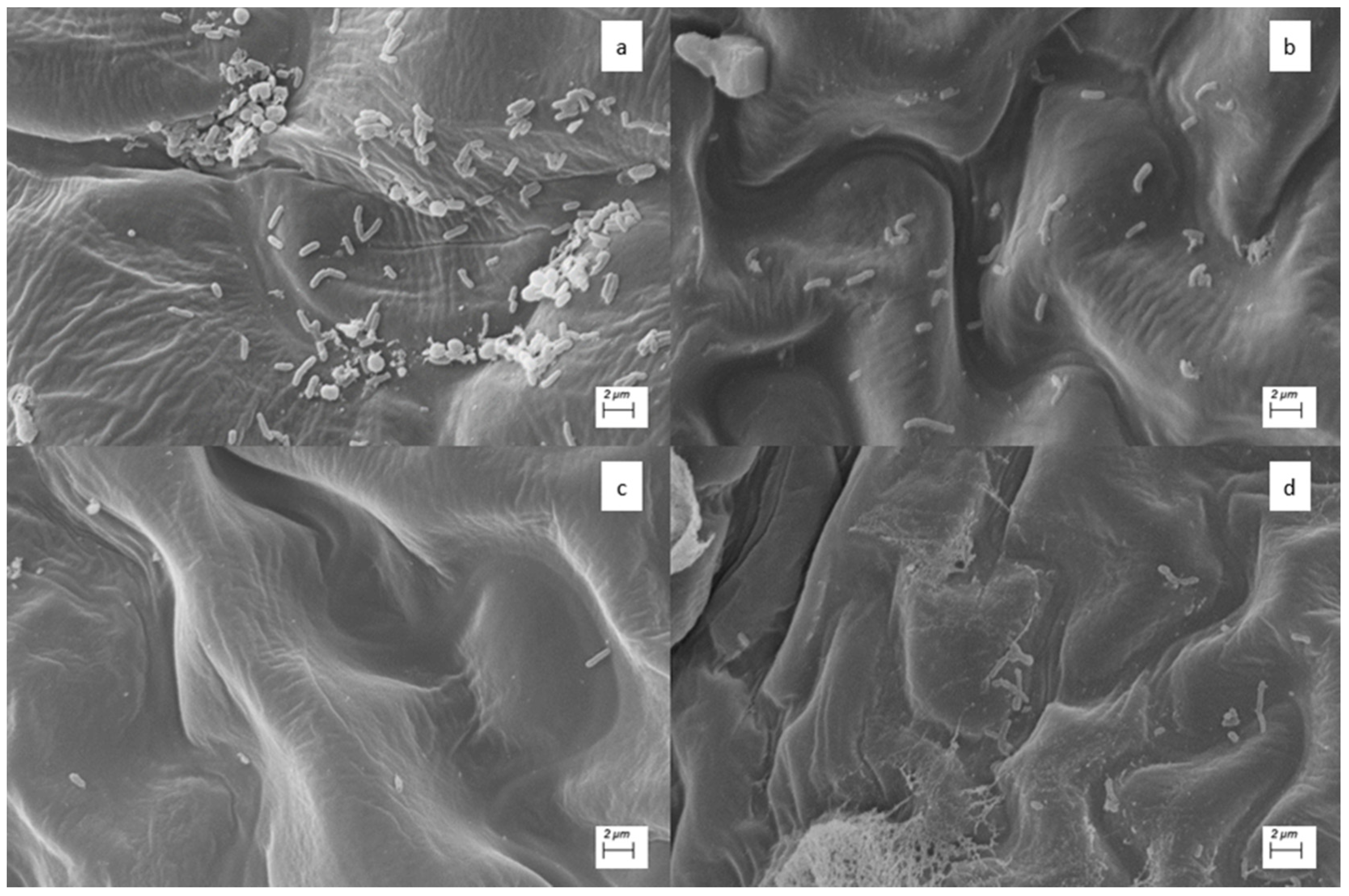
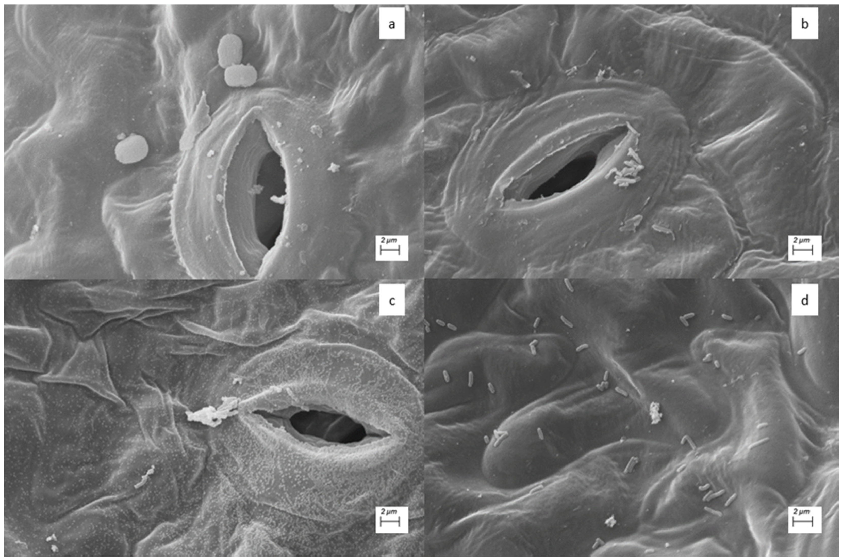
3.5. Ultrastructural Analysis by Scanning Electron Microscopy

In the Figure 6 are the electron micrographs showed the presence of microorganisms Acinetobacter septicus strain 23 (a); Acinetobacter ursingii strain 24 (b); A. ursingii strain 25 (c); A. ursingii strain 26 (d) of the ‘OHxF87′ rootstock after in vitro development and before the acclimatization period. No colonies were found in the treatment that consisted of synthetic auxin control, or zero control for this rootstock.
However, for the ‘PDW’ rootstock, the electron micrographs revealed the presence of bacteria in treatments with A. ursingii strain 24 and A. ursingii strain 26, in addition to colonies in the negative control and in synthetic IBA. No bacterial colonies were found in the portion observed for the treatments with A. septicus strain 23 and A. ursingii strain 25 (Figure 7).

4. Discussion

The objective of this work was to isolate and identify the endophytic microorganisms capable of producing auxin in the rootstocks of two pear cultivars. After identifying the isolates with the highest auxin production, the objective was to use them as an inoculant, in order to compare their effect on in vitro rooting and acclimatization of the two rootstocks, with the effects of the addition of synthetic auxin. An effect similar to that of the addition of synthetic auxin was observed for several agronomic traits evaluated after in vitro cultivation and acclimatization.

4.1. Isolation and Identification of Endophytic Bacteria in Pyrus communis Rootstocks

In the process of plant micropropagation, the absence of microorganisms is expected, as it is guaranteed by the aseptic culture processes; however, the results showed that even in these controlled conditions, microorganisms may be present, as also observed by Pirtillä et al. [26] and Pohjanen et al. with Pinus sylvestris and by Quambusch et al. [24,25] with Prunus avium.
Because in vitro culture was performed under controlled conditions, there was no possibility of interactions with the complex microbial community that naturally occurs in the soil, which explains the low microbial diversity found in the studied genotypes. The same was observed in a study conducted by Quambusch et al. [25] with six P. avium genotypes, where only five morphologically distinct isolates were identified from plants derived from tissue culture. The genus Acinetobacter is ubiquitous in nature and can be isolated from soil and/or water, and its high concentration, to the detriment of other genera, may be linked to its resistance to antibiotics and heavy metals [55,56,57].

4.2. Selection of IAA-Promoting Bacteria

The colonization of plants by these microorganisms may be related to the innate difficulty of woody plants in emitting adventitious roots. Plants and bacteria, as well as other living organisms, create symbiotic relationships to meet the needs of both. The production of auxin by microorganisms enables and facilitates the symbiosis between plants and microorganisms, since this compound is very important for the growth and development of plants [33,34], which is one of the most used mechanisms to explain the positive effects of plant growth-promoting bacteria [31,32].
Tests of in vitro production of auxins showed that 17 isolates have, to some degree, the ability to produce this phytohormone. Different bacteria with this ability have been identified, such as Azospirillum brasilense [39], Bacillus megaterium [38], Bacillus siamensis [37], Bacillus methylotrophicus [58], Pseudomonas veronii [45], Acinetobacter johnsonii [59], and Acinetobacter baumannii [42]. The auxin production is variable and dependent on the microorganism’s cultivation conditions. In an assay carried out with A. johnsonii, the production of auxin among the strains varied between 10.12 and 126.97 μg mL−1 in the different cultivation conditions studied [59].
The genus Acinetobacter, predominant among the bacteria identified in the evaluated rootstocks, is also widely found in the environment. In addition to the auxin production demonstrated in this work, species belonging to this genus were also described regarding their ability to fix nitrogen, solubilize minerals, and produce siderophores, characteristics that are desirable for plant growth promoters [60].

4.3. Evaluation of In Vitro Inoculation: In Vitro Development and Acclimatization

The results showed different responses to the same treatments among the evaluated genotypes. The treatment of rootstocks with bacterial isolates showed the same efficiency as the treatment with synthetic auxin for the different variables analyzed and allowed good development during the period of acclimatization of the seedlings. In a study investigating the vegetative growth of strawberries (Rosaceae), Andrade et al. [61] also observed that the use of growth-promoting bacteria, such as Azospirillum brasilense, Burkholderia cepacia, and Enterobacter cloacae, promoted shoot growth similar to that found in plants cultivated with fertilizers. The similarities observed between chemical and biological fertilizers allow the adoption of microorganisms to reduce production costs and improve the sustainability of the cultivation process.
Shoot growth and leaf formation are essential for plants, since individuals with more leaves, under ideal conditions, have a higher photosynthetic capacity, which benefits their growth and development [62]. For these variables, the strains 24 and 26 of A. ursingii, and 23 of A. septicus contributed to the formation of shoots in the evaluated clones. In addition, auxin-producing bacteria have been used especially to stimulate seed germination and accelerate root growth [63,64,65].
Other studies have also reported positive results for the in vitro rooting of woody plants from the Rosaceae family. Larraburu et al. [44] used a combination of synthetic microorganisms and auxins for the rooting of Photinia. These authors observed a key role of microorganisms in the root organogenesis of the plants, proposing an alternative protocol for micropropagation of the species. In Prunus avium, the use of Rhodopseudomonas sp. and Microbacterium sp. promoted in vitro rooting rates that were higher than zero control, ranging between 30 and 92.5%, and the formation of 0.7 to 7.2 roots depending on the cultivar [25]. The use of microorganisms such as Pseudomonas veronii R4 and P. fluorescens CHA0, both of which can synthesize IAA, also induced in vitro root formation in grape leaves [44,45].
Furthermore, ex vitro experiments showed that the success of acclimatization of P. communis plants depends on hormonal stimuli, as can be observed in the studies by Aygun & Dumanoglu [10], and Lizárraga et al. [66], and in the acclimatization of Musa spp. with Buttiauxella agrestis [67]. The results found in this work regarding the rooting of rootstocks confirm the findings of Quambusch et al. [25], who showed that the manipulation of the endogenous bacterial population, through their inoculation as growth promoters, has a similar effect to the use of synthetic auxin. Therefore, the use of microorganism-plant interactions and their mechanisms, such as auxin production, can lead to an improvement in plant survival and growth promotion during critical phases of plant tissue culture.

4.4. Ultrastructural Analysis by Scanning Electron Microscopy

The best results regarding plant growth and rooting were obtained with the A. ursingii strains 24 and 25 for ‘OH×F87′ rootstock. Electron micrographs showed the presence of bacteria in ‘OH×F87′ rootstocks that received treatments with the microorganisms. Bacteria’s presence close to the stomata can be observed, including inside them, similarly to what was observed by Gilbert et al. [68].

4.5. General Discussion

The results found during acclimatization corroborate evidence showing that rooting induction is not only related to the inoculation of microorganisms since even plants that did not have roots during in vitro growth showed good development and rooting in the ex vitro phase. The induction of ex vitro rooting may also be related to the increase in the plant’s metabolic activity, since acclimatized plants have a higher amount of plant tissue, and consequently more photosynthetic area. Therefore, an increase in primary and secondary metabolism is to be expected because of higher net photosynthesis.
The results found in this work suggest that the inoculation with microorganisms enabled growth similar to that promoted by synthetic auxin in pear plants, which can make them a potential substitute for the artificial auxin used in vitro. Some microorganisms increased the survival rate of the plants, especially during the acclimatization phase. Additional research can be done in order to refine the methodology, and the understanding of the use of beneficial microorganisms in vitro. However, based on our results, we can predict that this new approach can bring benefits for plant producers by reducing costs associated with the micropropagation process, especially regarding the purchase of synthetic auxin, and by improving the quality of the clones propagated through this system.

5. Conclusions

Based on this study, it can be concluded that it is possible to promote the growth and emission of roots in pear rootstocks, through a modulation of the endophytic microbial population prior to the introduction of the explant into the in vitro culture medium. From the inoculation with bacteria identified as capable of producing auxin, it was possible to obtain results similar to the treatment of materials with auxin with regard to rooting of the rootstocks. This process can be used to replace the inclusion of synthetic auxin in the production chain of vegetatively propagated woody fruits, such as pears.
None of the bacterial isolates tested was notably more promising, but the general similarity of treatments containing A. septicus and A. ursingii strains with the treatment with a synthetic auxin, suggests the possibility of its use on a large scale, allowing the adoption of a method cheaper for rooting rootstocks.
This work opens the way for new research using microbial isolates already described as the most promising in the production of plant hormones and growth promotion in general, also including research in the improvement of microbial strains exclusively focused on the production of auxins for agriculture. The constant search for low-cost agricultural systems with less environmental impact also generates demand for lower-cost microorganism cultivation techniques, such as low-cost means obtained from agro-industrial residues.

Author Contributions

Conceptualization, M.C.N. and G.M.d.R.F.; Data curation, M.C.N. and G.M.d.R.F.; Formal analysis, M.C.N. and G.M.d.R.F.; Funding acquisition, L.R., J.M.Q.L., J.D. and M.P.; Investigation, M.C.N. and G.V.S.A.; Methodology, M.C.N., G.M.d.R.F. and C.M.d.S.; Supervision, A.D.M., J.D. and M.P.; Visualization, M.C.N., G.V.S.A., V.H.B., F.A.R., C.M.d.S., A.D.M. and G.M.d.R.F.; Writing—original draft preparation, M.C.N., J.M.Q.L., J.D. and M.P.; Writing—review and editing, G.M.d.R.F., F.A.R., A.D.M., V.H.B., L.R., J.D., J.M.Q.L. and M.P. All authors have read and agreed to the published version of the manuscript.

Funding

This study was financed in part by the Coordenação de Aperfeiçoamento de Pessoal de Nível Superior—Brasil (CAPES), Conselho Nacional de Desenvolvimento Científico e Tecnológico—Brasil (CNPq), and Fundação de Amparo à Pesquisa do Estado de Minas Gerais (FAPEMIG) for financial support.

Institutional Review Board Statement

Not applicable.

Informed Consent Statement

Not applicable.

Data Availability Statement

All data that support the findings of this study are available from the corresponding author upon reasonable request.

Acknowledgments

The authors acknowledge the assistance of Laboratory Technician Vantuil Antônio Rodrigues, and anonymous referees for their comments and constructive suggestions for improving the manuscript quality.

Conflicts of Interest

The authors declare no conflict of interest.

References

  1. Rufato, L.; Marcon Filho, J.L.; Marodin, G.A.B.; Kretzschmar, A.A.; Miqueluti, D.J. Intensidade e Épocas de Poda Verde Em Pereira “Abate Fetel” Sobre Dois Porta-Enxertos. Rev. Bras. Frutic. 2012, 34, 475–481. [Google Scholar] [CrossRef] [Green Version]
  2. Hawerroth, F.J.; Herter, F.G.; Prezotto, M.E.; Haas, L.B.; Pretto, A. Aumento Da Produção de Pereira Asiática Pelo Uso de Fitorreguladores. Ciência Rural. St. Maria 2011, 41, 1750–1754. [Google Scholar] [CrossRef]
  3. Hartmann, H.T.; Kester, D.E.; Davies Junior, F.T.; Geneve, R.L. Plant Propagation: Principles and Practices, 7th ed.; Prentice Hall: Hoboken, NJ, USA, 2002. [Google Scholar]
  4. Park, S. Plant Tissue Culture; Elsevier: Amsterdam, The Netherlands, 2021; ISBN 978-0-12-821120-5. [Google Scholar]
  5. Ercisli, S.; Esitken, A.; Orhan, E.; Ozdemir, O. Rootstocks Used for Temperate Fruit Trees in Turkey: An Overview. Sodinink. Daržinink. 2006, 25, 27–33. [Google Scholar]
  6. Dimitrova, N.; Nacheva, L. An Optimized Micropropagation Protocol by Ex Vitro Rooting of Pear Rootstock OHF 333 (Pyrus communis L.). Acta Agrobot. 2021, 74, 744. [Google Scholar] [CrossRef]
  7. Apple & Pear Australia LTD (APAL) Rootstocks. Available online: https://apal.org.au/industry-info/intensive-pear-production/rootstocks/#pr (accessed on 15 July 2019).
  8. Washington State University (WSU) Rootstocks for Pear. Available online: http://treefruit.wsu.edu/web-article/pear-rootstocks/ (accessed on 15 July 2019).
  9. Grimaldi, F.; Grohskopf, M.A.; Muniz, A.W.; Frederico, A. Enraizamento in Vitro de Frutíferas Da Família Rosaceae. Rev. Ciências Agroveterinárias 2008, 7, 160–168. [Google Scholar]
  10. Aygun, A.; Dumanoglu, H. In Vitro Shoot Proliferation and In Vitro and Ex Vitro Root Formation of Pyrus Elaeagrifolia Pallas. Front. Plant Sci. 2015, 6, 225. [Google Scholar] [CrossRef]
  11. Yang, Y.; Wang, D.; Wang, C.; Wang, X.; Li, J.; Wang, R. Construction of High Efficiency Regeneration and Transformation Systems of Pyrus Ussuriensis Maxim. Plant Cell Tissue Organ Cult. 2017, 131, 139–150. [Google Scholar] [CrossRef]
  12. Norton, M.E. The Role of Auxin and Cytokinin in the Control of Morphogenesis In Vitro in the Plant Families “Rosaceae” and “Ericaceae”; University of St. Andrews: St. Andrews, UK, 1984. [Google Scholar]
  13. Orlikowska, T.; Nowak, K.; Reed, B. Bacteria in the Plant Tissue Culture Environment. Plant Cell Tissue Organ Cult. 2017, 128, 487–508. [Google Scholar] [CrossRef]
  14. Elnahal, A.S.M.; El-Saadony, M.T.; Saad, A.M.; Desoky, E.-S.M.; El-Tahan, A.M.; Rady, M.M.; AbuQamar, S.F.; El-Tarabily, K.A. The Use of Microbial Inoculants for Biological Control, Plant Growth Promotion, and Sustainable Agriculture: A Review. Eur. J. Plant Pathol. 2022, 162, 759–792. [Google Scholar] [CrossRef]
  15. Souza, J.A.; Bettoni, J.C.; Costa, M.D.; Baldissera, T.C.; dos Passos, J.F.M.; Primieri, S. In Vitro Rooting and Acclimatization of ‘Marubakaido’ Apple Rootstock Using Indole-3-Acetic Acid from Rhizobacteria. Commun. Plant Sci. 2022, 12, 16–23. [Google Scholar] [CrossRef]
  16. Erturk, Y.; Ercisli, S.; Haznedar, A.; Cakmakci, R. Effects of Plant Growth Promoting Rhizobacteria (PGPR) on Rooting and Root Growth of Kiwifruit (Actinidia deliciosa) Stem Cuttings. Biol. Res. 2010, 43, 91–98. [Google Scholar] [CrossRef] [Green Version]
  17. Montero-Calasanz, M.C.; Santamaría, C.; Albareda, M.; Daza, A.; Duan, J.; Glick, B.R.; Camacho, M. Alternative Rooting Induction of Semi-Hardwood Olive Cuttings by Several Auxin-Producing Bacteria for Organic Agriculture Systems. Span. J. Agric. Res. 2013, 11, 146–154. [Google Scholar] [CrossRef]
  18. Pretty, J. Agricultural Sustainability: Concepts, Principles and Evidence. Philos. Trans. R. Soc. B Biol. Sci. 2008, 363, 447–465. [Google Scholar] [CrossRef] [PubMed] [Green Version]
  19. Bizos, G.; Papatheodorou, E.M.; Chatzistathis, T.; Ntalli, N.; Aschonitis, V.G.; Monokrousos, N. The Role of Microbial Inoculants on Plant Protection, Growth Stimulation, and Crop Productivity of the Olive Tree (Olea europea L.). Plants 2020, 9, 743. [Google Scholar] [CrossRef] [PubMed]
  20. Keswani, C.; Singh, S.P.; Cueto, L.; García-Estrada, C.; Mezaache-Aichour, S.; Glare, T.R.; Borriss, R.; Singh, S.P.; Blázquez, M.A.; Sansinenea, E. Auxins of Microbial Origin and Their Use in Agriculture. Appl. Microbiol. Biotechnol. 2020, 104, 8549–8565. [Google Scholar] [CrossRef]
  21. Ali, M.; Boonerjee, S.; Islam, M.N.; Saha, M.L.; Hoque, M.I.; Sarker, R.H. Endogenous Bacterial Contamination of Plant Tissue Culture Materials: Identification and Control Strategy. Plant Tissue Cult. Biotechnol. 2018, 28, 99–108. [Google Scholar] [CrossRef] [Green Version]
  22. Örgeç, M.Ö.; Pehlivan Karakaş, F.; Şahin, G.; Ağıl, F.; Zencirci, N. Einkorn (Triticum monococcum ssp. monococcum) in vitro propagation sterilization protocol. Int. J. Second. Metab. 2018, 5, 67–74. [Google Scholar] [CrossRef]
  23. Quambusch, M.; Winkelmann, T. Bacterial Endophytes in Plant Tissue Culture: Mode of Action, Detection, and Control. In Plant Cell Culture Protocols, Methods in Molecular Biology; Humana Press: New York, NY, USA, 2018; Volume 1815, pp. 69–88. ISBN 9781493985944. [Google Scholar]
  24. Quambusch, M.; Brümmer, J.; Haller, K.; Winkelmann, T.; Bartsch, M. Dynamics of endophytic bacteria in plant in vitro culture: Quantification of three bacterial strains in Prunus avium in different plant organs and in vitro culture phases. Plant Cell Tissue Organ Cult. 2016, 126, 305–317. [Google Scholar] [CrossRef]
  25. Quambusch, M.; Pirttila, A.M.; Tejesvi, M.V.; Winkelmann, T.; Bartsch, M. Endophytic Bacteria in Plant Tissue Culture: Differences between Easy- and Difficult-to-Propagate Prunus Avium Genotypes. Tree Physiol. 2014, 34, 524–533. [Google Scholar] [CrossRef]
  26. Pirttilä, A.M.; Podolich, O.; Koskimäki, J.J.; Hohtola, E.; Hohtola, A. Role of Origin and Endophyte Infection in Browning of Bud-Derived Tissue Cultures of Scots Pine (Pinus sylvestris L.). Plant Cell Tissue Organ Cult. 2008, 95, 47–55. [Google Scholar] [CrossRef]
  27. Pohjanen, J.; Koskimaki, J.J.; Sutela, S.; Ardanov, P.; Suorsa, M.; Niemi, K.; Sarjala, T.; Haggman, H.; Pirttila, A.M. Interaction with ectomycorrhizal fungi and endophytic Methylobacterium affects nutrient uptake and growth of pine seedlings in vitro. Tree Physiol. 2014, 34, 993–1005. [Google Scholar] [CrossRef] [PubMed]
  28. Tsavkelova, E.A.; Klimova, S.Y.; Cherdyntseva, T.A.; Netrusov, A.I. Microbial Producers of Plant Growth Stimulators and Their Practical Use: A Review. Appl. Biochem. Microbiol. 2006, 42, 117–126. [Google Scholar] [CrossRef]
  29. Naik, K.; Mishra, S.; Srichandan, H.; Singh, P.K.; Sarangi, P.K. Plant Growth Promoting Microbes: Potential Link to Sustainable Agriculture and Environment. Biocatal. Agric. Biotechnol. 2019, 21, 101326. [Google Scholar] [CrossRef]
  30. Basu, A.; Prasad, P.; Das, S.N.; Kalam, S.; Sayyed, R.Z.; Reddy, M.S.; El Enshasy, H. Plant Growth Promoting Rhizobacteria (PGPR) as Green Bioinoculants: Recent Developments, Constraints, and Prospects. Sustainability 2021, 13, 1140. [Google Scholar] [CrossRef]
  31. Olanrewaju, O.S.; Glick, B.R.; Babalola, O.O. Mechanisms of Action of Plant Growth Promoting Bacteria. World J. Microbiol. Biotechnol. 2017, 33, 197. [Google Scholar] [CrossRef] [Green Version]
  32. Rana, K.L.; Kour, D.; Kaur, T.; Devi, R.; Yadav, A.N.; Yadav, N.; Dhaliwal, H.S.; Saxena, A.K. Endophytic Microbes: Biodiversity, Plant Growth-Promoting Mechanisms and Potential Applications for Agricultural Sustainability. Antonie Leeuwenhoek 2020, 113, 1075–1107. [Google Scholar] [CrossRef]
  33. Spaepen, S.; Vanderleyden, J. Auxin and Plant-Microbe Interactions. Cold Spring Harb. Perspect. Biol. 2011, 3, a001438. [Google Scholar] [CrossRef] [Green Version]
  34. Spaepen, S.; Vanderleyden, J.; Remans, R. Indole-3-Acetic Acid in Microbial and Microorganism-Plant Signaling. FEMS Microbiol. Rev. 2007, 31, 425–448. [Google Scholar] [CrossRef] [Green Version]
  35. Malhotra, M.; Srivastava, S. Stress-Responsive Indole-3-Acetic Acid Biosynthesis by Azospirillum brasilense SM and Its Ability to Modulate Plant Growth. Eur. J. Soil Biol. 2009, 45, 73–80. [Google Scholar] [CrossRef]
  36. Ahmed, A.; Hasnain, S. Auxin-producing Bacillus sp.: Auxin quantification and effect on the growth of Solanum tuberosum. Pure Appl. Chem. 2010, 82, 313–319. [Google Scholar] [CrossRef]
  37. Hossain, M.T.; Khan, A.; Harun-Or-Rashid, M.; Chung, Y.R. A volatile producing endophytic Bacillus siamensis YC7012 promotes root development independent on auxin or ethylene/jasmonic acid pathway. Plant Soil 2019, 439, 309–324. [Google Scholar] [CrossRef]
  38. López-Bucio, J.; Campos-Cuevas, J.C.; Hernández-Calderón, E.; Velásquez-Becerra, C.; Farías-Rodríguez, R.; Macías-Rodríguez, L.I.; Valencia-Cantero, E. Bacillus Megaterium Rhizobacteria Promote Growth and Alter Root-System Architecture through an Auxin- and Ethylene-Independent Signaling Mechanism in Arabidopsis Thaliana. Mol. Plant-Microbe Interact. 2007, 20, 207–217. [Google Scholar] [CrossRef] [PubMed] [Green Version]
  39. Rivera, D.; Mora, V.; Lopez, G.; Rosas, S.; Spaepen, S.; Vanderleyden, J.; Cassan, F. New insights into indole-3-acetic acid metabolism in Azospirillum brasilense. J. Appl. Microbiol. 2018, 125, 1774–1785. [Google Scholar] [CrossRef] [PubMed]
  40. Ortiz-Castro, R.; Campos-García, J.; López-Bucio, J. Pseudomonas putida and Pseudomonas fluorescens Influence Arabidopsis Root System Architecture through an Auxin Response Mediated by Bioactive Cyclodipeptides. J. Plant Growth Regul. 2019, 39, 254–265. [Google Scholar] [CrossRef]
  41. Karadeniz, A.; Topcuoğlu, Ş.F.; Inan, S. Auxin, Gibberellin, Cytokinin and Abscisic Acid Production in Some Bacteria. World J. Microbiol. Biotechnol. 2006, 22, 1061–1064. [Google Scholar] [CrossRef]
  42. Lin, H.-R.; Shu, H.-Y.; Lin, G.-H. Biological roles of indole-3-acetic acid in Acinetobacter baumannii. Microbiol. Res. 2018, 216, 30–39. [Google Scholar] [CrossRef]
  43. Foo, E.; Plett, J.M.; Lopez-Raez, J.A.; Reid, D. Editorial: The Role of Plant Hormones in Plant-Microbe Symbioses. Front. Plant Sci. 2019, 10, 1391. [Google Scholar] [CrossRef]
  44. Larraburu, E.E.; Carletti, S.M.; Cáceres, E.A.R.; Llorente, B.E. Micropropagation of photinia employing rhizobacteria to promote root development. Plant Cell Rep. 2007, 26, 711–717. [Google Scholar] [CrossRef]
  45. Peñafiel-Jaramillo, M.; Torres-Navarrete, E.D.; Barrera-Álvarez, A.E.; Prieto-Encalada, H.; Morante Carriel, J.; Canchignia Martínez, H.F. Producción de Ácido Indol-3-Acético Por Pseudomonas Veronii R4 y Formación de Raíces En Hojas de Vid “Thompson Seedless” in vitro. Cienc. Tecnol. 2016, 9, 31. [Google Scholar] [CrossRef]
  46. Russo, A.; Vettori, L.; Felici, C.; Fiaschi, G.; Morini, S.; Toffanin, A. Enhanced micropropagation response and biocontrol effect of Azospirillum brasilense Sp245 on Prunus cerasifera L. clone Mr.S 2/5 plants. J. Biotechnol. 2008, 134, 312–319. [Google Scholar] [CrossRef]
  47. Vettori, L.; Russo, A.; Felici, C.; Fiaschi, G.; Morini, S.; Toffanin, A. Improving micropropagation: Effect of Azospirillum brasilense Sp245 on acclimatization of rootstocks of fruit tree. J. Plant Interact. 2010, 5, 249–259. [Google Scholar] [CrossRef]
  48. Leblay, C.; Chevreau, E.; Raboin, L.M. Adventitious shoot regeneration from in vitro leaves of several pear cultivars (Pyrus communis L.). Plant Cell Tissue Organ Cult. 1991, 25, 99–105. [Google Scholar] [CrossRef]
  49. Murashige, T.; Skoog, F. A Revised Medium for Rapid Growth and Bio Assays with Tobacco Tissue Cultures. Physiol. Plant. 1962, 15, 473–497. [Google Scholar] [CrossRef]
  50. Carvalho, B.; Ávila, C.; Bernardes, T.; Pereira, M.N.; Santos, C.; Schwan, R. Fermentation profile and identification of lactic acid bacteria and yeasts of rehydrated corn kernel silage. J. Appl. Microbiol. 2017, 122, 589–600. [Google Scholar] [CrossRef]
  51. Glickmann, E.; Dessaux, Y. A critical examination of the specificity of the salkowski reagent for indolic compounds produced by phytopathogenic bacteria. Appl. Environ. Microbiol. 1995, 61, 793–796. [Google Scholar] [CrossRef] [Green Version]
  52. Lane, D.J. 16S/23S RRNA Sequencing. In Nucleic Acid Techniques in Bacterial Systematic; Stackbrandt, E., Goodfellow, M., Eds.; John Wiley and Sons: New York, NY, USA, 1991; pp. 115–175. ISBN 0-471-92906-9. [Google Scholar]
  53. Bozzola, J.J.; Russel, L.D. Electron Microscopy: Principles and Techniques for Biologists; Jones & Bartlett Learning: Burlington, MA, USA, 1999; ISBN 0763701920. [Google Scholar]
  54. Alves, E. Curso Introdutório à Microscopia Eletrônica de Varredura; Editora Universitária-Uniersidade Federal de Lavras: Lavras, Brazil, 2005. [Google Scholar]
  55. Nemec, A.; Radolfová-Křížová, L.; Maixnerová, M.; Shestivska, V.; Španělová, P.; Higgins, P.G. Acinetobacter silvestris sp. nov. discovered in forest ecosystems in Czechia. Int. J. Syst. Evol. Microbiol. 2022, 72, 005383. [Google Scholar] [CrossRef]
  56. Mujumdar, S.; Joshi, P.; Karve, N. Production, characterization, and applications of bioemulsifiers (BE) and biosurfactants (BS) produced by Acinetobacter spp.: A review. J. Basic Microbiol. 2019, 59, 277–287. [Google Scholar] [CrossRef]
  57. Guardabassi, L.; Dalsgaard, A.; Olsen, J.E. Phenotypic characterization and antibiotic resistance of Acinetobacter spp. isolated from aquatic sources. J. Appl. Microbiol. 1999, 87, 659–667. [Google Scholar] [CrossRef]
  58. Pérez-Flores, P.; Valencia-Cantero, E.; Altamirano-Hernández, J.; Pelagio-Flores, R.; López-Bucio, J.; García-Juárez, P.; Macías-Rodríguez, L. Bacillus methylotrophicus M4-96 isolated from maize (Zea mays) rhizoplane increases growth and auxin content in Arabidopsis thaliana via emission of volatiles. Protoplasma 2017, 254, 2201–2213. [Google Scholar] [CrossRef]
  59. Shi, Y.; Lou, K.; Li, C. Growth promotion effects of the endophyte Acinetobacter johnsonii strain 3-1 on sugar beet. Symbiosis 2011, 54, 159–166. [Google Scholar] [CrossRef]
  60. Sachdev, D.; Nema, P.; Dhakephalkar, P.; Zinjarde, S.; Chopade, B. Assessment of 16S rRNA gene-based phylogenetic diversity and promising plant growth-promoting traits of Acinetobacter community from the rhizosphere of wheat. Microbiol. Res. 2010, 165, 627–638. [Google Scholar] [CrossRef] [PubMed]
  61. de Andrade, F.M.; Pereira, T.D.A.; Souza, T.P.; Guimarães, P.H.S.; Martins, A.D.; Schwan, R.; Pasqual, M.; Dória, J. Beneficial effects of inoculation of growth-promoting bacteria in strawberry. Microbiol. Res. 2019, 223–225, 120–128. [Google Scholar] [CrossRef] [PubMed]
  62. Taiz, L.; Zeiger, E.; Moller, I.M.; Murphy, A. Fisiologia e Desenvolvimento Vegetal, 6th ed.; Artmed: Porto Alegre, Brazil, 2017. [Google Scholar]
  63. Martínez-Viveros, O.; Jorquera, M.; Crowley, D.; Gajardo, G.; Mora, M. Mechanisms and Practical Considerations Involved in Plant Growth Promotion by Rhizobacteria. J. Soil Sci. Plant Nutr. 2010, 10, 293–319. [Google Scholar] [CrossRef] [Green Version]
  64. Singh, I. Plant Growth Promoting Rhizobacteria (PGPR) and Their Various Mechanisms for Plant Growth Enhancement in Stressful Conditions: A Review. Eur. J. Biol. Res. 2018, 8, 191–213. [Google Scholar] [CrossRef]
  65. Etesami, H.; Maheshwari, D.K. Use of Plant Growth Promoting Rhizobacteria (PGPRs) with Multiple Plant Growth Promoting Traits in Stress Agriculture: Action Mechanisms and Future Prospects. Ecotoxicol. Environ. Saf. 2018, 156, 225–246. [Google Scholar] [CrossRef]
  66. Lizárraga, A.; Fraga, M.; Ascasíbar, J.; González, M.L. In Vitro Propagation and Recovery of Eight Apple and Two Pear Cultivars Held in a Germplasm Bank. Am. J. Plant Sci. 2017, 8, 2238–2254. [Google Scholar] [CrossRef] [Green Version]
  67. de Araújo, R.C.; Rodrigues, F.A.; Nadal, M.C.; de Souza Ribeiro, M.; Antônio, C.A.C.; Rodrigues, V.A.; de Souza, A.C.; Pasqual, M.; Dória, J. Acclimatization of Musa spp. Seedlings Using Endophytic Bacillus spp. and Buttiauxella agrestis Strains. Microbiol. Res. 2021, 248, 126750. [Google Scholar] [CrossRef]
  68. Gilbert, S.; Poulev, A.; Chrisler, W.; Acosta, K.; Orr, G.; Lebeis, S.; Lam, E. Auxin-Producing Bacteria from Duckweeds Have Different Colonization Patterns and Effects on Plant Morphology. Plants 2022, 11, 721. [Google Scholar] [CrossRef]
Figure 1. Experiment workflow.
Agronomy 12 01226 g001
Figure 2. Microbial community isolated from in vitro plants of Pyrus communis and identified using MALDI-TOF MS.
Agronomy 12 01226 g002
Figure 3. Phylogenetic analysis of auxin-producing microorganisms using 16S rDNA gene sequencing technique. Microorganisms followed by an asterisk * correspond to the five strains with the highest auxin production, which were used in the inoculation.
Agronomy 12 01226 g003
Figure 4. Boxplot and statistical tests for in vitro inoculation and acclimatization of ‘OH×F87′ rootstock. Legend: Statistics: T—means followed by the same letter do not differ by the Tukey test at the 5% confidence level; KN—means followed by the same letter do not differ by the Kruskal-Wallis-Nemenyi test at the 5% confidence level. Lowercase letters indicate significant differences.
Agronomy 12 01226 g004
Figure 5. Boxplot and statistical tests for in vitro inoculation and acclimatization of ‘PDW’ rootstock. Legend: Statistics: T—means followed by the same letter do not differ by the Tukey test at the 5% confidence level; KN—means followed by the same letter do not differ by the Kruskal-Wallis-Nemenyi test at the 5% confidence level. Lowercase letters indicate significant differences.
Agronomy 12 01226 g005
Figure 6. Electron micrograph of the ‘OhxF87′ rootstock after in vitro development. Acinetobacter septicus strain 23 (a); Acinetobacter ursingii strain 24 (b); A. ursingii strain 25 (c); A. ursingii strain 26 (d).
Agronomy 12 01226 g006
Figure 7. Electron micrograph of the ‘PDW’ rootstock. Acinetobacter ursingii strain 24 (a); Acinetobacter ursingii strain 26 (b); no inoculation no IBA (c); syntetc auxin (IBA) (d).
Agronomy 12 01226 g007
Table 1. Production of auxin by bacteria isolated from pear (Pyrus communis) rootstocks ‘OH×F87′ an ‘PDW’.
MicroorganismsRootstocksIAA Production (mg L−1)
Acinetobacter septicus strain 23 *‘OH×F87′19.48
Buttiauxella sp. strain 27 *‘OH×F87′10.82
Acinetobacter ursingii strain 24 *‘PDW’10.16
Acinetobacter ursingii strain 25 *‘PDW’8.99
Acinetobacter ursingii strain 26 *‘OH ×F87′8.60
Acinetobacter ursingii strain 5 **‘PDW’7.61
Acinetobacter ursingii strain 6 **‘PDW’6.50
Acinetobacter ursingii strain 7 **‘OH×F87′5.55
Acinetobacter ursingii strain 8 **‘OH×F87′5.16
Acinetobacter ursingii strain 9 **‘OH×F87′4.00
Acinetobacter ursingii strain 10 **‘OH×F87′3.44
Acinetobacter ursingii strain 11 **‘OH×F87′2.89
Acinetobacter ursingii strain 12 **‘OH×F87′2.45
Acinetobacter ursingii strain 13 **‘OH×F87′2.06
Acinetobacter ursingii strain 14 **‘OH×F87′1.78
Acinetobacter ursingii strain 15 **‘OH×F87′1.39
Acinetobacter ursingii strain 16 **‘OH×F87′1.28
* Microorganisms identified through 16 S. ** Microorganisms identified through MALDI-TOF.
Publisher’s Note: MDPI stays neutral with regard to jurisdictional claims in published maps and institutional affiliations.

