Article

miR-129-5p Participates in Hair Follicle Growth by Targeting HOXC13 in Rabbit

College of Animal Science and Technology, Yangzhou University, Yangzhou 225009, China
*
Author to whom correspondence should be addressed.
Genes 2022, 13(4), 679; https://doi.org/10.3390/genes13040679
Submission received: 22 March 2022 / Revised: 6 April 2022 / Accepted: 10 April 2022 / Published: 12 April 2022
(This article belongs to the Section Animal Genetics and Genomics)

Abstract

:
Mammalian hair formation is critically determined by the growth of hair follicles (HF). MiRNAs are crucial in the periodic development of hair follicles; they maintain epidermal homeostasis by targeting genes and influencing the activity of signaling pathways and related regulators. Our study discovered miR-129-5p to be overexpressed in the skin of Angora rabbits during catagen, and was negatively correlated with HOXC13 expression (Pearson’s R = −0.313, p < 0.05). The dual-Luciferase reporter gene detection system and Western blotting confirmed that miR-129-5p targeted HOXC13. In addition, miR-129-5p overexpression was found to significantly inhibit the expression of hair follicle development-related genes (HFDRGs), such as BCL2, WNT2, CCND1, and LEF1 (p < 0.01), and promoted the expression of SFRP2, TGF-β1, and FGF2 (p < 0.01), which was the same as the knockdown of HOXC13. In contrast, the knockout of miR-129-5p was the opposite, and it demonstrated similar results to the overexpression of HOXC13. CCK8 and flow cytometry demonstrated that miR-129-5p mimics significantly promoted the apoptosis of dermal papilla cells (DPCs) and inhibited proliferation (p < 0.01), while the inhibitor was found to reduce the apoptosis of DPCs and promote proliferation (p < 0.01). These results showed that miR-129-5p can participate in the periodic development of HF by targeting HOXC13, and it can induce apoptosis and inhibit proliferation of DPCs. These results will help to understand the role and mechanism of miR-129-5p in the periodic development of HF, and will provide support for subsequent studies, not only providing a theoretical basis for genetically improving the quality of hair in animals in the future, but also a new theory and method for diagnosing and treating hair loss in humans.

1. Introduction

MicroRNAs are 20–25 bp long endogenous non-coding small RNAs, which execute mRNA degradation or transcriptional inhibition by binding with the complementary region of the mRNA of the target gene, thus affecting the growth and development of animals [1,2]. Recent studies have identified miRNA as an effective regulator of various biological processes related to hair growth, the hair follicle (HF) cycle, and skin keratinocytes differentiation in animals [3,4]. For example, miR-31 expression increased significantly during the anagen of mouse HF, but the effect was reversed in catagen; the expression of miR-205, which is involved in regulating the periodic growth of HF, was low in anagen of mouse HF, but was the highest in catagen [2,5]. In mouse skin, the overexpression of miR-22 could inhibit HF transformation from telogen to anagen, as well as promote the transformation of HF from anagen to catagen, which finally leads to hair loss in mice [6]. In addition, miR-129-5p is also a regulator of mammalian melanin biosynthesis, which influenced the structure of goat hair color by decreasing the expression of TYR [7]. Although miRNA is immediately correlated with the development of HF, the effect of miR-129-5p on HF development remains elusive.
The synergistic relationship between a large number of cells, such as follicle stem cells (FSCs), dermal papilla cells (DPCs), and keratinocytes, determines the growth of hair and HF [8,9]. In addition, there are other signaling pathways that participate in HF growth, such as the Wnt/β-catenin, TGF-β, and Shh signaling pathways [10,11,12]. The HOX protein is evolutionarily a highly conserved homologous protein, which is indispensable for developing and regenerating organs [13]. HOXC13 is majorly involved in morphogenesis, periodic growth, and the formation of hair traits of skin HF. The deletional mutation of the HOXC13 gene led to PHNED (pure hair and nail ectodermal dysplasia) [14,15]. HOXC13 overexpression in goat fibroblasts promoted the expression of HFDRGs (hair follicle development-related genes) (TGF-βR II, Rorα2, Nanog, Wnt10b, and PdgfrA), which positively influenced HF growth, and inhibited the expression of HFDRGs (Bmp2, Msx2, Ntrk3, and Detal), which negatively affected HF growth [16]. HOXC13 blocked the conversion of somatostatin by inhibiting the signal transduction of TGF-β1 [17,18]. Concurrently, HOXC13 inhibited the development of mouse HF by targeting Soat1 [19]. In addition, HOXC13 directly regulated keratin and keratin-associated proteins to regulate the growth and development of HF. For instance, HOXC13 controlled the growth and development of cashmere by regulating KAP9.2 and KAP11.1 [20]. Additionally, HOXC13 adjusted the promoter activities of KRT1, KRT2, KRT38, and KRT84 [21]. Studies have already shown that HOXC13 exerts its regulatory effect as a transcription factor on HFDRGs, but its upstream regulatory factors are yet to be reported.
MiRNAs are essential for the periodic development of HF. This study has defined the expression patterns and rules of miR-129-5p and HOXC13, verified that they targeted HOXC13, and highlighted their effects on the proliferation and apoptosis of HFDRGs and DPCs. Our study suggests that miR-129-5p may regulate HF growth by targeting HOXC13, and may regulate the proliferation and apoptosis of DPCs.

2. Materials and Methods

2.1. Animals

This study selected 15 healthy rabbits of the same weight, and their back skin was shaved with a push. During sampling, the rabbits were administered with a Zoletil-50 (6 mg/kg) injection into the ear vein, ensuring that the muscle relaxants used in anesthesia did not inhibit respiration. The tissue samples of the rabbits’ back skin on days 30, 60, 90, 120, 130, and 150 were collected and directly frozen in liquid nitrogen, followed by storage at −80 °C. The rabbits were killed after the last sampling. The animal experiments in this paper complied with animal ethics and were approved by the Animal Care and Use Committee of Yangzhou University.

2.2. Cell Culture and Transfection Assay

The rabbit DPCs of hair follicle were isolated as part of a previous study [22], while the RAB-9 cells were bought from ATCC. DPCs were used for subsequent experiments, and the RAB-9 cells were used for the dual-Luciferase report assay only. In an incubator at 37 °C and with 5% CO2, the DPCs were cultured in the MSCM complete medium, and RAB-9 cells were cultured in MEM (with 10% FBS). Then, the adherent cells were separated using 0.25% trypsin. When cells grew to an 80% confluence in 24-well plates, pcDNA3.1-HOXC13, siRNA-HOXC13, miR-129-5p mimics, the inhibitor, and the negative control were transfected into the DPCs using Lipofectamine 2000 (Invitrogen, Carlsbad, CA, USA). According to the previous pre-experiment, the final concentration of miR-129-5p was 50 nM, siRNA-HOXC13 was 20 nM, and pcDNA3.1-HOXC13 was 2 ng/mL.

2.3. Quantitative Real-Time Polymerase Chain Reaction

According to the manufacturer’s instructions, an RNAsimple Total RNA Kit (Tiangen, China) was used to extract the total RNA from the skin tissues of the Angora rabbits and the DPCs. A HiScript III RT SuperMix for qPCR and miRNA 1st Strand cDNA Synthesis Kit (by stem-loop) (Vazyme, Nanjing, China) was reverse transcribed into cDNA, and the stem-loop primer sequence of miR-129-5p was 5′-GTCGTATCCAGTGCAGGGTCCGAGGTATTCGCACTGGATACGACGCAAGC-3′. A ChamQ SYBR qPCR Master Mix (Vazyme, Nanjing, China) was used for qPCR and a miRNA Universal SYBR qPCR Master Mix (Vazyme, Nanjing, China) was used for qPCR of miRNA. The qPCR primer sequence of miR-129-5p was as follows: F: 5′-CGCTTTTTGCGGTCTGG-3′, R: 5′-AGTGCAGGGTCCGAGGTATT-3′. The data were analyzed using QuantStudio® 5 (Thermo Fisher Scientific, Cleveland, OH, USA) software. The relative levels of gene expression were calculated using the 2−ΔΔCt method [23], while GAPDH was used as a control to normalize the expression of related genes, and U6 was used to normalize the miR-129-5p expression. The primer sequences used are represented in Table 1.

2.4. Construction of pcDNA3.1-HOXC13

The rabbit HOXC13 sequence was obtained from NCBI (Gene ID: 100352321). The CDS region of HOXC13 was cloned from the Angora rabbits’ skin tissues and inserted between HindIII and EcoRI restriction sites of pcDNA3.1 (F: 5′-ctagcgtttaaacttaagcttATGACGACTTCGCTGCTCCTG-3′, R: 5′-tgctggatatctgcagaattcTCAGGTGGAGTGGAGGTGCG-3′).

2.5. Western Blot

A cell lysis buffer for Western and IP (Beyotime, Shanghai, China) was used for collecting the total protein. The protein loading amount was 30 μg, and the protein was detected using ChemiDoc™ Touch (Bio-Rad, Hercules, CA, USA). The following antibodies were used: 1:20000 anti-GAPDH mouse monoclonal antibody (Abcam, Cambridge, MA, USA), and 1:1000 anti-HOXC13 mouse monoclonal antibody (Santa Cruz Biotechnology, Santa Cruz, CA, USA).

2.6. Dual-Luciferase Reporter Gene Assay

To construct the HOXC13-3′UTR wild-type plasmid, the HOXC13 3′UTR region containing the miR-129-5p binding site was cloned and inserted between the SpeI and HindIII restriction sites of the pMIR-report Luciferase vector (F: 5′-tgatgaaagctgcgcactagtCTCCACCTCCCTACATCCAGG-3′, R: 5′-aaaagatcctttattaagcttCACTCTGCTACTACTACCTCCCCAC-3′). On the other hand, the binding site on the wild-type plasmid was mutated from CGTTTTT to ATGGCGG using a rapid mutation kit (Vazyme, Nanjing, China), to construct the HOXC13-3′UTR mutant plasmid. The constructed pMIR-report plasmid, miR-129-5p mimics, and pRL-TK were co-transfected into the RAB-9 cells, and 48 h after transfection, 1× PLB (passive lysis buffer) was used to lyse the cells and the double luciferase activity was measured. The relative fluorescence activity of the fluorescent vector plasmid was tested using the Dual-Luciferase Reporter System (Promega, Madison, WI, USA).

2.7. Apoptosis Assay

The cells were treated with an Annexin V-FITC Apoptosis Detection Kit (Vazyme, Nanjing, China) and detected using flow cytometry FACSAria SORP (Becton Dickinson, San Jose, CA, USA). The test results were then analyzed using FlowJo V10 (Flowjo, LCC, Ashland, OR, USA) software.

2.8. Detection of Cell Proliferation

The proliferation of DPCs was detected by the CCK-8 method, using Cell Counting Kit-8 (Vazyme, Nanjing, China). After 6 h of transfection, the DPCs were digested and inoculated into 96-well plates and cells were recorded as 0 h after adhesion. The absorbance (OD) values of cells at a wavelength of 450 nm were measured at 0, 24, 48, and 72 h using the Infinite M200 Pro (Tecan, Mannedorf, Switzerland), and the cell proliferation curve was subsequently plotted.

2.9. Statistical Analysis

The data were analyzed using SPSS 25.0 (SPSS, Inc., Chicago, IL, USA) and plotted using the Graphpad 6.0 (GraphPad Software, San Diego, CA, USA) software. p < 0.05 indicated that the data have significant differences, while p < 0.01 indicated that the data have extremely significant differences. There were at least three biological and three operational repetitions per experiment, and the error bars represented the mean ± SD.

3. Results

3.1. MiR-129-5p Regulates HOXC13 Expression

In the previous study, we established the HF synchronization model, then the qRT-PCR results showed that miR-129-5p reached the highest expression level on the 120th day, while HOXC13 reached the highest expression on the 60th day, which means that miR-129-5p expressed in telogen and HOXC13 expressed in anagen. The analysis of correlation indicated that the expressions of miR-129-5p and HOXC13 were significantly negatively correlated (Pearson’s R = −0.313, p < 0.05) (Figure 1a). After miR-129-5p was transfected into the DPCs, the expression of the HOXC13 protein in the DPCs was observed by a fluorescence inverted microscope (Figure 1b). The overexpression of miR-129-5p inhibited the expression of the HOXC13 protein, while the knockout of it promoted the expression of the HOXC13 protein in the DPCs.

3.2. MiR-129-5p Directly Targets the Regulation of HOXC13

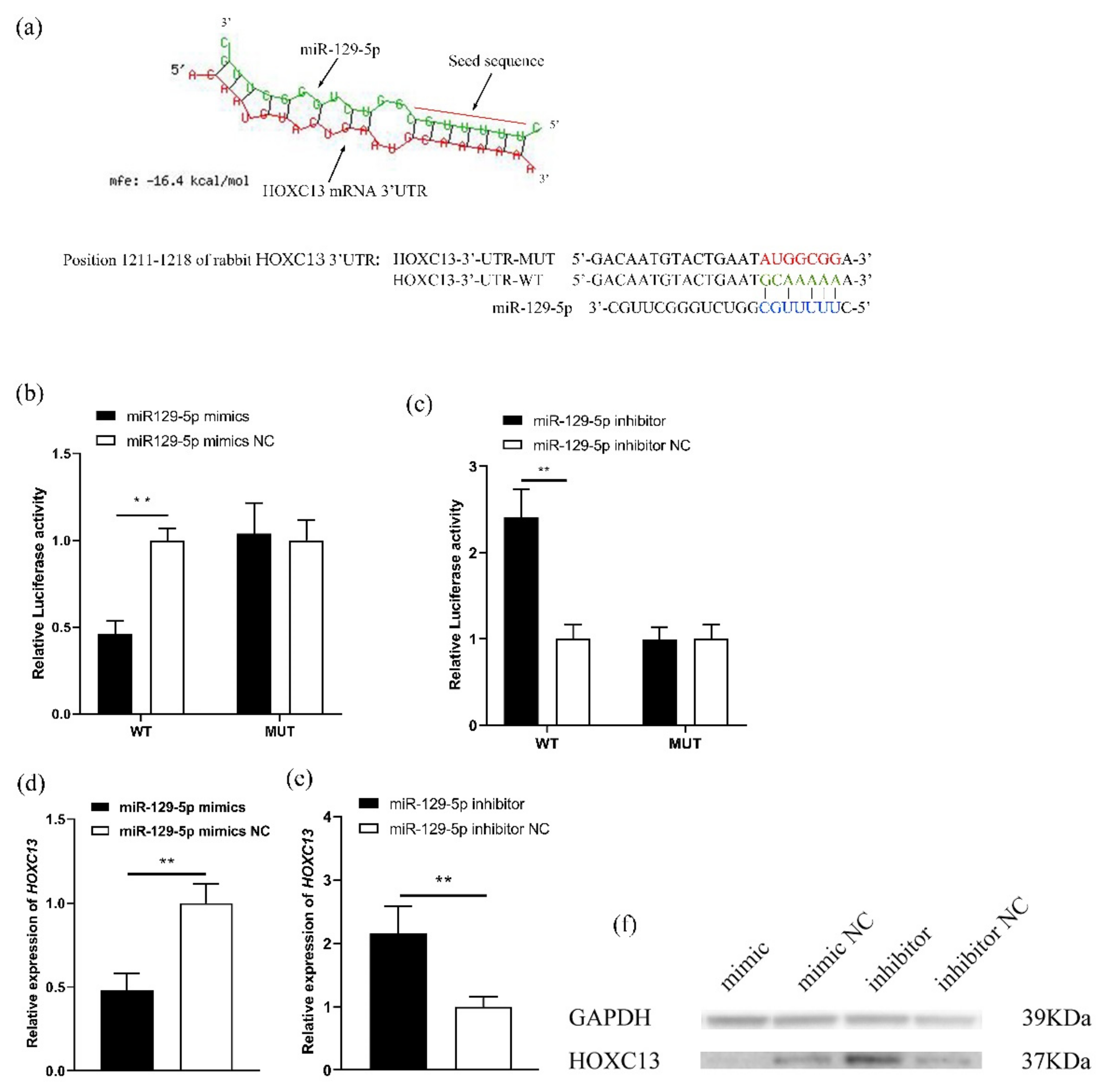
To further investigate the relationship between miR-129-5p and HOXC13, according to the prediction of bioinformatics on HOXC13-3′UTR and miRNA targeting binding sites, the binding sites of miR-129-5p and HOXC13 were determined (Figure 2a). In addition, the Dual-Luciferase Reporter Gene System was constructed to verify that it targeted HOXC13 3′-UTR. The luciferase activity of the RAB-9 cells was detected after pMIR-HOXC13-3′-UTR-WT and pMIR-HOXC13-3′-UTR-MUT were co-transfected with miR-129-5p mimics. The luciferase analysis indicated that the overexpression of miR-129-5p significantly decreased the luciferase activity of HOXC13, including a wild-type 3′UTR, and the knockdown of it was the opposite (p < 0.05) (Figure 2b,c). In addition, miR-129-5p significantly inhibited HOXC13 mRNA expression (p < 0.01) (Figure 2d). However, after the miR-129-5p inhibitor was transfected into the DPCs, the HOXC13 mRNA expression was found to be markedly up-regulated (p < 0.01) (Figure 2e). The Western blot results confirmed that it could inhibit HOXC13 protein expression (Figure 2f).

3.3. MiR-129-5p Targeted HOXC13 to Regulate the Expression of the HFDRGs

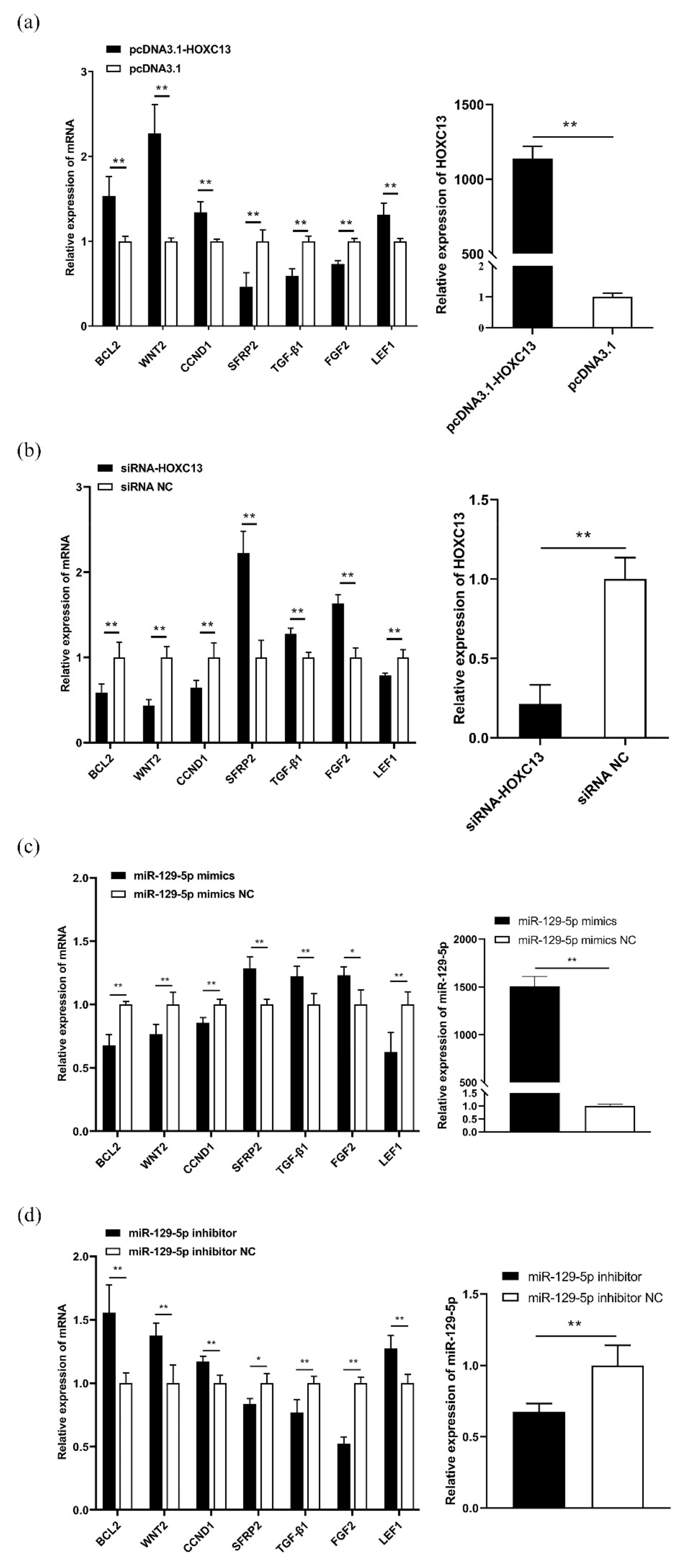
The DPCs were transfected with pcDNA3.1-HOXC13 and siRNA-HOXC13 to detect the expression of HOXC13 and the HFDRGs. The results indicated that HOXC13 could significantly promote the expression of HFDRGs, such as BCL2, WNT2, CCND1, and LEF1 (p < 0.01), and markedly reduce the expression of SFRP2, TGF-β1, and FGF2 (p < 0.01) (Figure 3a). Conversely, knocking down HOXC13 was found to markedly reduce the expression of BCL2, WNT2, CCND1, and LEF1, as well as inhibit the expression of SFRP2, TGF-β1, and FGF2 (p < 0.01) (Figure 3b). The DPCs were transfected with the miR-129-5p mimics and inhibitor to detect the expression of miR-129-5p and the HFDRGs. The results indicated that miR-129-5p could markedly reduce the expression of BCL2, WNT2, CCND1, and LEF1 (p < 0.01), and markedly promote the expression of SFRP2 and TGF-β1 (p < 0.01). It was also found to promote the expression of FGF2 (p < 0.05) (Figure 3c). Conversely, knocking down miR-129-5p was found to markedly promote the expression of BCL2, WNT2, CCND1, and LEF1, as well as inhibit the expression of SFRP2, TGF-β1, and FGF2 (p < 0.01) (Figure 3d).

3.4. MiR-129-5p Enhances Apoptosis and Inhibits Proliferation of DPCs

The miR-129-5p mimics and inhibitor were transfected into the DPCs to detect the apoptosis and proliferation of the DPCs. The results indicated that the overexpression of miR-129-5p promoted the apoptosis of DPCs (p < 0.01) (Figure 4a), while the knockdown of it was the opposite (p < 0.01) (Figure 4b). In addition, the overexpression of miR-129-5p inhibited the proliferation of the DPCs from 48 to 72 h (p < 0.01) (Figure 4c), while the knockdown of it promoted the proliferation of the DPCs from 24 to 72 h (p < 0.01) (Figure 4d).

3.5. HOXC13 Inhibits Apoptosis and Enhances the Proliferation of DPCs

PcDNA3.1-HOXC13 and siRNA-HOXC13 were transfected into the DPCs to detect the apoptosis and proliferation of the DPCs. The results indicated that the overexpression of HOXC13 inhibited the apoptosis of the DPCs (p < 0.01) (Figure 5a), while the knockdown of HOXC13 was the opposite (p < 0.01) (Figure 5b). Additionally, the overexpression of HOXC13 promoted the proliferation of the DPCs from 48 to 72 h (p < 0.01) (Figure 5c), while the knockdown of HOXC13 inhibited the proliferation of the DPCs from 48 to 72 h (p < 0.01) (Figure 5d).

4. Discussion

Skin is an important organ of mammals, with functions that involve resisting environmental hazards, maintaining homeostasis, preventing cold and warming, camouflage, and the exchange of interspecies information. Animal hair is a by-product of the HF cycle. Both the HF cycles and skin cycles are complexly coordinated by multiple genes and related pathways, along with a large number of regulatory factors. MiRNAs, as endogenous non-coding RNAs, widely exist in animals and regulate the expression of the genes and proteins [25]. Studies have shown miRNAs to significantly affect the morphogenesis, proliferation, and apoptosis of HF, as well as present in various diseases of HF [26,27,28,29,30].
Recent studies have represented that miR-129-5p can significantly affect oncogenes and adjust cancer cells. For example, it had been found that the growth and migration of chondrosarcoma cells could be restrained by targeting SOX4 and inhibiting glioma cells’ proliferation by targeting TGIF2 [31,32]. HOXC13, which is one of the miR-129-5p target genes and functions as a transcription factor, was primarily related to HF development and occurrence [33]. In addition, HOXC13 directly regulated keratin and keratin-associated proteins for controlling the growth and development of HF [34,35,36]. HOXC13 promoted the growth of cancer cells and inhibited their apoptosis [37,38]. In our study, miR-129-5p reached the highest expression level on the 120th day, indicating that it may play a role in catagen [39], while HOXC13 was found to be at the highest expression level on the 60th day, indicating that it may play a key role in anagen [39], and the expression of miR-129-5p and HOXC13 is significantly negatively correlated. Our data also showed that miR-129-5p can directly target rabbit HOXC13-3′-UTR to reduce the expression of HOXC13 and the HOXC13 protein.
In addition, the development of HF was regulated by related genes, such as WNT2, SFRP2, TGF-β1, and LEF1, which regulated skin morphogenesis and hair growth significantly [40,41]. Among these genes, LEF1 possessed a major role in HF morphogenesis and periodic growth [42]. TGF-β1 regulated the degeneration of HF through high expression [43]. SFRP2 was a key factor in HF development in catagen [44]. Our study indicated that the expression of these related genes changed after changing the expression of miR-129-5p and HOXC13, suggesting that miR-129-5p may affect the expression of these genes to participate in HF development by targeting HOXC13.
DPCs provide sufficient nutrition and signal support for the periodic regeneration of HF. This study showed that miR-129-5p restrained apoptosis and reduced the proliferation of DPCs, while the inhibition of it reduced apoptosis and promoted the proliferation of DPCs. However, the results of the overexpression of HOXC13 were opposite to those of the overexpression of miR-129-5p, indicating that miR-129-5p may regulate the proliferation and apoptosis of DPCs by targeting HOXC13.
In summary, miR-129-5p can induce apoptosis and inhibit the proliferation of DPCs, and participate in maintaining skin development, the HF cycle, and hair growth balance by targeting HOXC13. These results help to further understand the role and mechanism of miR-129-5p in the periodic development of hair follicles, which not only provides a theoretical basis for genetically improving the quality of hair in animals in the future, but also a new theory and method for diagnosing and treating hair loss in humans.

Author Contributions

Conceptualization, F.Y. and Y.C.; methodology, Y.C. and B.Z.; software, F.Y.; validation, F.Y. and S.H.; formal analysis, F.Y.; investigation, S.B., R.J. and C.Z.; resources, F.Y.; data curation, F.Y.; writing—original draft preparation, F.Y.; writing—review and editing, Y.C.; visualization, F.Y.; supervision, B.Z.; project administration, B.Z.; funding acquisition, X.W. All authors have read and agreed to the published version of the manuscript.

Funding

This work was supported by National Natural Science Foundation of China (Grant No. 32072724), the China Agriculture Research System of MOF and MARA (CARS-43-A-1) and Zhejiang Science and Technology Major Program on Agricultural New Variety Breeding (2021C02068-7).

Institutional Review Board Statement

The animal experiments in this paper complied with animal ethics and were approved by the Animal Care and Use Committee of Yangzhou University (approval No.202010025).

Informed Consent Statement

Not applicable.

Data Availability Statement

Forecast of targeted binding sites between miR-129-5p and HOXC13. Available online: https://bibiserv.cebitec.uni-bielefeld.de/ (accessed on 11 April 2022).

Conflicts of Interest

The authors declare no conflict of interest.

References

  1. Ambros, V. The Functions of Animal MicroRNAs. Nature 2004, 431, 350–355. [Google Scholar] [CrossRef] [PubMed]
  2. Huntzinger, E.; Izaurralde, E. Gene Silencing by MicroRNAs: Contributions of Translational Repression and MRNA Decay. Nat. Rev. Genet. 2011, 12, 99–110. [Google Scholar] [CrossRef] [PubMed]
  3. Dong, C.; Wang, H.; Xue, L.; Dong, Y.; Yang, L.; Fan, R.; Yu, X.; Tian, X.; Ma, S.; Smith, G.W. Coat Color Determination by MiR-137 Mediated down-Regulation of Microphthalmia-Associated Transcription Factor in a Mouse Model. RNA 2012, 18, 1679–1686. [Google Scholar] [CrossRef] [Green Version]
  4. Mardaryev, A.N.; Ahmed, M.I.; Vlahov, N.V.; Fessing, M.Y.; Gill, J.H.; Sharov, A.A.; Botchkareva, N.V. Micro-RNA-31 Controls Hair Cycle-Associated Changes in Gene Expression Programs of the Skin and Hair Follicle. FASEB J. 2010, 24, 3869–3881. [Google Scholar] [CrossRef] [PubMed] [Green Version]
  5. Wang, D.; Zhang, Z.; O’Loughlin, E.; Wang, L.; Fan, X.; Lai, E.C.; Yi, R. MicroRNA-205 Controls Neonatal Expansion of Skin Stem Cells by Modulating the PI(3)K Pathway. Nat. Cell Biol. 2013, 15, 1153–1163. [Google Scholar] [CrossRef]
  6. Yuan, S.; Li, F.; Meng, Q.; Zhao, Y.; Chen, L.; Zhang, H.; Xue, L.; Zhang, X.; Lengner, C.; Yu, Z. Post-Transcriptional Regulation of Keratinocyte Progenitor Cell Expansion, Differentiation and Hair Follicle Regression by MiR-22. PLoS Genet. 2015, 11, 1005253. [Google Scholar] [CrossRef]
  7. Li, J.L.; Liu, L.B.; Zhang, J.P.; Cheng, L.; Ren, L.T.; Zhao, Y.J. The expression of miR-129-5p and its target genes in the skin of goats. Anim. Biotechnol. 2020, 32, 573–579. [Google Scholar] [CrossRef]
  8. Greco, V.; Chen, T.; Rendl, M.; Schober, M.; Pasolli, H.A.; Stokes, N.; dela Cruz-Racelis, J.; Fuchs, E. A Two-Step Mechanism for Stem Cell Activation during Hair Regeneration. Cell Stem Cell 2009, 4, 464. [Google Scholar] [CrossRef] [Green Version]
  9. Rendl, M.; Lewis, L.; Fuchs, E. Molecular Dissection of Mesenchymal-Epithelial Interactions in the Hair Follicle. PLoS Biol. 2005, 3, 1910–1924. [Google Scholar] [CrossRef]
  10. Andl, T.; Reddy, S.T.; Gaddapara, T.; Millar, S.E. WNT Signals Are Required for the Initiation of Hair Follicle Development. Dev. Cell. 2002, 2, 643–653. [Google Scholar] [CrossRef]
  11. Niimori, D.; Kawano, R.; Felemban, A.; Niimori-Kita, K.; Tanaka, H.; Ihn, H.; Ohta, K. Tsukushi Controls the Hair Cycle by Regulating TGF-Β1 Signaling. Dev. Biol. 2012, 372, 81–87. [Google Scholar] [CrossRef] [PubMed] [Green Version]
  12. St-Jacques, B.; Dassule, H.R.; Karavanova, I.; Botchkarev, V.A.; Li, J.; Danielian, P.S.; Mcmahon, J.A.; Lewis, P.M.; Paus, R.; Mcmahon, A.P. Sonic Hedgehog Signaling Is Essential for Hair Development. Curr. Biol. 1998, 8, 1058–1069. [Google Scholar] [CrossRef] [Green Version]
  13. Stenn, K.S.; Paus, R. Controls of Hair Follicle Cycling. Physiol. Rev. 2001, 81, 449–494. [Google Scholar] [CrossRef] [PubMed]
  14. Lin, Z.; Chen, Q.; Shi, L.; Lee, M.; Giehl, K.A.; Tang, Z.; Wang, H.; Zhang, J.; Yin, J.; Wu, L.; et al. Loss-of-Function Mutations in HOXC13 Cause Pure Hair and Nail Ectodermal Dysplasia. Am. J. Hum. Genet. 2012, 91, 906–911. [Google Scholar] [CrossRef] [Green Version]
  15. Farooq, M.; Kurban, M.; Fujimoto, A.; Fujikawa, H.; Abbas, O.; Nemer, G.; Saliba, J.; Sleiman, R.; Tofaili, M.; Kibbi, A.G.; et al. A Homozygous Frameshift Mutation in the HOXC13 Gene Underlies Pure Hair and Nail Ectodermal Dysplasia in a Syrian Family. Hum. Mutat. 2013, 34, 578–581. [Google Scholar] [CrossRef] [PubMed]
  16. Jiang-hong, W.; Yan-jun, Z.; Jia-xin, Z.; Zi-li, C.; Jin-quan, L.; Zu-wei, Y.; Wen-guang, Z. Hoxc13/β-Catenin Correlation with Hair Follicle Activity in Cashmere Goat. J. Integr. Agric. 2012, 2012, 11. [Google Scholar]
  17. Qiu, W.; Lei, M.; Tang, H.; Yan, H.; Wen, X.; Zhang, W.; Tan, R.; Wang, D.; Wu, J. Hoxc13 Is a Crucial Regulator of Murine Hair Cycle. Cell Tissue Res. 2016, 364, 149–158. [Google Scholar] [CrossRef]
  18. Kahata, K.; Dadras, M.S.; Moustakas, A. TGF-β Family Signaling in Epithelial Differentiation and Epithelial-Mesenchymal Transition. Cold Spring Harb. Perspect. Biol. 2018, 10, 022194. [Google Scholar] [CrossRef] [Green Version]
  19. Potter, C.S.; Kern, M.J.; Baybo, M.A.; Pruett, N.D.; Godwin, A.R.; Sundberg, J.P.; Awgulewitsch, A. Dysregulated Expression of Sterol O-Acyltransferase 1 (Soat1) in the Hair Shaft of Hoxc13 Null Mice. Exp. Mol. Pathol. 2015, 99, 441–444. [Google Scholar] [CrossRef] [Green Version]
  20. Wang, X.; Xu, H.R.; Li, T.; Qu, L.; Zhao, Z.D.; Zhang, Z.Y. Expression Analysis of KAP9.2 and Hoxc13 Genes during Different Cashmere Growth Stages by QRT-PCR Method. Mol. Biol. Rep. 2014, 41, 5665–5668. [Google Scholar] [CrossRef]
  21. Wang, S.; Luo, Z.; Zhang, Y.; Yuan, D.; Ge, W.; Wang, X. The Inconsistent Regulation of HOXC13 on Different Keratins and the Regulation Mechanism on HOXC13 in Cashmere Goat (Capra Hircus). BMC Genom. 2018, 19, 630. [Google Scholar] [CrossRef] [PubMed]
  22. Li, J.L.; Zhao, B.H.; Zhang, C.; Zhang, X.Y.; Dai, Y.Y.; Hu, S.S.; Yang, N.S.; Chen, Y.; Wu, X.S. Establishment and Functional Characterization of Immortalized Rabbit Dermal Papilla Cell Lines. 2021; preprint. [Google Scholar]
  23. Livak, K.J.; Schmittgen, T.D. Analysis of relative gene expression data using real-time quantitative PCR and the 2−ΔΔCT method. Methods 2001, 25, 402–408. [Google Scholar] [CrossRef] [PubMed]
  24. Rehmsmeier, M.; Steffen, P.; Höchsmann, M.; Giegerich, R. Fast and effective prediction of microRNA/target duplexes. RNA 2004, 10, 1507–1517. [Google Scholar] [CrossRef] [PubMed] [Green Version]
  25. Bartel, D.P. MicroRNAs: Genomics, Biogenesis, Mechanism, and Function. Cell 2004, 116, 281–297. [Google Scholar] [CrossRef] [Green Version]
  26. Liu, Z.; Xiao, H.; Li, H.; Zhao, Y.; Lai, S.; Yu, X.; Cai, T.; Du, C.; Zhang, W.; Li, J. Identification of Conserved and Novel MicroRNAs in Cashmere Goat Skin by Deep Sequencing. PLoS ONE 2012, 7, e50001. [Google Scholar] [CrossRef] [PubMed]
  27. Iorio, M.V.; Ferracin, M.; Liu, C.G.; Veronese, A.; Spizzo, R.; Sabbioni, S.; Magri, E.; Pedriali, M.; Fabbri, M.; Campiglio, M.; et al. MicroRNA Gene Expression Deregulation in Human Breast Cancer. Cancer Res. 2005, 65, 7065–7070. [Google Scholar] [CrossRef] [Green Version]
  28. Murakami, Y.; Yasuda, T.; Saigo, K.; Urashima, T.; Toyoda, H.; Okanoue, T.; Shimotohno, K. Comprehensive Analysis of MicroRNA Expression Patterns in Hepatocellular Carcinoma and Non-Tumorous Tissues. Oncogene 2006, 25, 2537–2545. [Google Scholar] [CrossRef]
  29. Polytarchou, C.; Hommes, D.W.; Palumbo, T.; Hatziapostolou, M.; Koutsioumpa, M.; Koukos, G.; Van Der Meulen-De Jong, A.E.; Oikonomopoulos, A.; Van Deen, W.K.; Vorvis, C.; et al. MicroRNA214 Is Associated With Progression of Ulcerative Colitis, and Inhibition Reduces Development of Colitis and Colitis-Associated Cancer in Mice. Gastroenterology 2015, 149, 981–992.e11. [Google Scholar] [CrossRef] [Green Version]
  30. Ma, H.; Pan, J.S.; Jin, L.X.; Wu, J.; Ren, Y.D.; Chen, P.; Xiao, C.; Han, J. MicroRNA-17~92 Inhibits Colorectal Cancer Progression by Targeting Angiogenesis. Cancer Lett. 2016, 376, 293–302. [Google Scholar] [CrossRef]
  31. Zhang, P.; Li, J.; Song, Y.; Wang, X. MiR-129-5p Inhibits Proliferation and Invasion of Chondrosarcoma Cells by Regulating SOX4/Wnt/β-Catenin Signaling Pathway. Cell. Physiol. Biochem. 2017, 42, 242–253. [Google Scholar] [CrossRef] [PubMed] [Green Version]
  32. Diao, Y.; Jin, B.; Huang, L.; Zhou, W. MiR-129-5p Inhibits Glioma Cell Progression in Vitro and in Vivo by Targeting TGIF2. J. Cell. Mol. Med. 2018, 22, 2357–2367. [Google Scholar] [CrossRef] [Green Version]
  33. Wu, J.H.; Yan, Z.W.; Zhang, W.G.; Li, J.Q. Hoxc13 and the Development of Hair Follicle. Yi Chuan = Hereditas 2010, 32, 656–662. [Google Scholar] [PubMed]
  34. Jave-Suarez, L.F.; Winter, H.; Langbein, L.; Rogers, M.A.; Schweizer, J. HOXC13 Is Involved in the Regulation of Human Hair Keratin Gene Expression. J. Biol. Chem. 2002, 277, 3718–3726. [Google Scholar] [CrossRef] [Green Version]
  35. Pruett, N.D.; Tkatchenko, T.V.; Jave-Suarez, L.; Jacobs, D.F.; Potter, C.S.; Tkatchenko, A.V.; Schweizer, J.; Awgulewitsch, A. Krtap16, Characterization of a New Hair Keratin-Associated Protein (KAP) Gene Complex on Mouse Chromosome 16 and Evidence for Regulation by Hoxc13. J. Biol. Chem. 2004, 279, 51524–51533. [Google Scholar] [CrossRef] [PubMed] [Green Version]
  36. Ahlawat, S.; Arora, R.; Sharma, R.; Sharma, U.; Kaur, M.; Kumar, A.; Singh, K.V.; Singh, M.K.; Vijh, R.K. Skin Transcriptome Profiling of Changthangi Goats Highlights the Relevance of Genes Involved in Pashmina Production. Sci. Rep. 2020, 10, 6050. [Google Scholar] [CrossRef] [Green Version]
  37. Yao, Y.; Luo, J.; Sun, Q.; Xu, T.; Sun, S.; Chen, M.; Lin, X.; Qian, Q.; Zhang, Y.; Cao, L.; et al. Original Article HOXC13 Promotes Proliferation of Lung Adenocarcinoma via Modulation of CCND1 and CCNE1. Am. J. Cancer Res. 2017, 7, 1820. [Google Scholar]
  38. Kasiri, S.; Ansari, K.I.; Hussain, I.; Bhan, A.; Mandal, S.S. Antisense Oligonucleotide Mediated Knockdown of HOXC13 Affects Cell Growth and Induces Apoptosis in Tumor Cells and over Expression of HOXC13 Induces 3D-Colony Formation. RSC Adv. 2013, 3, 3260–3269. [Google Scholar] [CrossRef]
  39. Zhao, B.H.; Chen, Y.; Yang, N.S.; Chen, Q.R.; Bao, Z.Y.; Liu, M.; Hu, S.S.; Li, J.L.; Wu, X.S. miR-218-5p regulates skin and hair follicle development through Wnt/β-catenin signaling pathway by targeting SFRP2. J. Cell. Physiol. 2019, 234, 20329–20341. [Google Scholar] [CrossRef]
  40. Fuchs, E.; Raghavan, S. Getting under the Skin of Epidermal Morphogenesis. Nat. Rev. Genet. 2002, 3, 199–209. [Google Scholar]
  41. Liu, Y.; Zhou, Q.; Zhou, D.; Huang, C.; Meng, X.; Li, J. Secreted Frizzled-Related Protein 2-Mediated Cancer Events: Friend or Foe? Pharmacol. Rep. 2017, 69, 403–408. [Google Scholar] [CrossRef] [PubMed]
  42. Zhou, P.; Byrne, C.; Jacobs, J.; Fuchs, E. Lymphoid Enhancer Factor 1 Directs Hair Folhcle Patterning and Epithelial Cell Fate. Genes Dev. 1995, 9, 700–713. [Google Scholar] [CrossRef] [PubMed] [Green Version]
  43. Foitzik, K.; Lindner, G.; Mueller-Roever, S.; Maurer, M.; Botchkareva, N.; Botchkarev, V.; Handjiski, B.; Metz, M.; Hibino, T.; Soma, T.; et al. Control of Murine Hair Follicle Regression (Catagen) by TGF-β1 in Vivo. FASEB J. 2000, 14, 752–760. [Google Scholar] [CrossRef] [PubMed]
  44. Kim, B.K.; Yoon, S.K. Expression of Sfrp2 Is Increased in Catagen of Hair Follicles and Inhibits Keratinocyte Proliferation. Ann. Dermatol. 2014, 26, 79–87. [Google Scholar] [CrossRef] [Green Version]
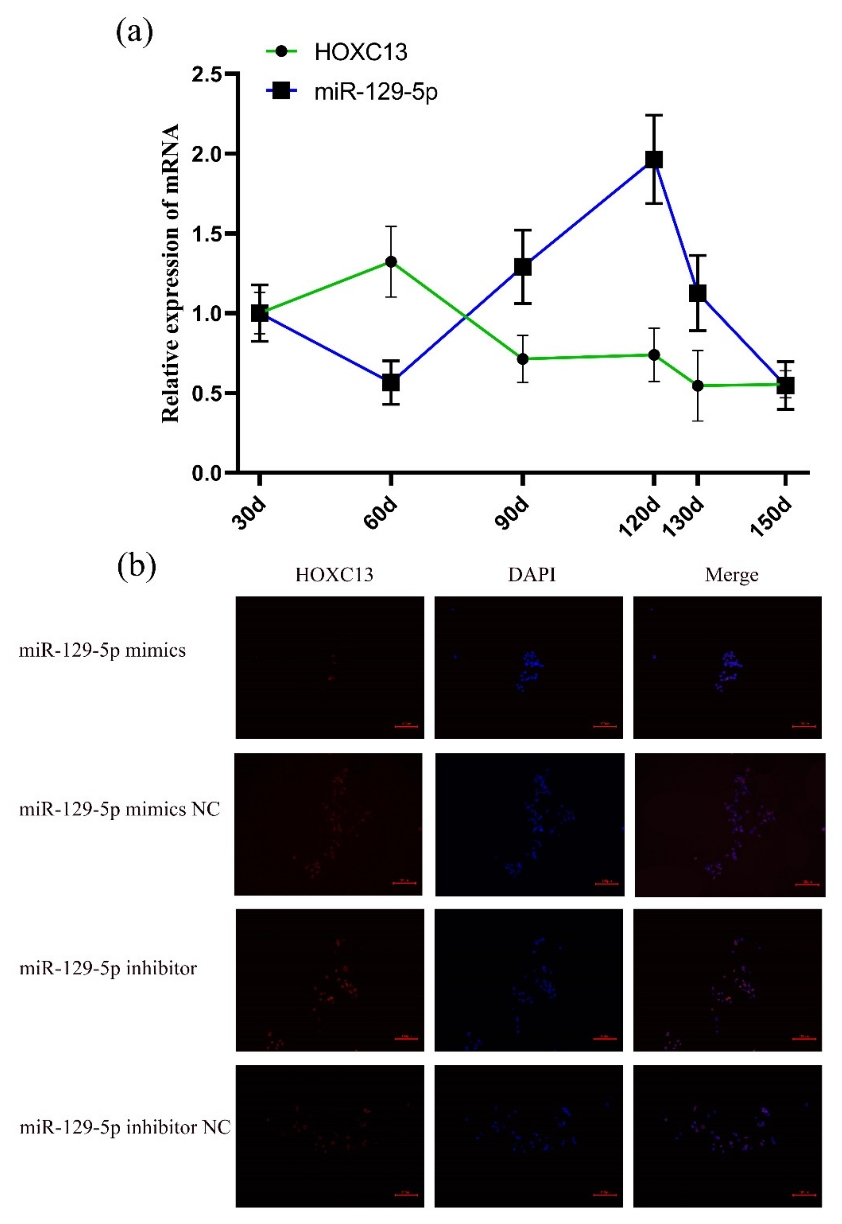
Figure 1. MiR-129-5p regulated HOXC13 expression. (a) The expression of miR-129-5p and HOXC13 in different stages of HF development. (b) MiR-129-5p regulated HOXC13 expression in DPCs; the scale in Figure 1b is 100 μm.
Genes 13 00679 g001
Figure 2. MiR-129-5p directly targeted HOXC13. (a) Prediction and consequential pairing of HOXC13 and miR-29-5p, and pMIR-HOXC13-3′-UTR-WT and pMIR-HOXC13-3′-UTR-MUT were constructed according to the assumed miR-129-5p binding site. Forecast of targeted binding sites between miR-129-5p and HOXC13 used RNAhybrid online software (https://bibiserv.cebitec.uni-bielefeld.de/accessed on 11 April 2022) [24]. (b,c) The Dual-Luciferase Reporter Gene System verified that miR-129-5p targeted HOXC13 3′-UTR. (d,e) MiR-129-5p affected the expression of HOXC13 in DPCs. (f) MiR-129-5p affected the expression of the HOXC13 protein in DPCs. ** p < 0.01; mfe: minimum free energy; WT: wild-type; MUT: mutant-type; mimic: miR-129-5p mimics; inhibitor: miR-129-5p inhibitor; DPCs: dermal papilla cells.
Genes 13 00679 g002
Figure 3. MiR-129-5p and HOXC13 affected the expression of HFDRGs. (a) PcDNA3.1-HOXC13 regulates the mRNA expression of the HFDRGs and overexpression of HOXC13 in DPCs. (b) SiRNA-HOXC13 regulates the mRNA expression of the HFDRGs and the knockdown of HOXC13 in DPCs. (c) MiR-129-5p mimics regulate the mRNA expression of the HFDRGs and overexpression of miR-129-5p in DPCs. (d) MiR-129-5p inhibitor regulates the mRNA expression of the HFDRGs and knockdown of miR-129-5p in DPCs. HFDRGs: hair follicle development-related genes. * p < 0.05; ** p < 0.01.
Genes 13 00679 g003
Figure 4. MiR-129-5p promoted apoptosis and inhibited the proliferation of DPCs. (a) MiR-129-5p mimics promoted the apoptosis of DPCs. (b) MiR-129-5p inhibitor inhibited the apoptosis of DPCs. (c) After miR-129-5p mimics were transfected into DPCs, the effect of this on the proliferation of DPCs was determined after 0, 24, 48, and 72 h. (d) After miR-129-5p inhibitor was transfected into DPCs, the effect of it on the proliferation of DPCs was determined after 0, 24, 48, and 72 h. ** p < 0.01. DPCs: dermal papilla cells; NC: negative control. In (a, b), the first quadrant represents the cell debris, the second quadrant represents late apoptotic cells, the third quadrant is living cells, and the fourth quadrant is early apoptotic cells. The apoptotic rate is Q2 + Q4.
Genes 13 00679 g004
Figure 5. HOXC13 inhibited apoptosis and promoted the proliferation of DPCs. (a) HOXC13 inhibited apoptosis of DPCs. (b) SiRNA-HOXC13 promoted apoptosis of DPCs. (c) After pcDNA3.1-HOXC13 was transfected into DPCs, the effect of HOXC13 on the proliferation of DPCs was determined after 0, 24, 48, and 72 h. (d) After siRNA-HOXC13 was transfected into DPCs, the effect of HOXC13 on the proliferation of DPCs was determined after 0, 24, 48, and 72 h. ** p < 0.01. DPCs: dermal papilla cells; NC: negative control.
Genes 13 00679 g005
Table 1. Primer sequences used in qPCR.
GenesPrimers Sequence(5′→3′)
GAPDHF: TCACCATCTTCCAGGAGCGA
R: CACAATGCCGAAGTGGTCGT
BCL2F: ACATCGCCCTGTGGATGACTG
R: CGAGGGTGATGCAAGCTCCTAT
WNT2F: AGCCATCCAGGTCGTCATGAACCAG
R: TGCACACACGACCTGCTGTACCC
CCND1F: GAACGCTACCTTCCCCAGTGCTC
R: CCTCACAGACCTCCAGCATCCAG
SFRP2F: CCAGCCCGACTTCTCCTACAAGC
R: TCCAGCACCTCTTTCATGGTCT
TGF-β1F: CAGGTCCTTGCGGAAGTCAA
R: CTGGAACGGGCTCAACATCTA
FGF2F: GTGTGTGCAAACCGTTACCTT
R: TCGTTTCAGTGCCACATACCAG
LEF1F: CATCTCGGGTGGATTCAGG
R: ATGAGGGATGCCAGTTGTG
HOXC13F: TGGAGAAAGAGTACGCAGCC
R: TCTTCTCTTTGACTCGGCGG
miR-129-5pCTTTTTGCGGTCTGGGCTTGC
U6CAAGGATGACACGCAAATTCG
Note: F: forward primer; R: reverse primer.
Publisher’s Note: MDPI stays neutral with regard to jurisdictional claims in published maps and institutional affiliations.