Article

Phytochemical Characterization, and Antioxidant and Antimicrobial Properties of Agitated Cultures of Three Rue Species: Ruta chalepensis, Ruta corsica, and Ruta graveolens

1
Department of Pharmaceutical Botany, Faculty of Pharmacy, Jagiellonian University Medical College, 30-688 Krakow, Poland
2
Department of Chemical, Biological, Pharmaceutical and Environmental Sciences, University of Messina, Viale F. Stagno d’Alcontres, 31, 98166 Messina, Italy
*
Author to whom correspondence should be addressed.
Antioxidants 2022, 11(3), 592; https://doi.org/10.3390/antiox11030592
Submission received: 23 February 2022 / Revised: 17 March 2022 / Accepted: 18 March 2022 / Published: 20 March 2022

Abstract

:
The in vitro cultures of the following three species of the genus Ruta were investigated: R. chalepensis, R. corsica, and R. graveolens. The dynamics of biomass growth and accumulation of secondary metabolites in the 3-, 4-, 5-, 6-, and 7-week growth cycle were analysed. The antioxidant capacity of the methanol extracts obtained from the biomass of the in vitro cultures was also assessed by different in vitro assays: 1,1-diphenyl-2-picrylhydrazil (DPPH), reducing power, and Fe2+ chelating activity assays. Moreover, a preliminary screening of the antimicrobial potential of the extracts was performed. The extracts were phytochemically characterized by high-performance liquid chromatography (HPLC), which highlighted the presence of linear furanocoumarins (bergapten, isoimperatorin, isopimpinellin, psoralen, and xanthotoxin) and furoquinoline alkaloids (γ-fagarine, 7-isopentenyloxy-γ-fagarine, and skimmianine). The dominant group of compounds in all the cultures was coumarins (maximum content 1031.5 mg/100 g DW (dry weight), R. chalepensis, 5-week growth cycle). The results of the antioxidant tests showed that the extracts of the three species had varied antioxidant capacity: in particular, the R. chalepensis extract exhibited the best radical scavenging activity (IC50 = 1.665 ± 0.009 mg/mL), while the R. graveolens extract displayed the highest chelating property (IC50 = 0.671 ± 0.013 mg/mL). Finally, all the extracts showed good activity against Staphylococcus aureus with MIC values of 250 μg/mL for the R. corsica extract and 500 μg/mL for both R. graveolens and R. chalepensis extracts.

1. Introduction

The genus Ruta belongs to the Rutaceae family [1]. The best-known species of the genus are Ruta graveolens L. and Ruta chalepensis L. Other rue species are less known and may occur, for example, as endemic species such as Ruta corsica D. C. [2]. The individual rue species differ slightly in terms of morphology.
R. graveolens is a plant whose natural habitats were originally located in south-eastern and eastern Europe, and the species then spread to the area of southern Europe and the African coast of the Mediterranean Sea. R. chalepensis is a common species on the Mediterranean coast. It grows on the Italian, Albanian, Greek, Spanish and French coasts, as well as in North Africa (e.g., in Algeria and Libya). It also grows in the areas of India, Latin America, and Western Asia, including countries such as Syria, Iran, Turkey, and Israel. R. corsica is a species endemic to the Corsican mountains [3,4].
The chemical composition of the investigated rue species has been studied to varying extents. The chemical composition of R. graveolens is well known, while that of R. corsica is least known.
The herb of R. graveolens is rich in bioactive compounds. It contains different subgroups of coumarins: simple coumarins (coumarin, herniarin, umbelliferon, methoxycoumarin, scopoletin); psoralen-type furanocoumarins (xanthotoxin, bergapten, psoralen, imperatorin, isoimperatorin, isopimpinellin, rutarin), dihydrofuranocoumarins (rutamarine, rutaretin), coumarin dimers (daphnoretin, methoxydaphnoretin), pyranocoumarins (xanthylethine); phenolic acids (vanillic acid, chlorogenic acid, ferulic acid, protocatechuic acid, p-coumaric acid, p-hydroxybenzoic acid, syringic acid, caffeic acid, gentisic acid); alkaloids: acridine (arborinin, rutacridone, gravacridondiol), quinoline (graveolin), furoquinoline (dictamine, cocusaginine, skimmianine, γ-fagarine), dihydrofuroquinoline (ribalin, platydesmin), pyroquinoline (rutaline); flavonoids (rutoside, quercetin, isorhamnetin, kaempferol, myricetin); and essential oil (0.2–0.7%). The simultaneous presence of alkaloids and essential oil is a unique case in the plant kingdom [5,6,7,8].
R. graveolens exhibits a broad spectrum of activity, such as antioxidant, anti-inflammatory, spasmolytic, sedative, antibacterial, antifungal, and antidiabetic effects [9,10]. R. graveolens is a valuable source of linear furanocoumarins (derivatives of psoralen) that have been used for treating skin diseases such as vitiligo and psoriasis in the so called PUVA therapy (psoralens and UVA irradiation) [11,12].
R. chalepensis herb contains secondary metabolites from various chemical groups: coumarins: simple coumarins (coumarin, scopoletin, umbelliferon), coumarin dimers (daphnorin), furanocoumarins (bergapten, chalepensin, chalepin, isopimpinellin, xanthotoxin), dihydrofuranocoumarins (rutamarine, isorutarin); alkaloids: acridine (arborinin, rutacridone), quinoline (graveolin, graveolinin, 3-hydroxygraveoline), furoquinoline (dictamine, isotaifine, cocusaginine, maculosidine, skimmianine, taifine, γ-fagarine, 8-methoxytaifine); flavonoids (hesperidin, rutoside); and essential oil (approximately 0.7%) [13,14,15].
R. corsica herb contains isoflavones (daidzein, genistein, prunetin, formononetin). In the root of this species, the following was found: simple coumarins (corsicarin, isoscopoletin), furanocoumarins (psoralen, xanthotoxin, isopimpinellin); furoquinoline alkaloids (skimmianine, dictamine); and essential oil [16,17,18].
In vitro plant cultures can provide high-quality plant material for pharmaceutical or cosmetic purposes. In addition, the use of natural products as screening libraries has recently come of interest and may contribute to the advancement of drug discovery [19]. In vitro cultures of R. graveolens are a convenient material for conducting multidirectional research because they exhibit a significant increase in biomass within a short time and show a high capacity for organogenesis and accumulation of many secondary metabolites. Therefore, numerous studies are conducted to determine the quantitative and qualitative composition of the biomass from in vitro cultures of this plant species and subspecies R. graveolens ssp. divaricata [20,21,22,23,24,25].
The cultures of other Ruta species have not been thoroughly investigated. There are few reports on the chemical constituents in R. chalepensis in vitro cultures [26,27]. However, there are no studies on the in vitro cultures of R. corsica in the available literature. Therefore, the present study aimed to investigate the biosynthetic, antioxidant, and antimicrobial potential of the in vitro cultures of R. chalepensis and R. corsica and to compare them with the potential of cultures of the most known species, R. graveolens.

2. Materials and Methods

2.1. In Vitro Cultures

The shoot cultures of Ruta ssp. were initiated in the Department of Pharmaceutical Botany UJ CM in Cracow: R. graveolens—from hypocotyl segments of sterile seedlings derived from the Botanical Garden at the Purkyny University in Brno (Czech Republic); and R. chalepensis and R. corsica—from seeds derived from the Botanical Garden at the Maria Skłodowska-Curie University in Lublin (Poland).
Stationary liquid cultures were grown on Linsmaier and Skoog medium [28] containing 1 mg/L auxin: NAA (α-naphthaleneacetic acid) and 1 mg/L cytokinin: BAP (6-benzylaminopurine). The experimental agitated cultures were initiated from the stationary liquid cultures.
The cultures were maintained in Erlenmeyer flasks (500 mL), containing 150 mL medium. Initial biomass was 1 g. Cultures were performed on a shaker (Altel) with a rotation frequency of 140/min. The cultures were maintained under artificial light with an intensity of 4 W/m2 at 25 ± 2 °C. The cultures were performed in Linsmaier and Skoog (LS) medium with the following content of plant growth regulators: NAA—0.1 mg/L; and BAP—0.1 mg/L. The culturing period was 3, 4, 5, 6, and 7 weeks (3 replicates were performed for each species in each week). After this period the biomass was separated from the medium and dried at 38 °C.

2.2. High-Performance Liquid Chromatography Analyses

High-performance liquid chromatography (HPLC) analysis was used to determine the metabolite content of the methanol extracts (sonication, 30 °C, 20 min, three times) obtained from 1 g of dried biomass. RP-HPLC analysis was performed as described elsewhere [29] on a Merck-Hitachi liquid chromatograph (LaChrom Elite, Hitachi, Tokyo, Japan) equipped with a DAD detector L-2455 and a Purospher ® RP-18e 250 × 4 mm/5 μm column (Merck, Darmstadt, Germany). The analysis was conducted at 25 °C, with the mobile phase consisting of A—methanol, B—methanol: 0.5% acetic acid 1:4 (v/v), gradient elution at the flow rate of 1 mL min−1, and the gradient was as follows: 100% B for 0–20 min; 100–80% B for 20–35 min; 80–60% B for 35–55 min; 60–0% B for 55–70 min; 0% B for 70–75 min; 0–100% B for 75–80 min; 100% B for 80–90 min; wavelength range 200–400 nm. The quantification was performed at λ = 254 and 330 nm for phenolic acids, catechins, coumarins, and alkaloids, and at 330 and 370 nm for flavonoids.
The following standards were purchased: bergapten, imperatorin, xanthotoxin, and psoralen from Roth (Karlsruhe, Germany); caffeic acid, chlorogenic acid, cinnamic acid, ellagic acid, gallic acid, gentizic acid, isoferulic acid, neochlorogenic acid, o-coumaric acid, protocatechuic acid, rosmarinic acid, salicylic acid, sinapic acid, syringic acid, apigenin, apigetrin (apigenin 7-glucoside), hyperoside (quercetin 3-O-galactoside), isoquercetin (quercetin 3-O-glucoside), isorhamnetin, kaempferol, luteolin, myricetin, populnin (kaempferol 7-O-glucoside), robinin (kaempferol 3-O-robinoside-7-O-rhamnoside), quercetin, quercitrin (quercetin 3-O-rhamnoside), rhamnetin, rutoside, vitexin, 5,7-dimethoxycoumarin, 4-hydroxy-6-methylcoumarin, 6-methylcoumarin, osthole, and umbelliferone from Sigma Aldrich (St Louis, MO, USA); p-coumaric acid, vanillic acid, ferulic acid, p-hydroxybenzoic acid, coumarin, and scopoletin from Fluka (Bucha, Switzerland), caftaric acid, cryptochlorogenic acid, isochlorogenic acid, catechin, epigallocatechin, epicatechin gallate, epicatechin, epigallocatechin gallate, cinaroside (luteolin 7-O-glucoside), osthenol, 4-methylumbelliferone, 4,6-dimethoxy-2H-1-benzopyran-2-one, and skimmianine from ChromaDex (Irvine, CA, USA); 4-O-feruloylquinic acid, apigetrin (apigenin 7-O-glucoside), apigenin 7-O-glucuronide, astragalin (kaempferol 3-O-glucoside), avicularin (quercetin 3-O-α-l-arabinofuranoside), trifolin (kaempferol 3-O-galactoside), isopimpinellin, isoimperatorin, daphnetin 7-methyl ether, rutaretin, daphnetin, osthenol, bergaptol, daphnetin dimethyl ether, γ-fagarine, and 7-isopentenyloxy-γ-fagarine from ChemFaces (Wuhan, China)

2.3. Antioxidant Activity

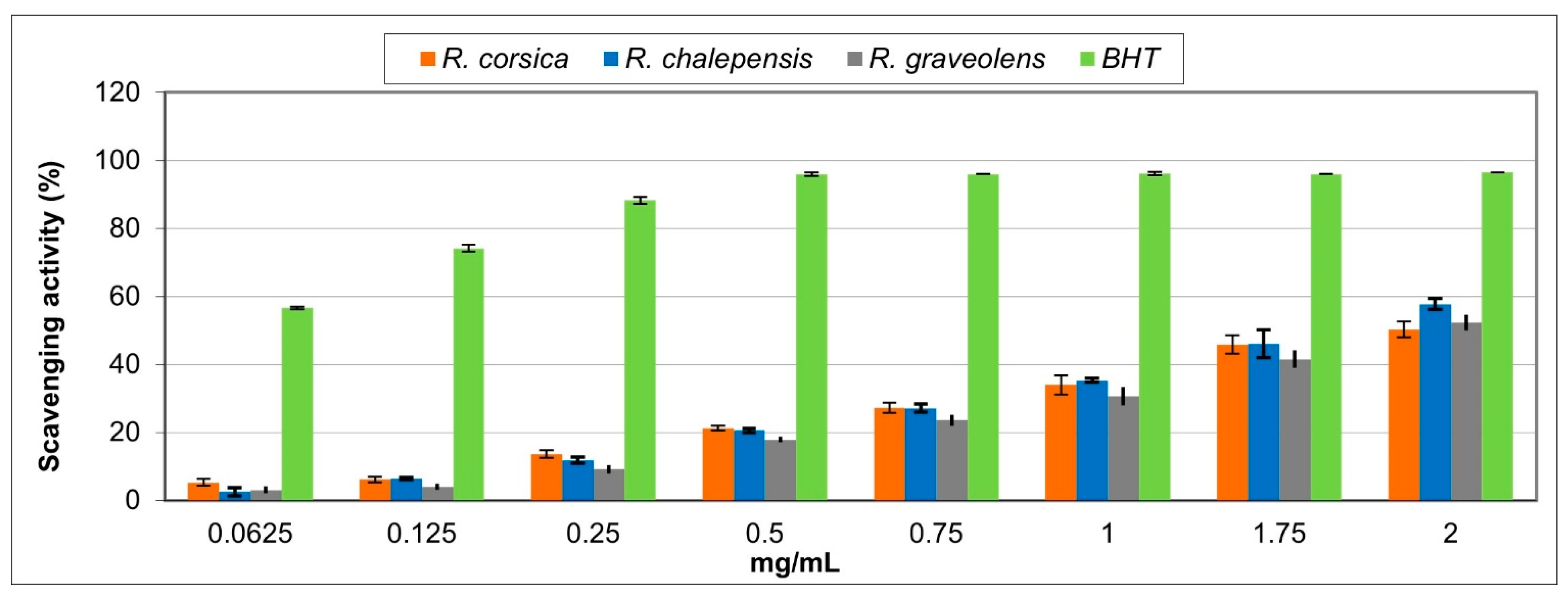
2.3.1. Free Radical Scavenging Activity

The free radical scavenging activity of the Ruta spp. extracts was determined using the DPPH (1,1-diphenyl-2-picrylhydrazyl) method [30]. The extracts were tested at different concentrations (0.0625–2 mg/mL). An aliquot (0.5 mL) of the methanol solution containing different amounts of sample solution was added to 3 mL of daily prepared methanol DPPH solution (0.1 mM). The change in optical density at 517 nm was measured 20 min after the initial mixing by using a model UV-1601 spectrophotometer (Shimadzu). Butylated hydroxytoluene (BHT) was used as a reference.
The scavenging activity was measured as the decrease in the absorbance of the samples versus the DPPH standard solution. Results were expressed as radical scavenging activity percentage (%) of the DPPH, defined by the formula ((Ao−Ac)/Ao) × 100, where Ao is the absorbance of the control and Ac is the absorbance in the presence of the sample or standard.
The results obtained from the average of three independent experiments are reported as the mean radical scavenging activity percentage (%) ± standard deviation (SD) and mean 50% inhibitory concentration (IC50) ± SD.

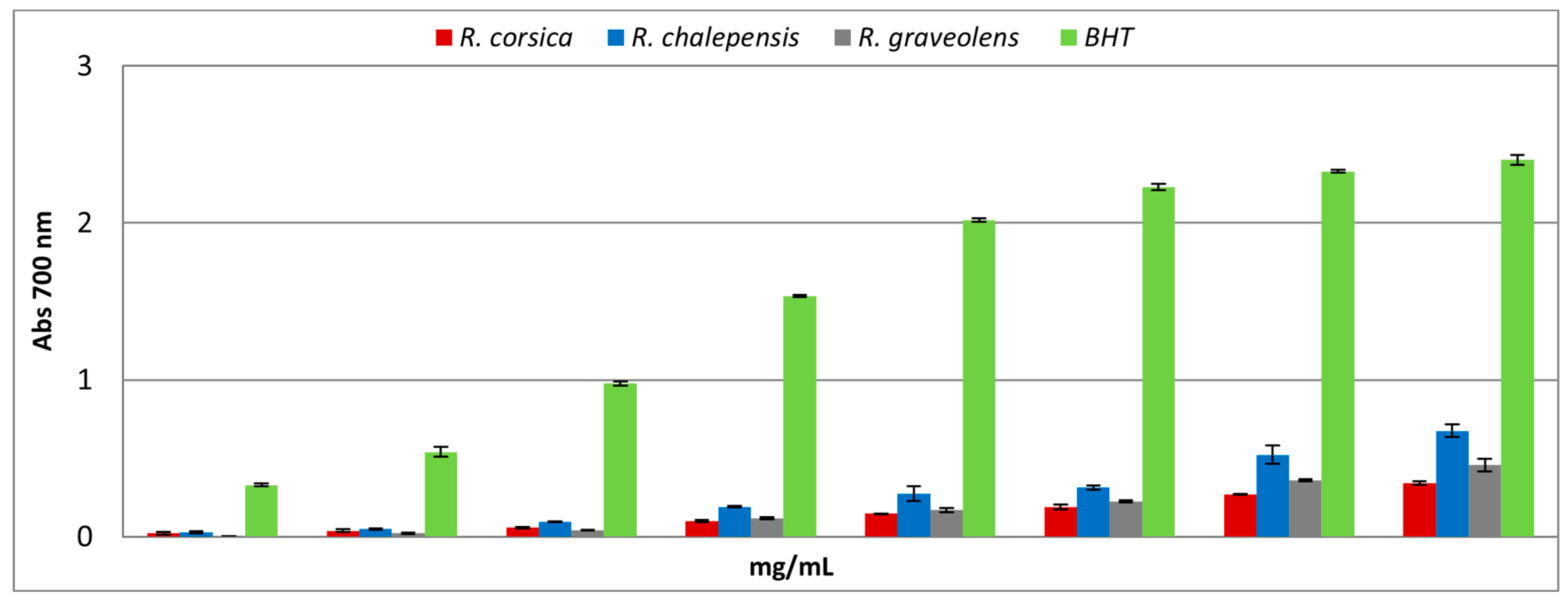
2.3.2. Reducing Power Assay

The reducing power of the Ruta spp. extracts was evaluated by spectrophotometric detection of Fe3+-Fe2+ transformation [31]. The extracts were tested at different concentrations (0.0625–2 mg/mL). Varying amounts of samples in 1 mL solvent were mixed with 2.5 mL of phosphate buffer (0.2 M, pH 6.6) and 2.5 mL of 1% potassium ferricyanide (K3Fe(CN)6). The mixture was incubated at 50 °C for 20 min. The resulting solution was cooled rapidly, mixed with 2.5 mL of 10% trichloroacetic acid, and centrifuged at 3000× g rpm for 10 min. The resulting supernatant (2.5 mL) was mixed with 2.5 mL of distilled water and 0.5 mL of 0.1% fresh ferric chloride (FeCl3), and the absorbance was measured at 700 nm after 10 min. The increased absorbance of the reaction mixture indicates an increase in reducing power. As a blank, an equal volume (1 mL) of water was mixed with a solution prepared as described above. Ascorbic acid and BHT were used as a reference. The results, obtained from the average of three independent experiments, are expressed as the mean absorbance values ± SD. The reducing power was also expressed as the ascorbic acid equivalent (ASE/mL).

2.3.3. Ferrous Ions (Fe2+) Chelating Activity

The ferrous (Fe2+) ion chelating activity of Ruta spp. extracts was estimated by measuring the formation of the Fe2+-ferrozine complex, according to the method described previously [32]. The extracts were tested at different concentrations (0.0625–2 mg/mL). Briefly, varying concentrations of each sample in 1 mL solvent were mixed with 0.5 mL of methanol and 0.05 mL of 2 mM FeCl2. The reaction was initiated by adding 0.1 mL of 5 mM ferrozine. The mixture was then shaken vigorously and left to stand at room temperature for 10 min. The absorbance of the solution was measured spectrophotometrically at 562 nm. The control contained the FeCl2 and ferrozine complex. Ethylenediaminetetraacetic acid (EDTA) was used as a reference. The percentage of inhibition of the ferrozine- (Fe2+) complex formation was calculated by the formula ((Ao − Ac)/Ao) × 100, where Ao is the absorbance of the control and Ac is the absorbance in the presence of the sample or standard. The results obtained from the average of three independent experiments are reported as mean inhibition of the ferrozine-(Fe2+) complex formation (%) ± SD and IC50 ± SD.

2.4. Antimicrobial Activity

The antimicrobial properties of the Ruta spp. extracts were tested against the following microbial strains: Staphylococcus aureus ATCC 65381, Escherichia coli ATCC 25922, Pseudomonas aeruginosa ATCC 9027, and the yeast Candida albicans ATCC 10231; these strains were obtained from the in-house culture collection of the Department of Chemical, Biological, Pharmaceutical and Environmental Sciences, University of Messina (Italy). Overnight cultures of the bacterial strains and C. albicans were grown at 37 °C in Mueller-Hinton Broth (MHB, Oxoid) and Roswell Park Memorial Institute (RPMI) 1640 medium, respectively. The Minimum Inhibitory Concentration (MIC) and the Minimum Bactericidal/Fungicidal Concentration (MBC/MFC) of the extracts were established according to the guidelines of the Clinical and Laboratory Standards Institute [33,34], with some modifications. The methanol extracts were dissolved in dimethyl sulfoxide (DMSO) and further diluted using MHB or RPMI 1640 to obtain a final concentration of 1 mg/mL. Two-fold serial dilutions were prepared in a 96-well plate. The tested concentrations ranged from 500 to 0.49 μg/mL. Working cultures were adjusted to the required inoculum of 1 × 10⁵ CFU/mL for bacteria and 1 × 10³ CFU/mL for yeast. Growth controls (medium with the inoculum but without the extracts) and vehicle controls (medium with the inoculum and DMSO) were also included. The solvent (DMSO) did not exceed 1% concentration. Tetracycline (TC), as a positive control, was tested against all the bacterial strains at concentrations ranging from 32 to 0.016 μg/mL, while itraconazole (ITC) was tested against the yeast. The MIC of TC was considered as the lowest concentration of the extracts at which there was no bacterial growth. The MIC of ITC was defined as the lowest drug concentration that inhibited ≥50% growth as compared to the control [35]. To determine MBC/MFC values, bacterial or yeast aliquots (10 µL) were taken from each well and cultured on Mueller-Hinton Agar (MHA, Oxoid) or Sabouraud Dextrose Agar (SDA, Oxoid), respectively. The cultures were incubated for 24–48 h at 37 °C.

2.5. Statistical Analysis

Statistical comparison of data was performed using one-way analysis of variance (ANOVA) followed by Tukey–Kramer multiple comparisons test (GraphPAD Prism Software for Science LLC, San Diego, CA, USA). p-values less than 0.05 were considered statistically significant.

3. Results and Discussion

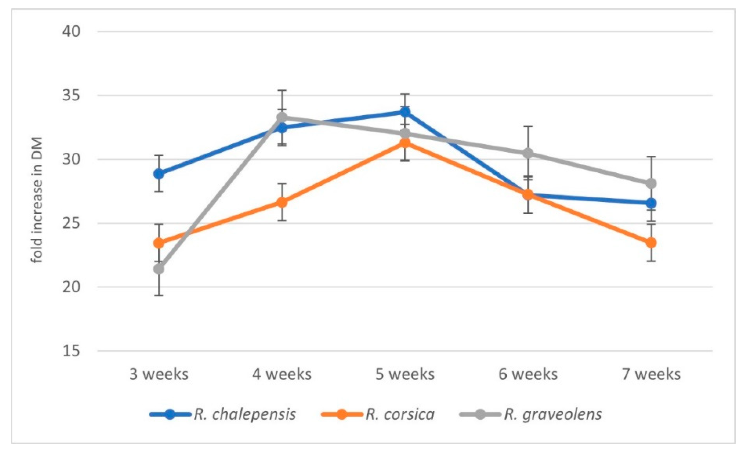
3.1. Biomass Increments

The in vitro cultures of R. graveolens, R. corsica, and R. chalepensis were maintained as agitated shoot cultures in a 7-week growth cycle (inoculum mass: 1 g). The cultures were characterized by a similar increase in biomass. The cultures showed a good biomass growth up to the fifth week of cultivation, except for R. graveolens cultures, which reached their maximum growth after four weeks of breeding. In the sixth week the growth gradually stopped, and after 7 weeks of cultivation, the shoots started to show signs of dieback. The best (33.7-fold) increase in biomass was observed in R. chalepensis cultures after 5 weeks of cultivation. For R. graveolens and R. corsica, the following biomass increase was recorded: 33.3-fold (after 4 weeks) and 31.3-fold (after 5 weeks), respectively (Figure 1).

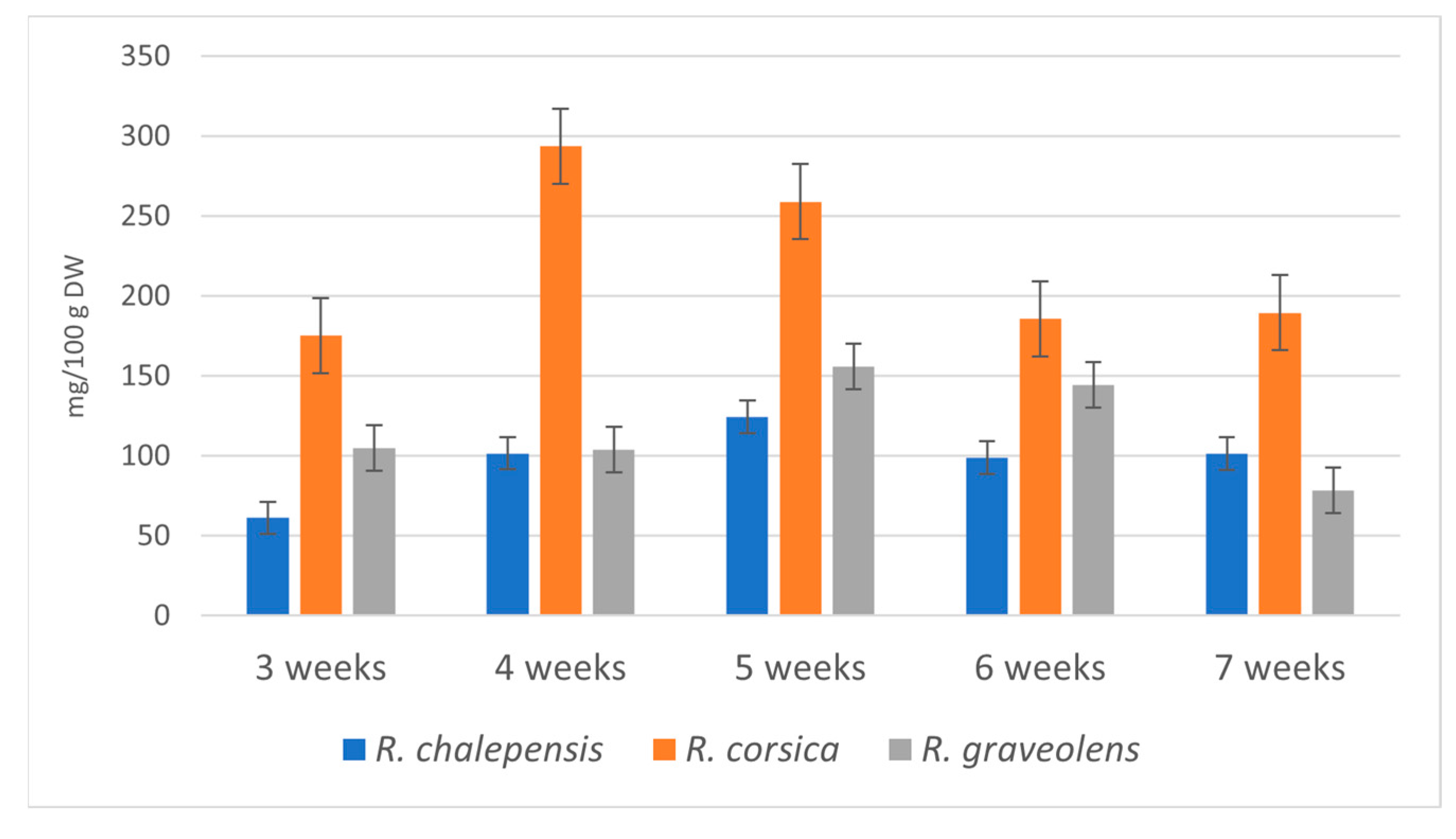
3.2. Accumulation of Bioactive Metabolites

The HPLC analysis of methanolic extracts obtained from the microshoots of the investigated in vitro cultures of the three rue species confirmed the accumulation of linear furanocoumarins (bergapten retention time (RT) = 63.72 min, isoimperatorin RT = 70.55 min, isopimpinellin RT = 60.91 min, psoralen RT = 53.38 min, xanthotoxin RT = 54.18 min) and furoquinoline alkaloids (γ-fagarine RT = 64.68 min, 7-isopentenyloxy-γ-fagarine RT = 71.92 min, skimmianine RT = 62.36 min). Sample chromatograms of the extracts from the R. corsica, R chalepensis, and R. graveolens in vitro cultures are included in Supplementary Materials Figures S1–S3.
No flavonoids were detected. Phenolic acids were accumulated in very small amounts. The dynamics of accumulation of individual metabolites varied according to the time of the culture and species. Generally, the production of most compounds increased between the third and sixth week and decreased in the seventh week. Table 1 shows the mean contents (mg/100 g DW) of individual metabolites depending on the rue species and the growth cycle. Among the coumarins, xanthotoxin was accumulated in the greatest amount (maximum content: R. chalepensis—509.8 mg/100 g DW; R. corsica—375.9 mg/100 g DW, 5-week growth cycle; R. graveolens—428.3 mg/100 g DW, 6-week growth cycle). Bergapten was the next coumarin that was accumulated in high amounts with the maximum content obtained after the 6-week growth cycle in R. chalepensis and R. graveolens cultures and after the 7-week growth cycle in R. corsica cultures (281.4; 186.6; 174.7 mg/100 g DW, respectively). The quantity of psoralen was also high, with the maximum content of 268.8 mg/100 g DW in R. chalepensis microshoots (5-week growth cycle) and 278.7 and 129.3 mg/100 g DW in R. corsica and R. graveolens microshoots (6-week growth cycle). The amounts of isopimpinellin were lower and reached the maximum content of 145.25 and 57.45 mg/100 g DW in R. graveolens and R. corsica extracts of biomass (5-week growth cycle), respectively, and 77.1 mg/100 g DW in the R. chalepensis extract (6-week growth cycle). Isoimperatorin was accumulated in the lowest amount of 46.8, 43.1, and 23.9 mg/100 g DW in R. corsica, R. chalepensis, and R. graveolens microshoots, respectively (6-week growth cycle).
Among the confirmed furoquinoline alkaloids, γ-fagarine was accumulated in the highest amount (293.7 mg/100 g DW in R. corsica cultures after the 4-week growth cycle, while its content was 155.9 and 124.3 mg/100 g DW in R. graveolens and R. chalepensis cultures after the 5-week growth cycle, respectively). The maximum skimmianine content was 133.4 mg/100 g DW in R. corsica cultures (4-week growth cycle) 94.6 mg/100 g DW in R. graveolens cultures (5-week growth cycle) and 55.5 mg/100 g DW in R. chalepensis cultures (7-week growth cycle). Isopentenyloxy-γ-fagarine was accumulated in the lowest amount: 26.5 mg/100 g DW in R. corsica microshoots (4-week growth cycle) and 6.8 and 4.4 mg/100 g DW in R. graveolens and R. chalepensis microshoots, respectively (5-week growth cycle) (Table 1).
The highest total content (1031.5 mg/100 g DW) of the estimated coumarins was detected in the cultures of R. chalepensis (5-week growth cycle). Very high amounts of coumarins were also confirmed in the cultures of R. graveolens (917.23 mg/100 g DW, 5-week growth cycle) and R. corsica (878.05 mg/100 g DW, 6-week growth cycle) (Table 1). During the breeding cycle, the total content of coumarins in the biomass of R. chalepensis cultures increased from 617.5 mg/100 g DW (3-week growth cycle) to 1031.5 mg/100 g DW (5-week growth cycle) (1.38-fold), in the biomass of R. graveolens cultures from 461.4 mg/100 g DW (3-week growth cycle) to 917.2 mg/100 g DW (5-week growth cycle) (1.99-fold), and in the biomass of R. corsica cultures from 533.5 mg/100 g DW (3-week growth cycle) to 878.05 mg/100 g DW (6-week growth cycle) (1.65-fold) (Table 1, Figure 2).
Shoot cultures of R. corsica showed the highest ability to accumulate alkaloids. The highest total content (293.7 mg/100 g DW) of the estimated alkaloids was detected after the 4-week growth cycle and was 1.88-fold higher than the maximal content of alkaloids in R. graveolens cultures (155.9 mg/100 g DW, 5-week growth cycle) and 2.36-fold higher than the maximal content of alkaloids in R. chalepensis cultures (124.3 mg/100 g DW, 5-week growth cycle) (Table 1). During the breeding cycle the total content of alkaloids in the biomass of R. corsica cultures increased from 175.4 mg/100 g DW (3-week growth cycle) to 293.7 mg/100 g DW (5-week growth cycle) (1.67-fold), in the biomass of R. graveolens cultures from 105.1 mg/100 g DW (3-week growth cycle) to 155.9 mg/100 g DW (5-week growth cycle) (1.48-fold), and in the biomass of R. chalepensis cultures from 61.25 mg/100 g DW (3-week growth cycle) to 124.3 mg/100 g DW (5-week growth cycle) (2.03-fold) (Table 1, Figure 3).
The production of secondary metabolites from the group of linear furanocoumarins and some alkaloids was observed in the in vitro cultures of the studied rue species. A characteristic of in vitro cultures is that their metabolic profile may differ from that of their parent plants. Our previous studies on in vitro cultures of R. graveolens confirmed that the shoot cultures of this plant species accumulated mainly linear furanocoumarins and alkaloids, and significantly lower amounts of phenolic acids [21,23]. The results of current studies also confirmed this ability of the R. graveolens shoot culture. The same biosynthetic potential was now documented by us for two other rue species—R. chalepensis and R. corsica. The shoots of these species also produced mainly linear furanocoumarins and some alkaloids. However, some differences were noted between the in vitro cultures of the three rue species. R. corsica shoots produced higher amounts of alkaloids than R. chalepensis and R. graveolens shoot cultures.
Previous studies [36] analysed the selected secondary metabolites (coumarins and alkaloids) in R. graveolens shoot stationary liquid cultures (LS medium containing 2 mg/L each of NAA and BAP). Seven secondary metabolites were identified. These included linear furanocoumarins (psoralen, bergapten, xanthotoxin, isopimpinellin), linear dihydrofuranocoumarin (rutamarine), and furoquinoline alkaloids (cocusaginine and skimmianine). Five of these were confirmed in the current HPLC analysis of R. graveolens biomass extracts. Additionally, we confirmed two furoquinoline alkaloids (γ-fagarine, isopentenyloxy-γ-fagarine) and isoimperatorin (furanocoumarin).
The results of dynamics of metabolites accumulation during the growth cycles showed that the maximal total amounts of furanocoumarins were obtained after the 5-week growth cycle. In our previous studies on agitated shoot cultures of R. graveolens, which were grown in a 6-week growth cycle, the maximal content (520.8 mg/g DW) of linear furanocoumarin was confirmed after 6 weeks (LS medium NAA/BAP 0.1/0.1 mg/L) [21].
The maximal accumulation (966 mg/100 g DW) of furanocoumarins in the stationary liquid culture of R. graveolens was confirmed after 4 weeks of growth (LS medium NAA/BAP: 2/2 mg/L) [20].
Baumert et al. showed the presence of the following metabolites in in vitro callus agar culture of R. chalepensis: furanocoumarins: bergapten and isopimpinellin; alkaloids: skimmianine, γ-fagarine, rutacridone, rutacridone epoxide, gravacridone, and arborinin. The authors, however, did not quantify these compounds [26].
In the study of Fischer et al., the authors isolated and identified three metabolites from R. chalepensis suspension cultures: isorutarin (dihydrofuranocoumarin), rutarensin—an unknown ester of daphnorin (coumarin dimer) and 3-hydroxy-3-methylglutaric acid. The authors, however, did not isolate the linear furanocoumarins detected in our present study [27].
As part of this work, the R. chalepensis shoot cultures were conducted for the first time. The abovementioned authors investigated other types of cultures (callus and suspension cultures). A comparison of the results of previous studies with those obtained in the present study revealed significant qualitative differences in the accumulated compounds from the coumarin group between different types of cultures. The shoot cultures studied in the present research accumulated high amounts of xanthotoxin and psoralen. However, they were not found in callus and suspension cultures.
There are no studies on in vitro cultures of the endemic species R. corsica in the available literature. In the present study, the metabolic profile of the R. corsica shoot cultures was investigated for the first time.
On the basis of the obtained results, all the studied rue cultures can be proposed as an alternative, rich, in vitro-controlled source of linear furanocoumarins. Xanthotoxin and bergapten in particular are objects of interest in dermatology. These compounds have photosensitizing effects on human skin and are used for their pigmentation stimulating and antiproliferative properties in the symptomatic treatment of vitiligo, psoriasis, and mycosis fungoides [37]. The obtained results showed very high levels of xanthotoxin in R. chalepensis and R. graveolens cultures (above 500 and 420 mg/100 g DW) and approximately 400 mg/100 g DW in R. corsica cultures. The obtained maximal amounts of bergapten were also high (above 280 mg/100 g DW in R. chalepensis cultures, above 180 mg/100 g DW in R. graveolens cultures, and above 170 mg/100 g DW in R. corsica cultures). For comparison, the content of xanthotoxin and bergapten in plants cultivated in the field is as follows: for R. graveolens—xanthotoxin 100 mg/100 g DW, bergapten 160 mg/100 g DW; for R. chalepensis—xanthotoxin 10 mg/100 g DW, bergapten 260 mg/100 g DW [29]. The biosynthetic potential for the production of furoquinoline alkaloids is the highest for R. corsica cultures (above 130 mg/100 g DW). These cultures can therefore be considered as a source of furoquinoline alkaloids: γ-fagarine and skimmianine. Furoquinoline alkaloids show a number of biological activities, including antifungal and antibacterial properties, inhibitory activity towards AchE (acetylcholinesterase), and 5-HT2 receptor-inhibiting properties [38].

3.3. Antioxidant Activity

Antioxidants compounds neutralize free radicals and their negative effects by preventing their production, intercepting them, and repairing the damage caused by these molecules; these compounds use various mechanisms, such as functioning as reducing agents by trapping free radicals, donating hydrogen, acting as chelators, and quenching singlet oxygen. Antioxidants can be divided into primary (or chain-breaking) and secondary (or preventive), and the primary antioxidant reactions can be classified as hydrogen-atom transfer (HAT) and single-electron transfer (SET). The HAT mechanism occurs when an antioxidant scavenges free radicals by donating hydrogen atoms; while in the SET mechanism, an antioxidant acts by transferring a single electron to reduce any compound.
Antioxidants can act through various mechanisms, such as binding of transition metal ion catalysts, reducing capacity and radical scavenging, prevention of chain initiation, decomposition of peroxides, and prevention of continued hydrogen abstraction. Therefore, it is important to use methodologies with distinct mechanisms to evaluate the antioxidant capacity of plant-derived phytocomplexes or isolated compounds [39].
To determine the in vitro antioxidant capacity of methanolic extracts of Ruta spp., three in vitro tests based on different mechanisms of determination of the antioxidant capacity were used: the DPPH (1,1-diphenyl-2-picrylhydrazyl) method, (involving both HAT and SET mechanisms); the reducing power assay (based on SET mechanism); and the ferrous ion chelating activity assay, to assess secondary antioxidant ability.
The results of the DPPH assay showed that all Ruta spp. extracts exhibited the radical scavenging effect that increased with the increasing concentrations (Figure 4). The R. chalepensis extract showed the best radical scavenging properties, reaching 60% of the activity at the higher concentration tested (Figure 4). As indicated by the IC50 values, the activity of the extracts was found to be lower than that of butylated hydroxytoluene (BHT) used as a standard drug and it decreased in the following order: BHT > R. chalepensis > R. graveolens > R. corsica (Table 2).
The antiradical activity of the extracts obtained from R. chalepensis grown in different countries has been extensively studied. Fakhfakh et al. (2012) found IC50 values of 0.12 and 0.22 mg/mL for R. chalepensis ethanol and aqueous extracts, respectively [40]. Ereifej et al. (2015) reported a DPPH radical scavenging ability of Tunisian leaf extract with an IC50 value of 70.01 μg/mL [41]. Similar results were obtained by Rached et al. (2010) who reported an IC50 value of 61.41 μg/mL for an Algerian R. chalepensis leaf ethanol extract and by Loizzo et al. (2018) who reported an IC50 value of 60.2 μg/mL for the methanol leaf extract obtained from Tunisian samples of this species [42,43]. Kacem et al. (2015) highlighted a decreasing DPPH radical scavenging potential in different extracts of R. chalepensis from Tunisia: ethanol extract > methanol/water (1/1) extract > methanol extract > ethyl acetate extract > water extract > hexane extract [14]. In contrast, other authors found a higher IC50 value for the methanolic extract of the aerial part of R. chalepensis from Saudi Arabia (320.7 μg/mL) [44]. Interestingly, the IC50 values in the DPPH test were found for decoction and ethanol extracts of stem and leaves of R. chalepensis from El Hamma (Tunisia) (IC50 value of 2.26 μg/mL for the leaf decoction) [45].
Regarding the antioxidant activity of R. graveolens, Molnar et al. (2017) reported that the ethanolic extracts obtained from commercial samples of the plant showed a higher DPPH scavenging activity (% inhibition = 59.3%) than the hexane extract (% inhibition = 16.8%) at the same concentration (250 μg/mL) [46]. Furthermore, methanolic and ethanolic extracts of wild-growing and cultivated Serbian R. graveolens at the beginning and end of the flowering season exhibited significant antioxidant potential. The methanolic extract of wild plants collected at the end of the flowering season showed the strongest antioxidant activity in DPPH tests (IC50 = 36.36 μg/mL) [47].
As shown in Figure 5, the Ruta spp. extracts showed a weak, dose-dependent reducing power. A comparison of the ASE/mL values showed that the reducing power of the Ruta spp. extracts and the standard decreased in the following order: BHT > R. chalepensis > R. corsica > R. graveolens (Table 2).
In the Fe2+ chelating activity assay, the Ruta spp. extracts exhibited good, dose-dependent chelating properties. R. graveolens showed the best activity, reaching approximately 80% at the highest tested concentration (Figure 6). Based on the IC50 values, the activity of the Ruta spp. extracts and the standard decreased in the following order EDTA > R. graveolens > R. corsica > R. chalepensis (Table 2). Loizzo et al. (2018) conducted a study on the methanolic leaf extract of Tunisian R. graveolens and found that this extract showed low metal chelating activity (14.6%) as compared to EDTA (97.8%) [43].
On the basis of the obtained results, it is evident that the methanolic extracts of the Ruta spp. cultures act as weak primary antioxidants and possess good secondary antioxidant properties.
The genus Ruta is rich in bioactive compounds. Phytochemical investigations conducted on extracts obtained from Ruta spp. grown in fields have detected the presence of compounds belonging to different chemical classes, such as coumarins, phenolic acids, flavonoids, alkaloids, and tannins [48].
In our HPLC study of in vitro cultures of Ruta spp., a different metabolic profile with respect to that of the parent plant was highlighted. In all the extracts, flavonoids were undetectable and phenolic acids were present only in small amounts. The dominant group of compounds in all cultures were coumarins followed by alkaloids. Among the coumarins, xanthotoxin was accumulated in the highest amount. It was reported that coumarins imperatorin, xanthotoxin, and bergapten did not exhibit anti-radical activity against DPPH, while they showed a moderate level of reducing power, moreover, they were effective in the ferrous ion-chelation test [49]. Coumarins can reduce the level of oxidative stress through chelation of redox-active Cu and Fe, thus suppressing the formation of ROS through the Fenton reaction [50].
Thus, the good secondary antioxidant properties observed for R. graveolens, R. Corsica, and R. chalepensis methanol extracts could be mainly related to the presence of coumarins, but it cannot be excluded that other compounds present in the phytocomplex could also contribute to the observed effects.

3.4. Antimicrobial Activity

The development of antimicrobial resistance in pathogens has prompted extensive research to find alternative therapeutics. Staphylococcus aureus is the first pathogen that has become resistant to all known antibiotics and thus it is essential that new antimicrobial agents are discovered to combat this problem [51]. Plants rich in secondary metabolites are one of the go-to reservoirs for the discovery of potential resources to alleviate this problem [52,53].
The preliminary antimicrobial screening of the Ruta spp. extracts obtained from in vitro cultured biomass was performed against a small representative set of Gram-positive and Gram-negative bacterial strains and the yeast Candida albicans according to the protocols recommended by the Clinical and Laboratory Standards Institute [33,34]. The obtained results showed that all the methanolic extracts had a good bacteriostatic activity against the Gram-positive S. aureus at the tested concentrations. Among the extracts, the R. corsica extract showed the highest activity (MIC = 250 μg/mL) followed by R. graveolens and R. chalepensis extracts (MIC = 500 μg/mL). None of the tested strains were inhibited by DMSO (maximum 0.5% v/v), used as a negative control. The MIC of tetracycline, used as positive control, was 1 μg/mL.
Several studies have reported the antibacterial and antifungal potential of Ruta spp., with R. chalepensis and R. graveolens being the most frequently investigated [48].
Our results showed good antibacterial activity of the methanolic extract obtained from Ruta spp. in vitro cultured biomass against S. aureus at concentrations lower (MIC range: 250–500 μg/mL) than those reported by Alzoreky and Nakahara (2003) for wild or cultivated R. chalepensis and R. graveolens (MIC = 2640 mg/mL) [54]. There are no data on R. corsica.
The in vitro cultures studied in the present research accumulated mainly metabolites from the group of coumarins and alkaloids. Alkaloids exhibit antimicrobial activity by enzyme activity inhibition, intracellular molecular imbalance, and cell membrane disturbances, which can cause cell death [55]. All the extracts obtained from Ruta spp. in vitro cultured biomass contained furoquinoline alkaloids, namely, γ-fagarine, 7-isopentenyloxy-γ-fagarine, and skimmianine, whose strong antibacterial activity against S. aureus has been widely demonstrated [56,57,58].
The highest total content of the identified alkaloids was found in the R. corsica extract, which verified the greater activity shown by this extract than the other two extracts.
Coumarins possess a wide variety of biological activities [59]; however, only a limited number of coumarin structures have been assessed for antimicrobial activity [48]. Among these, the furanocoumarin xanthotoxin proved to be effective in inhibiting the growth of S. aureus while bergapten was not active [53].
Thus, it can be hypothesized that the good antibacterial activity observed for all Ruta spp. extracts against S. aureus could depend mainly on the presence of the alkaloids γ-fagarine and skimmianine and the coumarin xanthotoxin contained in the phytocomplex.

4. Conclusions

The present research proved a similar biosynthetic potential of shoot cultures of three rue species: R. chalepensis, R. corsica, and R. graveolens. The metabolism of the in vitro cultures of these species differs from that of the parent plants and is mainly directed towards the production of linear furanocoumarin compounds and furoquinoline alkaloids. The antioxidant and antibacterial activities of all rue cultures were confirmed. These activities are derived from the presence of the dominant metabolites from the furanocoumarin group and alkaloids, but they may also be related to the action of other compounds present in the phytocomplex. The obtained contents of the described groups of metabolites are higher than those in plants grown in the field. Therefore, in vitro cultures of Ruta spp. can be proposed as an alternative source of obtaining these metabolites: all rue cultures tested can be used to obtain furanocoumarins, while the most effective source for obtaining alkaloids is an R. corsica culture.

Supplementary Materials

The following are available online at https://www.mdpi.com/article/10.3390/antiox11030592/s1, Figure S1: Sample chromatogram of the extract from Ruta corsica in vitro cultures; Figure S2: Sample chromatogram of the extract from Ruta chalepensis in vitro cultures; Figure S3: Sample chromatogram of the extract from Ruta graveolens in vitro cultures.

Author Contributions

Conceptualization, A.S.; investigation, A.M., A.S., J.M. and N.M.; data curation, A.M., A.S. and N.M.; writing—original draft preparation, A.M., A.S. and N.M.; writing—review and editing, A.S., H.E. and N.M. All authors have read and agreed to the published version of the manuscript.

Funding

The research was realized as a part of the research project N42/DBS/000135 supported by the Polish Ministry of Science and Higher Education. The publication was created with the use of equipment co-financed by the qLIFE Priority Research Area under the program “Excellence Initiative—Research University” at Jagiellonian University.

Institutional Review Board Statement

Not applicable.

Informed Consent Statement

Not applicable.

Data Availability Statement

The data presented in this study are available in the article and Supplementary Materials.

Conflicts of Interest

The authors declare no conflict of interest.

References

  1. Engler, A. Syllabus Der Pflanzenfamilien, 12th ed.; Gebruder Borntraeger Verlag: Berlin, Germany, 1964. [Google Scholar]
  2. Meloni, M.; Dettori, C.A.; Reid, A.; Bacchetta, G.; Hugot, L.; Conti, E. High genetic diversity and presence of genetic structure characterise the endemics Ruta corsica and Ruta lamarmorae (Rutaceae). Caryologia 2020, 73, 11–26. [Google Scholar]
  3. Tutin, T.G.; Heywood, V.H.; Burges, N.A. Flora Europea; University Press Cambridge: Cambridge, UK, 1968; p. 2. [Google Scholar]
  4. Hammiche, V.; Azzouz, M. Les rues: Ethnobotanique, phytopharmacologie et toxicité. Phytotherapie 2013, 11, 22–30. [Google Scholar] [CrossRef]
  5. Asgarpanah, J.; Khoshkam, R. Phytochemistry and pharmacological properties of Ruta graveolens L. J. Med. Plants Res. 2012, 6, 3942–3949. [Google Scholar] [CrossRef]
  6. Kostova, I.; Ivanova, A.; Mikhova, B.; Klaiber, I. Alkaloids and Coumarins from Ruta graveolens. Mon. Für Chem. 1999, 130, 703–707. [Google Scholar] [CrossRef]
  7. Smolarz, H.D.; Sokołowska-Woźniak, A.; Zgórka, G. Phenolic acids from herb of Ruta graveolens L. Acta Pol. Pharm. Drugs Res. 1997, 54, 161–163. [Google Scholar]
  8. Ben Hadj Fredj, B.; Marzouk, B.; Chraief, I. Analysis of Tunisian Ruta graveolens L. oils from Jemmel. J. Food Agric. Environ. 2007, 5, 52–55. [Google Scholar]
  9. Giresha, A.S.; Anitha, M.G.; Dharmappa, K.K. (2015). Phytochemical composition, antioxidant and in-vitro anti-inflammatory activity of ethanol extract of Ruta graveolens L. leaves. Int J Pharm Pharm Sci 2015, 7, 272–276. [Google Scholar]
  10. Pollio, A.; De Natale, A.; Appetiti, E.; Aliotta, G.; Touwaide, A. Continuity and change in the Mediterranean medical tradition: Ruta spp. (rutaceae) in Hippocratic medicine and present practices. J. Ethnopharmacol. 2008, 116, 469–482. [Google Scholar] [CrossRef] [PubMed]
  11. Roelandts, R. Photo(chemo) therapy for vitiligo. Photodermatol. Photoimmunol. Photomed. 2003, 19, 1–4. [Google Scholar] [CrossRef] [PubMed]
  12. Wolf, P. Psoralen-ultraviolet A endures as one of the most powerful treatments in dermatology: Reinforcement of this ‘triple-product therapy’ by the 2016 British guidelines. Br. J. Dermatol. 2016, 174, 11–14. [Google Scholar] [CrossRef] [PubMed]
  13. Gunaydin, K.; Savci, S. Phytochemical studies on Ruta chalepensis (Lam.) Lamarck. Nat. Prod. Res. 2005, 19, 203–210. [Google Scholar] [CrossRef] [PubMed]
  14. Kacem, M.; Kacem, I.; Simon, G.; Ben Mansour, A.; Chaabouni, S.; Elfeki, A.; Bouaziz, M. Phytochemicals and biological activities of Ruta chalepensis L. growing in Tunisia. Food Biosci. 2015, 12, 73–83. [Google Scholar] [CrossRef]
  15. Mejri, J.; Abderrabba, M.; Mejri, M. Chemical composition of the essential oil of Ruta chalepensis L: Influence of drying, hydro-distillation duration and plant parts. Ind. Crop. Prod. 2010, 32, 671–673. [Google Scholar] [CrossRef]
  16. Bertrand, C.; Fabre, N.; Moulis, C. A new coumarin glucoside, coumarins and alkaloids from Ruta corsica roots. Fitoterapia 2004, 75, 242–244. [Google Scholar] [CrossRef] [PubMed]
  17. Bertrand, C.; Fabre, N.; Moulis, C.; Bessiere, J.-M. Composition of the Essentials Oil of Ruta Corsica DC. J. Essent. Oil Res. 2003, 15, 98–99. [Google Scholar] [CrossRef]
  18. Koblovská, R.; Mackova, Z.; Vitkova, M.; Kokoska, L.; Klejdus, B.; Lapcik, O. Isoflavones in the Rutaceae family: Twenty selected representatives of the genera Citrus, Fortunella, Poncirus, Ruta and Severinia. Phytochem. Anal. 2008, 19, 64–70. [Google Scholar] [CrossRef] [PubMed]
  19. Tian, S.; Wang, J.; Li, Y.; Li, D.; Xu, L.; Hou, T. The application of in silico drug-likeness predictions in pharmaceutical research. Adv. Drug Deliv. Rev. 2015, 86, 2–10. [Google Scholar] [CrossRef] [PubMed]
  20. Ekiert, H.; Chołoniewska, M.; Gomółka, E. Accumulation of furanocoumarins in Ruta graveolens L. shoot culture. Biotechnol. Lett. 2001, 23, 543–545. [Google Scholar] [CrossRef]
  21. Ekiert, H.; Czygan, F.-C. Accumulation of biologically active furanocoumarins in agitated cultures of Ruta graveolens L. and Ruta graveolens ssp. divaricata (Tenore) Gams. Die Pharm. 2005, 60, 623–626. [Google Scholar]
  22. Ekiert, H.; Abou-Mandour, A.A.; Czygan, F.C. Accumulation of biologically active furanocoumarins in Ruta graveolens ssp. divaricata (Tenore) Gams in vitro culture. Die Pharm. 2005, 60, 66–68. [Google Scholar]
  23. Ekiert, H.; Szewczyk, A.; Kus, A. Free phenolic acids in Ruta graveolens L. in vitro culture. Pharmazie 2009, 64, 694–696. [Google Scholar] [CrossRef] [PubMed]
  24. Ekiert, H.; Piekoszewska, A.; Muszyńska, B.; Baczyńska, S. Accumulation of p-coumaric acid and other bioactive phenolic acids in in vitro culture of Ruta graveolens ssp. divaricata (Tenore) Gams. MIR 2014, 26, 24–30. [Google Scholar]
  25. Szopa, A.; Ekiert, H.; Szewczyk, A.; Fugas, E. Production of bioactive phenolic acids and furanocoumarins in in vitro cultures of Ruta graveolens L. and Ruta graveolens ssp. divaricata (Tenore) Gams. under different light conditions. Plant Cell Tissue Organ Cult. (PCTOC) 2012, 110, 329–336. [Google Scholar] [CrossRef] [Green Version]
  26. Baumert, A.; Gröger, D.; Kuzovkina, I.N.; Reisch, J. Secondary metabolites produced by callus cultures of various Ruta species. Plant Cell Tissue Organ Cult. 1992, 28, 159–162. [Google Scholar] [CrossRef]
  27. Fischer, H.; Römer, A.; Ulbrich, B.; Arens, H. A New Biscoumarin Glucoside Ester fromRuta ChalepensisCell Cultures. Planta Medica 1988, 54, 398–400. [Google Scholar] [CrossRef] [PubMed]
  28. Linsmaier, E.M.; Skoog, F. Organic Growth Factor Requirements of Tobacco Tissue Cultures. Physiol. Plant 1965, 18, 100–127. [Google Scholar] [CrossRef]
  29. Sułkowska-Ziaja, K.; Maślanka, A.; Szewczyk, A.; Muszyńska, B. Physiologically Active Compounds in Four Species of Phellinus. Nat. Prod. Commun. 2017, 12, 363–366. [Google Scholar] [CrossRef] [PubMed] [Green Version]
  30. Ohnishi, M.; Morishita, H.; Iwahashi, H.; Toda, S.; Shirataki, Y.; Kimura, M.; Kido, R. Inhibitory effects of chlorogenic acids on linoleic acid peroxidation and haemolysis. Phytochemistry 1994, 36, 579–583. [Google Scholar] [CrossRef]
  31. Oyaizu, M. Studies on products of browning reaction. Antioxidative activities of products of browning reaction prepared from glucosamine. Jpn. J. Nutr. Diet. 1986, 44, 307–315. [Google Scholar] [CrossRef] [Green Version]
  32. Decker, E.A.; Welch, B. Role of ferritin as a lipid oxidation catalyst in muscle food. J. Agric. Food Chem. 1990, 38, 674–677. [Google Scholar] [CrossRef]
  33. Standard M27-A3; Clinical and Laboratory Standards Institute. Reference Method for Broth Dilution Antifungal Susceptibility Testing of Yeasts. CLSI: Wayne, PA, USA, 2008.
  34. Standard M07-Ed11; Methods for Dilution Antimicrobial Susceptibility Tests for Bacteria That Grow Aerobically. CLSI: Wayne, PA, USA, 2018.
  35. Marino, A.; Nostro, A.; Mandras, N.; Roana, J.; Ginestra, G.; Miceli, N.; Taviano, M.F.; Gelmini, F.; Beretta, G.; Tullio, V. Evaluation of antimicrobial activity of the hydrolate of Coridothymus capitatus (L.) Reichenb. fil. (Lamiaceae) alone and in combination with antimicrobial agents. BMC Complement. Med. Ther. 2020, 20, 89. [Google Scholar] [CrossRef] [PubMed]
  36. Ekiert, H.; Kisiel, W. Coumarins and alkaloids in shoot culture of Ruta graveolens L. Acta Soc. Bot. Pol. 1997, 66, 329–332. [Google Scholar] [CrossRef] [Green Version]
  37. Milesi, S.; Massot, B.; Gontier, E.; Bourgaud, F.; Guckert, A. Ruta graveolens L.: A promising species for the production of furanocoumarins. Plant Sci. 2001, 161, 189–199. [Google Scholar] [CrossRef]
  38. Adamska-Szewczyk, A.; Głowniak, K.; Baj, T. Furochinoline alkaloids in plants from Rutaceae family—A review. Curr. Issues Pharm. Med. Sci. 2016, 29, 33–38. [Google Scholar] [CrossRef] [Green Version]
  39. Gali, L.; Bedjou, F. Antioxidant and anticholinesterase effects of the ethanol extract, ethanol extract fractions and total alkaloids from the cultivated Ruta chalepensis. S. Afr. J. Bot. 2018, 120, 163–169. [Google Scholar] [CrossRef]
  40. Fakhfakh, N.; Zouari, S.; Zouari, M.; Loussayef, C.; Zouari, N. Chemical composition of volatile compounds and antioxidant activities of essential oil, aqueous and ethanol extracts of wild Tunisian Ruta chalepensis L. (Rutaceae). J. Med. Plants Res. 2012, 6, 593–600. [Google Scholar]
  41. Ereifej, K.I.; Feng, H.; Rababah, T.; Almajwal, A.; Alu’Datt, M.; Gammoh, S.I.; Oweis, L.I. Chemical Composition, Phenolics, Anthocyanins Concentration and Antioxidant Activity of Ten Wild Edible Plants. Food Nutr. Sci. 2015, 06, 581–590. [Google Scholar] [CrossRef] [Green Version]
  42. Rached, W.; Benamar, H.; Bennaceur, M.; Marouf, A. Screening of the Antioxidant Potential of Some Algerian Indigenous Plants. J. Biol. Sci. 2010, 10, 316–324. [Google Scholar] [CrossRef] [Green Version]
  43. Loizzo, M.R.; Falco, T.; Bonesi, M.; Sicari, V.; Tundis, R.; Bruno, M. Ruta chalepensis L. (Rutaceae) leaf extract: Chemical composition, antioxidant and hypoglicaemic activities. Nat. Prod. Res. 2018, 32, 521–528. [Google Scholar] [CrossRef] [PubMed]
  44. Alotaibi, S.M.; Saleem, M.S.; Al-humaidi, J.G. Phytochemical contents and biological evaluation of Ruta chalepensis L. growing in Saudi Arabia. Saudi Pharm. J. 2018, 26, 504–508. [Google Scholar] [CrossRef] [PubMed]
  45. Khadhri, A.; Bouali, I.; Belkhir, S.; Mokded, R.; Smiti, S.; Falé, P.; Araújo, M.E.M.; Serralheiro, M.L.M. In vitro digestion, antioxidant and antiacetylcholinesterase activities of two species of Ruta: Ruta chalepensis and Ruta montana. Pharm. Biol. 2017, 55, 101–107. [Google Scholar] [CrossRef] [PubMed] [Green Version]
  46. Molnar, M.; Jerković, I.; Suknović, D.; Rajs, B.B.; Aladić, K.; Šubarić, D.; Jokić, S. Screening of six medicinal plant extracts obtained by two conventional methods and supercritical CO2 extraction targeted on coumarin content, 2,2-diphenyl-1-picrylhydrazyl radical scavenging capacity and total phenols content. Molecules 2017, 22, 348. [Google Scholar] [CrossRef] [PubMed] [Green Version]
  47. Pavlović, D.R.; Vukelic, M.; Najman, S.; Kostic, M.; Zlatkovic, B.; Mihajilov-Krstev, T.; Kitic, D. Assessment of polyphenol content, in vitro antioxidant, antimicrobial and toxic potentials of wild growing and cultured rue. J. Appl. Bot. Food Qual. 2014, 181, 175–181. [Google Scholar] [CrossRef]
  48. Coimbra, A.T.; Ferreira, S.; Duarte, A.P. Genus Ruta: A natural source of high value products with biological and pharmacological properties. J. Ethnopharmacol. 2020, 260, 113076. [Google Scholar] [CrossRef] [PubMed]
  49. Senol, F.S.; Skalicka-Woźniak, K.S.; Khan, M.T.H.; Orhan, I.E.; Sener, B.; Głowniak, K. An in vitro and in silico approach to cholinesterase inhibitory and antioxidant effects of the methanol extract, furanocoumarin fraction, and major coumarins of Angelica officinalis L. fruits. Phytochem. Lett. 2011, 4, 462–467. [Google Scholar] [CrossRef]
  50. Simunkova, M.; Alwasel, S.H.; Alhazza, I.M.; Jomova, K.; Kollar, V.; Rusko, M.; Valko, M. Management of oxidative stress and other pathologies in Alzheimer’s disease. Arch. Toxicol. 2019, 93, 2491–2513. [Google Scholar] [CrossRef] [Green Version]
  51. Smyth, T.; Ramachandran, V.N.; Smyth, W.F. A study of the antimicrobial activity of selected naturally occurring and synthetic coumarins. Int. J. Antimicrob. Agents 2009, 33, 421–426. [Google Scholar] [CrossRef] [PubMed]
  52. Mahizan, N.A.; Yang, S.-K.; Moo, C.L.; Song, A.A.-L.; Chong, C.-M.; Chong, C.-W.; Abushelaibi, A.; Lim, S.-H.-E.; Lai, K.-S. Terpene Derivatives as a Potential Agent against Antimicrobial Resistance (AMR) Pathogens. Molecules 2019, 24, 2631. [Google Scholar] [CrossRef] [PubMed] [Green Version]
  53. Ojala, T.; Remes, S.; Haansuu, P.; Vuorela, H.; Hiltunen, R.; Haahtela, K.; Vuorela, P. Antimicrobial activity of some coumarin containing herbal plants growing in Finland. J. Ethnopharmacol. 2000, 73, 299–305. [Google Scholar] [CrossRef]
  54. Alzoreky, N.S.; Nakahara, K. Antibacterial activity of extracts from some edible plants commonly consumed in Asia. Int. J. Food Microbiol. 2003, 80, 223–230. [Google Scholar] [CrossRef]
  55. Othman, L.; Sleiman, A.; Abdel-Massih, R.M. Antimicrobial Activity of Polyphenols and Alkaloids in Middle Eastern Plants. Front. Microbiol. 2019, 10, 911. [Google Scholar] [CrossRef] [PubMed]
  56. O’Donnell, F.; Smyth, T.J.P.; Ramachandran, V.; Smyth, W. A study of the antimicrobial activity of selected synthetic and naturally occurring quinolines. Int. J. Antimicrob. Agents 2010, 35, 30–38. [Google Scholar] [CrossRef] [PubMed] [Green Version]
  57. Aniszewski, T. Biology of Alkaloids. In Alkaloids, 2nd ed.; Elsevier: Helsinki, Finland, 2015. [Google Scholar]
  58. Murugan, N.; Srinivasan, R.; Murugan, A.; Kim, M.; Natarajan, D. Glycosmis pentaphylla (Rutaceae): A Natural Candidate for the Isolation of Potential Bioactive Arborine and Skimmianine Compounds for Controlling Multidrug-Resistant Staphylococcus aureus. Front. Public Health 2020, 8, 176. [Google Scholar] [CrossRef] [PubMed]
  59. Widelski, J.; Luca, S.V.; Skiba, A.; Chinou, I.; Marcourt, L.; Wolfender, J.L.; Skalicka-Wozniak, K. Isolation and Antimicrobial Activity of Coumarin Derivatives from Fruits of Peucedanum luxurians Tamasha. Molecules 2018, 23, 1222. [Google Scholar] [CrossRef] [PubMed] [Green Version]
Figure 1. Average (n = 3, ±SD) biomass increments during the cultivation cycle (3, 4, 5, 6, and 7 weeks) of individual rue species.
Antioxidants 11 00592 g001
Figure 2. Total content of coumarins (mg/100 g DW) in the methanol extracts obtained from Ruta spp. in vitro cultured biomass in the consecutive weeks of the breeding cycle. Values are expressed as the mean ± SD (n = 3).
Antioxidants 11 00592 g002
Figure 3. Total content of alkaloids (mg/100 g DW) in the methanol extracts obtained from the Ruta spp. in vitro cultured biomass in the consecutive weeks of the breeding cycle. Values are expressed as the mean ± SD (n = 3).
Antioxidants 11 00592 g003
Figure 4. Free radical scavenging activity of the methanolic extracts of the Ruta spp. in vitro cultured biomass. Values are expressed as the mean ± SD (n = 3).
Antioxidants 11 00592 g004
Figure 5. Reducing power of the methanolic extracts of the Ruta spp. in vitro cultured biomass, evaluated by spectrophotometric detection of Fe3+-Fe2+ transformation. Values are expressed as the mean ± SD (n = 3).
Antioxidants 11 00592 g005
Figure 6. The chelating activity of the methanolic extracts of the Ruta spp. in vitro cultured biomass measured by inhibition of the ferrozine-Fe2+ complex formation. Values are expressed as the mean ± SD (n = 3).
Antioxidants 11 00592 g006
Table 1. Average contents of metabolites (mg/100 g DW) in methanol extracts obtained from biomass of cultures of three rue species depending on the duration of the culture (3, 4, 5, 6, and 7 weeks). Means of three measurements ± SD. Different letters indicate significant differences (p < 0.05).
Accumulated CompoundsGrowth PeriodR. chalepensisR. corsicaR. graveolens
bergapten3 weeks91.537 ± 11.656 ace89.049 ± 13.492 ace55.391 ± 3.004 be
4 weeks112.803 ± 8.821 adl135.548 ± 10.760 cdghl78.183 ± 6.148 abe
5 weeks184.083 ± 10.970 fhjk149.363 ± 6.694 dghjl162.236 ± 9.359 dfghj
6 weeks281.388 ± 8.330 i165.047 ± 7.075 fghk186.603 ± 2.905 fhjk
7 weeks130.150 ± 2.211 cdgl174.666 ± 12.736 fghjk67.990 ± 12.442 abe
isoimperatorin3 weeks15.128 ± 1.156 abh23.696 ± 4.974 abcd14.611 ± 1.951 abdh
4 weeks20.944 ± 2.184 abcd32.090 ± 10.191 bcdef23.299 ± 1.807 abcd
5 weeks28.573 ± 3.600 bcde36.684 ± 2.145 cdefg22.003 ± 4.661 abcd
6 weeks43.126 ± 1.183 cefg46.771 ± 1.435 efg23.931 ± 1.068 abcd
7 weeks22.742 ± 2.426 abcd42.981 ± 4.047 cefg6.610 ± 1.690 ah
isopimpinellin3 weeks37.867 ± 9.467 ab43.184 ± 1.851 abd92.809 ± 1.865 c
4 weeks31.680 ± 1.116 ab47.130 ± 1.859 abd76.459 ± 2.762 c
5 weeks40.157 ± 2.292 ab57.455 ± 1.945 bd145.251 ± 7.313 de
6 weeks77.140 ± 9.462 c46.760 ± 1.668 abd138.345 ± 4.211 e
7 weeks36.063 ± 5.455 ab44.033 ± 10.052 abd84.774 ± 8.748 c
psoralen3 weeks122.970 ± 15.264 abcdg98.344 ± 0.902 abcgi70.121 ± 9.557 abcgih
4 weeks177.943 ± 27.752 ade246.241 ± 51.342 def100.141 ± 9.547 abcgi
5 weeks268.823 ± 6.085 ef256.226 ± 37.091 ef105.269 ± 5.043 abcg
6 weeks98.766 ± 3.088 abcgi278.699 ± 26.560 ef129.266 ± 3.940 abcdg
7 weeks24.047 ± 1.514 chi179.779 ± 23.994 ade37.421 ± 22.530 bchi
xanthotoxin3 weeks349.997 ± 54.203 abdehijk279.242 ± 44.171 abcj228.435 ± 11.563 bcjk
4 weeks404.187 ± 42.136 adeghj397.999 ± 19.047 adehj385.553 ± 24.113 adehj
5 weeks509.827 ± 14.507 fgh375.924 ± 54.065 adehj385.480 ± 18.961 dfgh
6 weeks365.223 ± 20.883 abdehj340.774 ± 10.539 abdehj428.302 ± 19.060 adefgh
7 weeks101.527 ± 2.700 ik319.537 ± 27.264 abcdej171.908 ± 45.753 cik
Total coumarins3 weeks617.498 ± 28.808 abf533.515 ± 32.612 abc461.368 ± 9.025 bcm
4 weeks747.557 ± 59.484 defl849.022 ± 85.331 dehij663.636 ± 32.960 adfl
5 weeks1031.463 ± 21.196 gi875.651 ± 40.408 ehijl917.230 ± 32.550 eghijl
6 weeks865.643 ± 26.639 ehijl878.051 ± 40.412 ehij906.447 ± 15.819 ehij
7 weeks314.530 ± 5.806 kl760.995 ± 40.617 defhl368.703 ± 26.781 ckm
γ-fagarine3 weeks41.203 ± 9.563 acfhil83.388 ± 5.113 bdjk47.164 ± 5.480 acdfhil
4 weeks65.350 ± 5.152 bcdhik133.804 ± 7.766 eg34.663 ± 0.728 acfhl
5 weeks78.810 ± 6.455 bdjk114.682 ± 10.950 egj54.517 ± 3.776 acdhi
6 weeks58.341 ± 2.812 acdhik97.303 ± 6.759 bgj53.649 ± 0.803 acdfhi
7 weeks42.638 ± 5.295 acfhil74.105 ± 10.043 bdik31.106 ± 4.124 acfl
Isopentenyloxy-γ-fagarine3 weeks2.778 ± 0.714 aefh17.482 ± 1.638 b9.143 ± 0.503 ceg
4 weeks2.935 ± 0.100 aefh26.505 ± 2.900 d6.311 ± 0.705 acefg
5 weeks4.379 ± 0.357 aefgh20.397 ± 1.339 b6.788 ± 0.027 cefg
6 weeks3.015 ± 1.010 aefh25.669 ± 0.066 d5.479 ± 0.100 acefg
7 weeks3.337 ± 0.498 aefgh19.931 ± 2.741 b1.644 ± 0.088 afh
skimmianine3 weeks17.267 ± 5.937 ad74.520 ± 11.342 bfi48.836 ± 2.680 cdfgj
4 weeks33.336 ± 2.404 acdg133.412 ± 1.278 e63.099 ± 5.734 bcfj
5 weeks41.127 ± 6.753 cdgj123.816 ± 10.221 e94.635 ± 5.419 hi
6 weeks37.608 ± 4.668 cdgj62.912 ± 1.729 bcfj85.399 ± 3.297 bhi
7 weeks55.535 ± 5.968 cfgj95.575 ± 9.810 hi45.517 ± 3.340 cdfgj
Total alkaloids3 weeks61.247 ± 13.462 aj175.390 ± 9.103 bgh105.143 ± 6.920 cej
4 weeks101.621 ± 4.374 cej293.721 ± 8.627 d104.074 ± 6.286 cej
5 weeks124.316 ± 12.860 cei258.895 ± 12.770 f155.940 ± 7.130 bgi
6 weeks98.964 ± 6.569 cej185.883 ± 5.235 bh144.527 ± 2.506 egi
7 weeks101.510 ± 5.749 cej189.612 ± 20.186 bh78.267 ± 3.789 acj
Table 2. Free radical scavenging activity (DPPH test), reducing power, and ferrous ions (Fe2+) chelating activity of the methanolic extracts of the Ruta spp. in vitro cultured biomass.
Extracts DPPH TestIC50 (mg/mL)Reducing Power AssayASE/mLFe2+ Chelating ActivityIC50 (mg/mL)
Ruta chalepensis1.665 ± 0.009 a15.493 ± 0.207 a1.452 ± 0.012 a
Ruta corsica2.032 ± 0.001 b20.516 ± 0.016 b0.736 ± 0.100 b
Ruta graveolens1.883 ± 0.007 c26.012 ± 0.019 c0.671 ± 0.013 a c
StandardBHT0.065 ± 0.008 dBHT1.131 ± 0.037 dEDTA0.0067 ± 0.0003 d
Values are expressed as the mean ± SD. (n = 3). a–d Different letters within the same column indicate significant differences between mean values (p < 0.05).
Publisher’s Note: MDPI stays neutral with regard to jurisdictional claims in published maps and institutional affiliations.

